international journal of pharmaceuticscqm.uma.pt/cqm12/12-23_int_j_pharma.pdf · international...
TRANSCRIPT
C
Aa
b
a
ARRAA
KSCGT
1
edaegchldd
pfddvae
H
0h
International Journal of Pharmaceutics 434 (2012) 199– 208
Contents lists available at SciVerse ScienceDirect
International Journal of Pharmaceutics
jo ur nal homep a ge: www.elsev ier .com/ locate / i jpharm
alcium phosphate-mediated gene delivery using simulated body fluid (SBF)
lireza Nouria,∗, Rita Castroa, José L. Santosa,1, César Fernandesa,b, João Rodriguesa, Helena Tomása,∗∗
CQM – Centro de Química da Madeira, MMRG, Universidade da Madeira, Campus Universitário da Penteada, 9000-390 Funchal, PortugalLaboratório Regional de Engenharia Civil, IP-RAM, Rua Agostinho Pereira de Oliveira, 9000-264 Funchal, Portugal
r t i c l e i n f o
rticle history:eceived 5 April 2012eceived in revised form 18 May 2012ccepted 21 May 2012vailable online 1 June 2012
eywords:
a b s t r a c t
The present study aimed at developing a new approach in gene delivery of calcium phosphate nanopar-ticles through simulated body fluid (CaP-SBF). The physicochemical and biological characteristics of theCaP-SBF nanoparticles were compared with those made in pure water (CaP-water) via a similar proce-dure. The CaP-SBF and CaP-water solutions were then adjusted to two different pH values of 7.4 and8.0, mixed with plasmid DNA (pDNA), and added in varying amounts to human embryonic kidney (HEK293T) cells. The transfection efficiency and cell viability were studied in vitro by reporter gene (luciferase
imulated body fluid (SBF)alcium phosphateene deliveryransfection
and Enhanced Green Fluorescent Protein) expression and the resazurin reduction assay, respectively,24 and 48 h after the incubation with the nanoparticles. Our results indicated considerably high in vitrotransfection efficiency for CaP-SBF/DNA complexes at physiological pH (7.4) with high amounts of CaP.Additionally, the SBF solution exhibited the ability to reduce the rapid growth of CaP particles overtime, leading to higher transfection efficiency of CaP-SBF/DNA complexes than those made in water(CaP-water/DNA).
. Introduction
Gene therapy is becoming a rapidly growing therapeutic strat-gy for the treatment of both acquired and inherited diseases. Theevelopment of safe and efficient gene delivery vectors remainsn essential prerequisite for gene therapy. Viral vectors are highlyfficient but have significant drawbacks, including high immuno-enicity, limited DNA carrying capacity, recombination and highosts. As an alternative system to viral vectors, non-viral vectorsave become increasingly attractive for gene therapy due to their
ow or no immunogenicity, relatively simple preparation proce-ures, low cost and high flexibility to accommodate the size of theelivered transgene (Santos et al., 2011).
Amongst various non-viral delivery systems, calcium phos-hate (CaP) particles offer one of the most attractive methodsor gene therapy, and have widely been used as an in vitro geneelivery vector for over three decades. The CaP method was firstescribed as a technique to assay the infectivity of DNA from several
iruses, as an alternative to the DEAE–dextran method (Grahamnd Van Der Eb, 1973), however, it became more popular afterxtending its application to plasmid DNA (pDNA) and gene deliv-∗ Corresponding author. Tel.: +351 291705150; fax: +351 291705249.∗∗ Corresponding author.
E-mail addresses: [email protected] (A. Nouri), [email protected] (H. Tomás).1 Present address: Department of Materials Science and Engineering, Johnsopkins University, 3400 North Charles Street, Baltimore, MD 21218, United States.
378-5173/$ – see front matter © 2012 Elsevier B.V. All rights reserved.ttp://dx.doi.org/10.1016/j.ijpharm.2012.05.066
© 2012 Elsevier B.V. All rights reserved.
ery. The suitability of this method lies in its general efficiency for awide range of cell lines, simplicity, low cost, as well as biocompati-bility, and biodegradability of CaP particles (Jordan, 2000). Differentprocedures have been used for the synthesis of CaP/DNA com-plexes including, co-precipitation (Seelos, 1997), encapsulation(Bisht et al., 2005), multi-shell structures formation (Sokolova et al.,2006), and coating of CaP nanoparticles with pDNA (Welzel et al.,2004). According to the current state of knowledge, the CaP/DNAcomplex precipitates are incorporated into the cells through endo-cytosis by forming an intracellular vesicle. The vesicles merge withlysosomes and the CaP/DNA particles are released into the cyto-plasm. The entrapped DNA somehow escapes from the lysosomesinto cytoplasm; and the DNA molecules eventually enter the nucleiof the recipient cells to effect gene transfer and expression (Orrantiaand Chang, 1990).
Simulated body fluid (SBF) was first introduced by Kokubo et al.(1990) to analyze the changes in surface-structure of a bioactiveglass ceramic. Since then, the SBF solution has routinely beenused as an effective in vitro testing method to study CaP (apatite)formation or precipitation on the surfaces of different types of bio-materials, in order to predict their in vivo bone bioactivity (Chenet al., 2008b; Kokubo and Takadama, 2007; Nouri et al., 2007).The development of SBF is based on the concept that the essentialprerequisite to evaluate the bone-binding abilities of a biomate-
rial is the formation of bone-like apatite layer on its surface whenimplanted into the living body. Upon the formation of apatite nucleion a material, they can grow spontaneously by consuming the CaPions from the surrounding SBF. The inorganic ion concentration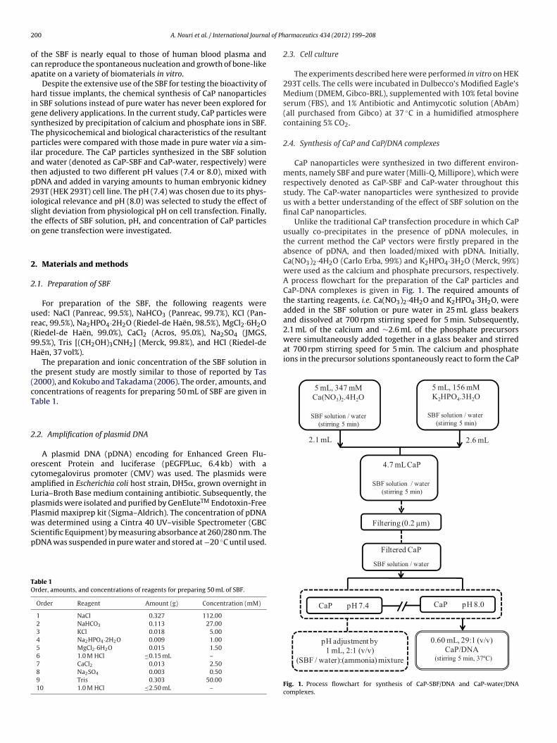
2 l of Ph
oca
higsTpiatp2isto
2
2
ur(9H
t(cT
2
ocaLpPwSp
TO
2.1 mL of the calcium and ∼2.6 mL of the phosphate precursorswere simultaneously added together in a glass beaker and stirredat 700 rpm stirring speed for 5 min. The calcium and phosphateions in the precursor solutions spontaneously react to form the CaP
5 mL, 347 mM
Ca(NO3)2.4H2O
SBF solution / water
(stirr ing 5 min)
5 mL, 156 mM
K2HPO4.3H2O
SBF solution / water
(stirri ng 5 min)
4.7 mL CaP
SBF solution / water
(stirr ing 5 min)
2.1 mL 2.6 mL
Filtering (0.2 µm)
00 A. Nouri et al. / International Journa
f the SBF is nearly equal to those of human blood plasma andan reproduce the spontaneous nucleation and growth of bone-likepatite on a variety of biomaterials in vitro.
Despite the extensive use of the SBF for testing the bioactivity ofard tissue implants, the chemical synthesis of CaP nanoparticles
n SBF solutions instead of pure water has never been explored forene delivery applications. In the current study, CaP particles wereynthesized by precipitation of calcium and phosphate ions in SBF.he physicochemical and biological characteristics of the resultantarticles were compared with those made in pure water via a sim-
lar procedure. The CaP particles synthesized in the SBF solutionnd water (denoted as CaP-SBF and CaP-water, respectively) werehen adjusted to two different pH values (7.4 or 8.0), mixed withDNA and added in varying amounts to human embryonic kidney93T (HEK 293T) cell line. The pH (7.4) was chosen due to its phys-
ological relevance and pH (8.0) was selected to study the effect oflight deviation from physiological pH on cell transfection. Finally,he effects of SBF solution, pH, and concentration of CaP particlesn gene transfection were investigated.
. Materials and methods
.1. Preparation of SBF
For preparation of the SBF, the following reagents weresed: NaCl (Panreac, 99.5%), NaHCO3 (Panreac, 99.7%), KCl (Pan-eac, 99.5%), Na2HPO4·2H2O (Riedel-de Haën, 98.5%), MgCl2·6H2ORiedel-de Haën, 99.0%), CaCl2 (Acros, 95.0%), Na2SO4 (JMGS,9.5%), Tris [(CH2OH)3CNH2] (Merck, 99.8%), and HCl (Riedel-deaën, 37 vol%).
The preparation and ionic concentration of the SBF solution inhe present study are mostly similar to those of reported by Tas2000), and Kokubo and Takadama (2006). The order, amounts, andoncentrations of reagents for preparing 50 mL of SBF are given inable 1.
.2. Amplification of plasmid DNA
A plasmid DNA (pDNA) encoding for Enhanced Green Flu-rescent Protein and luciferase (pEGFPLuc, 6.4 kb) with aytomegalovirus promoter (CMV) was used. The plasmids weremplified in Escherichia coli host strain, DH5�, grown overnight inuria–Broth Base medium containing antibiotic. Subsequently, thelasmids were isolated and purified by GenEluteTM Endotoxin-Freelasmid maxiprep kit (Sigma–Aldrich). The concentration of pDNAas determined using a Cintra 40 UV–visible Spectrometer (GBC
cientific Equipment) by measuring absorbance at 260/280 nm. TheDNA was suspended in pure water and stored at −20 ◦C until used.
able 1rder, amounts, and concentrations of reagents for preparing 50 mL of SBF.
Order Reagent Amount (g) Concentration (mM)
1 NaCl 0.327 112.002 NaHCO3 0.113 27.003 KCl 0.018 5.004 Na2HPO4·2H2O 0.009 1.005 MgCl2·6H2O 0.015 1.506 1.0 M HCl ≤0.15 mL –7 CaCl2 0.013 2.508 Na2SO4 0.003 0.509 Tris 0.303 50.0010 1.0 M HCl ≤2.50 mL –
armaceutics 434 (2012) 199– 208
2.3. Cell culture
The experiments described here were performed in vitro on HEK293T cells. The cells were incubated in Dulbecco’s Modified Eagle’sMedium (DMEM, Gibco-BRL), supplemented with 10% fetal bovineserum (FBS), and 1% Antibiotic and Antimycotic solution (AbAm)(all purchased from Gibco) at 37 ◦C in a humidified atmospherecontaining 5% CO2.
2.4. Synthesis of CaP and CaP/DNA complexes
CaP nanoparticles were synthesized in two different environ-ments, namely SBF and pure water (Milli-Q, Millipore), which wererespectively denoted as CaP-SBF and CaP-water throughout thisstudy. The CaP-water nanoparticles were synthesized to provideus with a better understanding of the effect of SBF solution on thefinal CaP nanoparticles.
Unlike the traditional CaP transfection procedure in which CaPusually co-precipitates in the presence of pDNA molecules, inthe current method the CaP vectors were firstly prepared in theabsence of pDNA, and then loaded/mixed with pDNA. Initially,Ca(NO3)2·4H2O (Carlo Erba, 99%) and K2HPO4·3H2O (Merck, 99%)were used as the calcium and phosphate precursors, respectively.A process flowchart for the preparation of the CaP particles andCaP-DNA complexes is given in Fig. 1. The required amounts ofthe starting reagents, i.e. Ca(NO3)2·4H2O and K2HPO4·3H2O, wereadded in the SBF solution or pure water in 25 mL glass beakersand dissolved at 700 rpm stirring speed for 5 min. Subsequently,
Fil tered CaP
SBF solution / water
CaP pH 7.4 CaP pH 8.0
pH adjustment by
1 mL, 2:1 (v/v)
(SBF / wat er):(amm onia) mixture
0.60 mL, 29 :1 (v/v)
CaP/DN A(stirr ing 5 min, 37ºC)
Fig. 1. Process flowchart for synthesis of CaP-SBF/DNA and CaP-water/DNAcomplexes.
l of Ph
poCtClsoo82saiD
2
ntpRbl(w
ptawt
pdwpat1basdptpcopp
2
dcwtpow7ra
A. Nouri et al. / International Journa
recipitates upon mixing together. No pH adjustment was carriedut before and during the synthesis. The final pH values of theaP-SBF and CaP-water solutions after the synthesis and beforehe filtering process were, respectively, 7.1 and 5.8. The resultantaP-SBF or CaP-water solutions were filtered through a 0.2 �m cel-
ulose acetate filter (VWR International). The pH of the filteredolutions was adjusted to 7.4 and 8.0 using 2:1 (v/v) of the SBFr water with NH4OH solution (Riedel-de Haën, 33 vol%). 580 �Lf each CaP-SBF and CaP-water solutions, in either pH of 7.4 or.0, were separately added to 20 �L of pDNA solution (containing0 �g of pDNA), placed in a 37 ◦C water bath, and stirred at 700 rpmtirring speed for 5 min. The appropriate amounts of CaP-SBF/DNAnd CaP-water/DNA complexes (i.e. 25, 50, and 100 �L) were thenmmediately added to the appointed wells containing 500 �L of theMEM culture medium, supplemented with 10% FBS and 1% AbAm.
.5. Characterization of CaP nanoparticles
The X-ray diffraction patterns of the CaP-SBF and CaP-wateranoparticles were recorded using a powder X-ray diffractome-er (D8 Advance, Bruker AG). Prior to the filtering process, the CaParticles were collected and centrifuged 3 times (8850 × g, 5 min,T). The CaP powders were thoroughly washed with pure wateretween each centrifugation process and dried by a conventional
yophilization technique. The diffractometer used Cu K� radiation� = 1.5418 A) and was operated at 30 kV and 40 mA. All samplesere run for 1 h in the 2� range 5–100◦.
Fourier transform infrared spectroscopy (FTIR) of the CaParticles was assessed using attenuated total reflectance (ATR)echnique by a Vertex 70, Bruker. Specac Golden GateTM was useds an ATR accessory. During the test, the samples were purgedith dry, CO2-free nitrogen. All of the spectra were collected in
he 4000–400 cm−1 wavenumber range.In order to find the difference in final mass between the CaP
articles made in the SBF solution and water, an experiment wasevised. Firstly, 9.4 mL of either CaP-SBF or CaP-water solutionsas synthesized by adding 4.2 mL of calcium and 5.2 mL of phos-hate precursors in their respective environments. The resultantqueous suspensions of particles were transferred into centrifugeubes (30 mL) and were subjected to centrifugation for 10 min at0,000 × g and at room temperature. The deposit was washed twicey re-suspension in pure water and centrifuged, and finally driedt 37 ◦C. The obtained dried mass of the CaP-SBF and CaP-waterolutions was 0.131 and 0.086 g, respectively. This considerableifference between the masses suggests the denser aqueous sus-ensions of particles in the CaP-SBF solution, and it can be due tohe fact that the SBF solution already contained calcium and phos-hate ions. A slight turbidity was observed upon the addition ofalcium and phosphate precursors into the SBF solution as a resultf an immediate reaction between the pre-existing calcium andhosphate ions in the SBF solution with their counterparts in therecursors.
.6. PicoGreen intercalation assay
The interaction between pDNA and the CaP nanoparticles wasetermined using the PicoGreen® (Invitrogen) assay. The fluores-ence intensity of PicoGreen increases significantly upon bindingith free pDNA. Complexes between pDNA and varying concen-
rations (0–10.15 �L) of CaP-SBF and CaP-water at two differentH values of 7.4 and 8.0 were prepared in pure water using 0.2 �gf pDNA in a final volume of 100 �L. Meanwhile, PicoGreen reagent
as 200-fold diluted in TE buffer (10 mM Tris, 1 mM EDTA, pH.5) and 100 �L of the solution was added to each complex. Theesultant mixtures were allowed to stand for 5 min at room temper-ture and protected from the light. The mixtures were added to an
armaceutics 434 (2012) 199– 208 201
opaque 96-well plate and PicoGreen fluorescent emission was mea-sured in the microplate reader (model Victor3 1420, PerkinElmer)at �ex = 485 nm, �em = 535 nm.
The measurements were carried out in triplicates and therelative fluorescence percentage (%F) was determined using thefollowing equation:
%F = Fcomplex − Fblank
Ffree DNA − Fblank× 100
It is notable that the fluorescence from free DNA is considered100%.
2.7. Particle size analysis and Zeta potential measurement
Dynamic light scattering (DLS) with a Zetasizer Nano-ZS(Malvern) was used to measure the particle size and zeta poten-tial of the CaP-SBF/DNA and CaP-water/DNA complexes. Keepingthe same ratio of CaP solution to pDNA (29:1), a total volume of800 �L of complexes was prepared as described earlier. The particlesize of the freshly prepared complexes was measured at 25 ◦C witha scattering angle of 173◦, and the zeta potential was determinedby the standard capillary electrophoresis cell of Zetasizer Nano ZSwith a detection angle of 17◦ at 25 ◦C. All the average values wereperformed with the data from three separate measurements. Thegrowth of CaP nanoparticles, in the presence and absence of DMEMculture medium, was also monitored by measuring the change insize at different time intervals including 1, 3, 6, and 24 h.
2.8. Optical density (OD) measurements
The OD measurement was carried out to quantify the rate of CaPparticle growth by measuring the turbidity of the CaP solutions inthe presence or absence of DMEM at varying incubation time peri-ods of 1, 3, 6, and 24 h. For this reason, two well plates (24 wellseach) were chosen and in each well 200 �L of either CaP-SBF orCaP-water solutions were added to 1 mL of DMEM. Prior to additionof CaP solutions, the pH of DMEM was adjusted to 7.4 to resemblethe real conditions of the test. The samples in 24 well plates wereincubated at 37 ◦C in a humidified atmosphere containing 5% CO2and their absorbance was measured at different time intervals of1, 3, 6, and 24 h using UV spectrometer at 320 nm. Meanwhile, thegrowth of CaP-SBF and CaP-water nanoparticles at room tempera-ture and without using DMEM was monitored by measuring theirOD at similar time intervals.
2.9. Cytotoxicity tests
The cytotoxicity of CaP-SBF/DNA and CaP-water/DNA com-plexes was determined 24 h post-transfection using the HEK 293Tcell line. Cytotoxicity was evaluated by determining the percentageof cell viability using the resazurin reduction assay that estab-lishes a correlation between the cellular metabolic activity andthe number of viable cells in culture (Page et al., 1993). 50 �L ofresazurin reagent was added to the each well and the plate wasincubated for 3 h in a humidified atmosphere with 5% CO2 at 37 ◦C.Subsequently, the resultant medium was transferred to 96-wellopaque plates (Nunc) using 100 �L/well and the resorufin fluores-
cence (�ex = 530 nm, �em = 590 nm) was measured in the microplatereader. Untreated cells were taken as control with 100% viabil-ity. All experiments were run in triplicate from two independentmeasurements.
2 l of Pharmaceutics 434 (2012) 199– 208
2m
oaoOwSS5im
aaarptaedtar
2
uEo
2
tmtg
3
3
osoaCfCpt
iFftcasa
CaP/DNA, suggesting more tightly condensed complexes at largeramount of CaP. The CaP-water at pH (7.4) appeared to be the mosttightly bound to pDNA than other tested samples, as indicatedby higher percent reduction in fluorescence intensity. However,
02 A. Nouri et al. / International Journa
.10. In vitro transfection procedure and luciferase activityeasurement
A day before transfection, HEK 293T cells were seeded at densityf 6 × 10⁴ cells/well onto 24-well tissue culture plates (Corning)long with 1 mL of DMEM medium containing serum and antibi-tics. Cells were transfected at approximately 60% confluence.ne hour before transfection, the cell culture medium in eachell was replaced with 500 �L fresh serum-containing medium.
ubsequently, the transfection was performed by adding the CaP-BF/DNA (or CaP-water/DNA) complexes in varying amounts of 25,0 and 100 �L to each well. The complexes were allowed to remain
n the cell culture medium overnight, after which the cell cultureedium was replaced with 1 mL serum-containing medium.Forty-eight hours post transfection, the medium was removed
nd 100 �L Reporter Lysis Buffer 1× (RLB) was added to each wellnd the plates were stored at −80 ◦C overnight. Cell lysates werenalyzed for luciferase activity with Promega’s luciferase assayeagent. The relative light units (RLU) were normalized againstrotein concentration in the cell extracts. The protein concen-ration was determined using the bicinchoninic acid assay (BCAssay) with bovine serum albumin as a standard. The transfectionfficiency was characterized by firefly luciferase expression andenoted as relative light units per mg of protein (RLU mg−1 pro-ein) ± standard deviation. All samples were carried out in triplicatend two independent experiments were performed to verify theeproducibility.
.11. Fluorescence microscopy
EGFP expression studies were carried out 48 h after transfection,sing a fluorescent inverted microscope (TE2000-E, Nikon). The NISlements Advanced Research software was used to acquire picturesf the transfected cells.
.12. Statistics
All statistical analyses were performed using IBM SPSS Statis-ics 19 software (IBM Inc., Armonk). Results are reported as
ean ± standard deviation. One-way ANOVA with Tukey Post Hocest were used to assess the statistical differences between theroup means.
. Results
.1. Characterization of CaP nanoparticles
The X-ray diffraction patterns (Fig. 2) revealed a high degreef crystallinity for CaP-SBF and CaP-water, as represented byharp diffraction peaks of the materials. From the angle of thebtained peaks, we have evaluated that the major peaks are char-cteristic of the dicalcium phosphate dihydrate structure (DCPD;aHPO4·2H2O), commonly known as brushite. No significant dif-
erence was found in the XRD patterns between CaP-SBF andaP-water particles. However, the CaP-SBF patterns revealed smalleaks of NaCl, due to the high concentration of NaCl (112 mM) inhe SBF solution.
FTIR analyses were performed to further characterize the chem-cal structure of CaP-SBF and CaP-water nanoparticles, as shown inig. 3. Similar to the XRD patterns, no remarkable difference wasound between the FTIR spectra of CaP nanoparticles prepared inhe SBF solution and water. The FTIR spectra of the CaP nanoparti-
les showed intense and broad bands assigned to O H stretchingnd bending of H2O (1645 and 3435 cm−1), and two bands corre-ponding to CO32− groups (�3 mode at 1645 cm−1 and �2 modet 874 cm−1), indicating the presence of CaCO3·PO4
3− groups were
Fig. 2. XRD spectra of CaP particles synthesized in (a) SBF solution (CaP-SBF); and(b) pure water (CaP-water).
located at 1216–984 cm−1 (�3 mode), and at 874 cm−1 (�1 mode).The absorption band located at around 870 cm−1 is a common bandin all spectra which can be due to either HPO4 group or due to�2 band of CO3
2−. FTIR spectra verified previous XRD analysesthat phosphate and hydrogen phosphate bands are characteristicof brushite structure (1220, 1130, 1055, and 986 cm−1 for P O �3mode, 874 cm−1 for P O(H) �1 mode and 520 cm−1 for P O �4mode) (Barrere et al., 2002).
3.2. Characterization of CaP/DNA complexes
The extent of pDNA condensation with CaP particles was eval-uated by performing the PicoGreen test. As shown in Fig. 4,the fluorescence intensity decreased with increasing amount of
Fig. 3. FTIR spectra of CaP particles synthesized in (a) SBF solution (CaP-SBF); and(b) pure water (CaP-water).

A. Nouri et al. / International Journal of Pharmaceutics 434 (2012) 199– 208 203
0
20
40
60
80
100
120
10.158.77.255.84.352.91.450
Rela�
veFluo
rescen
ce(%
)
CaPamoun t (μL)
CaP-SBF/DNA (pH7.4)CaP-SBF/DNA (pH8.0)CaP-water/DNA (pH7.4)CaP-water/DNA (pH8.0)
* *
Fig. 4. The percentage of bound pDNA with CaP nanoparticles was quantified byPicoGreen (PG) assay. The results are reported as the relative percentage of PG flu-orescence, where 100% intensity was observed for a CaP-free solution (only pDNA).*s
abv
3
igtaCpeCww8cip
SCvbpaar
TPp
T
p
0
200
400
600
800
1000
1200
1400
24631
Size(nm)
Time(h)
CaP-SBF(pH7.4)
CaP-SBF(pH8.0)
CaP-water(pH7.4)
CaP-water(pH8.0)*
*
*
Fig. 5. Determination of particle growth by measuring the CaP particle diameter atpH values of 7.4 and 8.0. The CaP-SBF and CaP-water solutions were incubated inDMEM culture medium at 37 ◦C and 5% CO2 and the particle size was assessed atdifferent incubation times of 1, 3, 6 and 24 h. *p < 0.05 when the CaP-SBF particles
p < 0.05 when the CaP-SBF/DNA complexes are compared to CaP-water/DNA at theame CaP amount and pH value.
t CaP amount of ≥4.35 �L, the CaP-SBF (pH 8.0) showed slightlyetter pDNA binding ability than CaP-water at the same pHalue.
.3. Particle size analysis and zeta potential
In the present study, the size of CaP particles was measuredmmediately after synthesis to avoid any aggregation or particlerowth that might occur over time. Table 2 summarizes the par-icle size and zeta potential of CaP-SBF and CaP-water particlesnd complexes at two different pH values. As listed in Table 2,aP-SBF (pH 7.4) particles exhibited smaller size than CaP-waterarticles at the same pH value, with the average particle diam-ter (z-average) of 105 nm and 220 nm, respectively. The size ofaP particles increased upon mixing with pDNA, yet a smaller sizeas obtained for those made in the SBF solution. A similar trendas also observed with increasing pH value of the CaP solutions to
.0, indicating smaller particle size for CaP-SBF and CaP-SBF/DNAompared to their counterparts made in water. However, increas-ng pH (8.0) resulted in larger CaP particles than those formed atH (7.4).
The average zeta potential for the CaP-SBF particles and CaP-BF/DNA complexes was more negative (or less positive) thanaP-water particles and CaP-water/DNA complexes for both pHalues. No considerable difference in zeta potential was observedetween pH values in a given CaP solution. CaP particles withoutDNA carry almost no charge or slightly positive charge. Complex-tion with pDNA renders the particles strongly negative, with the
verage of −18 and −14 mV for CaP-SBF/DNA and CaP-water/DNA,espectively.able 2article size and zeta potential of CaP-SBF and CaP-water particles and their com-lexes with pDNA at two pH values of 7.4 and 8.0.
Sample Particle size (nm) Zeta potential (mV)
CaP-SBF (pH 7.4) 104.9 ± 19.8* −0.8 ± 1.6CaP-SBF (pH 8.0) 348.6 ± 28.8 0.3 ± 1.8CaP-water (pH 7.4) 219.9 ± 21.5 2.8 ± 1.1CaP-water (pH 8.0) 384.3 ± 32.2 2.9 ± 1.4CaP-SBF/DNA (pH 7.4) 323.1 ± 34.8 −18.9 ± 1.3CaP-SBF/DNA (pH 8.0) 444.9 ± 55.0 −18.8 ± 2.0*
CaP-water/DNA (pH 7.4) 390.2 ± 32.4 −14.7 ± 0.9CaP-water/DNA (pH 8.0) 461.4 ± 56.6 −14.1 ± 1.5
he data is presented as average size ± standard error (n = 3).* In a statistical analysis, p < 0.05 when CaP-SBF particles and complexes are com-
ared to CaP-water particles and complexes, respectively, and at the same pH value.
are compared to CaP-water particles at the same time period and pH value.
3.4. CaP particle growth
DLS measurements also indicated a growth in particle size overtime for all CaP samples. Fig. 5 demonstrates the particle size asa function of time for CaP-SBF and CaP-water particles at pH val-ues of 7.4 and 8.0. The CaP particles were immersed in DMEM andincubated at 37 ◦C and 5% CO2; and the growth in particle size wasevaluated 1, 3, 6, and 24 h after incubation. As shown in Fig. 5, themost rapid growth occurred between 1 and 3 h in all tested samples.The CaP particles grew larger when prepared in water, as indicatedby steeper and larger size of CaP-water particles than those made inthe SBF at varying time intervals. This difference in particle growthis particularly more noticeable at pH 7.4 between CaP-SBF and CaP-water. Increasing the pH value to 8.0, enhanced the growth rate ofCaP particles for both the SBF- and water-made samples. However,after 24 h, the CaP-SBF (pH 7.4) particles showed the lowest growthrate with the mean particle diameter of 620 nm. The CaP-water par-ticles experienced the most dramatic growth rates over the entireperiod.
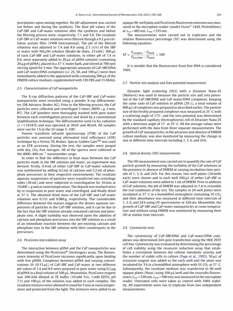
The growth in particle size and density of CaP particles canalso be determined by measuring the OD of CaP solutions asaddressed by Jordan (2000) and Jordan and Wurm (2004). TheOD, also referred as turbidity test, was quantified by measuringthe absorbance at 320 nm with a UV spectrophotometer. Normally,higher OD values are typical for mixtures with an increasing parti-cle size or/and an increasing number of particles, which render thesolution visibly opaque. Fig. 6 represents the OD values (320 nm) ofthe CaP particles incubated in the presence and absence of DMEMat different time periods of 1, 3, 6, and 24 h. In the presence ofDMEM (as illustrated in columns) and at pH value of 7.4, the ODvalues of the CaP-water solution were considerably higher thanCaP-SBF solution, indicating the higher turbidity (i.e. particle size)of the former. However, a somewhat similar trend was observedbetween the CaP-SBF and CaP-water solutions at higher pH valueof 8.0. Increasing the pH also resulted in higher OD values. The ODvalues (320 nm) of CaP particles at different time periods were alsomeasured at ambient temperature and without using the DMEMculture medium. The results have been represented in Fig. 6 withstraight lines and markers. The CaP-water (pH 7.4) showed aver-agely 23% increase in its OD values as compared to that of CaP-SBF(pH 7.4). It can also be seen that the turbidity of the CaP-water atpH of 7.4 was even higher than CaP-SBF at pH of 8.0. Likewise, theCaP-water (pH 8.0) presented the highest absorbance among the
samples. An increase in absorption at 320 nm indicates the growthof precipitates (or higher density), which could be confirmed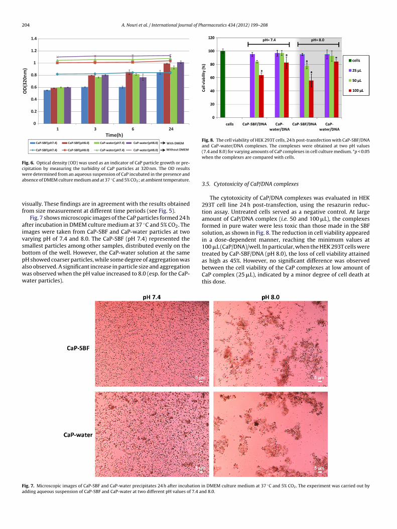
204 A. Nouri et al. / International Journal of Pharmaceutics 434 (2012) 199– 208
0
0.2
0.4
0.6
0.8
1
1.2
1.4
24631
OD(320
nm)
Time(h)CaP-SBF(pH7.4) Ca P-SBF(pH8.0) Ca P-water(pH7.4) Ca P-water(pH8.0)
CaP-SBF(pH7.4) Ca P-SBF(pH8.0) Ca P-water(pH7.4) Ca P-water(pH8.0) Without DMEM
With DMEM
Fig. 6. Optical density (OD) was used as an indicator of CaP particle growth or pre-cipitation by measuring the turbidity of CaP particles at 320 nm. The OD resultswa
vf
aivsbpaww
Fig. 8. The cell viability of HEK 293T cells, 24 h post-transfection with CaP-SBF/DNAand CaP-water/DNA complexes. The complexes were obtained at two pH values(7.4 and 8.0) for varying amounts of CaP complexes in cell culture medium. *p < 0.05
Fa
ere determined from an aqueous suspension of CaP incubated in the presence andbsence of DMEM culture medium and at 37 ◦C and 5% CO2; at ambient temperature.
isually. These findings are in agreement with the results obtainedrom size measurement at different time periods (see Fig. 5).
Fig. 7 shows microscopic images of the CaP particles formed 24 hfter incubation in DMEM culture medium at 37 ◦C and 5% CO2. Themages were taken from CaP-SBF and CaP-water particles at twoarying pH of 7.4 and 8.0. The CaP-SBF (pH 7.4) represented themallest particles among other samples, distributed evenly on theottom of the well. However, the CaP-water solution at the sameH showed coarser particles, while some degree of aggregation was
lso observed. A significant increase in particle size and aggregationas observed when the pH value increased to 8.0 (esp. for the CaP-ater particles).ig. 7. Microscopic images of CaP-SBF and CaP-water precipitates 24 h after incubation idding aqueous suspension of CaP-SBF and CaP-water at two different pH values of 7.4 an
when the complexes are compared with cells.
3.5. Cytotoxicity of CaP/DNA complexes
The cytotoxicity of CaP/DNA complexes was evaluated in HEK293T cell line 24 h post-transfection, using the resazurin reduc-tion assay. Untreated cells served as a negative control. At largeamount of CaP/DNA complex (i.e. 50 and 100 �L), the complexesformed in pure water were less toxic than those made in the SBFsolution, as shown in Fig. 8. The reduction in cell viability appearedin a dose-dependent manner, reaching the minimum values at100 �L (CaP/DNA)/well. In particular, when the HEK 293T cells weretreated by CaP-SBF/DNA (pH 8.0), the loss of cell viability attainedas high as 45%. However, no significant difference was observedbetween the cell viability of the CaP complexes at low amount of
CaP complex (25 �L), indicated by a minor degree of cell death atthis dose.n DMEM culture medium at 37 ◦C and 5% CO2. The experiment was carried out byd 8.0.

A. Nouri et al. / International Journal of Ph
Fig. 9. In vitro luciferase expression in HEK 293T cells transfected with CaP-SBF/DNAand CaP-water/DNA complexes prepared at two pH values of 7.4 and 8.0. Cells werehwt
3
w(im5dcso
aep7pnchc
pCiwwot
4
4
sptBa6Gbwi
arvested 48 h after transfection, and luciferase activities were determined. *p < 0.05hen the CaP-SBF/DNA complexes are compared to CaP-water/DNA complexes at
he same amount and pH value.
.6. In vitro transfection studies
The gene transfection efficiency of the CaP/DNA complexesas assessed in vitro using luciferase and GFP reporter genes
pEGFP-Luc) into HEK 293T cells. The CaP/DNA complexes weremmediately added to wells, containing 500 �L DMEM (supple-
ented with serum and antibiotic), at varying amounts (i.e. 25,0 and 100 �L) and incubated overnight. Luciferase activity wasetermined 48 h-post transfection and normalized against proteinontent (Fig. 9). The CaP/DNA amounts were chosen based on aeries of preliminary experiments to test the effect of CaP amountsn transfection efficiency.
Enhanced transfection efficiency was observed by increasingmount of CaP/DNA complexes, showing the highest transfectionfficiency at 100 �L for CaP-SBF (pH 7.4). The CaP-SBF/DNA com-lexes showed higher transfection efficiency at both pH values of.4 and 8.0, as compared to CaP-water/DNA complexes in similarH values. The difference in transfection efficiency was more pro-ounced at pH (8.0) between CaP-SBF/DNA and CaP-water/DNAomplexes at larger amount of CaP/DNA complex. Nevertheless,igher transfection efficiency was achieved when the CaP/DNAomplexes at pH (7.4) were used.
Fig. 10 shows fluorescence micrographs of the CaP/DNA com-lexes synthesized at two pH values (7.4 and 8.0) and three variousaP/DNA amounts of 25, 50 and 100 �L. The obtained fluorescence
mages were in agreement with the luciferase reporter assay data. Itas noticeable that the number of green fluorescent cells increasedith increasing amount of CaP/DNA complexes in wells. It was also
bserved that at higher amounts of CaP, larger number of cells wasransfected with CaP-SBF/DNA complexes than CaP-water/DNA.
. Discussion
.1. Characterization of CaP nanoparticles
The drop in pH values of the SBF solution and water after theynthesis of CaP particles lies in the formation of calcium andhosphate particles upon incorporation of hydroxyl groups (OH)o crystal lattice, which in fact favors the formation of brushite.rushite is the most predominant CaP phase often formed in morecidic aqueous solutions. Formation of brushite at pH lower than.5 has been reported in several studies (Cheng and Pritzker, 1983;
iocondi et al., 2010; Wang and Nancollas, 2008). Theoretically,rushite should form even at physiological pH and temperature,hen the product of the concentrations of total calcium and totalnorganic phosphate is sufficiently large (Cheng and Pritzker, 1983).
armaceutics 434 (2012) 199– 208 205
Brushite is endowed with an excellent biocompatibility, grows anddissolves readily, and has a comparatively fast nucleation rate dueto its low surface energy (Giocondi et al., 2010).
4.2. Size and zeta potential
The size and surface charge of particles are considered to be thekey factors for successful gene delivery into mammalian cells. Smalland positively charged particles are easily taken up by cells andwould lead to high gene expression (Chowdhury et al., 2004; Eppleet al., 2010). The surface charge of particles is mainly controlledby adsorption of multiply charged ions, the ionic strength of thesolution, pH, as well as the particle size and shape (Zhu et al., 2004).
The increase in ionic strength will reduce the Debye length, andwill subsequently shield the surface charge and reduce the zetapotential (Morgan, 2010). In an experiment with seawater, Beckettand Le (1990) noted that NaCl on its own cannot account for thereduction in surface charge of the suspended particles and the pres-ence of minor divalent cations such as Mg2+ and Ca2+ in the solutionare more effective in reducing the surface charge of the particlesthan the major monovalent cations like Na+. Thus, the lower zetapotential of the CaP-SBF particles and complexes can be contributedto the higher ionic strength and also multiply charged ions of theSBF solution when compared to pure water.
Calcium-deficient and/or relatively phosphate-rich condition ofthe CaP particles can also give rise to their net negative surfaces(Zhu et al., 2004). The magnitude of the negative zeta potentialincreases in response to the decrease in Ca/P ratio. In a theoreti-cal study performed by Bastos et al. (2008), interactions betweencalcium and two buffer systems, namely Tris and Bis-tris, wereinvestigated in standard and modified SBF solutions. The resultsrevealed that the Tris/Bis-tris-buffered SBF solutions reduce thecontent of available calcium ion, with a stronger trend towardsBis-tris. They reasoned that with buffer, the complexation reac-tion Ca2+ + PO4
3− � CaPO4− is slightly displaced towards to free
phosphate direction, making the phosphate more available. Theabove results had also been experimentally confirmed by Serroand Saramago (2003). They prepared a SBF solution with the samecomposition of Kokubo SBF (Kokubo and Takadama, 2006), butwithout the buffer Tris. It was shown that the Tris-buffered SBFsolution formed soluble complexes with several cations, includ-ing the most important Ca2+. Thus, it is envisaged that the Ca2+
ions chelated by Tris in the SBF solution lowers Ca/P molar ratioand consequently, affect the surface charge and binding abilities ofthe CaP-SBF particles as compared with CaP-water particles. How-ever, one should not expect a large difference in Ca/P molar ratiobetween the CaP-SBF and CaP-water particles since the XRD analy-ses of the both particles showed similar diffraction peaks revealingbrushite as their major peaks. Brushite has a Ca/P molar ratio of 1, incomparison with the stoichiometric value of 1.6 for hydroxyapatite.
The negatively charged pDNA reversed the initially positive orneutral surface charge of the CaP particles to negative values, asseen by drastically lower zeta potential of the CaP particles aftercomplexation with pDNA (Table 2). On the other hand, a fairly sim-ilar trend in surface charge variation of the particles with pH isindicating that the surface charges of the CaP particles after thesynthesis were not affected by the adsorption of H+ and OH− ions.It is believed that pDNA binding ability is associated with the sur-face charge of the particles. Thus, it comes as no surprise that themore positively charged CaP-water particles promote higher bind-ing ability to the negatively charged pDNA, as seen in Fig. 4.
4.3. Size and particle growth
The variation in size of CaP-SBF and CaP-water particlesstems from the difference in rate of CaP crystal growth between

206 A. Nouri et al. / International Journal of Pharmaceutics 434 (2012) 199– 208
F ransfe8 to ceA
SgmlcwoboIuptacai
Ctcorg(
ig. 10. Enhanced Green Fluorescent Protein (EGFP) expression in HEK 293T cells t.0. The CaP/DNA complexes were added in varying amounts of 25, 50 and 100 �LbAm). The fluorescence microscopy images were taken 48 h post-transfection.
BF- and water-mediated solutions. This uncontrollable rapidrowth of CaP crystals is one of the major limitations of the CaPethod, resulting in poor reproducibility and the formation of
arge aggregates (>�m) to considerably reduce the transfection effi-iency. Conditions which affect the solubility of CaP precipitatesould directly affect the rate of CaP crystal growth. The solubility
f CaP precipitates in a solution/culture medium, is in turn affectedy several parameters including ionic strength, the compositionf the solution, and the pH value of the solution (Jordan, 2000).ncreasing the NaCl content, i.e. ionic strength, enhances the sol-bility of the CaP precipitates and decrease its growth and rapidrecipitation, since NaCl simply reduces the driving force for reac-ion (Nancollas and Tomazic, 1974). Barrere et al. (2002) reported
delay in CaP precipitation on a Ti alloy soaked in a 5 times con-entrated SBF solution (SBF × 5), by increasing the ionic strength. Itppeared that increasing NaCl content lowers the nucleation sitesn the solution and thereby decrease the CaP precipitation.
The composition of a solution also affects the solubility ofaP precipitates. For instance, increasing the calcium concentra-ion decreases solubility, and lead to rapid precipitation of CaPrystals, especially at basic pH values (Jordan, 2000). As previ-
usly pointed out, Tris forms soluble complexes with Ca2+ andeduces the concentration of free Ca2+ ions in SBF. It can also inhibitrowth through physical adsorption on the surface of CaP crystalsKanzaki et al., 1998). In addition, the pH value of the Tris-bufferedcted using CaP-SBF/DNA and CaP-water/DNA complexes with pH values of 7.4 andll culture wells containing 500 �L of DMEM (supplemented with 10% FBS and 1%
solution remains at a stable, relatively low value (7.4 ± 0.1), hinder-ing CaP precipitation (Marques et al., 2003). In general, biologicalfluids contain ions and molecules that interfere with CaP precipita-tion in vitro. Molecules and ions are usually adsorbed on the surfaceof CaP precipitates and, thus, reducing the formation and/or growthof CaP critical nuclei and crystals (Blumenthal, 1989). As reportedby Lu and Leng (2005), the higher concentration of HCO3
− (27 mM)in SBF than pure water can also reduce the driving force for pre-cipitation. It is also very well known that the solubility of CaP ina solution increases with a decrease of pH (Elliot, 1994). Similarly,the lack of ionic strength and interfering ions in water-mediatedsolutions resulted in the rapid growth and aggregation of particlesover time (as seen in Fig. 7 for CaP-water particles). However, boththe CaP-SBF and CaP-water particles grew in size with increasingincubation time due to further consumption of the calcium andphosphate ions from the solution.
4.4. Cytotoxicity of CaP/DNA complexes
Although the CaP method is classified as a relatively safegene delivery system (since this is in the GRAS list of FDA) (Roy
et al., 2003), the CaP nanoparticles can adversely affect cell via-bility. In the CaP method, the inhibiting cell proliferation andcell damage typically continue in a dose- and time-dependentmanner and increase with higher amount of CaP nanoparticles
l of Ph
aotct
iSps(tambp(
wpcE(Sav
4
otftttlaemopctRie
Ccai
bfaiofoaawme
A. Nouri et al. / International Journa
nd longer incubation time (Motskin et al., 2009). Larger amountf CaP nanoparticles can produce too much Ca2+ for the cello survive. Cells need to maintain a very low intracellular Ca2+
oncentration, and an overload in cellular Ca2+ has been suggestedo be the pathway of cell death (Yu et al., 2001).
The chemical composition of the transfection medium plays anmportant role in cell survival. The lower cell viability of the CaP-BF/DNA than CaP-water/DNA complexes can be attributed to theresence of multiple ions and molecules in the SBF solution. In atudy on the effect of ions in dispersion of rat liver cells, Seglen1973) reported the maximum stimulations by higher concentra-ions of divalent cations such as Ca2+ and Mg2+, while K+, SO4
2−
nd HPO42− appeared to have less or no effect in inhibiting enzy-
atic dispersion. The buffer Tris, present in SBF, is also known toe inhibitory in certain biological systems due to the availability ofrimary amine and hydroxyl groups on Tris molecule for reactivityFernandez et al., 1993).
Although the precise pH value for optimal cell growth variesith the individual cell lines, any significant deviation from thehysiological pH range (i.e. 7.2–7.6) may lead to impediment ofell growth/proliferation and alter the metabolic rate (Ceccarini andagle, 1971), as seen by lower degree of cell viability at higher pH8.0) than pH (7.4) for both complexes and in particular for CaP-BF/DNA. As a result, the CaP-SBF/DNA complex at amounts of 25nd 50 �L/well at two different pHs lie in an acceptable level of celliability.
.5. In vitro transfection studies
Despite the lower DNA-binding ability and lower zeta potentialf CaP-SBF particles than CaP-water particles, in vitro transfec-ion studies on HEK 293T cells revealed higher gene expressionor the former. This is surprising since a decrease in zeta poten-ial represents a reduction in the electrostatic interaction betweenhe CaP particles and the pDNA. On the other hand, the lowerhe DNA-binding ability (lower extent of DNA condensation), theess effectively is the DNA bound to the CaP nanoparticles, whichlso results in low transfection efficiency. Therefore one mightxpect a different response in vitro. Such discrepancy could beainly attributed to the smaller particle size and lower degree
f particle growth in CaP-SBF particle as compared to CaP-waterarticles. In another words, the resulting lower transfection effi-iency in CaP-water/DNA complexes is indicating that most ofhe CaP-water/DNA complexes were too large to enter the cells.apid growth of the CaP-water particles resulted in sharp increase
n diameter which is a big hurdle for efficient gene delivery andxpression into the cells.
However, due to the more CaP nuclei per unit volume in the SBF-aP than CaP-water solution, it would be rational to expect thatovering cells with numerous small CaP/DNA complexes instead of
few larger ones increase the chances of pDNA to be transfectednto the nucleus.
The uncontrollable rapid growth of CaP particles always leads toulk and highly aggregated precipitates, which induces low trans-ection efficiencies (Wang et al., 2010). The results from DLS and ODt different incubation time periods revealed more physical stabil-ty for CaP-SBF solution at pH (7.4), reducing the rapid formationf coarse precipitates. Thus, the CaP-SBF/DNA complex is graduallyormed in the culture medium during incubation and precipitatesn the cells. Physical stability is an important advantage for in vivopplications, as nanoparticles do not have to be used immediately
fter preparation. In a high pH environment, the CaP/DNA mixtureill form coarse precipitates that aggregate into large clumps in theedium and significantly reduce the transfection efficiency (Chent al., 2008a).
armaceutics 434 (2012) 199– 208 207
The valuable aspect of SBF solution in gene delivery applicationswas its impact in reducing rapid particle growth and aggregation insolution and culture medium. The resulting formation of relativelysmall and stable CaP-SBF particles/complexes at longer incubationtime gave rise to high transfection efficiency in HEK 293T cell lineat physiological pH.
5. Conclusions
In an attempt to investigate the application of simulated bodyfluid (SBF) in gene delivery, calcium and phosphate (CaP) nanopar-ticles were synthesized in a SBF solution and mixed with pDNA.The in vitro gene transfection studies revealed that the pEGFP-Lucplasmid could effectively be transfected by the CaP-SBF/DNA com-plex at physiological pH (7.4). The results were also compared withthose using CaP particles made in pure water (CaP-water) via a sim-ilar procedure. Despite the lower DNA-binding ability and lowerzeta potential of CaP-SBF particles in comparison with CaP-waterparticles, the in vitro studies showed higher transfection efficiencyfor CaP-SBF/DNA complexes than for CaP-water/DNA. The SBF solu-tion showed the ability to reduce the rapid growth of CaP particlesover time, leading to smaller particles as compared to those synthe-sized in pure water. These findings provide new insights into therole of SBF solution in synthesis of CaP particles for gene deliveryapplications.
Acknowledgments
The Fundac ão para a Ciência e a Tecnologia (FCT, Portugal)is acknowledged for funding through the project PEst-OE/QUI/UI0674/2011 (CQM, Portuguese Government funds).The support of FCT through the Post-Doc grant awarded to A.Nouri (SFRH/BPD/47369/2008) is also acknowledged. Additionally,authors are grateful to Dr. T. Segura (UCLA, USA) and to INEB(Porto, Portugal) for kindly providing the EGFPLuc-pDNA and thecell line, respectively. The authors also wish to thank the supportof UMa, through the Santander bank Chair in Nanotechnology, andLREC (Laboratório Regional de Engenharia Civil–Madeira).
References
Barrere, F., van Blitterswijk, C.A., de Groot, K., Layrolle, P., 2002. Influence of ionicstrength and carbonate on the Ca–P coating formation from SBF × 5 solution.Biomaterials 23, 1921–1930.
Bastos, I.N., Platt, G.M., Andrade, M.C., Soares, G.D., 2008. Theoretical study of Trisand Bistris effects on simulated body fluids. J. Mol. Liq. 139, 121–130.
Beckett, R., Le, N.P., 1990. The role or organic matter and ionic composition in deter-mining the surface charge of suspended particles in natural waters. Colloids Surf.44, 35–49.
Bisht, S., Bhakta, G., Mitra, S., Maitra, A., 2005. pDNA loaded calcium phosphatenanoparticles: highly efficient non-viral vector for gene delivery. Int. J. Pharm.288, 157–168.
Blumenthal, N.C., 1989. Mechanisms of inhibition of calcification. Clin. Orthop. Relat.Res. 247, 279–289.
Ceccarini, C., Eagle, H., 1971. pH as a determinant of cellular growth and contactinhibition. Proc. Natl. Acad. Sci. U.S.A. 68, 229–233.
Chen, C.Y.A., Ezzeddine, N., Shyu, A.B., 2008a. Messenger RNA half-life measure-ments in mammalian cells. Methods Enzym. 448, 335–357.
Chen, X.B., Nouri, A., Li, Y.C., Lin, J.G., Hodgson, P.D., Wen, C.E., 2008b. Effect of surfaceroughness of Ti, Zr, and TiZr on apatite precipitation from simulated body fluid.Biotechnol. Bioeng. 101, 378–387.
Cheng, P.T., Pritzker, K.P.H., 1983. Solution Ca/P ratio affects calcium phosphatecrystal phases. Calcif. Tissue Int. 35, 596–601.
Chowdhury, E.H., Kunou, M., Nagaoka, M., Kundu, A.K., Hoshiba, T., Akaike, T., 2004.High-efficiency gene delivery for expression in mammalian cells by nanopre-cipitates of Ca–Mg phosphate. Gene 341, 77–82.
Elliot, J.C., 1994. Structure and Chemistry of the Apatites and Other CalciumOrthophosphates. Elsevier, Amsterdam, The Netherlands.
Epple, M., Ganesan, K., Heumann, R., Klesing, J., Kovtun, A., Neumann, S., Sokolova, V.,2010. Application of calcium phosphate nanoparticles in biomedicine. J. Mater.Chem. 20, 18–23.
2 l of Ph
F
G
G
J
J
K
K
K
K
L
M
M
M
N
08 A. Nouri et al. / International Journa
ernandez, R.D., Yoshimizu, M., Ezura, Y., Kimura, T., 1993. Comparative growthresponse of fish cell lines in different media, temperatures, and sodium chlorideconcentrations. Fish Pathol. 28, 27–34.
iocondi, J.L., El-Dasher, B.S., Nancollas, G.H., Orme, C.A., 2010. Molecular mecha-nisms of crystallization impacting calcium phosphate cements. Philos. Trans. R.Soc. A 368, 1937–1961.
raham, F.L., Van Der Eb, A.J., 1973. A new technique for the assay of infectivity ofhuman adenovirus 5 DNA. Virology 52, 456–467.
ordan, M., 2000. Transient gene expression in mammalian cells based on the cal-cium phosphate transfection method. In: Al-Rubeai, M. (Ed.), Cell Engineering:Transient Expression. Kluwer Academic Publishers, pp. 56–79.
ordan, M., Wurm, F.M., 2004. Transfection of adherent and suspended cells bycalcium phosphate. Methods 33, 136–143.
anzaki, N., Onuma, K., Ito, A., Teraoka, K., Tateishi, T., Tsutsumi, S., 1998. Directgrowth rate measurement of hydroxyapatite single crystal by moire phase shiftinterferometry. J. Phys. Chem. B 102, 6471–6476.
okubo, T., Kushitani, H., Sakka, S., Kitsugi, T., Yamamuro, T., 1990. Solutions able toreproduce in vivo surface-structure changes in bioactive glass–ceramic A–W. J.Biomed. Mater. Res. 24, 721–734.
okubo, T., Takadama, H., 2006. How useful is SBF in predicting in vivo bone bioac-tivity? Biomaterials 27, 2907–2915.
okubo, T., Takadama, H., 2007. Simulated body fluid (SBF) as a standard tool totest the bioactivity of implants. In: Epple, M., Baeuerlein, E. (Eds.), Handbookof Biomineralization: Medical and Clinical Aspects. Wiley-VCH, Weinheim, pp.97–108.
u, X., Leng, Y., 2005. Theoretical analysis of calcium phosphate precipitation insimulated body fluid. Biomaterials 26, 1097–1108.
arques, P.A.A.P., Serro, A.P., Saramago, B.J., Fernandes, A.C., Magalhães, M.C.F., Cor-reia, R.N., 2003. Mineralisation of two calcium phosphate ceramics in biologicalmodel fluids. J. Mater. Chem. 13, 1484–1490.
organ, T.T., 2010. Thesis: Synthesis and characterization of bioresorbable calciumphosphosilicate nanocomposite particles for fluorescence imaging and biomed-ical applications. Department of Chemistry, The Pennsylvania State University.
otskin, M., Wright, D.M., Muller, K., Kyle, N., Gard, T.G., Porter, A.E., Skepper, J.N.,
2009. Hydroxyapatite nano and microparticles: correlation of particle propertieswith cytotoxicity and biostability. Biomaterials 30, 3307–3317.ancollas, H., Tomazic, B., 1974. Growth of calcium phosphate on hydroxyap-atite crystals. Effect of supersaturation and ionic medium. J. Phys. Chem. 78,2218–2225.
armaceutics 434 (2012) 199– 208
Nouri, A., Chen, X.B., Hodgson, P.D., Long, J.M., Yamada, Y., Wen, C.E., 2007. Prepa-ration of bioactive porous Ti–Sn–Nb alloy for biomedical applications. In: 5thInternational Conference on Porous Metals and Metallic Foams (MetFoam), pp.307–310.
Orrantia, E., Chang, P.L., 1990. Intracellular distribution of DNA internalized throughcalcium phosphate precipitation. Exp. Cell Res. 190, 170–174.
Page, B., Page, M., Noel, C., 1993. A new fluorometric assay for cytotoxicity measure-ments in vitro. Int. J. Oncol. 3, 473–476.
Roy, I., Mitra, S., Maitra, A., Mozumdar, S., 2003. Calcium phosphate nanoparticlesas novel non-viral vectors for targeted gene delivery. Int. J. Pharm. 250, 25–33.
Santos, J.L., Pandita, D., Rodrigues, J., Pêgo, A.P., Granja, P.L., Tomás, H., 2011.Non-viral gene delivery to mesenchymal stem cells: methods, strategies andapplication in bone tissue engineering and regeneration. Curr. Gene Ther. 11,46–57.
Seelos, C., 1997. A critical parameter determining the aging of DNA–calcium-phosphate precipitates. Anal. Biochem. 245, 109–111.
Seglen, P.O., 1973. Preparation of rat liver cells. II. Effects of ions and chelators ontissue dispersion. Exp. Cell Res. 76, 25–30.
Serro, A.P., Saramago, B., 2003. Influence of sterilization on the mineralization oftitanium implants induced byincubation in various biological model fluids. Bio-materials 24, 4749–4760.
Sokolova, V.V., Radtke, I., Heumann, R., Epple, M., 2006. Effective transfection ofcells with multi-shell calcium phosphate-DNA nanoparticles. Biomaterials 27,3147–3153.
Tas, A.C., 2000. Synthesis of biomimetic Ca-hydroxyapatite powders at 37 ◦C in syn-thetic body fluids. Biomaterials 21, 1429–1438.
Wang, K.W., Zhou, L.Z., Sun, Y., Wu, G.J., Gu, H.C., Duan, Y.R., Chen, F., Zhu, Y.J., 2010.Calcium phosphate/PLGA-mPEG hybrid porous nanospheres: a promising vec-tor with ultrahigh gene loading and transfection efficiency. J. Mater. Chem. 20,1161–1166.
Wang, L., Nancollas, G.H., 2008. Calcium orthophosphates: crystallization and dis-solution. Chem. Rev. 108, 4628–4669.
Welzel, T., Radtke, I., Meyer-Zaika, W., Heumann, R., Epple, M., 2004. Transfectionof cells with custom-made calcium phosphate nanoparticles coated with DNA.
J. Mater. Chem. 14, 2213–2217.Yu, S.P., Canzoniero, L.M.T., Choi, D.W., 2001. Ion homeostasis and apoptosis. Curr.Opin. Cell Biol. 13, 405–411.
Zhu, P., Masuda, Y., Koumoto, K., 2004. The effect of surface charge on hydroxyapatitenucleation. Biomaterials 25, 3915–3921.