international, evidence-based consensus treatment ... · goodman,21 christian hoffmann, 22 pier...
TRANSCRIPT
doi:10.1182/blood-2018-07-862334Prepublished online September 4, 2018;
Corey Casper, Eric Oksenhendler and David C. FajgenbaumWong, Jean-Francois Rossi, Sunita Nasta, Kazuyuki Yoshizaki, Razelle Kurzrock, Thomas S. Uldrick,Hoffmann, Pier Luigi Zinzani, Simone Ferrero, Louis Terriou, Yasuharu Sato, David Simpson, Raymond Amy Chadburn, Megan S. Lim, Kojo S. Elenitoba-Johnson, Vera Krymskaya, Aaron Goodman, ChristianShanmuganathan Chandrakasan, Raj Jayanthan, Elaine S. Jaffe, Heather Leitch, Naveen Pemmaraju, Mukherjee, Dustin Shilling, Katie Stone, Amy Greenway, Jason Ruth, Mary Jo Lechowicz,Nikhil Munshi, Stephen Schey, Matthew Streetly, Sheila K. Pierson, Helen L. Partridge, Sudipto Frits van Rhee, Peter Voorhees, Angela Dispenzieri, Alexander Fosså, Gordan Srkalovic, Makoto Ide, idiopathic multicentric Castleman diseaseInternational, evidence-based consensus treatment guidelines for
http://www.bloodjournal.org/site/misc/rights.xhtml#repub_requestsInformation about reproducing this article in parts or in its entirety may be found online at:
http://www.bloodjournal.org/site/misc/rights.xhtml#reprintsInformation about ordering reprints may be found online at:
http://www.bloodjournal.org/site/subscriptions/index.xhtmlInformation about subscriptions and ASH membership may be found online at:
digital object identifier (DOIs) and date of initial publication. indexed by PubMed from initial publication. Citations to Advance online articles must include final publication). Advance online articles are citable and establish publication priority; they areappeared in the paper journal (edited, typeset versions may be posted when available prior to Advance online articles have been peer reviewed and accepted for publication but have not yet
Copyright 2011 by The American Society of Hematology; all rights reserved.Hematology, 2021 L St, NW, Suite 900, Washington DC 20036.Blood (print ISSN 0006-4971, online ISSN 1528-0020), is published weekly by the American Society of
For personal use only.on September 6, 2018. by guest www.bloodjournal.orgFrom
1
International, Evidence-based Consensus Treatment Guidelines for Idiopathic Multicentric
Castleman Disease
Frits van Rhee,1 Peter Voorhees,2 Angela Dispenzieri,3 Alexander Fosså,4 Gordan Srkalovic,5 Makoto
Ide,6 Nikhil Munshi,7 Stephen Schey,8 Matthew Streetly,8 Sheila K. Pierson,9 Helen L. Partridge,9 Sudipto
Mukherjee,10 Dustin Shilling,9 Katie Stone,1 Amy Greenway,1 Jason Ruth,11 Mary Jo Lechowicz,12
Shanmuganathan Chandrakasan,13 Raj Jayanthan,14 Elaine S. Jaffe,15 Heather Leitch,16 Naveen
Pemmaraju,17 Amy Chadburn,18 Megan S. Lim,19 Kojo S. Elenitoba-Johnson,19 Vera Krymskaya,20 Aaron
Goodman,21 Christian Hoffmann,22 Pier Luigi Zinzani,23 Simone Ferrero,24 Louis Terriou,25 Yasuharu
Sato,26 David Simpson,27 Raymond Wong,28 Jean-Francois Rossi,29 Sunita Nasta,30 Kazuyuki
Yoshizaki,31 Razelle Kurzrock,32 Thomas S. Uldrick,33 Corey Casper,34 Eric Oksenhendler,35 and David
C. Fajgenbaum9
1Myeloma Center, University of Arkansas for Medical Sciences, Little Rock, AR; 2Department of
Hematologic Oncology and Blood Disorders, Levine Cancer Institute, Atrium Health, Charlotte, NC;
3Division of Hematology/Oncology, Mayo Clinic, Rochester, MN; 4Department of Oncology, Oslo
University Hospital–Norwegian Radium Hospital, Oslo, Norway; 5Herbert-Herman Cancer Center,
Michigan State University College of Human Medicine, Lansing, MI; 6Department of Hematology,
Takamatsu Red Cross Hospital, Takamatsu, Kagawa, Japan; 7Dana Farber Cancer Institute, VA Boston
Healthcare System, Harvard Medical School, Boston, MA; 8Guys and St. Thomas' NHS Foundation
Trust, London, United Kingdom; 9Division of Translational Medicine and Human Genetics, Perelman
School of Medicine, University of Pennsylvania, Philadelphia, PA; 10Department of Hematology and
Medical Oncology, Taussig Cancer Institute, Cleveland Clinic, Cleveland, OH, USA; 11Department of
Medical Oncology, Dana-Farber Cancer Institute, Harvard Medical School, Boston, MA; 12Department of
Hematology and Medical Oncology, Emory University School of Medicine, Atlanta, GA; 13Aflac Cancer
and Blood Disorders Center, Children’s Healthcare of Atlanta, Emory University, Atlanta, GA;
Blood First Edition Paper, prepublished online September 4, 2018; DOI 10.1182/blood-2018-07-862334
Copyright © 2018 American Society of Hematology
For personal use only.on September 6, 2018. by guest www.bloodjournal.orgFrom

2
14Department of Medicine, Baylor College of Medicine, Houston, TX; 15National Cancer Institute,
National Institutes of Health, Bethesda, MD; 16Division of Hematology, Department of Medicine, St.
Paul’s Hospital, University of British Columbia, Vancouver, BC, Canada; 17 Department of Leukemia,
Division of Cancer Medicine, The University of Texas MD Anderson Cancer Center, Houston, TX;
18Department of Pathology, Weill Cornell Medical College, New York, NY; 19Department of Pathology
and Laboratory Medicine, Perelman School of Medicine, University of Pennsylvania, Philadelphia, PA; 20
Penn Center for Pulmonary Biology, Pulmonary, Allergy, and Critical Care Division, Perelman School of
Medicine, University of Pennsylvania, Philadelphia, PA; 21Department of Medicine, Division of
Hematology/Oncology, University of California San Diego, La Jolla, CA, USA; 22University of
Schleswig Holstein, Campus Kiel, Kiel, and ICH Study Center, Hamburg, Germany; 23Institute of
Hematology "L. e A. Seràgnoli", University of Bologna, Bologna, Italy; 24Division of Hematology,
Department of Molecular Biotechnologies and Health Sciences University of Torino/AOU “Città della
Salute e della Scienza di Torino”, Turin, Italy; 25Department of Internal Medicine - Hematology, Hôpital
Claude Huriez - CHRU Lille, Lille, France; 26 Division of Pathophysiology, Okayama University
Graduate School of Health Sciences, Okayama, Japan; 27North Shore Hospital, Auckland, New Zealand;
28Sir Y.K. Pao Centre for Cancer and Department of Medicine & Therapeutics, Prince of Wales Hospital,
The Chinese University of Hong Kong, Hong Kong; 29Department of Hematology, CHU de Montpellier,
Montpellier, France. INSERM U1040, Montpellier, France. Université Montpellier I, Montpellier, France;
30Division of Hematology/Oncology, Perelman School of Medicine, University of Pennsylvania,
Philadelphia, PA; 31Department of Organic Fine Chemicals, The Institute of Scientific and Industrial
Research, Osaka University, Osaka, Japan; 32Center for Personalized Therapy and Clinical Trials Office,
UC San Diego Moore’s Cancer Center, La Jolla, CA; 33Fred Hutch Global Oncology, Fred Hutchinson
Cancer Research Center, Seattle, WA; 34Infectious Disease Research Institute and the Departments of
Medicine and Global Health, University of Washington, Seattle, WA; 35Department of Clinical
Immunology, Hôpital Saint-Louis, Paris, France
For personal use only.on September 6, 2018. by guest www.bloodjournal.orgFrom

3
Research Support: None
Corresponding author:
Frits van Rhee MD PhD MRCP(UK) FRCPath FACP
Professor of Medicine & Clinical Director,
Charles and Clydene Scharlau Chair for Hematologic Malignancies Research,
Myeloma Center
University of Arkansas for Medical Sciences,
Mail slot 816
Little Rock, AR 72205
E-mail: [email protected]
Fax: (501) 526-2273
Phone: (501) 804-7020 (cell); (501) 526-2873 (office)
Running Head: Therapy for Idiopathic Castleman Disease
Word Count Abstract: 247
Word Count Text: 3966
Table Count: 3
Figure Count: 3
Reference Count: 87
Disclaimers: The Castleman Disease Collaborative Network (CDCN) coordinated the meetings during
which the treatment guidelines were developed. The authors of the guidelines had full responsibility for
For personal use only.on September 6, 2018. by guest www.bloodjournal.orgFrom

4
the consensus-building process/methods, data interpretations, treatment recommendations, and writing of
the report.
For personal use only.on September 6, 2018. by guest www.bloodjournal.orgFrom

5
Abstract
Castleman Disease (CD) describes a group of heterogeneous hematologic disorders with characteristic
histopathological features. CD can present with unicentric or multicentric (MCD) regions of lymph node
enlargement. Some cases of MCD are caused by human herpesvirus-8 (HHV-8) whereas others are HHV-
8-negative/idiopathic (iMCD). Treatment of iMCD is challenging and outcomes can be poor since no
uniform treatment guidelines exist, few systematic studies have been conducted, and no agreed upon
response criteria have been described. The purpose of this paper is to establish consensus, evidence-based
treatment guidelines based on the severity of iMCD to improve outcomes. An international Working
Group of 42 experts from ten countries was convened by the Castleman Disease Collaborative Network to
establish consensus guidelines for the management of iMCD based on published literature, review of
treatment effectiveness for 344 cases and expert opinion. The anti-interleukin-6 monoclonal antibody
siltuximab (or tocilizumab, if siltuximab is not available) with or without corticosteroids is the preferred
first-line therapy for iMCD. In the most severe cases, adjuvant combination chemotherapy is
recommended. Additional agents are recommended, tailored by disease severity, as second- and third-line
therapies for treatment failures. Response criteria were formulated to facilitate the evaluation of treatment
failure or success. These guidelines should help treating physicians to stratify patients based on disease
severity in order to select the best available therapeutic option. An international registry for patients with
CD (ACCELERATE, NCT02817997) was established in October 2016 to collect patient outcomes to
increase the evidence base for selection of therapies in the future.
For personal use only.on September 6, 2018. by guest www.bloodjournal.orgFrom

6
Introduction
Dr. Benjamin Castleman described the first case of Castleman Disease (CD) involving a single lymph
node station, which is now referred to as unicentric CD (UCD).1 Characteristic histopathological features
observed in CD lymph nodes include hyaline vascular (HV), plasmacytic (PC), and mixed variants.2,3 CD
was later observed to affect multiple lymph node stations, which is known as multicentric CD (MCD).4 In
1995, Human herpesvirus 8 (HHV-8) was found to be the etiologic agent of a plasmablastic variant of
MCD occurring most commonly in HIV-infected or otherwise immunocompromised individuals.5-10 In
HHV-8-associated MCD, viral interleukin-6 (IL-6), a homologue of human IL-6, promotes a pro-
inflammatory state accounting for clinical symptomatology and laboratory abnormalities such as anemia,
hypoalbuminemia, and elevated C-reactive protein (CRP). In HHV-8-negative MCD, which comprises
33-58% of MCD cases, human IL-6 is the most common pathological driver, but the exact etiology is
unknown; this entity is also referred to as “idiopathic MCD” (iMCD).11-15 We have proposed four
etiological hypotheses, including autoimmune, auto-inflammatory, neoplastic, and pathogenic
mechanisms, which are now being actively investigated through the Castleman Disease Collaborative
Network (CDCN).12,16
The presentation of iMCD is quite varied with some patients having mild constitutional symptoms,
whilst others develop a life-threatening cytokine storm, organ failure, and death. The diverse clinical
presentation calls for a treatment stratagem that takes into account the severity of the disease. Further
complicating treatment recommendations is the existence of distinct iMCD sub-types. Some patients
experience thrombocytopenia (T), anasarca (A), fever (F), reticulin fibrosis of the bone marrow (R), and
organomegaly (O), but generally have normal gamma-globulin levels, which has recently been referred to
as the TAFRO subtype of iMCD.17,18 Other patients have more classic iMCD with features attributed to
IL-6 excess such as thrombocytosis and hyper-gammaglobulinemia, but less extreme anasarca.18,19 The
TAFRO subtype often has more severe clinical symptomatology and worse outcome.18,20-22 We and others
have reported that TAFRO patients have highly vascular lymph nodes and exhibit a different cytokine
For personal use only.on September 6, 2018. by guest www.bloodjournal.orgFrom
7
spectrum with elevated vascular endothelial growth factor (VEGF) levels, but milder elevation of IL-6.23-
25
Four recent studies in HIV-negative HHV-8 status unknown (believed to be HHV-8-negative) MCD
reported 5-year overall survival (OS) rates of 55-77%, reminiscent of the outcomes of malignant
disorders, though a large series from tertiary specialty centers reported one-year survival exceeding
90%.11,26-30 The poor outcome of iMCD may be due to several factors. First, there were no diagnostic
criteria for iMCD prior to 2017, when the CDCN published the first-ever consensus diagnostic criteria for
iMCD.23 Second, iMCD is a complex orphan disease with an incidence of 1,000-1,500 cases in the
USA.31 Consequently, few physicians have substantive experience managing iMCD and clinical trials are
difficult to conduct. Third, there is a paucity of systematic studies to guide the treating physician, further
compounded by the lack of uniform response criteria, hampering evaluation of treatment efficacy. Finally,
there are no existing recommendations on how to utilize available treatment modalities in the context of
disease severity.
iMCD has been treated with a wide variety of agents including corticosteroids, rituximab, and
chemotherapies. More recently, monoclonal antibodies (mAbs) targeting IL-6 directly (siltuximab) or the
IL-6 receptor (tocilizumab) have been approved for iMCD therapy.32,33 However, a significant proportion
of patients do not benefit from anti-IL-6 mAbs and additional therapeutic options are needed for non-
responders, especially severely-afflicted patients. Herein, we establish comprehensive guidance on the
treatment of iMCD based on review of data from 344 patients, published literature, and expert opinion
provided by a panel of physicians from the CDCN. The management of HHV-8-associated MCD and
POEMS syndrome-associated MCD is well-established and has been reported elsewhere. 34-41
Methods
An international group of 42 participants from the USA, Japan, China, France, United Kingdom,
Germany, Italy, Canada, Norway, and New Zealand, comprised of experts in Hematology/Oncology,
For personal use only.on September 6, 2018. by guest www.bloodjournal.orgFrom

8
Hematopathology, Infectious Diseases, and Immunology, as well as two physician-patients with iMCD,
embarked on the establishment of treatment guidelines for iMCD. The Working Group first met in
December 2016 with a follow-up meeting in December 2017. Three additional web-based teleconferences
were held in August 2017, November 2017, and March 2018. All relevant English language publications
from 1954 to 2017 were identified through PUBMED and other databases using as MESH headings
Castleman Disease, Multicentric, and TAFRO. All age groups, including pediatric cases, were included.
Clinical trials conducted with siltuximab (NCT00412321, NCT01024036 and NCT01400503) and
tocilizumab were also reviewed. Five large datasets as well as individual case reports (see
Supplementary Appendix 1) served as the primary evidence base.11,21,32,33,39
Based on the panel’s expert opinion, the impact of different therapeutic interventions was assessed in
the context of disease severity, and recommendations for classification of severity and response criteria
for evaluation of treatment were derived from the literature. The consensus focused on three main topics:
(1) development of iMCD severity criteria, (2) treatment of iMCD, and (3) development of iMCD
response criteria. Categories of evidence and consensus were modeled after those developed by the
National Comprehensive Cancer Network
(https://www.nccn.org/professionals/physician_gls/categories_of_consensus.aspx). A modified Delphi
process comprising the integration of evidence provided by the literature and expert opinions was used to
generate the final consensus statement contained in this paper, which was approved by all authors.
Data Sharing Statement: All data reviewed for the purposes of generating the consensus criteria was
sourced from publicly-available journal articles. A table describing the aggregate data as well as outcome
calculations is available as a supplemental appendix.
Results
Management of iMCD
For personal use only.on September 6, 2018. by guest www.bloodjournal.orgFrom

9
To serve as the evidence base for the development of iMCD management guidelines, a dataset of
iMCD clinical cases (n = 344) and treatment regimens (n = 479) was assembled (summarized in Table 1,
complete dataset in Supplementary Appendix 1).
Evaluation of iMCD Severity. If a patient is suspected to have iMCD, a comprehensive set of testing
is recommended to determine if the patient meets the consensus iMCD diagnostic criteria and assess
disease severity (Table 2).23 Laboratory testing for inflammatory markers and organ dysfunction are
indicated. Computed tomography (CT) should be performed to visualize the extent of the disease; CT-
positron emission tomography (PET) scanning is a useful alternative and high standardized uptake values
(>6) should raise the suspicion of an alternative diagnosis e.g. lymphoma. The severity of iMCD spans a
wide spectrum with some patients exhibiting mild symptomatology, while others experience life-
threatening organ failure. Based on expert opinion and review of the evidence base, we recommend
assessing the severity of iMCD according to simple criteria (Figure 1) to inform the appropriate treatment
choice as defined in Figure 2. These criteria are intended to segment patients according to their
performance status and extent of organ dysfunction into two broad categories: non-severe and severe.
Patients with severe iMCD have evidence of organ dysfunction such as renal failure, anasarca, severe
anemia, and pulmonary dysfunction resulting in poor performance status likely requiring critical care.
Laboratory features include very high CRP levels ( 100g/dL), marked hypoalbuminemia ( 2.0g/dL), and
thrombocytopenia ( 100 x 1012/L). Patients with lymphocytic interstitial pneumonitis can also progress to
end-stage pulmonary fibrosis if inadequately treated.
Non-severe iMCD. iMCD patients who are not severely sick are typically diagnosed in the outpatient
setting and have a good performance status without evidence of abnormal organ function, whilst other
patients are more symptomatic and often exhibit an IL-6-driven inflammatory response that interferes
significantly with their ability to function and work. Clinical symptoms may be intense enough to require
hospitalization, albeit not in intensive care.
For personal use only.on September 6, 2018. by guest www.bloodjournal.orgFrom

10
We recommend (category 1) using anti-IL-6 mAb therapy with siltuximab (11mg/kg every three
weeks) for all patients with non-severe iMCD based on the high proportion of responders, the rigorous
nature of the studies underlying the evidence base, and the low side effect profile relative to other
interventions. Siltuximab, which has been evaluated in a Phase I trial (n = 34), a long-term safety study (n
= 19), and a randomized, double-blind placebo-controlled Phase II trial (n =79), is presently approved in
the USA, Canada, EU, and Brazil, among other countries.33,42-47 In the Phase II study, the only
randomized controlled trial performed in iMCD to date, 79 patients were allocated to siltuximab 11mg/kg
every three weeks or placebo. Durable tumor and symptomatic responses were achieved in 18 of 53
patients in the siltuximab arm [34%; one complete response (CR), 17 partial responses (PRs)] versus zero
of 26 in the placebo arm. Nearly 60% of patients had a durable symptomatic response and 31 patients
continued to receive unblinded siltuximab.33 While elevated pre-treatment IL-6 levels are associated with
a trend towards an increased likelihood of response to siltuximab, IL-6 levels should not be used to guide
treatment decisions. In the Phase II trial, there were iMCD patients with low/normal IL-6 levels who
responded to siltuximab while others with high IL-6 levels did not.45
If siltuximab is not available, tocilizumab (8mg/kg every two weeks) may be used (category 2A).
Tocilizumab, which has undergone an open-label, non-randomized prospective study of 35 patients and
been reported extensively in the literature, is approved for the treatment of iMCD in Japan. Like
siltuximab, responding patients showed improvement in constitutional symptoms, normalization of
abnormal laboratory markers such as CRP, hemoglobin, albumin, and IgG, and reduction in
lymphadenopathy with few significant adverse events.32,48,49 The most common side effects of both
siltuximab and tocilizumab are mild thrombocytopenia, hypertriglyceridemia, hypercholesteremia, and
pruritus. The availability of siltuximab and tocilizumab varies amongst countries, and the choice between
the two drugs is currently more dependent on indication within that country and access, as no head-to-
head trials have been performed to compare efficacy.
For personal use only.on September 6, 2018. by guest www.bloodjournal.orgFrom

11
If needed, first-line therapy with anti-IL6 mAb should be accompanied by corticosteroid therapy for
initial disease control. The existing data on corticosteroid monotherapy do not support its use due to
limited long-term control and frequent relapses, except in countries where there is no access to mAb
therapy.11,29,50-55 Combining data from published series, we noted a high treatment failure rate at 54%
(Table 1). Nevertheless, corticosteroids can augment iMCD symptom control along with anti-IL-6
mAbs.32,33 Patients with more indolent disease can be treated with lower doses of adjunctive
corticosteroids (e.g. prednisone 1mg/kg, or equivalent for 4-8 weeks followed by tapering; category 2B),
whilst patients who are more symptomatic may require higher initial doses of corticosteroids (e.g.
methylprednisolone 2mg/kg or equivalent) and more gradual tapering.
Careful inspection of the siltuximab and tocilizumab data and the clinical experience of the expert
panel suggests that patients with a clear inflammatory syndrome as manifested by symptomatology and
biochemical abnormalities are most likely to derive benefit from anti-IL-6 mAb therapy. In the
tocilizumab studies virtually all patients had increases in CRP, ESR, and fibrinogen as well significant
anemia and hypoalbuminemia.32,48,49 Though no formal response criteria were employed, 86% of patients
remained on therapy for at least five years.49 In contrast, the symptomatic response rates in patients
treated with siltuximab were about 60%, and the combined stringently-defined endpoint of durable
symptomatic and lymph node response was 34%. However, the patients in the siltuximab arm of the
randomized trial were less severely-affected as reflected by low scores on the MCD symptom scale as
well as modest elevations in CRP and fibrinogen, and a median serum albumin that was in the normal
range.33,45,47 Strict exclusion criteria for organ dysfunction and patient selection bias in the randomized
siltuximab trial due to a placebo arm likely contributed to the milder phenotype in these patients. Of note,
ad hoc analysis of the Phase II data revealed that patients demonstrating more clinical and laboratory
abnormalities included in the minor criteria of the iMCD diagnostic criteria had a greater response rate
than those who did not.23
For personal use only.on September 6, 2018. by guest www.bloodjournal.orgFrom

12
Responses in clinical symptomatology occur rapidly with anti-IL-6 mAb therapy and should be
apparent after 3-4 doses.32,46 Laboratory indicators including hemoglobin, CRP, ESR, and albumin should
mirror clinical improvements and be followed initially weekly and then bi-weekly until
normalization.33,42,45 Of note, both siltuximab and tocilizumab give rise to spuriously elevated IL-6 levels
for 18-24 months following the last dose. Therefore, serum IL-6 levels should not be used to assess
response.14 Resolution of lymphadenopathy can be slow with anti-IL-6 mAb therapy with a median time
to lymph node response of five months.33,46 This is because anti-IL-6 mAbs merely abrogate an important
growth signal for lymphocytes and plasma cells, but do not have direct cytotoxic effects. Early response
to therapy should be judged using the criteria provided in Figure 3, defining symptomatic and
biochemical response rather than relying on reduction in lymph node size. Patients should be followed by
serial CT-scanning every three months until maximum response has occurred, after which the frequency
of imaging can be reduced to six and later 12 months.
Clinical experience of the expert working group with siltuximab and data reported by Nishimoto et al.
for tocilizumab suggest that relapses occur on cessation of therapy.49 Indefinite continuation of anti-IL6
mAb therapy in responding patients is therefore recommended. However, dosing intervals were safely
extended to six weeks in 40% of iMCD patients in the long-term safety study of siltuximab, suggesting
that dosing may be spaced out in some patients.44 If used in combination with other agents, steroids
should be discontinued as early as possible to minimize side-effects.
We recommend rituximab (375mg/m2 x 4-8 doses) as a first-line alternative to anti-IL6 mAb therapy
for patients with non-severe iMCD who do not have marked cytokine-driven symptomatology based on a
more limited dataset, since rituximab has not been subjected to systematic study in iMCD and data are
confined to case reports or small series (category 2B evidence).56-61 Most papers report the use of
rituximab along with conventional chemotherapies; Table 1 presents combined data on cytotoxic
chemotherapy, which often includes rituximab as a component. In a recently-published study of iMCD
patients, the CR and PR rates with rituximab or rituximab-based chemotherapy regimens as first-line
For personal use only.on September 6, 2018. by guest www.bloodjournal.orgFrom

13
therapy were 20% and 48%, respectively. Rituximab-treated patients had inferior progression-free
survival compared to those managed with siltuximab.21 In two further series, approximately half of the
iMCD patients failed rituximab.11,39 Despite the lack of rigorous evaluation, the available data and expert
opinion do support a role for rituximab monotherapy in the treatment of non-severe iMCD patients for
whom it would be reasonable to give a limited course of therapy rather than indefinite anti-IL-6 mAb
treatment.
It is important to note that approximately 50% of iMCD patients will not achieve a satisfactory
response to first-line anti-IL6 therapy. Failure to achieve a satisfactory response, defined as PR or CR
(Figure 3), to first-line therapy should prompt re-evaluation of the original diagnosis to rule out an
alternative diagnosis such as lymphoma. Anti-IL6 mAb treatment does not need to be continued if it was
not effective in first-line. Second-line therapy should comprise rituximab to which
immunomodulatory/immunosuppressive agents (see below) and steroids may be added. Thalidomide has
been combined with rituximab and steroids since it downregulates IL-6 expression and has anti-
angiogenic properties by modulating VEGF. Thalidomide has induced remissions in iMCD as a single
agent and has also been valuable in combination with rituximab in both HHV-8-associated MCD and
iMCD (Schey, personal communication).62-64
Third-line therapy for patients who fail both anti-IL-6 mAbs and rituximab is less well-defined.
Cytotoxic chemotherapies have a high response rate in our pooled data analysis (78%), but treatment
failure with relapses are common (42%) and toxicities are significant (Table 1). Therefore, the consensus
opinion is to avoid cytotoxic chemotherapy unless the patient progresses to severe iMCD. We recommend
use of an immunomodulatory/immunosuppressive agent since these agents have less toxicity than
chemotherapy and have similar efficacy (69% response), albeit in fewer case reports. 62-64,66-71,72 These
agents include cyclosporine A, sirolimus, thalidomide, lenalidomide, bortezomib, the IL-1 receptor
antagonist anakinra, retinoic acid derivatives, and interferon- .62,63,65-72,73 Cyclosporine A has been used
most extensively in iMCD-TAFRO cases, particularly to improve persistent ascites and
For personal use only.on September 6, 2018. by guest www.bloodjournal.orgFrom

14
thrombocytopenia.20,74-77 Anakinra, which blocks the IL-1 receptor and presumably NF-kB signaling,
has been reported as successful treatment of a siltuximab-refractory iMCD patient.67,68
Severe iMCD: How to Treat the Critically Ill Patient. Based on published data, the proportion of
patients with severe iMCD, who have marked organ dysfunction, poor performance status, and require
critical care, is estimated to be 10-20%.11,21 These patients should be promptly started on a high-dose
steroid regimen (e.g. methylprednisolone 500mg daily) together with siltuximab. For pharmacokinetic
reasons, an accelerated, weekly dosing schedule of siltuximab may be used for one month. Patients who
immediately respond should continue on siltuximab at every three week intervals indefinitely and slowly
taper steroids.
There is consensus in the Working Group that patients with severe iMCD are at significant risk of
mortality and expert advice should be sought. Severe iMCD may not respond immediately to high-dose
steroids and anti-IL-6 mAbs, which can take weeks to achieve steady state concentration. Still others may
never respond to anti-IL-6 mAbs. Therefore, aggressive intervention with multi-agent chemotherapy
should be considered as early as necessary (any sign of deterioration or after one week of no response to
siltuximab, whichever comes first) to ablate the hyperactivated immune system and stem the cytokine
storm. Chemotherapy regimens including those for lymphoma: R-CHOP (rituximab, cyclophosphamide,
doxorubicin, vincristine, prednisone), CVAD (cyclophosphamide, vincristine, doxorubicin,
dexamethasone), or CVP (cyclophosphamide, vincristine, prednisone); myeloma: VDT-ACE-R
(bortezomib, dexamethasone, thalidomide, doxorubicin, cyclophosphamide, etoposide, rituximab); or
etoposide/cyclophosphamide-containing regimens as used for hemophagocytic lymphohistiocytosis have
all been employed.18,50,54,78 Combination chemotherapy is appropriate in poor performance status patients,
including those requiring treatment in the intensive care unit, as control of the cytokine storm can be life-
saving and bring about rapid improvement. As per Table 1, cytotoxic chemotherapy has the highest
overall response rate (78%), but considerable toxicities and frequent relapses deter its use outside of the
most severe setting when the risk/benefit analysis is skewed.29,52,79
For personal use only.on September 6, 2018. by guest www.bloodjournal.orgFrom

15
The subsequent management of severe iMCD patients who fail to respond to anti-IL-6 mAb or the
first cytotoxic chemotherapy regimen, or those who relapse, is not well-defined and is mostly done on an
ad hoc basis taking into account any previous response, clinical status, co-morbidities, and cytokine
profile. Patients who have elevation of IL-6 prior to starting anti-IL-6 mAb therapy may still benefit from
extended therapy with anti-IL-6 mAb, even if they did not respond during the acute episode, whilst others
may respond to immunomodulators/immunosuppressants or salvage cytotoxic therapy more commonly
used in plasma cell malignancies e.g. VTD (bortezomib, thalidomide, dexamethasone). Autologous and
allogeneic stem cell transplantation have only been reported in a few cases with mixed results and are
therapies of last resort.80-83
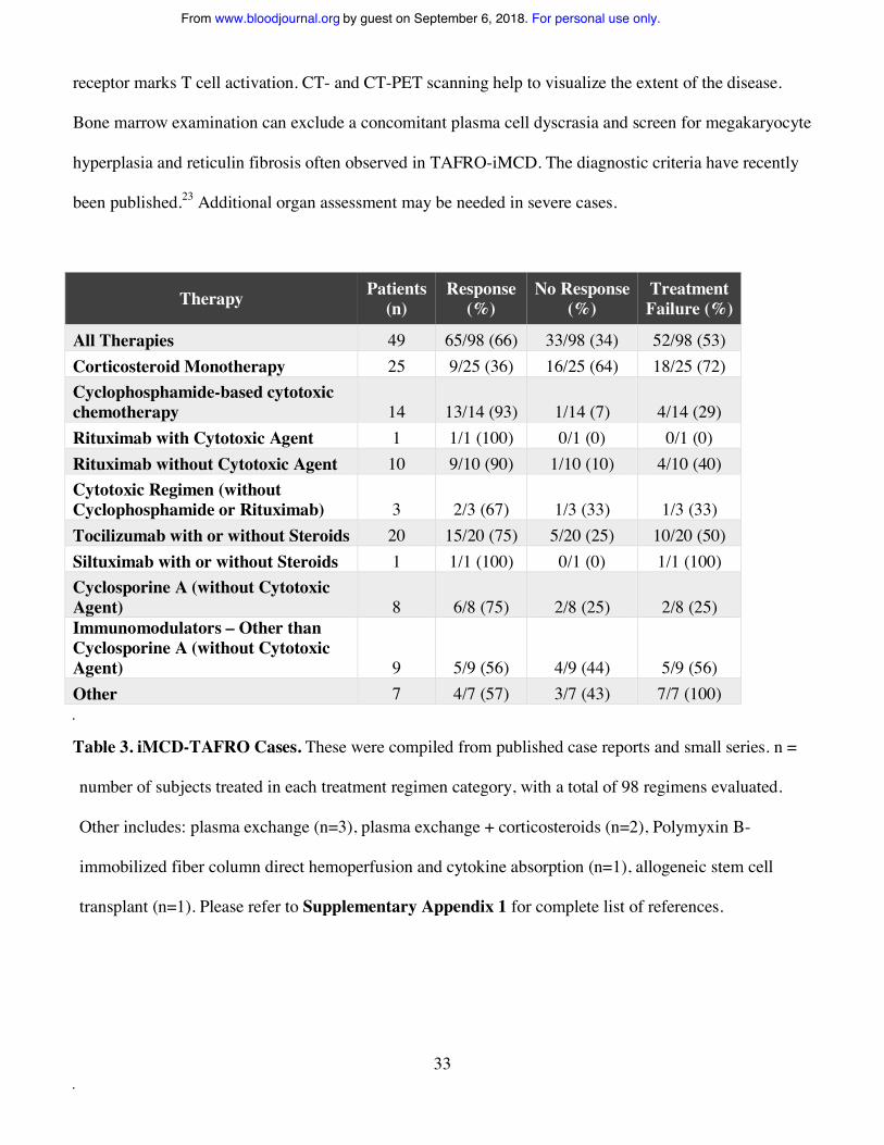
Severe iMCD often presents as the TAFRO subtype. Our analysis of 49 published iMCD-TAFRO
cases with treatment data revealed that corticosteroids, anti-IL-6 mAbs, cytotoxic chemotherapies, and
cyclosporine A are most often used. These agents demonstrate initial similar efficacy to the other cohorts,
but higher rates of treatment failures and relapses (Table 3). Based on the available evidence, we
recommend following the same treatment algorithm as for other cases of iMCD that is dependent on
disease severity and initiate therapy with anti-IL-6 mAb therapy with or without corticosteroids. Among
TAFRO cases, cyclosporine A can be useful therapy for anti-IL-6-refractory cases particularly to improve
persistent ascites and thrombocytopenia.21,74-77 The Japanese TAFRO research group recommends high-
dose steroids, tocilizumab and cyclosporine A for patients with TAFRO syndrome.84 A comprehensive
analysis of a treatment-refractory iMCD-TAFRO patient who sustained multiple relapses after repeated
cycles of chemotherapy showed upregulation of the mTOR pathway, and remission was successfully
maintained with sirolimus.85 Early data suggest that the proteomic profiles of classical and TAFRO-
iMCD are different, supporting the notion that there may be diverse chemokines/cytokines driving the
symptomatology across the iMCD spectrum.24,25
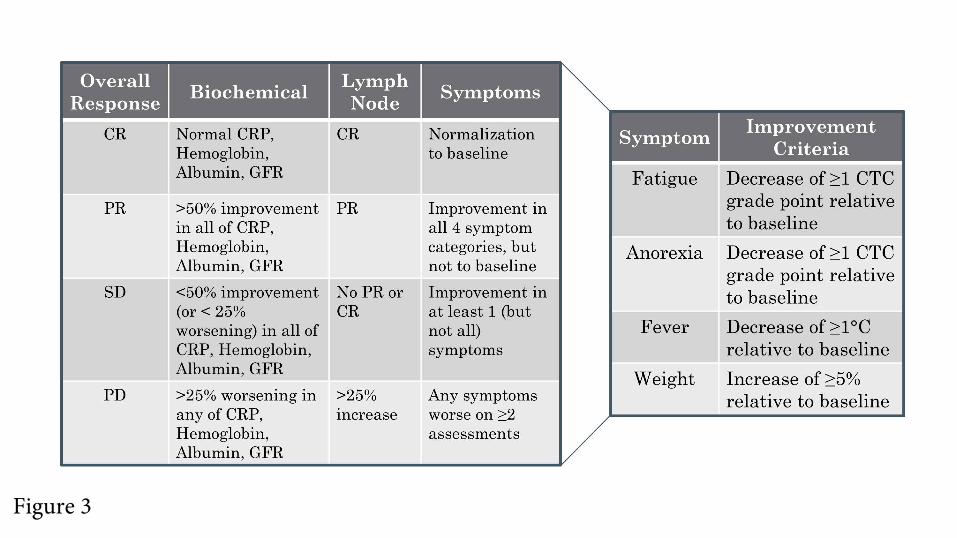
Evaluation of Response. As is evident from the review of published literature, criteria for response to
treatment of iMCD have thus far not been agreed upon. In the tocilizumab study, the primary endpoint
For personal use only.on September 6, 2018. by guest www.bloodjournal.orgFrom

16
was based on improvements in specific laboratory tests, but there was no aggregated response definition.32
The Phase I siltuximab study used Cheson criteria for lymph node response modified to assess the skin
lesions of iMCD. This trial introduced a clinical benefit response assessing six iMCD-related clinical
features.42,86 In the Phase II registration study of siltuximab, lymphadenopathy was similarly assessed, but
the symptomatic response was evaluated by the investigators using a complex 34 iMCD-related symptom
score.33
The FDA, in its approval of siltuximab, commented on the necessity of a composite response
assessment for iMCD.87 Therefore, our expert panel established a composite end-point for evaluating
response taking into account all cardinal features of the disease: a) objective biochemical markers of
inflammatory response and organ function (hemoglobin, CRP, albumin, estimated glomerular filtration
rate), b) lymph node size, and c) clinical symptoms (fatigue, anorexia, fever, weight change) as assessed
by the clinician (Figure 3).
A biochemical CR requires normalization of all values compared to baseline. In a PR, there is 50-
99% improvement in all laboratory values. In patients with stable disease (SD), there is a <50%
improvement in all laboratory values or <25% worsening in any laboratory indicators. Progressive disease
(PD) indicates a >25% worsening in any of the laboratory markers. Lymph node response is assessed
using modified Cheson criteria as previously published.42,86 Lastly, four important clinical symptoms are
assessed using the National Cancer Institute Common Terminology Criteria of Adverse Events (version
4). A symptomatic CR requires normalization of all symptoms. PR requires improvement in the grades of
all 4 symptoms, but they do not have to return to baseline. SD requires not meeting the criteria for PR or
PD, which occurs if any symptoms worsen on 2 assessments four weeks apart. Evaluation of overall
response requires integration of the three response categories as defined in Figure 3.
For personal use only.on September 6, 2018. by guest www.bloodjournal.orgFrom
17
Discussion
The published diagnostic criteria of iMCD, together with the recognition of the TAFRO-iMCD
subtype, provides a framework for recognizing different clinical entities on the CD spectrum.18,23 We
present the first formal guidelines for the treatment of iMCD, depending on symptom severity. Based on
the response criteria used in the literature and our clinical expertise, we propose composite response
criteria addressing all relevant features of the disease to evaluate treatment in both clinical practice and
future studies. The present guidelines should assist physicians with selecting therapy and evaluating
response, thereby improving patient outcomes. The preferred treatment for non-severe iMCD is
siltuximab, whilst for some patients with limited symptomatology, a short course of rituximab is an
alternative option. Patients with severe iMCD are a challenge and may require early intervention with
combination chemotherapy to avoid a fatal outcome. Not all patients will benefit from siltuximab therapy,
especially those who have a very mild inflammatory syndrome, or on the other end of the spectrum,
severely-ill patients who require a rapid response. Lastly, it has become clear that iMCD has a
pleomorphic cytokine profile and that the disease is not driven by IL-6 in all.
There are several important limitations to our treatment recommendations. It is important to highlight
that most recommendations were reached by consensus and are not supported by prospective, randomized
data. Due to the rarity of the disease, there are no clinical studies available comparing treatment
modalities such as chemotherapy, rituximab, and anti-IL6 mAbs. Though the evidence base included
clinical trial data and the largest collection of treatment data analyzed to date, it should be noted that case
reports and retrospective case series with short follow-up durations make up a large portion of cases,
which are subject to publication bias of successful uses of novel agents. Additionally, the various studies
used different criteria for assessing response (CR, PR, “response”); e.g. the threshold for a CR in a
randomized controlled trial is likely different from that in a case report. Therefore, we aggregated all
response categories into one global response category, which is listed in Table 1. We included a broad
range of data from multiple sources to minimize these limitations. Although anti-IL-6 mAbs are an
For personal use only.on September 6, 2018. by guest www.bloodjournal.orgFrom

18
important contribution to the therapeutic armamentarium for iMCD, treatment must be continued long
term. The CDCN established an international registry (www.CDCN.org/ACCELERATE), which collects
data pertaining to treatment and outcome, to increase the evidence base for selection of therapies in the
future. Ongoing research will focus on defining the etiology and pathogenesis of this complex and rare
disease to promote the development of better and more targeted therapies, particularly for patients who do
not benefit from anti-IL-6 mAb administration.
Authorship and Conflict of Interest Statements
Contribution: All authors were responsible for the conceptualization of this manuscript and participated in
the generation of these consensus treatment guidelines; F.v.R. and D.C.F. wrote the paper; K.S. and A.G.
edited the paper and made figures and tables.
Conflict-of-interest disclosure: F.v.R. has received research funding from Bristol Myers-Squibb and
Janssen Pharmaceuticals, and has served on advisory boards from Janssen Pharmaceuticals. D.C.F. has
received research funding from Janssen Pharmaceuticals. R.W. and C.C. have received research funding
from Janssen Pharmaceuticals and served on advisory boards for Janssen Pharmaceuticals. P.V. has
served on advisory boards for Janssen Pharmaceuticals. S.F. has received consultancy fees and speaker
honoraria and served on advisory boards for Janssen Pharmaceuticals. A.F. has received honoraria from
Janssen Pharmaceuticals. T.S.U. has received research funding Genentech, Merck, and Celgene via
Cooperative Research and Development Agreements with the National Cancer Institute, and has a patent
for an immunomodulatory compound for KSHV malignancies (Inst). R.K. has received research funding
from Incyte, Genentech, Merck Serono, Pfizer, Sequenom, Foundation Medicine, Guardant Health, and
Konica Minolta, consultant fees from LOXO, Actuate Therapeutics, Genentech and NeoMed, as well as
speaker fees from Roche, and has an ownership interest in Curematch, Inc. D.S. has received honoraria
and research funding from Janssen Pharmaceuticals. All other authors have nothing to disclose.
For personal use only.on September 6, 2018. by guest www.bloodjournal.orgFrom
19
REFERENCES
1. Castleman B, Towne VW. Case Records of the Massachusetts General Hospital; Weekly
Clinicopathological Exercises; Founded by Richard C. Cabot. N. Engl. J. Med. 1954;251(10):396-
400.
2. Flendrig J. Benign Giant Lymphoma: The Clincal Signs and Symptoms and the Morphological
Aspects. Folia Medica. 1969;12:119-120.
3. Keller AR, Hochholzer L, Castleman B. Hyaline-Vascular and Plasma-Cell Types of Giant Lymph
Node Hyperplasia of the Mediastinum and Other Locations. Cancer. 1972;29(3):670-683.
4. Gaba AR, Stein RS, Sweet DL, Variakojis D. Multicentric Giant Lymph Node Hyperplasia. Am. J.
Clin. Pathol. 1978;69(1):86-90.
5. Lachant NA, Sun NC, Leong LA, Oseas RS, Prince HE. Multicentric Angiofollicular Lymph
Node Hyperplasia (Castleman's Disease) Followed by Kaposi's Sarcoma in Two Homosexual
Males with the Acquired Immunodeficiency Syndrome (AIDS). Am. J. Clin. Pathol.
1985;83(1):27-33.
6. Soulier J, Grollet L, Oksenhendler E, et al. Kaposi's Sarcoma-Associated Herpesvirus-Like DNA
Sequences in Multicentric Castleman's Disease. Blood. 1995;86(4):1276-1280.
7. Oksenhendler E, Duarte M, Soulier J, et al. Multicentric Castleman's Disease in HIV Infection: A
Clinical and Pathological Study of 20 Patients. AIDS (London, England). 1996;10(1):61-67.
8. Aoki Y, Tosato G, Fonville TW, Pittaluga S. Serum Viral Interleukin-6 in AIDS-Related
Multicentric Castleman Disease. Blood. 2001;97(8):2526-2527.
9. Dossier A, Meignin V, Fieschi C, Boutboul D, Oksenhendler E, Galicier L. Human Herpesvirus 8-
Related Castleman Disease in the Absence of HIV Infection. Clinical Infectious Diseases : An
Official Publication of the Infectious Diseases Society of America. 2013;56(6):833-842.
For personal use only.on September 6, 2018. by guest www.bloodjournal.orgFrom
20
10. Nicoli P, Familiari U, Bosa M, et al. HHV8-Positive, HIV-Negative Multicentric Castleman's
Disease: Early and Sustained Complete Remission with Rituximab Therapy without Reactivation
of Kaposi Sarcoma. Int. J. Hematol. 2009.
11. Liu AY, Nabel CS, Finkelman BS, et al. Idiopathic Multicentric Castleman's Disease: A
Systematic Literature Review. The Lancet. Haematology. 2016;3(4):e163-175.
12. Fajgenbaum DC, van Rhee F, Nabel CS. HHV-8-Negative, Idiopathic Multicentric Castleman
Disease: Novel Insights into Biology, Pathogenesis, and Therapy. Blood. 2014;123(19):2924-
2933.
13. Yoshizaki K, Matsuda T, Nishimoto N, et al. Pathogenic Significance of Interleukin-6 (IL-6/BSF-
2) in Castleman's Disease. Blood. 1989;74(4):1360-1367.
14. Nishimoto N, Terao K, Mima T, Nakahara H, Takagi N, Kakehi T. Mechanisms and Pathologic
Significances in Increase in Serum Interleukin-6 (IL-6) and Soluble IL-6 Receptor after
Administration of an Anti-IL-6 Receptor Antibody, Tocilizumab, in Patients with Rheumatoid
Arthritis and Castleman Disease. Blood. 2008;112(10):3959-3964.
15. Stone K, Woods E, Szmania SM, et al. Interleukin-6 Receptor Polymorphism is Prevalent in HIV-
Negative Castleman Disease and is Associated with Increased Soluble Interleukin-6 Receptor
Levels. PLoS One. 2013;8(1):e54610.
16. Fajgenbaum DC, Ruth JR, Kelleher D, Rubenstein AH. The Collaborative Network Approach: A
New Framework to Accelerate Castleman's Disease and Other Rare Disease Research. The Lancet.
Haematology. 2016;3(4):e150-152.
17. Iwaki N, Sato Y, Takata K, et al. Atypical Hyaline Vascular-Type Castleman's Disease with
Thrombocytopenia, Anasarca, Fever, and Systemic Lymphadenopathy. Journal of Clinical and
Experimental Hematopathology : JCEH. 2013;53(1):87-93.
For personal use only.on September 6, 2018. by guest www.bloodjournal.orgFrom

21
18. Iwaki N, Fajgenbaum DC, Nabel CS, et al. Clinicopathologic Analysis of TAFRO Syndrome
Demonstrates a Distinct Subtype of HHV-8-Negative Multicentric Castleman Disease. Am. J.
Hematol. 2016;91(2):220-226.
19. Kawabata H, Kotani S, Matsumura Y, et al. Successful Treatment of a Patient with Multicentric
Castleman's Disease Who Presented with Thrombocytopenia, Ascites, Renal Failure and
Myelofibrosis Using Tocilizumab, an Anti-Interleukin-6 Receptor Antibody. Internal Medicine
(Tokyo, Japan). 2013;52(13):1503-1507.
20. Takai K, Nikkuni K, Shibuya H, Hashidate H. [Thrombocytopenia with Mild Bone Marrow
Fibrosis Accompanied by Fever, Pleural Effusion, Ascites and Hepatosplenomegaly]. [Rinsho
ketsueki] The Japanese Journal of Clinical Hematology. 2010;51(5):320-325.
21. Yu L, Tu M, Cortes J, et al. Clinical and Pathological Characteristics of HIV- and HHV-8-
Negative Castleman Disease. Blood. 2017;129(12):1658-1668.
22. Igawa T, Sato Y. TAFRO Syndrome. Hematol. Oncol. Clin. North Am. 2018;32(1):107-118.
23. Fajgenbaum DC, Uldrick TS, Bagg A, et al. International, Evidence-Based Consensus Diagnostic
Criteria for HHV-8-Negative/Idiopathic Multicentric Castleman Disease. Blood.
2017;129(12):1646-1657.
24. Iwaki N, Gion Y, Kondo E, et al. Elevated Serum Interferon Gamma-Induced Protein 10 kDa is
Associated with TAFRO Syndrome. Sci. Rep. 2017;7:42316.
25. Pierson S, Stonestrom A, Ruth J, et al. Quantification of Plasma Proteins from Idiopathic
Multicentric Castleman Disease Flares and Remissions Reveals 'Chemokine Storm' and Separates
Clinical Subtypes. ASH Annual Meeting Abstracts. 2017;130:3592.
26. Talat N, Schulte KM. Castleman's Disease: Systematic Analysis of 416 Patients from the
Literature. The Oncologist. 2011;16(9):1316-1324.
27. Dispenzieri A, Armitage JO, Loe MJ, et al. The Clinical Spectrum of Castleman's Disease. Am. J.
Hematol. 2012;87(11):997-1002.
For personal use only.on September 6, 2018. by guest www.bloodjournal.orgFrom
22
28. Shin DY, Jeon YK, Hong YS, et al. Clinical Dissection of Multicentric Castleman Disease. Leuk.
Lymphoma. 2011;52(8):1517-1522.
29. Seo S, Yoo C, Yoon DH, et al. Clinical Features and Outcomes in Patients with Human
Immunodeficiency Virus-Negative, Multicentric Castleman's Disease: A Single Medical Center
Experience. Blood Res. 2014;49(4):253-258.
30. Robinson D, Jr., Reynolds M, Casper C, et al. Clinical Epidemiology and Treatment Patterns of
Patients with Multicentric Castleman Disease: Results from Two US Treatment Centres. Br. J.
Haematol. 2014;165(1):39-48.
31. Simpson D. Epidemiology of Castleman Disease. Hematol. Oncol. Clin. North Am. 2018;32(1):1-
10.
32. Nishimoto N, Kanakura Y, Aozasa K, et al. Humanized Anti-Interleukin-6 Receptor Antibody
Treatment of Multicentric Castleman Disease. Blood. 2005;106(8):2627-2632.
33. van Rhee F, Wong RS, Munshi N, et al. Siltuximab for Multicentric Castleman's Disease: A
Randomised, Double-Blind, Placebo-Controlled Trial. Lancet Oncol. 2014;15(9):966-974.
34. Marcelin AG, Aaron L, Mateus C, et al. Rituximab Therapy for HIV-Associated Castleman
Disease. Blood. 2003;102(8):2786-2788.
35. Hoffmann C, Schmid H, Muller M, et al. Improved Outcome with Rituximab in Patients with
HIV-Associated Multicentric Castleman Disease. Blood. 2011;118(13):3499-3503.
36. Bower M, Powles T, Williams S, et al. Brief Communication: Rituximab in HIV-Associated
Multicentric Castleman Disease. Ann. Intern. Med. 2007;147(12):836-839.
37. Gerard L, Michot JM, Burcheri S, et al. Rituximab Decreases the Risk of Lymphoma in Patients
with HIV-Associated Multicentric Castleman Disease. Blood. 2012;119(10):2228-2233.
38. Uldrick TS, Polizzotto MN, Aleman K, et al. Rituximab Plus Liposomal Doxorubicin in HIV-
Infected Patients with KSHV-Associated Multicentric Castleman Disease. Blood.
2014;124(24):3544-3552.
For personal use only.on September 6, 2018. by guest www.bloodjournal.orgFrom

23
39. Oksenhendler E, Boutboul D, Fajgenbaum D, et al. The Full Spectrum of Castleman Disease: 273
Patients Studied over 20 Years. Br. J. Haematol. 2018;180(2):206-216.
40. Bower M. How I Treat HIV-Associated Multicentric Castleman Disease. Blood.
2010;116(22):4415-4421.
41. Dispenzieri A. POEMS Syndrome: 2017 Update on Diagnosis, Risk Stratification, and
Management. Am. J. Hematol. 2017;92(8):814-829.
42. van Rhee F, Fayad L, Voorhees P, et al. Siltuximab, a Novel Anti-Interleukin-6 Monoclonal
Antibody, for Castleman's Disease. J. Clin. Oncol. 2010;28(23):3701-3708.
43. Kurzrock R, Voorhees PM, Casper C, et al. A Phase I, Open-Label Study of Siltuximab, an Anti-
IL-6 Monoclonal Antibody, in Patients with B-Cell Non-Hodgkin Lymphoma, Multiple Myeloma,
or Castleman Disease. Clinical Cancer Research : An Official Journal of the American
Association for Cancer Research. 2013;19(13):3659-3670.
44. van Rhee F, Casper C, Voorhees PM, et al. A Phase 2, Open-Label, Multicenter Study of the
Long-Term Safety of Siltuximab (an Anti-Interleukin-6 Monoclonal Antibody) in Patients with
Multicentric Castleman Disease. Oncotarget. 2015;6(30):30408-30419.
45. Casper C, Chaturvedi S, Munshi N, et al. Analysis of Inflammatory and Anemia-Related
Biomarkers in a Randomized, Double-Blind, Placebo-Controlled Study of Siltuximab (Anti-IL6
Monoclonal Antibody) in Patients with Multicentric Castleman Disease. Clinical Cancer Research
: An Official Journal of the American Association for Cancer Research. 2015;21(19):4294-4304.
46. van Rhee F, Munshi N, Wong R, et al. Efficacy of Siltuximab in Patients with Previously Treated
Multicentric Castleman's Disease (MCD). J. Clin. Oncol. 2014;32(5s):suppl; abstr 8514, Oral
Abstract Session.
47. van Rhee F, Rothman M, Ho KF, et al. Patient-Reported Outcomes for Multicentric Castleman's
Disease in a Randomized, Placebo-Controlled Study of Siltuximab. Patient. 2015;8(2):207-216.
For personal use only.on September 6, 2018. by guest www.bloodjournal.orgFrom

24
48. Nishimoto N, Sasai M, Shima Y, et al. Improvement in Castleman's Disease by Humanized Anti-
Interleukin-6 Receptor Antibody Therapy. Blood. 2000;95(1):56-61.
49. Nishimoto N, Honda O, Sumikawa H, Johkoh T, Aozasa K, Kanakura Y. A Long-Term (5-Year)
Sustained Efficacy of Tocilizumab for Multicentric Castleman's Disease and the Effect on
Pulmonary Complications. Blood. 2007;110.
50. Herrada J, Cabanillas F, Rice L, Manning J, Pugh W. The Clinical Behavior of Localized and
Multicentric Castleman Disease. Ann. Intern. Med. 1998;128(8):657-662.
51. Bowne WB, Lewis JJ, Filippa DA, et al. The Management of Unicentric and Multicentric
Castleman's Disease: A Report of 16 Cases and a Review of the Literature. Cancer.
1999;85(3):706-717.
52. Chronowski GM, Ha CS, Wilder RB, Cabanillas F, Manning J, Cox JD. Treatment of Unicentric
and Multicentric Castleman Disease and the Role of Radiotherapy. Cancer. 2001;92(3):670-676.
53. Kessler E. Multicentric Giant Lymph Node Hyperplasia. A Report of Seven Cases. Cancer.
1985;56(10):2446-2451.
54. Weisenburger DD, Nathwani BN, Winberg CD, Rappaport H. Multicentric Angiofollicular
Lymph Node Hyperplasia: A Clinicopathologic Study of 16 Cases. Hum. Pathol. 1985;16(2):162-
172.
55. Frizzera G, Peterson BA, Bayrd ED, Goldman A. A Systemic Lymphoproliferative Disorder with
Morphologic Features of Castleman's Disease: Clinical Findings and Clinicopathologic
Correlations in 15 Patients. J. Clin. Oncol. 1985;3(9):1202-1216.
56. Ocio EM, Sanchez-Guijo FM, Diez-Campelo M, et al. Efficacy of Rituximab in an Aggressive
Form of Multicentric Castleman Disease Associated with Immune Phenomena. Am. J. Hematol.
2005;78(4):302-305.
For personal use only.on September 6, 2018. by guest www.bloodjournal.orgFrom
25
57. Ide M, Ogawa E, Kasagi K, Kawachi Y, Ogino T. Successful Treatment of Multicentric
Castleman's Disease with Bilateral Orbital Tumour Using Rituximab. Br. J. Haematol.
2003;121(5):818-819.
58. Gholam D, Vantelon JM, Al-Jijakli A, Bourhis JH. A Case of Multicentric Castleman's Disease
Associated with Advanced Systemic Amyloidosis Treated with Chemotherapy and Anti-CD20
Monoclonal Antibody. Ann. Hematol. 2003;82(12):766-768.
59. Ide M, Kawachi Y, Izumi Y, Kasagi K, Ogino T. Long-Term Remission in HIV-Negative Patients
with Multicentric Castleman's Disease Using Rituximab. Eur. J. Haematol. 2006;76(2):119-123.
60. Mian H, Leber B. Mixed Variant Multicentric Castleman Disease Treated with Rituximab: Case
Report. J. Pediatr. Hematol. Oncol. 2010;32(8):622.
61. Adam Z, Szturz P, Koukalova R, et al. [PET-CT Documented Remission of Multicentric
Castleman Disease after Treatment with Rituximab: Case Report and Review]. Vnitrni lekarstvi.
2015;61(3):251-259.
62. Lee FC, Merchant SH. Alleviation of Systemic Manifestations of Multicentric Castleman's
Disease by Thalidomide. Am. J. Hematol. 2003;73(1):48-53.
63. Starkey CR, Joste NE, Lee FC. Near-Total Resolution of Multicentric Castleman Disease by
Prolonged Treatment with Thalidomide. Am. J. Hematol. 2006;81(4):303-304.
64. Ramasamy K, Gandhi S, Tenant-Flowers M, et al. Rituximab and Thalidomide Combination
Therapy for Castleman Disease. Br. J. Haematol. 2012;158(3):421-423.
65. Tatekawa S, Umemura K, Fukuyama R, et al. Thalidomide for Tocilizumab-Resistant Ascites with
TAFRO Syndrome. Clin Case Rep. 2015;3(6):472-478.
66. Zhou X, Wei J, Lou Y, et al. Salvage Therapy with Lenalidomide Containing Regimen for
Relapsed/Refractory Castleman Disease: A Report of Three Cases. Front. Med. 2017;11(2):287-
292.
For personal use only.on September 6, 2018. by guest www.bloodjournal.orgFrom

26
67. Galeotti C, Tran TA, Franchi-Abella S, Fabre M, Pariente D, Kone-Paut I. IL-1Ra Agonist
(Anakinra) in the Treatment of Multifocal Castleman Disease: Case Report. J. Pediatr. Hematol.
Oncol. 2008;30(12):920-924.
68. El-Osta H, Janku F, Kurzrock R. Successful Treatment of Castleman's Disease with Interleukin-1
Receptor Antagonist (Anakinra). Mol. Cancer Ther. 2010;9(6):1485-1488.
69. Hess G, Wagner V, Kreft A, Heussel CP, Huber C. Effects of Bortezomib on Pro-Inflammatory
Cytokine Levels and Transfusion Dependency in a Patient with Multicentric Castleman Disease.
Br. J. Haematol. 2006;134(5):544-545.
70. Yuan ZG, Dun XY, Li YH, Hou J. Treatment of Multicentric Castleman's Disease Accompanying
Multiple Myeloma with Bortezomib: A Case Report. J. Hematol. Oncol. 2009;2:19.
71. Lin Q, Fang B, Huang H, et al. Efficacy of Bortezomib and Thalidomide in the Recrudescent
Form of Multicentric Mixed-Type Castleman's Disease. Blood Cancer Journal. 2015;5:e298.
72. Rieu P, Droz D, Gessain A, Grunfeld JP, Hermine O. Retinoic Acid for Treatment of Multicentric
Castleman's Disease. Lancet. 1999;354(9186):1262-1263.
73. Miltenyi Z, Toth J, Gonda A, Tar I, Remenyik E, Illes A. Successful Immunomodulatory Therapy
in Castleman Disease with Paraneoplastic Pemphigus Vulgaris. Pathology Oncology Research :
POR. 2009;15(3):375-381.
74. Takasawa N, Sekiguchi Y, Takahashi T, Muryoi A, Satoh J, Sasaki T. A Case of TAFRO
Syndrome, a Variant of Multicentric Castleman's Disease, Successfully Treated with
Corticosteroid and Cyclosporine A. Mod Rheumatol. 2016:1-5.
75. Yamaga Y, Tokuyama K, Kato T, et al. Successful Treatment with Cyclosporin a in Tocilizumab-
Resistant TAFRO Syndrome. Internal Medicine (Tokyo, Japan). 2016;55(2):185-190.
76. Konishi Y, Takahashi S, Nishi K, et al. Successful Treatment of TAFRO Syndrome, a Variant of
Multicentric Castleman's Disease, with Cyclosporine A: Possible Pathogenetic Contribution of
Interleukin-2. The Tohoku Journal of Experimental Medicine. 2015;236(4):289-295.
For personal use only.on September 6, 2018. by guest www.bloodjournal.orgFrom
27
77. Inoue M, Ankou M, Hua J, Iwaki Y, Hagihara M, Ota Y. Complete Resolution of TAFRO
Syndrome (Thrombocytopenia, Anasarca, Fever, Reticulin Fibrosis and Organomegaly) after
Immunosuppressive Therapies Using Corticosteroids and Cyclosporin A : A Case Report. Journal
of Clinical and Experimental Hematopathology : JCEH. 2013;53(1):95-99.
78. Frizzera G. Castleman's Disease and Related Disorders. Semin. Diagn. Pathol. 1988;5(4):346-364.
79. Zhu SH, Yu YH, Zhang Y, Sun JJ, Han DL, Li J. Clinical Features and Outcome of Patients with
HIV-Negative Multicentric Castleman's Disease Treated with Combination Chemotherapy: A
Report on 10 Patients. Med Oncol. 2013;30(1):492.
80. Jerkeman M, Linden O. Long-Term Remission in Idiopathic Castleman's Disease with
Tocilizumab Followed by Consolidation with High-Dose Melphalan--Two Case Studies. Eur. J.
Haematol. 2016;96(5):541-543.
81. Tal Y, Haber G, Cohen MJ, et al. Autologous Stem Cell Transplantation in a Rare Multicentric
Castleman Disease of the Plasma Cell Variant. Int. J. Hematol. 2011;93(5):677-680.
82. Ogita M, Hoshino J, Sogawa Y, et al. Multicentric Castleman Disease with Secondary AA Renal
Amyloidosis, Nephrotic Syndrome and Chronic Renal Failure, Remission after High-Dose
Melphalan and Autologous Stem Cell Transplantation. Clin. Nephrol. 2007;68(3):171-176.
83. Angenendt L, Kerkhoff A, Wiebe S, et al. Remissions of Different Quality Following Rituximab,
Tocilizumab and Rituximab, and Allogeneic Stem Cell Transplantation in a Patient with Severe
Idiopathic Multicentric Castleman's Disease. Ann. Hematol. 2015;94(7):1241-1243.
84. Masaki Y, Kawabata H, Takai K, et al. Proposed Diagnostic Criteria, Disease Severity
Classification and Treatment Strategy for TAFRO Syndrome, 2015 Version. Int. J. Hematol.
2016;103(6):686-692.
85. Fajgenbaum D, Shilling D, Partridge HL, et al. Prolonged Remission Achieved in a Relapsing
Idiopathic Multicentric Castleman Disease Patient with a Novel, Targeted Treatment Approach.
ASH Annual Meeting Abstracts. 2017;130:3593.
For personal use only.on September 6, 2018. by guest www.bloodjournal.orgFrom

28
86. Cheson BD, Horning SJ, Coiffier B, et al. Report of an International Workshop to Standardize
Response Criteria for Non-Hodgkin's Lymphomas. NCI Sponsored International Working Group.
J. Clin. Oncol. 1999;17(4):1244.
87. Deisseroth A, Ko CW, Nie L, et al. Fda Approval: Siltuximab for the Treatment of Patients with
Multicentric Castleman Disease. Clinical Cancer Research : An Official Journal of the American
Association for Cancer Research. 2015;21(5):950-954.
For personal use only.on September 6, 2018. by guest www.bloodjournal.orgFrom

29
Figure Legends.
Figure 1. CDCN Severity Classification for Rapid Assessment and Allocation of Therapy. Patients
with severe iMCD must have at least two of the five criteria listed above. Patients should be classified as
non-severe iMCD if the above criteria are not met.
Figure 2. Treatment Algorithm for iMCD. iMCD patients should be stratified for disease severity per
Figure 1. Non-Severe iMCD: Siltuximab is recommended as frontline therapy for patients with non-
severe iMCD. Tocilizumab can be used if siltuximab is not available or approved. Steroids are useful
adjunctive therapy and the dose can be tailored according to the severity of the disease. Patients
responding to anti-IL6 mAb therapy should be continued indefinitely. *For patients with mild
symptomatology, a limited course of rituximab is an alternative option. Patients not responding to anti-
IL6 mAb therapy should be considered for rituximab-based therapy + steroids ±
immunomodulatory/immunosuppressive agents. Immunomodulatory/immunosuppressive agents for
second or third line therapy include thalidomide, cyclosporine A, sirolimus, anakinra, or bortezomib, but
we recommend consulting with an expert at this stage.
Severe iMCD: Severe disease must be closely monitored, as life-threatening events may occur in this
population. Severely ill patients should be treated with siltuximab and high-dose steroids, but if no clear
response occurs within one week (or if status worsens at any time), then combination chemotherapy
should be considered. When possible, expert advice should be sought to identify the most appropriate
therapy for a given patient. Further therapy is best individualized. †Examples of chemotherapy include
R-CHOP (rituximab, cyclophosphamide, doxorubicin, vincristine, prednisone), R-VDT-PACE
(rituximab, bortezomib, dexamethasone, thalidomide, cisplatin, doxorubicin, cyclophosphamide,
etoposide), or etoposide/ cyclophosphamide/rituximab. Siltuximab is the preferred anti-IL-6 therapy.
However, in countries where siltuximab is not available or approved, tocilizumab can be used instead.
Supporting evidence category 1, green boxes, category 2A, yellow boxes; category 2B, blue boxes.
For personal use only.on September 6, 2018. by guest www.bloodjournal.orgFrom

30
Figure 3. CDCN Response Criteria Based on Evaluation of Biochemical, Lymph Node, and
Symptom Response. Biochemical, lymph node, and response criteria have been detailed in the text. For
lymph node response, Cheson criteria have been modified to include assessment of skin manifestations.42
An overall CR requires a complete biochemical, lymph node and symptomatic response. An overall PR
requires nothing less than a PR across all categories, but not meeting criteria for CR. Overall SD requires
no PD in any of the categories and not meeting the criteria for CR or PR. An overall PD occurs when any
category has a PD. Symptomatic and biochemical response evaluation should be done on a monthly basis
until maximum response has been achieved. Radiological assessment of lymph node response by CT-
scanning is first recommended at six weeks and at three-monthly intervals thereafter until maximum
regression of lymph nodes has occurred. Lymph node response may take several months in patients
treated with anti-IL-6 mAbs.
For personal use only.on September 6, 2018. by guest www.bloodjournal.orgFrom

31
Tables.
Therapy Patients (n)
Response/m* (%)
No Response/m*
(%)
Treatment Failure/m*
(%) Data Combined
From References
All Therapies 344 281/461 (61) 180/461 (39) 163/367 (44) 11,21,32,33,39, supplementary
appendix citations
Corticosteroid Monotherapy 117 53/114 (46) 61/114 (54) 62/115 (54)
22, 23, 44, supplementary
appendix citations Corticosteroid or Cytotoxic Chemotherapy (not distinguished)
19 12/19 (63) 7/19 (37) NA 21
Cytotoxic Chemotherapy (any time used)
135 102/131 (78) 29/131 (22) 44/105 (42) 7, 22, 23,
supplementary appendix citations
Anti-IL-6 mAb (without cytotoxic agent or rituximab)
147 88/144 (61) 56/144 (39) 32/100 (32) 7, 22, 23, 43, 44, supplementary
appendix citations Immunomodulator (without cytotoxic agent)
27 18/26 (69) 8/26 (31) 10/26 (38) 23, supplementary appendix citations
Other 16 8/13 (62) 5/13 (38) 12/15 (80) 23, supplementary appendix citations
No treatment/Follow-up Only
18 0/14 (0) 14/14 (100) 11/14 (79) 7, 22, 23,
supplementary appendix citations
Table 1. iMCD Clinical Case Series of 344 patients. Literature review of published case reports, small
series, and clinical trials were compiled to inform and substantiate the experience and opinion of the
Working Group authors. Cytotoxic chemotherapy regimens described may include the use of rituximab. n
= number of subjects treated in each treatment regimen category; m = total number of regimens evaluated
(479); m* = number regimens assessed for stated outcome. Treatment failure was defined as disease
progression while on treatment or insufficient response requiring additional treatments. The main series
included in this analysis are referenced. A detailed breakdown of the data is provided in Supplementary
Appendix 1. The TAFRO case reports are tabulated in the Table 3. MDACC= MD Anderson Cancer
Cancer Center case series. Other includes: plasma exchange (n=4), radiation (n=2), plasma exchange +
For personal use only.on September 6, 2018. by guest www.bloodjournal.orgFrom

32
corticosteroids (n=2), IVIg (n=2), Polymyxin B-immobilized fiber column direct hemoperfusion and
cytokine absorption (n=1), allogeneic stem cell transplant. (n=1), Cimetidine (n=1), antibiotics (n=1),
corticosteroids and etanercept (n=1), interferon-alpha (n=1).
Purpose Tests
Inflammatory Response
CBC, Renal Function, Liver Function, CRP, ESR, Fibrinogen, Immunoglobulins & Free Light Chains, Albumin, Ferritin*
Histopathology Hypervascular/ Mixed Cellularity/Plasmacytic Variant
Virologic Status HIV Serology, HHV8 qPCR (peripheral blood), EBER (lymph node), LANA-1 (lymph node)
Cytokine Profile IL6, VEGF, sIL2 Receptor†
Imaging CT-PET or CT Neck, Chest, Abdomen, Pelvis
Bone Marrow Evaluation MGUS, Myeloma, Reticulin Fibrosis
Immunology ANA, Rheumatoid Factor
Organ Function ECHO, Pulmonary Function
Table 2. Recommended Work-up of iMCD. Work-up should include excisional lymph node biopsy for
histopathologic examination to confirm features consistent with iMCD, establish histopathologic variety,
and to rule out EBV and HHV-8 infection by EBER and LANA-1 staining. Blood work is helpful to
exclude HIV infection, autoimmune disorders and monoclonal gammopathy of undetermined significance
(MGUS)/myeloma as well as measure inflammatory markers, determine organ function, and evaluate
cytokines levels including IL-6 and VEGF. *Ferritin is measured as an acute phase reactant. †Soluble IL2
For personal use only.on September 6, 2018. by guest www.bloodjournal.orgFrom
33
receptor marks T cell activation. CT- and CT-PET scanning help to visualize the extent of the disease.
Bone marrow examination can exclude a concomitant plasma cell dyscrasia and screen for megakaryocyte
hyperplasia and reticulin fibrosis often observed in TAFRO-iMCD. The diagnostic criteria have recently
been published.23 Additional organ assessment may be needed in severe cases.
Therapy Patients (n)
Response (%)
No Response (%)
Treatment Failure (%)
All Therapies 49 65/98 (66) 33/98 (34) 52/98 (53) Corticosteroid Monotherapy 25 9/25 (36) 16/25 (64) 18/25 (72) Cyclophosphamide-based cytotoxic chemotherapy 14 13/14 (93) 1/14 (7) 4/14 (29) Rituximab with Cytotoxic Agent 1 1/1 (100) 0/1 (0) 0/1 (0) Rituximab without Cytotoxic Agent 10 9/10 (90) 1/10 (10) 4/10 (40) Cytotoxic Regimen (without Cyclophosphamide or Rituximab) 3 2/3 (67) 1/3 (33) 1/3 (33) Tocilizumab with or without Steroids 20 15/20 (75) 5/20 (25) 10/20 (50) Siltuximab with or without Steroids 1 1/1 (100) 0/1 (0) 1/1 (100) Cyclosporine A (without Cytotoxic Agent) 8 6/8 (75) 2/8 (25) 2/8 (25) Immunomodulators – Other than Cyclosporine A (without Cytotoxic Agent) 9 5/9 (56) 4/9 (44) 5/9 (56) Other 7 4/7 (57) 3/7 (43) 7/7 (100)
Table 3. iMCD-TAFRO Cases. These were compiled from published case reports and small series. n =
number of subjects treated in each treatment regimen category, with a total of 98 regimens evaluated.
Other includes: plasma exchange (n=3), plasma exchange + corticosteroids (n=2), Polymyxin B-
immobilized fiber column direct hemoperfusion and cytokine absorption (n=1), allogeneic stem cell
transplant (n=1). Please refer to Supplementary Appendix 1 for complete list of references.
For personal use only.on September 6, 2018. by guest www.bloodjournal.orgFrom

For personal use only.on September 6, 2018. by guest www.bloodjournal.orgFrom

For personal use only.on September 6, 2018. by guest www.bloodjournal.orgFrom
For personal use only.on September 6, 2018. by guest www.bloodjournal.orgFrom