internal carotid artery
TRANSCRIPT
By Dr. Huda Moutaz Ismael College of dentistry /University of BaghdadDepartment of oral & maxillofacial surgery
Arteries of the head and neck

ICA start at … The internal carotid artery begins at the bifurcation of the common carotid artery at the level of the upper border of the thyroid cartilage.
supplies the brain, the eye, the forehead, and part of the nose.

Thyroid cartilage Superior laryngeal nerve

Embedded in the carotid sheath.

ICA pass into the cranial cavity through the carotid canal.
It then passes upward and forward in the cavernous sinus


The internal carotid artery then inclines backward, lateral to the optic chiasma, and terminates by dividing into the anterior and the middle cerebral arteries.


Relations of the Internal Carotid Artery in the Neck Anterolaterally: Below the digastric lie the skin, the fascia, the anterior border of the sternocleidomastoid, and the hypoglossal nerve. Posteriorly: The sympathetic trunk , the longus capitis muscle, and the transverse processes of the upper three cervical vertebrae Medially: The pharyngeal wall and the superior laryngeal nerve
Laterally: The internal jugular vein and the vagus nerve

Relation
ICA

Relations of the Internal Carotid Artery in the Neck
Laterally: The internal jugular vein and the vagus nerve

Branches:1. Ophthalmic ArteryThe ophthalmic artery arises from the internal carotid artery as it emerges from the cavernous sinus . It passes forward into the orbital cavity through the optic canal, and it gives off the central artery of the retina, which enters the optic nerve and runs forward to enter the eyeball. The central artery is an end artery and the only blood supply to the retina.

Course of Ophthalmic Artery

2. Posterior Communicating Artery
runs backward to join the posterior cerebral artery.

posterior cerebral artery.posterior cerebral artery supply this area

3. Anterior cerebral ArteryThe anterior cerebral artery is a terminal branch of the internal carotid artery. It passes forward between the cerebral hemispheres and supply the medial and the superolateral surfaces of the cerebral hemisphere. It is joined to the artery of the opposite side by the anterior communicating artery.


3. Middle Cerebral Arterythe largest terminal branch It supplies the entire lateral surface and the occipital pole

Middle Cerebral Artery Middle Cerebral Artery branches


Subclavian artery

Subclavian artery


The circle of Willis lies in the subarachnoid space at the base of the brain. It is formed by the anastomosis between the branches of the two internal carotid arteriesand the two vertebral arteries. The anterior communicating, anterior and posterior cerebral, and posterior communicating are all arteries that contribute to the circle. Cortical and central branches arise from the circle and supply the brain.

Parts:1. First partsBranches: A. Vertebral arteryB. Thyrocervical
trunkC. Internal thoracic

A. Vertebral arteryascends in the neck through the foramina in the transverse processes of the upper six cervical vertebrae. It passes medially above the posterior arch of the atlas and then ascends through the foramen magnum into the skull. On reaching the anterior surface of the medulla oblongata of the brain at the level of the lower border of the pons, it joins the vessel of the opposite side to form the basilar artery.

Vertebral artery


Basilar artery
The basilar artery ascends in a groove on the anterior surface of the pons. It gives off branches to the pons, the cerebellum, and the internal ear. It finally divides into the two posterior cerebral arteries. On each side, the posterior cerebral artery curves laterally and backward around the midbrain. Branches in the neck: Spinal and muscular arteries
PonsCerebellumInternal ear

Branches from basilar artery to Pons Branch from basilar artery to internal ear


B. Thyrocervical trunk
is a short trunk that gives off three terminal branches

Thyrocervical trunk branches to shoulder region
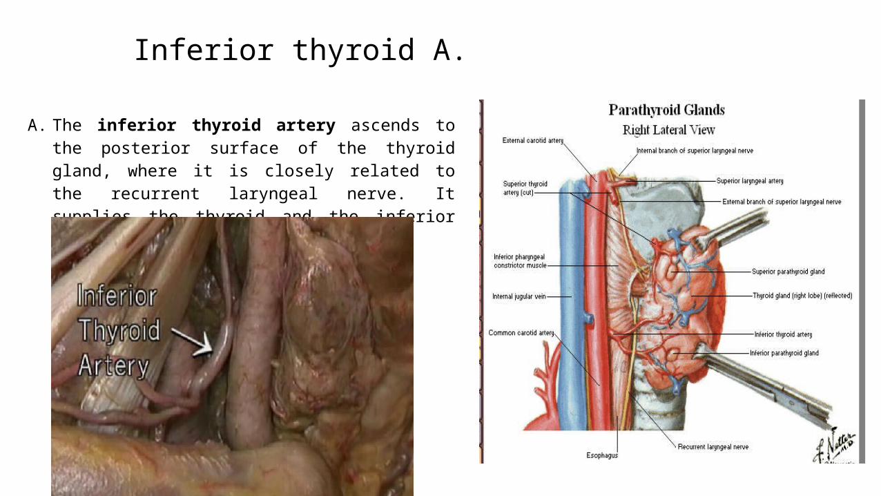
Inferior thyroid A.
A. The inferior thyroid artery ascends to the posterior surface of the thyroid gland, where it is closely related to the recurrent laryngeal nerve. It supplies the thyroid and the inferior parathyroid glands.

Transverse cervical Artery
A. The superficial cervical artery is a small branch that
crosses the brachial plexus.

Suprascapular artery
A. The suprascapular artery runs laterally over the brachial plexus and follows the suprascapular nerve onto the back of the scapula.
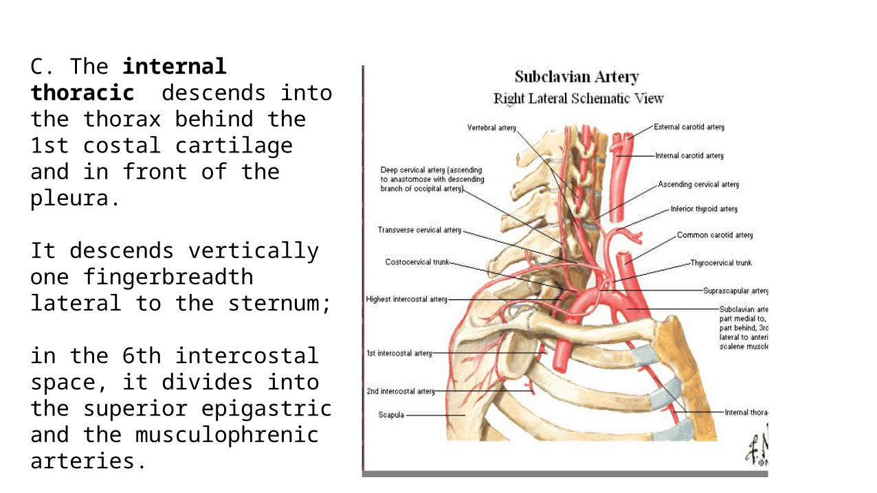
C. The internal thoracic descends into the thorax behind the 1st costal cartilage and in front of the pleura. It descends vertically one fingerbreadth lateral to the sternum;
in the 6th intercostal space, it divides into the superior epigastric and the musculophrenic arteries.
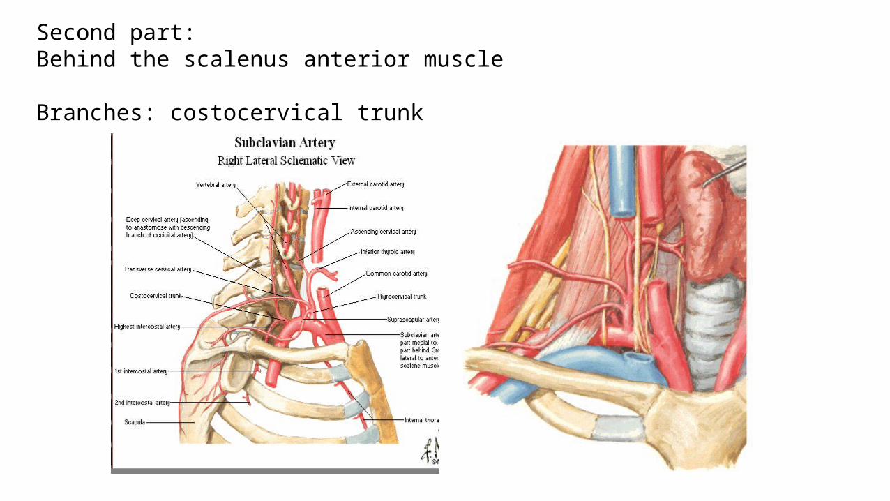
Second part: Behind the scalenus anterior muscle
Branches: costocervical trunk


the superior intercostals artery, which supplies the 1st and the 2nd intercostalspaces,
the deep cervical artery, which supplies the deep muscles of the neck.

Third part:
No branches
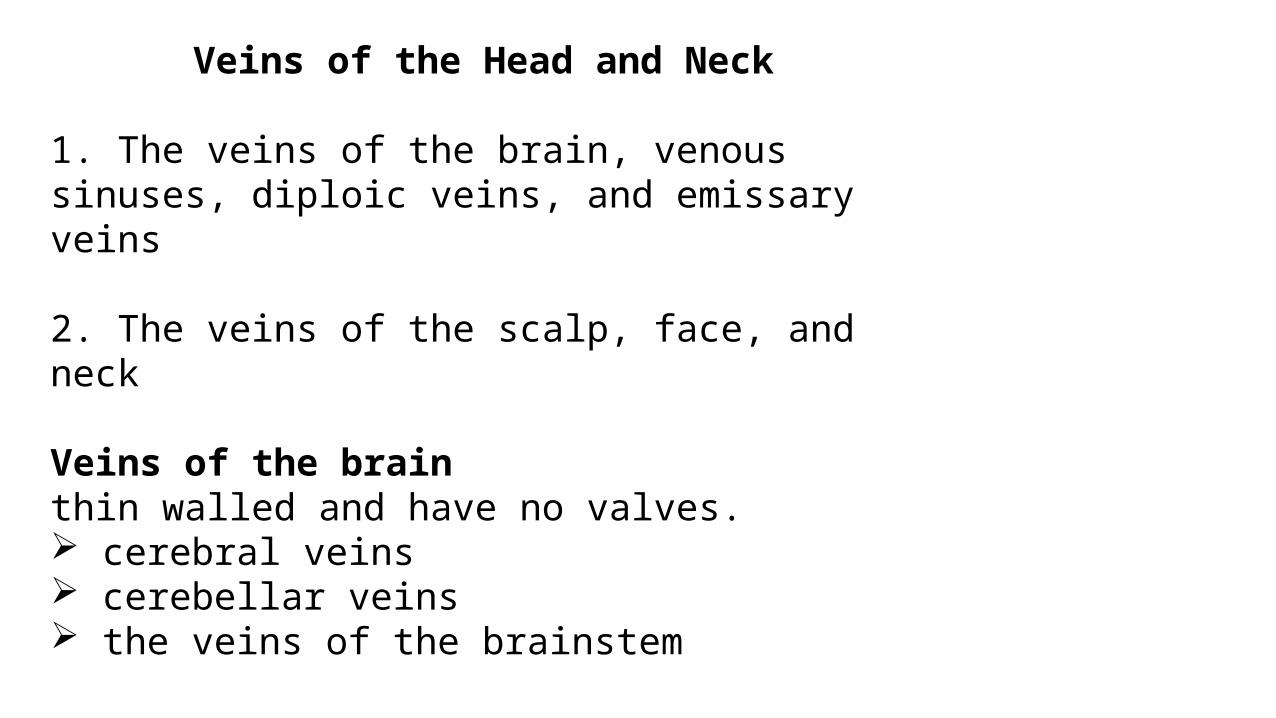
Veins of the Head and Neck
1. The veins of the brain, venous sinuses, diploic veins, and emissary veins
2. The veins of the scalp, face, and neck
Veins of the brain thin walled and have no valves. cerebral veins cerebellar veins the veins of the brainstem

Venous sinus
The venous sinuses are situated between the periosteal and the meningeal layer of the dura mater. They have thick, fibrous walls, but they possess no valves. They receive tributaries from the brain, the skull bones, the orbit, and the internal ear. The venous sinuses include the superior and inferior sagittal sinuses, the straight sinus, the transverse sinuses, the sigmoid sinuses, the occipital sinus, the cavernous sinuses, and the superior and inferior petrosal sinuses.

Falx cerebri Inferior sagittal sinus
Superior sagittal sinus


cavernous sinuses


Diploic veins and emissary veins
Diploic VeinsThe diploic veins occupy channels within the bones of the vault of the skull. Emissary VeinsThe emissary veins are valveless veins that pass through the skull bones. They connect the veins of the scalp to the venous sinuses (and are an important route for the spread of infection).

Internal Jugular Veinreceives blood from the brain, face, and neck . It starts as a continuation of the sigmoid sinus and leaves the skull through the jugular foramen.

Intraoperative photograph showing dilated internal jugular vein


IJV ends by joining the subclavian vein behind the medial end of the clavicle to form the brachiocephalic vein.
The vein has a dilatation at its upper end called the superior bulb and another near its termination called the inferior bulb.

Superior vena cava

Relations of the Internal Jugular VeinAnterolaterally: The skin, the fascia, the sternocleidomastoid, and the parotid salivary gland.
Posteriorly: The transverse processes of the cervical vertebrae, the levator scapulae, the scalenus medius, the scalenus anterior, the cervical plexus, the phrenic nerve, the thyrocervical trunk, the vertebral vein, and the first part of the subclavian artery. On the left side, it passes in front of the thoracic duct. Medially: Above lie the internal carotid artery and the 9th, 10th, 11th, and 12th cranial nerves. Below lie the common carotid artery and the vagus nerve.

Relations of the Internal Jugular Vein

Tributaries of the Internal Jugular Vein
Inferior petrosal sinus
Facial vein
Pharyngeal veins
Lingual vein
Superior thyroid vein
Middle thyroid vein


Subclavian VeinThe subclavian vein is a continuation of the axillary vein at the outer border of the 1st rib. It joins the internal jugular vein to form the brachiocephalic vein, and it receives the external jugular vein. In addition, it often receives the thoracic duct on the left side and the right lymphatic duct on the right.

RelationsAnteriorly: The clavicle
Posteriorly: The scalenus anterior muscle and the phrenic nerve
Inferiorly: The upper surface of the 1st rib

Scalene muscles


THANK U