interleukin-6/interleukin-21 signaling axis is critical in ... · conflict of interest statement:...
TRANSCRIPT

Interleukin-6/interleukin-21 signaling axis is critical inthe pathogenesis of pulmonary arterial hypertensionTakahiro Hashimoto-Kataokaa, Naoki Hosenb, Takashi Sonobec, Yoh Aritaa, Taku Yasuia, Takeshi Masakia,Masato Minamid, Tadakatsu Inagakic, Shigeru Miyagawae, Yoshiki Sawae, Masaaki Murakamif, Atsushi Kumanogohg,Keiko Yamauchi-Takiharaa, Meinoshin Okumurad, Tadamitsu Kishimotoh, Issei Komuroi,j, Mikiyasu Shiraic,Yasushi Sakataa, and Yoshikazu Nakaokaa,k,1
Departments of aCardiovascular Medicine, bCancer Stem Cell Biology Functional Diagnostic Science, dGeneral Thoracic Surgery, eCardiovascular Surgery, andgRespiratory Medicine, Allergy and Rheumatic Disease, Osaka University Graduate School of Medicine, Suita, Osaka, 565-0871, Japan; cDepartment ofCardiac Physiology, National Cerebral and Cardiovascular Center Research Institute, Suita, Osaka, 565-8565, Japan; fDepartment of MolecularNeuroimmunology, Institute for Genetic Medicine, Hokkaido University, Sapporo, Hokkaido, 060-0815, Japan; hLaboratory of Immune Regulation,Immunology Frontier Research Center, Osaka University, Suita, Osaka, 565-0871, Japan; iDepartment of Cardiovascular Medicine, The University of TokyoGraduate School of Medicine, Tokyo, 113-8656, Japan; and jCore Research for Evolutional Science and Technology and kPrecursory Research for EmbryonicScience and Technology, Japan Science Technology Agency, Kawaguchi, Saitama, 332-0012, Japan
Edited by Kari Alitalo, University of Helsinki, Helsinki, Finland, and approved April 16, 2015 (received for review December 26, 2014)
IL-6 is a multifunctional proinflammatory cytokine that is elevatedin the serum of patients with pulmonary arterial hypertension(PAH) and can predict the survival of patients with idiopathic PAH(IPAH). Previous animal experiments and clinical human studiesindicate that IL-6 is important in PAH; however, the molecularmechanisms of IL-6–mediated pathogenesis of PAH have been elu-sive. Here we identified IL-21 as a downstream target of IL-6signaling in PAH. First, we found that IL-6 blockade by the mono-clonal anti-IL-6 receptor antibody, MR16-1, ameliorated hypoxia-induced pulmonary hypertension (HPH) and prevented the hyp-oxia-induced accumulation of Th17 cells and M2 macrophages inthe lungs. Consistently, the expression levels of IL-17 and IL-21 genes,one of the signature genes for Th17 cells, were significantly up-reg-ulated after hypoxia exposure in the lungs of mice treated with con-trol antibody but not in the lungs of mice treated with MR16-1.Although IL-17 blockade with an anti–IL-17A neutralizing antibodyhad no effect on HPH, IL-21 receptor-deficient mice were resistantto HPH and exhibited no significant accumulation of M2macrophagesin the lungs. In accordance with these findings, IL-21 promoted thepolarization of primary alveolar macrophages toward the M2 pheno-type. Of note, significantly enhanced expressions of IL-21 and M2macrophage markers were detected in the lungs of IPAH patientswho underwent lung transplantation. Collectively, these findings sug-gest that IL-21 promotes PAH in association with M2 macrophagepolarization, downstream of IL-6-signaling. The IL-6/IL-21–signalingaxis may be a potential target for treating PAH.
pulmonary arterial hypertension | interleukin-21 | interleukin-6 |Th17 cells | M2 macrophage
Pulmonary arterial hypertension (PAH) is a debilitating dis-ease characterized by arteriopathy in the small to medium-
sized distal pulmonary arteries that is associated with arterialmuscularization, concentric intimal thickening, and the forma-tion of plexiform lesions (1, 2). Inflammation and autoimmunitycurrently are recognized as critical contributors to the patho-genesis of PAH (3, 4). Inflammatory cells such as T cells, B cells,and macrophages infiltrate the plexiform lesions in patients withadvanced PAH. Thus, proinflammatory cytokines produced by thesecells may be responsible for the hyperproliferation of pulmonaryartery endothelial cells (PAECs) and pulmonary artery smoothmuscle cells (PASMCs) (5, 6).IL-6 is a multifunctional proinflammatory cytokine linked to
numerous autoimmune diseases (7, 8). Patients with idiopathicPAH (IPAH) exhibit increased IL-6 serum levels, which correlatewith their prognoses (3, 9, 10). Consistent with these findings,lung-specific IL-6 transgenic mice display spontaneous pulmonaryhypertension in normoxia and develop greatly exaggerated hyp-oxia-induced pulmonary hypertension (HPH) (11), whereas IL-6–
deficient mice show resistance to HPH (12). These findings sug-gest that IL-6 has a significant role in the pathogenesis of pul-monary hypertension; however, the downstream target(s) of IL-6in HPH have been elusive.T-helper cells are comprised of three subsets, Th1, Th2, and
Th17 cells, which are defined by the cytokines they produce (13,14). Th17 cells, a subset of IL-17–producing effector T cells, playimportant roles in the pathogenesis of autoimmune diseases suchas rheumatoid arthritis (13, 14). IL-6 together with TGF-β pro-motes the differentiation of Th17 cells (14, 15). However, theinvolvement of Th17 cells in the pathogenesis of HPH has notbeen determined.Macrophages undergo differential polarization that generates
three distinct macrophage populations: the classically activated(M1), alternatively activated (M2), and anti-inflammatory (reg-ulatory) macrophages (16–18). Upon their activation by IFN-γand TNF-α, M1 macrophages exert microbicidal and tumoricidaleffects by producing inducible nitric oxide synthase and IL-12p40
Significance
Pulmonary arterial hypertension (PAH) is a serious disease char-acterized by vascular remodeling in pulmonary arteries. Althoughan elevated IL-6 serum level correlates with poor prognosis of PAHpatients, it is unclear how IL-6 promotes PAH. Here we identifiedIL-21 as a downstream target of IL-6 signaling in PAH. In mice withhypoxia-induced pulmonary hypertension (HPH), Th17 cells andM2 macrophages accumulate in the lungs after hypoxia exposure.IL-21 primarily derived from Th17 cells promotes M2 macrophagepolarization. Consistently, IL-21 receptor-deficient mice show re-sistance to HPH with no accumulation of M2 macrophages in thelungs. IL-21 andM2macrophage markers were upregulated in thelungs of patients with end-stage idiopathic PAH. These findingssuggest promising therapeutic strategies for PAH targeting IL-6/IL-21–signaling axis.
Author contributions: T.H.-K., T.K., I.K., M.S., and Y.N. designed research; T.H.-K., N.H., T.S.,Y.A., T.Y., T.M., M. Minami, T.I., S.M., K.Y.-T., M.S., and Y.N. performed research; M. Minami,Y. Sawa, M. Murakami, M.O., and T.K. contributed new reagents/analytic tools; T.H.-K., N.H.,T.S., M.Murakami, A.K., K.Y.-T., M.S., Y. Sakata, and Y.N. analyzed data; and T.H.-K. and Y.N.wrote the paper.
Conflict of interest statement: T.K. holds a patent for a monoclonal humanized anti-IL6Rantibody (tocilizumab). Y.N. is a consultant of the sponsor-initiated clinical trial (ChugaiPharmaceutical Co.) using tocilizumab for Takayasu arteritis.
This article is a PNAS Direct Submission.
Freely available online through the PNAS open access option.1To whom correspondence should be addressed. Email: [email protected].
This article contains supporting information online at www.pnas.org/lookup/suppl/doi:10.1073/pnas.1424774112/-/DCSupplemental.
www.pnas.org/cgi/doi/10.1073/pnas.1424774112 PNAS | Published online May 4, 2015 | E2677–E2686
MED
ICALSC
IENCE
SPN
ASPL
US
Dow
nloa
ded
by g
uest
on
Apr
il 23
, 202
0

(17, 18). In contrast, M2 macrophages are activated by IL-4 andIL-13 and characteristically express arginase-1 (Arg1), found ininflammatory zone-1 (Fizz1), chitinase 3-like-3 (Ym1), and man-nose receptor, C type lectin-1 (MRC-1) (16–18). Intriguingly, M2macrophages recently were reported to play a critical role in HPH(19); however, the molecular mechanisms underlying macrophagepolarization in HPH are unknown.Here, we examined the roles of IL-6 and its downstream sig-
naling targets in the pathogenesis of HPH in mice. We foundthat the IL-6/IL-21–signaling axis played critical roles in thedevelopment of HPH in association with M2 macrophage po-larization. Consistent with these findings in mice, large numbersof IL-21+ cells and M2 macrophages were detected in the lungsof patients with IPAH. Taken together, these findings indicate aconserved role for the IL-6/IL-21–signaling axis in mouse HPHand human PAH (HPAH) and suggest promising targets fortreating PAH.
ResultsHypoxia-Induced IL-6 Expression in Mouse Pulmonary Vessels. Wefirst examined the Il-6mRNA levels in the lungs of C57BL/6 miceafter hypoxia exposure using quantitative RT-PCR (qRT-PCR).The Il-6 mRNA levels peaked on day 2 and returned to basallevels by day 7 after hypoxia exposure (Fig. 1A). Consistent withthis finding, immunohistochemical analysis demonstrated strongIL-6 induction in the intima and medial layers of the arteriolesand small arteries of the lung on day 2 after hypoxia exposure(Fig. 1B).
IL-6 Blockade by MR16-1 Prevents HPH. IL-6 signaling is transducedprimarily by STAT3. Thus, we examined the tyrosine-phos-phorylation of STAT3 in the lungs of mice. Although tyrosine-phosphorylation of STAT3 was strongly induced by hypoxia ex-posure in the lungs of mice treated with control antibody, it wasattenuated in those treated with the anti–IL-6 receptor (IL-6R)antibody MR16-1 (Fig. 1C). Next, we investigated the effect ofIL-6 blockade on HPH development by measuring the rightventricular systolic pressure (RVSP), Fulton’s index [right/(left +
septum) ventricular weight], and the medial wall thickness index,which is estimated by elastic Van Gieson staining of the pul-monary arterioles (Fig. 1D). In control antibody-treated mice,hypoxia exposure for 4 wk induced significant increases in theRVSP, Fulton’s index, and medial wall thickness index, com-pared with the values observed under normoxic conditions (Fig.1 E–G). In contrast, the hypoxia-induced elevation of these pa-rameters was significantly inhibited by treatment with MR16-1(Fig. 1 E–G). Similarly, thickened medial vascular walls weredetected in the lung sections of control antibody-treated micebut not in those of mice treated with MR16-1 (Fig. 1H). Undernormoxia, there was no significant difference in the physiologicalparameters of mice treated with control antibody and thosetreated with MR16-1 (Table S1). These data suggest that IL-6blockade by MR16-1 effectively prevents HPH.
IL-6 Blockade Abrogates Hypoxia-Induced Accumulation of Th17 Cellsin the Lungs. IL-6–dependent accumulation of Th17 cells pro-motes the development of inflammatory diseases, such as rheu-matoid arthritis (20). Thus, we examined the involvement of Th17cells in HPH by measuring the mRNA levels of Il-17A, a Th17signature gene. Hypoxia-induced Il-17A in the lungs peaked onday 2 and declined on day 7 but remained slightly higher than thebasal level on and after day 7 (Fig. 2A). We next examined themRNA levels of IL-17A and other Th17 signature gene, such asRorc (retinoic acid receptor–related orphan receptor-c, also knownas “Ror-γt”), and IL-17–downstream chemokines, such as Cxcl1and Cxcl5, in the lungs of mice exposed to hypoxia for 2 d. ThemRNA levels of all these genes were significantly up-regulated inthe lungs of control antibody-treated mice but not in those ofmice treated with MR16-1 (Fig. 2 B–E). The IL-17A proteinlevels also were increased after hypoxia in the lungs of controlantibody-treated mice but not in those of mice treated withMR16-1 (Fig. 2 F and G). In addition, exposure to hypoxia for3 d led to significantly increased Th17 numbers in the lungs ofcontrol antibody-treated mice but not in those of mice treatedwith MR16-1 (Fig. 2 H and I). In contrast, there was no sub-stantial difference in Th1 and Th2 numbers in the lungs of mice
Fig. 1. Blockade of IL-6 signaling prevents HPH. (A) qRT-PCR analysis of Il-6 mRNA expression in the lungs of C57BL/6 WT mice after hypoxia exposure. Eachdata point represents the analysis of 5–10 mice. (B) Representative IL-6 immunostaining of lung sections from C57BL6 mice after exposure to hypoxia ornormoxia for 2 d (n = 5). (C) Western blot analysis of STAT3 phosphorylation (Tyr705) and total STAT3 in lung homogenates from mice treated with controlantibody or MR16-1 after exposure to normoxia or hypoxia for 2 d (n = 3). (D) Experimental protocol for examining the effect of antibody treatment on HPH.MR16-1 or control antibody was administered to C57BL/6 mice under normoxic or hypoxic conditions. (E–G) Assessment of antibody-treated mice. (E) RVSP(n = 8–12). (F) Fulton’s index (n = 8–12). (G) Medial wall thickness index (percent wall thickness) (n = 5–6). Distal acinar arterioles (50–100 μm in diameter) wereexamined. (H) Representative images of the vascular remodeling of distal acinar arterioles in lung sections subjected to elastic Van Gieson (EVG) staining.(Scale bars: 25 μm.) Br, bronchus; PA, pulmonary artery. Values shown are the mean ± SEM; *P < 0.05, **P < 0.01 calculated using ANOVA.
E2678 | www.pnas.org/cgi/doi/10.1073/pnas.1424774112 Hashimoto-Kataoka et al.
Dow
nloa
ded
by g
uest
on
Apr
il 23
, 202
0

treated with control antibody or with MR16-1 exposed to hyp-oxia (Fig. S1). These findings suggest that IL-6 is critical for thehypoxia-induced accumulation of Th17 cells in the lungs.We next examined the effect of IL-17 blockade on HPH (Fig.
2J). Treatment with an anti–IL-17A neutralizing antibody had noinhibitory effect on the hypoxia-induced elevation in RVSP andFulton’s index compared with control antibody treatment, in-dicating that IL-17 is not required for HPH development (Fig. 2K and L).
IL-6 Blockade Suppresses the Hypoxia-Induced Accumulation of IL-21–Producing Th17 Cells in the Lungs. IL-21 is an IL-2 family cytokineproduced by activated T cells, including Th17 cells, that regulatesimmune responses (21). We found that the Il-21 mRNA levelpeaked on day 2, remained elevated until day 14, and returnedto the basal levels on day 28 after hypoxia exposure (Fig. 3A).Hypoxia induced a significantly up-regulated Il-21 mRNA level inthe lungs of mice treated with control antibody but not in the lungsof mice treated with MR16-1 (Fig. 3B). Similarly, hypoxia-inducedIL-21 protein levels were increased significantly in the lungs ofmice treated with control antibody but not in those treated withMR16-1 (Fig. 3 C and D). In addition, hypoxia induced signifi-cantly increased numbers of Th17 cells, which produce both IL-17and IL-21, in the lungs of mice treated with control antibody butnot in those treated with MR16-1 (Fig. 3 E–G). These findingssuggest that IL-6 promotes the accumulation of IL-21–producingTh17 cells in the lungs after hypoxia exposure.
We also examined the effect of IL-17A blockade with anti–IL-17A neutralizing antibody on the level of IL-21 expression in thelungs after hypoxia exposure. IL-17A blockade significantly at-tenuated hypoxia-induced up-regulation of IL-21 in the lungs ofmice after hypoxia exposure (Fig. 2 M–O). These findings sug-gest that IL-17 is in part involved in the hypoxia-induced up-regulation of IL-21.
IL-21 Receptor Knockout Mice Are Resistant to HPH. We next eval-uated the effect of hypoxia on IL-21 receptor (IL-21R) knockout(IL-21RKO) mice (Fig. 3H) (22). WT mice exposed to hypoxia for4 wk exhibited significantly increased RVSP, Fulton’s index, andmedial wall thickness compared with mice exposed to normoxia(Fig. 3 I–K). In contrast, the hypoxia-induced elevation of theseparameters was inhibited significantly in IL-21RKO mice (Fig. 3I–K). Similarly, remodeling of the medial vascular walls followinghypoxia exposure in WT mice also was inhibited significantly inthe lungs of IL-21RKO mice (Fig. 3L). WT and IL-21RKO miceshowed no differences in physiological parameters under normoxia(Table S2). These findings suggest that IL-21R–deficient mice areresistant to HPH.
IL-6 Blockade Inhibits the Hypoxia-Induced Generation of M2Macrophagesin the Lung. Alveolar macrophages undergo M2 macrophage polari-zation in response to hypoxia (19). Therefore, we examined the effectof MR16-1 treatment on the macrophage polarization in the lungsafter hypoxia exposure. First, we examined a temporal profile of cell
Fig. 2. IL-6 blockade inhibits accumulation of Th17 cells in the murine lung after hypoxia exposure. (A) qRT-PCR analysis of Il-17A mRNA expression in thelungs of C57BL/6 WT mice after hypoxia exposure. The results are pooled data from at least three independent experiments with 5–10 mice per group. (B–E)qRT-PCR analysis of IL-17A (B), Rorc (C), Cxcl1 (D), and Cxcl5 (E) mRNA expression in the lungs of mice treated with control antibody or MR16-1 after exposureto hypoxia or normoxia for 2 d (n = 8). (F and G) Western blot analysis of IL-17A in the lung homogenates from mice treated with control antibody or MR16-1after exposure to hypoxia or normoxia for 2 d (n = 6). Relative levels of IL-17A protein (normalized to β-tubulin) compared with the normoxic control groupare shown. (H) Flow cytometric analysis of IL-17A–expressing cells in the CD4+ gated T-cell population isolated from the lungs of mice treated with controlantibody or MR16-1 after exposure to hypoxia or normoxia for 3 d (n = 6). (I) Percentage of IL-17A+ cells within the CD4+ population. Values shown are themean ± SEM; *P < 0.05, **P < 0.01 calculated using ANOVA. (J) Experimental protocol for examining the effect of anti–IL-17A antibody treatment on HPH. Ananti–IL-17A neutralizing monoclonal antibody or control antibody (IgG) was administered to C57BL/6 mice under hypoxic conditions. (K and L) Assessment ofantibody-treated mice. RVSP (n = 6) (K) and Fulton’s index (n = 6) (L) are shown. (M–O) IL-17A blockade significantly attenuates the hypoxia-induced IL-21 up-regulation in the murine lungs. (M) qRT-PCR analysis of IL-21 mRNA expression in the lungs of mice treated with control antibody or an anti–IL-17A neu-tralizing antibody after exposure to hypoxia or normoxia for 2 d (n = 3). (N and O) Western blot analysis of IL-21 in the lung homogenates from mice treatedwith control antibody or an anti–IL-17A neutralizing antibody after exposure to hypoxia or normoxia for 2 d (n = 3). Values shown are the mean ± SEM; *P <0.05, **P < 0.01 calculated using ANOVA. NS, not significant.
Hashimoto-Kataoka et al. PNAS | Published online May 4, 2015 | E2679
MED
ICALSC
IENCE
SPN
ASPL
US
Dow
nloa
ded
by g
uest
on
Apr
il 23
, 202
0

content in the bronchoalveolar lavage fluid (BALF) of C57BL6 miceafter hypoxia exposure. The percentage of the CD45+CD11c+F4/80+
cells in the BALF-isolated living cells was 83.7 ± 0.7% by flowcytometry analysis (Fig. S2 A and B). The percentage of the alveolarmacrophage lineage (CD11c+F4/80+) cells in the CD45+ cells was92.4 ± 0.9% (Fig. S2C). The mRNA levels of Fizz1 (also known as“Relmα, resistin-like molecule-α”), an M2 signature gene, in the al-veolar macrophages isolated from the BALF of C57BL6 mice afterhypoxia exposure peaked on day 4, were declining on day 7, butremained elevated until day 14 after hypoxia exposure (Fig. 4A),indicating that the hypoxia-induced M2 macrophage accumulation inthe lung occurred 2 d later than the increase of IL-6 and IL-21 onday 2.Next, we examined the mRNA levels of Fizz1 and other M2
signature genes, including Arg1 (arginase 1), Chi3l3 (chitinase3-like 3), Mrc1 (mannose receptor, C type 1) and Cxcl12 (alsoknown as “Sdf1”) in the alveolar macrophages isolated from theBALF after mice were exposed to hypoxia for 4 d. Hypoxiainduced significant M2 signature gene up-regulation in themice treated with control antibody but not in those treated withMR16-1 (Fig. 4 B–F). Similarly, hypoxia exposure for 1 wkinduced a significant up-regulation of Fizz1 protein levels in thelungs of mice treated with control antibody but not in those ofmice treated with MR16-1, as shown by Western blot (Fig. 4 Gand H) and immunohistochemical (Fig. 4 I and J) analyses. Incontrast, there were no significant differences in the mRNAlevels of M1 signature genes, including Nos2 (also known as
“iNos”), Il-12β (Il-12p40), and Tnf-α, in control- and MR16-1–treated mice following hypoxia exposure (Fig. S3 A–C). No-tably, treatment with IL-6, soluble IL-6R (sIL-6R), or bothIL-6 and sIL-6R had no effect on either M1 or M2 signaturegene expression in the alveolar macrophages isolated fromBALF (Fig. S4). Collectively, these data indicate that IL-6indirectly promotes the hypoxia-induced generation of M2macrophages in the lungs.
IL-21 Is Indispensable for Hypoxia-Induced M2 Polarization of AlveolarMacrophages in the Lungs. We hypothesized that IL-21, which hasbeen reported to be majorly secreted from Th17 cells in mice,might play a role in the hypoxia-induced generation of M2 mac-rophages in the lung. IL-21 treatment of alveolar macrophagesisolated from BALF significantly up-regulated the mRNA levels ofM2 signature genes, such as Fizz1, Arg1, and Cxcl12 (Fig. 5 A–C),but had no effect on the mRNA levels of M1 signature genes,including Nos2, Il-12β, and Tnf-α (Fig. S3 D–F). These results in-dicate that IL-21 specifically induces the M2 polarization ofalveolar macrophages.We next examined the effect of IL-21R deletion on the hyp-
oxia-induced up-regulation of M2 signature genes, includingFizz1, Arg1, and Cxcl12, in the alveolar macrophages isolatedfrom the BALF after exposure to hypoxia for 4 d. All these geneswere significantly up-regulated in the alveolar macrophagesisolated from WT mice but not in those from IL-21RKO mice(Fig. 5 D–F). Similarly, Fizz1 protein expression was increased
Fig. 3. IL-21, a downstream target of IL-6, mediates the development of HPH in mice. (A) qRT-PCR analysis of Il-21 mRNA expression in the lungs of C57BL/6 WTmice after hypoxia exposure. The results are pooled data from at least three independent experiments with 5–10 mice per group. (B) qRT-PCR analysis of IL-21mRNA expression in the lungs of mice treated with control antibody or MR16-1 after exposure to hypoxia or normoxia for 2 d (n = 8). (C and D) Relative levels ofIL-21 protein (normalized to β-tubulin) compared with the normoxic control group. A.U., arbitrary units. (E) Flow cytometric analysis of IL-17A+/IL-21+ cells in theCD4+ gated T-cell population isolated from the lungs of mice treated with control antibody or MR16-1 after exposure to hypoxia or normoxia for 3 d (n = 6).(F and G) Percentage of IL-21+ (F) and IL-17A+/IL-21+ cells (G) within the CD4+ population. (H) Experimental protocol for examining the effect of IL-21R deficiencyon HPH. (I–K) Assessment of WT and IL-21RKO (KO) mice. RVSP (n = 5–10) (I), Fulton’s index (n = 5–10) (J), and percent wall thickness (n = 5) (K) are shown. Distalacinar arterioles (50–100 μm in diameter) were examined. (L) Representative images of vascular remodeling of the distal acinar arterioles, subjected to elastic VanGieson staining. (Scale bar: 40 μm.) Values shown are the mean ± SEM; *P < 0.05, **P < 0.01 calculated using ANOVA.
E2680 | www.pnas.org/cgi/doi/10.1073/pnas.1424774112 Hashimoto-Kataoka et al.
Dow
nloa
ded
by g
uest
on
Apr
il 23
, 202
0

in the lungs of WT mice but not in those of IL-21RKO miceafter exposure to hypoxia for 1 wk (Fig. 5 G and H). In contrast,M1 signature gene expression was comparable in the WT and IL-21RKO mice after exposure to hypoxia (Fig. S3 G–I). Consistentwith these findings, the hypoxia-induced generation of M2 mac-rophages in the lung was inhibited significantly by treatment withan anti–IL-21 neutralizing antibody but not by treatment with acontrol antibody (Fig. 5 I and J). Collectively, these findingssuggest that IL-21 is essential for hypoxia-induced M2 macro-phage polarization in the lung.
IL-21–InducedM2Macrophage Polarization Is Required for the Hypoxia-Induced Proliferation of PASMCs in the Lung. We next investigatedthe molecular mechanism by which the IL-6/IL-21–signaling axisdrives the proliferation of PASMCs after hypoxia exposure. Treat-ment with IL-6, sIL-6R, or both did not promote the prolifera-tion of human PASMCs (HPASMCs), but treatment with FBS(5%) did (Fig. S5A). Similarly, treatment with IL-21 did notincrease the proliferation of HPASMCs (Fig. S5B). Intriguingly,treatment with the conditioned medium of the alveolar macro-phages that had been cultivated with IL-21 promoted HPASMCproliferation (Fig. 5K), indicating that soluble factors secretedby the M2 macrophages may promote HPASMC proliferation.CXCL12 is a chemokine that stimulates vascular smooth musclecell proliferation by binding to its receptor CXCR4 (23). Treat-ment with a selective CXCR4 antagonist, AMD3100, significantlyattenuated the proliferative effect of the alveolar macrophage-conditioned medium on the HPASMCs (Fig. 5L).We next examined the effect of IL-21 blockade on the in vivo
hypoxia-induced proliferation of PASMCs. Hypoxia exposuresignificantly increased the number of anti-Ki67 antibody–positivePASMCs in the lungs of WT mice but not in the lungs of IL-
21RKO mice (Fig. S5 C and D). Consistent with this finding,hypoxia exposure significantly increased the number of anti–phospho-histone H3 (p-HH3) antibody–positive PASMCs in thelungs of mice treated with control antibody but not in thosetreated with the anti–IL-21 neutralizing antibody (Fig. 5 M andN). Taken together, these data suggest that IL-21 plays a criticalrole in the hypoxia-induced proliferation of PASMCs in associ-ation with M2 macrophage skewing (see Fig. 7).
Increased Expression of IL-21 and M2 Macrophage Markers in the Lungsof Patients with IPAH. To validate the clinical significance of ourexperimental findings, we performed immunohistochemical anal-yses of the lung tissues from patients with IPAH undergoing lungtransplantation. Notably, an excessive infiltration of IL-21+ cellswas detected in the adventitia, obliterative intimal proliferativelesions, and plexiform lesions of the remodeled pulmonary arteries(Fig. 6 A and B), and more moderate infiltration was detected inthe alveolar areas of the lungs of IPAH patients (Fig. 6C). Incontrast, IL-21+ cell infiltration was barely detected in the lungs ofthe control patient (Fig. 6 A–C). We also observed both Arg1+
and MRC-1+ cells in all layers of the remodeled pulmonary ar-teries and alveolar areas examined in the lungs from patients withIPAH but not in the tissue from control patients (Fig. 6 D–I).Together, these findings strongly suggest that the IL-6/IL-21–sig-naling axis is essential for the pathogenesis of human PAH inassociation with M2 macrophage polarization (Fig. 7).
DiscussionHere, we demonstrate that IL-6 blockade by MR16-1 effectivelyprevented HPH in mice. IL-6 exhibited critical roles in the hyp-oxia-induced accumulation of Th17 cells and M2 macrophages inthe lungs and in the up-regulation of IL-17 and IL-21, which are
Fig. 4. IL-6 blockade ameliorates the hypoxia-induced generation of M2 macrophages in the murine lung. (A) qRT-PCR analysis of Fizz1 mRNA expression inthe alveolar macrophages isolated from the BALF of C57BL/6 WT mice after hypoxia exposure. The results are pooled data from three independent ex-periments with 6 mice per group. (B–F) qRT-PCR analysis of M2 macrophage marker genes, including Fizz1 (B), Arg1 (C), Chi3l3 (D), Mrc1 (E), and Cxcl12 (F) inthe alveolar macrophages isolated from mice treated with control antibody or MR16-1 after exposure to hypoxia or normoxia for 4 d (n = 6). (G) Western blotanalysis of Fizz1 in lung homogenates from mice treated with control antibody or MR16-1 after exposure to hypoxia or normoxia for 1 wk (n = 6). (H) RelativeFizz1 protein levels (normalized to β-tubulin level) compared with normoxic mice treated with control antibody. (I) Representative Fizz-1 immunostaining ofthe alveolar areas to detect the accumulation of M2 macrophages (arrowheads) in mice treated with control antibody or MR16-1 after exposure to hypoxia ornormoxia for 1 wk (n = 5). (Scale bars: 25 μm.) (J) Quantification of Fizz1+ cells in the alveolar areas. Data shown are the mean number of positive cells perhigh-power field ± SEM; *P < 0.05, **P < 0.01 calculated using ANOVA.
Hashimoto-Kataoka et al. PNAS | Published online May 4, 2015 | E2681
MED
ICALSC
IENCE
SPN
ASPL
US
Dow
nloa
ded
by g
uest
on
Apr
il 23
, 202
0

produced mainly by Th17 cells. Although treating mice with aneutralizing IL-17A antibody did not affect HPH development,IL-21R–deficient mice were resistant to HPH and showed no sig-nificant accumulation of M2 macrophages in the lungs. Intrigu-ingly, IL-21 was found to promote the M2 polarization of alveolarmacrophages in vitro. We also found enhanced expression of IL-21and M2 macrophage markers in the lung specimens of IPAHpatients. Collectively, these findings suggest that the IL-6/IL-21–signaling axis promotes the pathogenesis of PAH, in concert withthe accumulation of M2 macrophages in the lungs (Fig. 7).IL-6 blockade significantly inhibited the Th17-cell accumulation
in the lungs after hypoxia exposure (Fig. 2). IL-6, in cooperationwith TGF-β, is reported to be critical for the differentiation ofTh17 cells from naive CD4+ T cells (15, 24, 25). A humanizedmonoclonal anti–IL-6R antibody, tocilizumab, is an efficacioustherapeutic for patients with rheumatoid arthritis (26, 27). Mecha-nistically, the protective effect of IL-6 blockade in autoimmunearthritis is attributed primarily to the inhibition of Th17 differen-tiation (28, 29). Similarly, IL-6 blockade inhibits the induction of
Th17 and Th1 cells in an animal model of multiple sclerosis (30).A recent clinical study reported that genes related to IL-17 sig-naling were up-regulated significantly in lung specimens frompatients with IPAH undergoing lung transplantation (31). In addi-tion, previous clinical studies reported that the IL-6 level in theserum or plasma correlated with the severity of pulmonary hyper-tension in the patients with IPAH and chronic obstructive pulmo-nary disease (9, 10, 32). Collectively, these findings indicate that theIL-6/Th17–signaling axis plays a key role in the pathogenesis ofinflammatory diseases, including PAH.IL-6 blockade by MR16-1 also abrogated the hypoxia-induced
M2 macrophage polarization in the lungs (Fig. 4). M2 macrophagepolarization has been implicated in the pathogenesis of pulmonaryhypertension (19, 33, 34), and immunization with certain antigensevokes pulmonary arterial muscularization along with a Th2 re-sponse and the subsequent polarization of M2 macrophages (33).Notably, the lung-specific transgenic overexpression of hemeoxygenase-1 effectively inhibits the hypoxia-induced up-regulationof inflammatory cytokines (including IL-6), M2 macrophage
Fig. 5. IL-21–induced M2 macrophage polarization is associated with hypoxia-induced pulmonary vascular remodeling. (A–C) qRT-PCR analysis of M2 macrophagemarkers in IL-21–treated primary mouse alveolar macrophages. (D–F) qRT-PCR analysis of M2 macrophage markers in alveolar macrophages isolated from WT and IL-21RKO mice after exposure to hypoxia for 4 d (n = 6). (G and H) Fizz1 Western blot analysis of lung homogenates from mice exposed to hypoxia for 1 wk (n = 6).(I) Fizz1 immunostaining of lung sections from mice treated with control or an anti–IL-21 neutralizing antibody after exposure to hypoxia for 1 wk (n = 5).(J) Quantification of Fizz1+ cells in the alveolar areas. Values are themean number of Fizz1+ cells per high-power field± SEM. (K) Effects of the conditionedmedium (CM)obtained from primary mouse alveolar macrophages cultivated with IL-21 on the proliferation of HPASMCs. (L) Inhibitory effect of AMD3100 on the proliferation ofHPASMCs cultured with the conditioned medium of IL-21–treated primary mouse alveolar macrophages. IL-21-CM, conditioned medium of IL-21–treated alveolarmacrophages; Med-CM, conditioned medium of control alveolar macrophages. (M) p-HH3 (green) and α-smooth muscle actin (α-SMA) (red) immunostaining of lungsections frommice treated with an anti–mouse IL-21 neutralizing antibody after exposure to hypoxia for 1 wk (n = 5). (N) Percentage of p-HH3+ nuclei (arrowheads) inthe PASMCs. (Scale bars: 40 μm.) Br, bronchus; PA, pulmonary artery. Values shown are the mean ± SEM; *P < 0.05, **P < 0.01 calculated using ANOVA.
E2682 | www.pnas.org/cgi/doi/10.1073/pnas.1424774112 Hashimoto-Kataoka et al.
Dow
nloa
ded
by g
uest
on
Apr
il 23
, 202
0

accumulation, and the subsequent development of pulmonaryhypertension (19, 34). Taken together, these data suggest thatIL-6 blockade may prevent HPH, at least in part, by inhibiting M2macrophage polarization.IL-6 is reported to induce the up-regulation of IL-21 in naive
and memory CD4+ T cells (35, 36). In addition, IL-21 also isproduced by Th17 cells and drives Th17 differentiation in anautocrine manner (36–38). Here we found that hypoxia exposureinduced transient expression of IL-6 and prolonged expression ofIL-17 and IL-21 in the lungs (Figs. 1A, 2A, and 3A). The pro-longed induction of IL-17 and IL-21 may result from the autocrineproduction of IL-21 by Th17 cells. Taken together, these findingsindicate that IL-21 may be critical for the sustained inflammationthat promotes the development of pulmonary hypertension.The involvement of IL-21 in M2 macrophage polarization has
been reported previously (39, 40). In a colitis-associated coloncancer model, IL-21RKO mice show less M2 macrophage in-
filtration into tumors than WT model mice (40). IL-21RKO micealso exhibit a markedly reduced expression of several M2 mac-rophage-related genes in the lungs when infected with parasites,as compared with WT mice (40). Here, we found that IL-21RKOmice were resistant to HPH and failed to accumulate M2 mac-rophages in the lungs after hypoxia exposure, unlike WT mice(Figs. 3 H–L and 5 D–F). Furthermore, we found that the ex-pression levels of IL-21 and of M2 macrophage signature geneswere up-regulated in the lungs of IPAH patients (Fig. 6). Thislast finding supports the clinical significance of the link betweenthe IL-6/IL-21–signaling axis and the pathogenesis of PAHsuggested by the results of our animal studies.In conclusion, our findings indicate that the IL-6/IL-21–sig-
naling axis plays a critical role in the pathogenesis of PAH andsuggest that this signaling axis may yield potential therapeutictargets for treating patients with PAH.
Fig. 6. Significantly enhanced expression of IL-21 and M2 macrophage markers is detected in the lungs of IPAH patients who underwent lung trans-plantation. (A, D, and G) Representative IL-21, Arg1, and MRC-1 immunostaining of the pulmonary artery, plexiform lesion, and alveolar area from the lungsof a control patient and patients with IPAH. Arrowheads indicate IL-21+ (A), Arg1+ (D), and MRC-1+ (G) cells. (Scale bars: 100 μm.) (B, E, and H) The numbers ofIL-21+ (B), Arg1+ (E), and MRC-1+ (H) cells around one pulmonary artery of a control patient and IPAH patients. The horizontal line in the graph indicates themean number in each group. (C, F, and I) The numbers of IL-21+ (C), Arg1+ (F), and MRC-1+ (I) cells per high-power field in the alveolar areas of the controlpatient and IPAH patients. The horizontal line in the graph indicates the mean number in each group.
Hashimoto-Kataoka et al. PNAS | Published online May 4, 2015 | E2683
MED
ICALSC
IENCE
SPN
ASPL
US
Dow
nloa
ded
by g
uest
on
Apr
il 23
, 202
0
Materials and MethodsFull experimental procedures and associated references are available in SIMaterials and Methods.
Animals. All experiments were carried out under the guidelines of the OsakaUniversity Committee for Animal and Recombinant DNA Experiments and thelocal Animal Ethics Committee of the National Cerebral and CardiovascularCenter Research Institute (Osaka) and also were approved by the InstitutionalReviewBoard ofOsakaUniversity and theNational Cerebral and CardiovascularCenter Research Institute. Male C57BL/6 8-wk-old mice (average body weight:21.4 g) purchased from Japan SLC, Inc. were used in the experiments. Inmost ofthe experiments examining the effect of IL-6 blockade, the mice were dividedinto four groups: normoxic control antibody group (n = 8), normoxic MR16-1group (n = 8), hypoxic control antibody group (n = 10), and hypoxic MR16-1group (n = 12). IL-21RKO mice were kindly provided by Warren J. Leonard,National, Heart, Lung, and Blood Institute, Bethesda (22). IL-21RKO hetero-zygous mice were intercrossed, and male 8-wk-old littermates of WT and IL-21RKO mice were used in the following experiments. The average bodyweight of the male IL-21RKO mice was 21.5 g. In most of the experimentsexamining the effect of IL-21R deficiency, the mice were divided into fourgroups: normoxic WT group (n = 5), normoxic IL-21RKO group (n = 5), hypoxicWT group (n = 10), and hypoxic IL-21RKO group (n = 9). All mice were housedon a 12-h light/12-h dark cycle at 24 ± 1 °C and were given standard mousefood and water ad libitum. The mice either were housed under standardnormoxic conditions or were housed continuously in a hypoxic chamber (10% O2)for up to 4 wk, except for a 5-min interval twice a week when the chamberwas cleaned. The hypoxic gas mixture was delivered continuously to thechamber at a flow rate of ∼1 L/min. After chronic hypoxic exposure, the micewere subjected to hemodynamic recording and were killed for pathologicalanalysis of the heart and lungs.
Treatment of Mice with Neutralizing Antibodies.MR16-1, a rat IgG1monoclonalneutralizing antibody against murine IL-6R, was kindly provided by ChugaiPharmaceutical Co. (41). To determine the effect of the MR16-1–mediatedblockade of IL-6, mice were i.v. injected with 2 mg of MR16-1 or purified ratnonimmune isotype control IgG (MP Biomedicals) just before exposure tohypoxia or normoxia and subsequently were injected i.p. with 0.5 mg of MR16-1or control IgG, respectively, once a week. To block IL-17A, mice were injectedi.p. with 200 μg of a neutralizing anti–mouse IL-17A monoclonal antibody,MAB421 (R&D Systems) or isotype control IgG on days −1, 0, 1, 2, 3, 4, 7, 10, 13,16, 19, 22, and 25 after hypoxia exposure was initiated. To block IL-21, micewere injected i.p. with 100 μg of a neutralizing anti–mouse IL-21 monoclonalantibody (FFA21; eBioscience) or isotype control IgG on days −1, 0, 1, 2, 3, and4 after exposure to hypoxia or normoxia was initiated.
Surgical Preparation and Blood Pressure Measurements. Mice were anes-thetized with pentobarbital sodium (60 mg/kg i.p.) and an analgesic agent,
butorphanol tartrate (0.1–0.4 mg/kg i.p.). Supplementary doses of pento-barbital (15–20 mg·kg−1·h−1 i.p.) and butorphanol tartrate (0.03–0.05mg·kg−1·h−1 i.p.) were administered periodically to maintain a surgical levelof anesthesia. Throughout the procedure, the body temperature wasmaintained at 37–38 °C using a rectal thermistor coupled with a thermo-statically controlled heating pad. The trachea was cannulated, and the lungswere ventilated with a mouse ventilator (Minivent Type 845; Harvard Ap-paratus) (42). The inspirated gas was enriched with oxygen, and the venti-lator settings were adjusted (tidal volume 6 μL/g; frequency ∼170–190/min).A polyethylene tube was inserted into the right external jugular vein andadvanced into the right ventricle (RV) to measure right ventricular pressure(RVP). RVP signals were detected by a pressure transducer (MLT0670; ADInstruments), and the signals were relayed to pressure amplifiers (ML117; ADInstruments) and then were sampled continuously with a PowerLab system(AD Instruments) and recorded on a computer using Chart software (ADInstruments) (42). Heart rate (HR) was derived from the right ventricularsystolic peaks and was typically between 300 and 500 beats/min (bpm) underthese conditions. If the HR fell below 300 bpm, the measurements wereexcluded from the analysis. Systemic arterial blood pressure was obtainednoninvasively from conscious mice using a computerized tail-cuff system (BP-98A-L; Softron) as described previously (43).
Western Blot Analysis. Frozen lungs were homogenized in 50 mM Hepes,100 mM sodium fluoride, 2 mM sodium orthovanadate, 4 mM EDTA, 1%Tween-20, 0.1% SDS, and Complete protease inhibitor mixture (Roche Ap-plied Science) using a Polytron homogenizer (PT 10-35GT; Kinematica) asdescribed previously (44). The precleared lysates then were resolved by SDS/PAGE and subjected to Western blot analysis using a standard procedurewith the following antibodies: anti–pSTAT3-Tyr705 (clone D3A7; Cell Sig-naling), anti-STAT3 (clone 79D7; Cell Signaling), anti-RELM alpha (Fizz1)(ab39626; Abcam), anti–IL-17A (sc-52567; Santa Cruz), and anti–IL-21 (MAB594;R&D Systems). An anti–β-tubulin antibody (T5201; Sigma-Aldrich) was used asthe internal control. The blots were developed using HRP-coupled secondaryantibodies (Cell Signaling Technology) and the ECL system (GE Healthcare).
Morphometric Analysis. After hemodynamic recording, the mice were killedby anesthesia overdose, and the heart was excised. The atria were removed,and the RV wall was separated from the left ventricle (LV) and septum. Thetissues were blotted, weighed, and their weights normalized to a 100-g bodyweight. Fulton’s index, the ratio of RV to LV + septum weight [RV/(LV + S)]was used as an index of right ventricular hypertrophy (45). The lungs thenwere harvested for histological and immunohistochemical analyses. The pul-monary artery and trachea were perfused with 4% (wt/vol) paraformaldehyde(PFA) at constant pressure (100 cm H2O for the pulmonary artery and 25 cmH2O for the trachea) to distend fully the pulmonary blood vessels and airway,respectively. Excised lungs were fixed in 4% PFA overnight at 4 °C and thenwere embedded in paraffin and cut into 4-μm-thick sections. For pulmonary
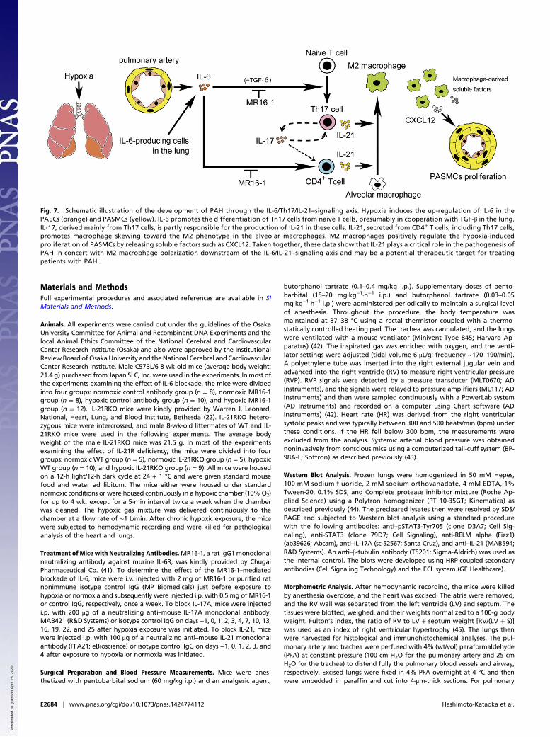
Fig. 7. Schematic illustration of the development of PAH through the IL-6/Th17/IL-21–signaling axis. Hypoxia induces the up-regulation of IL-6 in thePAECs (orange) and PASMCs (yellow). IL-6 promotes the differentiation of Th17 cells from naive T cells, presumably in cooperation with TGF-β in the lung.IL-17, derived mainly from Th17 cells, is partly responsible for the production of IL-21 in these cells. IL-21, secreted from CD4+ T cells, including Th17 cells,promotes macrophage skewing toward the M2 phenotype in the alveolar macrophages. M2 macrophages positively regulate the hypoxia-inducedproliferation of PASMCs by releasing soluble factors such as CXCL12. Taken together, these data show that IL-21 plays a critical role in the pathogenesis ofPAH in concert with M2 macrophage polarization downstream of the IL-6/IL-21–signaling axis and may be a potential therapeutic target for treatingpatients with PAH.
E2684 | www.pnas.org/cgi/doi/10.1073/pnas.1424774112 Hashimoto-Kataoka et al.
Dow
nloa
ded
by g
uest
on
Apr
il 23
, 202
0

vascular morphometry, paraffin-embedded lung sections were subjectedto elastic Van Gieson staining. Images of arteries were captured with afluorescence microscope (BZ-9000; Keyence). Morphometry was performedon lung sections obtained from five or six randomly chosen animals in eachtreatment group. Pulmonary remodeling was assessed by the percent wallthickness of parenchymal pulmonary arteries classified into the small arteries(terminal bronchioles) and arterioles (acini or alveolar ducts). The percentwall thickness was the medial wall thickness (the distance between the in-ternal and external lamina) × 2 divided by the diameter of the vessel (thedistance between the external lamina) × 100. For vessels with a single elasticlamina, the distance between the elastic and endothelial basement mem-brane was measured. Medial thickness was analyzed only for sectionedvessels exhibiting an approximately circular profile. The diameter of pul-monary arteries was determined by Image J software (National Institutes ofHealth). The percent wall thickness was calculated for at least 10 smallpulmonary arteries and arterioles for each mouse.
Lung Specimens from Patients with IPAH. All the experiments using humanspecimens were approved by the Institutional Review Board of Osaka Uni-versity, Suita, Japan. The lung tissues were obtained from four patients withIPAH undergoing lung transplantation and from a control patient un-dergoing surgery for lung cancer at a site far away from the tumor margins.All lung specimens were procured at Osaka University Hospital. Before sur-gery, all patients provided written consent for the use of their lung tissues forbiomedical research. The resected lung tissues were fixed in 10% PFA. Patientcharacteristics are shown in Table S3.
Histological and Immunohistochemical Analyses of the Pulmonary Vasculatures.For the immunohistochemical analyses of murine lung tissues, samples werefixed in 4% PFA/PBS for 1 h, cryoprotected with PBS containing 5–20% su-crose, frozen in OCT compound (Sakura), and cut into 10-μm-thick cryosectionsas described previously (46). Human lung samples, which were formaldehyde-fixed and paraffin-embedded, were cut into 5-μm-thick sections, deparaffi-nized, and rehydrated. The human sections were subjected to antigen retrievaleither by heat induction in 10 mM sodium citrate buffer, pH 6.0, or by pro-teinase K digestion for 3 min. All lung sections then were washed in PBS,treated with 1% hydrogen peroxide in methanol for 30 min, incubated inblocking solution containing either 1% BSA or 5% skim milk in PBS and 0.1%Triton-X (PBST) for 1 h, and incubated with primary antibody in PBST overnightat 4 °C or 25 °C. The following primary antibodies were used for immuno-staining: anti-RELM alpha (Fizz1) (1:100 dilution; ab39626; Abcam); anti–IL-6(1:200 dilution; ab6672; Abcam); anti– p-HH3 (Ser10) (1:100 dilution; 06–570;Upstate); anti-Ki67 (1:200 dilution; ab16667; Abcam); anti-Actin, alpha-SmoothMuscle-Cy3 (1:200 dilution; C6198; Sigma-Aldrich); anti-Arg1 (1:1,000 dilution;HPA024006; Sigma-Aldrich); anti-mannose receptor (MRC1) (1:1,000 dilution;ab64693; Abcam); and anti–IL-21 (1:200 dilution; NBP1-02706; Novus). Next,the sections were washed in PBST and incubated with fluorescence-conjugated(1:200 dilution; Alexa Fluor 488- or 546-coupled; Invitrogen) or HRP-coupled(1:200 or 1:400 dilution; 7074; Cell Signaling Technology) secondary antibodiesfor 1 h at room temperature, and, if necessary, were visualized using 3,3′-diaminobenzidine substrate (Sigma). Images were acquired with a fluorescencemicroscope (BZ-9000; Keyence). Negative control sections for the immuno-histochemical experiments received identical treatment except for exposure tothe primary antibodies and showed no specific staining. Fizz1+ cells werecounted in 10–20 high-power fields (magnification: 400×) per mouse. p-HH3+
cells and Ki67+ cells were counted in at least 10 pulmonary arteries for eachmouse. Arg1+, MRC1+, and IL-21+ cells were counted in at least 10 pulmonaryarteries or at least 10 high-power fields (magnification: 200×) in the alveolararea from each human sample.
Flow Cytometry Analysis. Lung tissuewas incubated directly for 40min to 1 h at37 °C in DMEM containing collagenase and DNase1 (1 U/mL) (Takara). Thetissue suspensions then were pressed through a 40-μm mesh, pelleted, resus-
pended in PBS-F (PBS containing 2% FBS), and the cells were washed twicewith PBS-F. The cells were preincubated with an antibody to CD16/32 to blockFcγ receptors and then were washed and incubated with the indicated fluo-rophore-conjugated antibody (CD4) for 30 min in a total volume of 100 μL PBS-F.For intracellular cytokine staining, the cells first were stimulated for 5 h incomplete medium in the presence of 25 ng/mL phorbol12-myristate13-acetate,1 μg/mL ionomycin, and 10 μg brefeldin A (Sigma-Aldrich) and then werestained for CD4, followed by fixation and permeabilization using the MouseFoxp3 Buffer Set (BD Pharmingen) according to the manufacturer’s in-structions. After two washes, the cells were stained for 30 min on ice withfluorophore-conjugated antibodies (anti–IFN-γ, anti–IL-4, anti–IL-17A, anti–IL-21) or the appropriate isotype control antibodies. The purity of the cells iso-lated from BALF was assessed by using the fluorophore-conjugated antibodiesagainst CD45, F4/80, and CD11c or the appropriate isotype control antibodies.Dead cells were excluded by propidium iodide. The cells then were analyzedon a FACSCanto (BD Biosciences), followed by analysis with FlowJo software(Tri-Star). The following antibodies were used for flow cytometry analyses:FITC-conjugated anti-CD4 (RM4-5), PE-conjugated anti–IFN-γ (XMG1.2), APC-conjugated anti–IL-4 (11B11), APC-conjugated anti–IL-17A (eBio17B7), PE-conjugated anti–IL-21 (mhalx21), and APC-conjugated anti-F4/80 (BM8) fromeBioscience and PE-conjugated anti-CD45 (30-F11) and FITC-conjugated anti-CD11c (HL3) from BD Biosciences.
qRT-PCR. Total RNA from the mouse lung or alveolar macrophages wasextracted using TRIzol reagent (Invitrogen). qRT-PCR was carried out usingthe QuantiFast SYBRGreen RT-PCR kit (Qiagen) as described previously (47).For each reaction, 80 ng of total RNA was transcribed for 10 min at 50 °C,followed by a denaturing step at 95 °C for 5 min and 40 cycles of 10 s at 95 °Cand 30 s at 60 °C. Fluorescence data were collected and analyzed using anABI PRISM 7900HT. The following primers were used: Gapdh: 5′-TCTCCA-CACCTATGGTGCAA-3′, 5′-CAAGAAACAGGGGAGCTGAG-3′; Fizz1: 5′-CCCTT-CTCATCTGCATCTCC-3′, 5′-AGGAGGCCCATCTGTTCATA-3′; Arg1: 5′-GTGAA-GAACCCACGGTCTGT-3′, 5′-CTGGTTGTCAGGGGAGTGTT-3′; Chi3I3: 5′-CCCA-CCAGGAAAGTACACAG-3′, 5′-GAGGGAAATGTCTCTGGTGA-3′; Mrc1: 5′-CG-CGAGGCAATTTTTAATCT-3′, 5′-ATTTGCATTGCCCAGTAAGG-3′; Cxcl12: 5′-GGTTC-TTCGAGAGCCACATC-3′, 5′-TAATTTCGGGTCAATGCACA-3′; Il17a: 5′-TCCAGAA-GGCCCTCAGACTA-3′, 5′-CTCGACCCTGAAAGTGAAGG-3′; Rorc: 5′-AACCAGG-CATCCTGAACTTG-3′, 5′-CGTAGAAGGTCCTCCAGTCG-3′; Cxcl1: 5′-GCCTATC-GCCAATGAGCTG-3′, 5′-TCTGAACCAAGGGAGCTTCA-3′; Cxcl5: 5′-CTGCCCCTT-CCTCAGTCATA-3′, 5′-TGGATCCAGACAGACCTCCT-3′; Il21: 5′-GGACAGTGGCCC-ATAAATCA -3′, 5′- CAGGGTTTGATGGCTTGAGT -3′; Il6: 5′- TGTGCAATGGCAATT-CTGAT-3′, 5′-GGTACTCCAGAAGACCAGAGGA-3′; Nos2: 5′-GCTCATGACATCGAC-CAGAA-3′, 5′-TGTTGCATTGGAAGTGAAGC-3′; Il12b: 5′-AGGTCACACTGGACCA-AAGG-3′, 5′-AGGGTACTCCCAGCTGACCT-3′; Tnf-α: 5′-TGCCTATGTCTCAGCCTCTTC-3′, 5′-GGTCTGGGCCATAGAACTGA-3′.
Statistics. All data are expressed as the mean ± SEM. Differences amongmultiple groups were compared by one-way ANOVA followed by a post hoccomparison tested with Scheffé’s method. The Student’s t test was used toanalyze differences between two groups. P < 0.05 was considered statisticallysignificant.
ACKNOWLEDGMENTS. We thank Yuka Yoshimoto and Yumi Yanagisawa forsecretarial assistance; Kaori Yamamoto and Manami Sone (National Cerebraland Cardiovascular Research Center) for technical assistance; Yoshito Takeda,Yingji Jin (Osaka University), Yasunobu Arima (Hokkaido University), HatsueUeda, Naoki Mochizuki (National Cerebral Cardiovascular Research Center),Susumu Nakae, and Tomokazu Sumida (University of Tokyo) for helpful discus-sions and suggestions; Warren J. Leonard (National, Heart, Lung, and BloodInstitute) for providing IL-21RKO mice; and Chugai Pharmaceutical Co. for pro-viding the anti–IL-6R antibody MR16-1. This work was supported in part byGrant KAKENHI-25670386 from the Ministry of Education, Science, Sports andCulture of Japan (to Y.N.) and by a grant from Precursory Research for Embry-onic Science and Technology, Japan Science Technology Agency (to Y.N.).
1. Pietra GG, et al. (2004) Pathologic assessment of vasculopathies in pulmonary hy-
pertension. J Am Coll Cardiol 43(12, Suppl S):25S–32S.2. Rabinovitch M (2012) Molecular pathogenesis of pulmonary arterial hypertension.
J Clin Invest 122(12):4306–4313.3. Dorfmüller P, Perros F, Balabanian K, Humbert M (2003) Inflammation in pulmonary
arterial hypertension. Eur Respir J 22(2):358–363.4. Schermuly RT, Ghofrani HA, Wilkins MR, Grimminger F (2011) Mechanisms of disease:
Pulmonary arterial hypertension. Nat Rev Cardiol 8(8):443–455.5. Tuder RM, Groves B, Badesch DB, Voelkel NF (1994) Exuberant endothelial cell growth
and elements of inflammation are present in plexiform lesions of pulmonary hyper-
tension. Am J Pathol 144(2):275–285.
6. Tuder RM, Voelkel NF (1998) Pulmonary hypertension and inflammation. J Lab Clin
Med 132(1):16–24.7. Kishimoto T (2006) Interleukin-6: Discovery of a pleiotropic cytokine. Arthritis Res
Ther 8(Suppl 2):S2.8. Kishimoto T, Akira S, Taga T (1992) Interleukin-6 and its receptor: A paradigm for
cytokines. Science 258(5082):593–597.9. Humbert M, et al. (1995) Increased interleukin-1 and interleukin-6 serum concentra-
tions in severe primary pulmonary hypertension. Am J Respir Crit Care Med 151(5):
1628–1631.10. Soon E, et al. (2010) Elevated levels of inflammatory cytokines predict survival in id-
iopathic and familial pulmonary arterial hypertension. Circulation 122(9):920–927.
Hashimoto-Kataoka et al. PNAS | Published online May 4, 2015 | E2685
MED
ICALSC
IENCE
SPN
ASPL
US
Dow
nloa
ded
by g
uest
on
Apr
il 23
, 202
0

11. Steiner MK, et al. (2009) Interleukin-6 overexpression induces pulmonary hyperten-sion. Circ Res 104(2):236–244.
12. Savale L, et al. (2009) Impact of interleukin-6 on hypoxia-induced pulmonary hyper-tension and lung inflammation in mice. Respir Res 10:6.
13. Iwakura Y, Ishigame H, Saijo S, Nakae S (2011) Functional specialization of inter-leukin-17 family members. Immunity 34(2):149–162.
14. Korn T, Bettelli E, Oukka M, Kuchroo VK (2009) IL-17 and Th17 Cells. Annu Rev Im-munol 27:485–517.
15. Bettelli E, et al. (2006) Reciprocal developmental pathways for the generation ofpathogenic effector TH17 and regulatory T cells. Nature 441(7090):235–238.
16. Biswas SK, Mantovani A (2010) Macrophage plasticity and interaction with lympho-cyte subsets: Cancer as a paradigm. Nat Immunol 11(10):889–896.
17. Mosser DM, Edwards JP (2008) Exploring the full spectrum of macrophage activation.Nat Rev Immunol 8(12):958–969.
18. Wynn TA, Chawla A, Pollard JW (2013) Macrophage biology in development, ho-meostasis and disease. Nature 496(7446):445–455.
19. Vergadi E, et al. (2011) Early macrophage recruitment and alternative activation arecritical for the later development of hypoxia-induced pulmonary hypertension. Cir-culation 123(18):1986–1995.
20. Tanaka T, Narazaki M, Kishimoto T (2012) Therapeutic targeting of the interleukin-6receptor. Annu Rev Pharmacol Toxicol 52:199–219.
21. Liu SM, King C (2013) IL-21-producing Th cells in immunity and autoimmunity.J Immunol 191(7):3501–3506.
22. Ozaki K, et al. (2002) A critical role for IL-21 in regulating immunoglobulin pro-duction. Science 298(5598):1630–1634.
23. Takeda Y, et al. (2011) Macrophage skewing by Phd2 haplodeficiency prevents is-chaemia by inducing arteriogenesis. Nature 479(7371):122–126.
24. Mangan PR, et al. (2006) Transforming growth factor-beta induces development ofthe T(H)17 lineage. Nature 441(7090):231–234.
25. Veldhoen M, Hocking RJ, Atkins CJ, Locksley RM, Stockinger B (2006) TGFbeta in thecontext of an inflammatory cytokine milieu supports de novo differentiation of IL-17-producing T cells. Immunity 24(2):179–189.
26. Choy EH, et al. (2002) Therapeutic benefit of blocking interleukin-6 activity with ananti-interleukin-6 receptor monoclonal antibody in rheumatoid arthritis: A random-ized, double-blind, placebo-controlled, dose-escalation trial. Arthritis Rheum 46(12):3143–3150.
27. Nishimoto N, et al. (2004) Treatment of rheumatoid arthritis with humanized anti-interleukin-6 receptor antibody: A multicenter, double-blind, placebo-controlled trial.Arthritis Rheum 50(6):1761–1769.
28. Fujimoto M, et al. (2008) Interleukin-6 blockade suppresses autoimmune arthritis in miceby the inhibition of inflammatory Th17 responses. Arthritis Rheum 58(12):3710–3719.
29. Iwanami K, et al. (2008) Crucial role of the interleukin-6/interleukin-17 cytokine axis inthe induction of arthritis by glucose-6-phosphate isomerase. Arthritis Rheum 58(3):754–763.
30. Serada S, et al. (2008) IL-6 blockade inhibits the induction of myelin antigen-specific
Th17 cells and Th1 cells in experimental autoimmune encephalomyelitis. Proc Natl
Acad Sci USA 105(26):9041–9046.31. Hsu E, et al. (2011) Lung tissues in patients with systemic sclerosis have gene ex-
pression patterns unique to pulmonary fibrosis and pulmonary hypertension. Arthritis
Rheum 63(3):783–794.32. Chaouat A, et al. (2009) Role for interleukin-6 in COPD-related pulmonary hyper-
tension. Chest 136(3):678–687.33. Daley E, et al. (2008) Pulmonary arterial remodeling induced by a Th2 immune re-
sponse. J Exp Med 205(2):361–372.34. Minamino T, et al. (2001) Targeted expression of heme oxygenase-1 prevents the
pulmonary inflammatory and vascular responses to hypoxia. Proc Natl Acad Sci USA
98(15):8798–8803.35. Dienz O, et al. (2009) The induction of antibody production by IL-6 is indirectly me-
diated by IL-21 produced by CD4+ T cells. J Exp Med 206(1):69–78.36. Nurieva R, et al. (2007) Essential autocrine regulation by IL-21 in the generation of
inflammatory T cells. Nature 448(7152):480–483.37. Korn T, et al. (2007) IL-21 initiates an alternative pathway to induce proinflammatory
T(H)17 cells. Nature 448(7152):484–487.38. Zhou L, et al. (2007) IL-6 programs T(H)-17 cell differentiation by promoting se-
quential engagement of the IL-21 and IL-23 pathways. Nat Immunol 8(9):967–974.39. Pesce J, et al. (2006) The IL-21 receptor augments Th2 effector function and alter-
native macrophage activation. J Clin Invest 116(7):2044–2055.40. Stolfi C, et al. (2011) Involvement of interleukin-21 in the regulation of colitis-asso-
ciated colon cancer. J Exp Med 208(11):2279–2290.41. Takagi N, et al. (1998) Blockage of interleukin-6 receptor ameliorates joint disease in
murine collagen-induced arthritis. Arthritis Rheum 41(12):2117–2121.42. Sonobe T, et al. (2011) Imaging of the closed-chest mouse pulmonary circulation using
synchrotron radiation microangiography. J Appl Physiol (1985) 111(1):75–80.43. Higuchi K, et al. (2012) Endothelial Gab1 deletion accelerates angiotensin II-dependent
vascular inflammation and atherosclerosis in apolipoprotein E knockout mice. Circ J
76(8):2031–2040.44. Nakaoka Y, et al. (2007) Gab family proteins are essential for postnatal mainte-
nance of cardiac function via neuregulin-1/ErbB signaling. J Clin Invest 117(7):
1771–1781.45. Ogura S, et al. (2013) Oxidative stress augments pulmonary hypertension in chroni-
cally hypoxic mice overexpressing the oxidized LDL receptor. Am J Physiol Heart Circ
Physiol 305(2):H155–H162.46. Arita Y, et al. (2014) Myocardium-derived angiopoietin-1 is essential for coronary vein
formation in the developing heart. Nat Commun 5:4552.47. Shioyama W, et al. (2011) Docking protein Gab1 is an essential component of post-
natal angiogenesis after ischemia via HGF/c-met signaling. Circ Res 108(6):664–675.
E2686 | www.pnas.org/cgi/doi/10.1073/pnas.1424774112 Hashimoto-Kataoka et al.
Dow
nloa
ded
by g
uest
on
Apr
il 23
, 202
0