interferences with urinary elimination. urinary elimination “ bones can break, muscles can...
TRANSCRIPT

Interferences Interferences with with
Urinary Elimination Urinary Elimination

Urinary EliminationUrinary Elimination
““Bones can break, muscles can Bones can break, muscles can atrophy, glands can loaf, even the atrophy, glands can loaf, even the
brain can go to sleep without brain can go to sleep without immediate danger to survival. But immediate danger to survival. But should the kidneys fail….neither should the kidneys fail….neither
bone, muscle, gland, nor brain could bone, muscle, gland, nor brain could carry on.”carry on.”

Urinary SystemUrinary System


KidneysKidneysMacrostructureMacrostructure
Paired, reddish, brown bean-shaped organsPaired, reddish, brown bean-shaped organs
Location: Location: Retroperitoneal on either side of the Retroperitoneal on either side of the
vertebral columnvertebral column1212thth thoracic vertebrae to 3 thoracic vertebrae to 3rdrd Lumbar Lumbar
Left kidney is 1.5 to 2 cm higher than rightLeft kidney is 1.5 to 2 cm higher than rightWeight: 115 – 175 gms (4-6 ounces)Weight: 115 – 175 gms (4-6 ounces)Adrenal Gland lies on top of each kidneyAdrenal Gland lies on top of each kidney


KidneysKidneysMicrostructureMicrostructure
NephronNephron -- Functional unit of kidney, forms -- Functional unit of kidney, forms urineurine
Each kidney has 1 million nephronsEach kidney has 1 million nephrons
Each Nephron is composed of: Each Nephron is composed of:
CortexCortex: glomerulus, Bowman’s capsule, : glomerulus, Bowman’s capsule, proximal convoluted tubule, distal convoluted proximal convoluted tubule, distal convoluted tubuletubule
MedullaMedulla: The loop of Henle and collecting ducts: The loop of Henle and collecting ducts


KidneysKidneysMicrostructureMicrostructure
GlomerulusGlomerulus – selective filtration – selective filtrationBowman’s Capsule – semipermeable membrane / Bowman’s Capsule – semipermeable membrane /
hydrostatic pressure changeshydrostatic pressure changesProximal tubuleProximal tubule: Active transport: Active transport
Reabsorption of 80% of electrolytes & waterReabsorption of 80% of electrolytes & waterReabsorption of all glucose & amino acidsReabsorption of all glucose & amino acidsReabsorption of HCO3- Acid-Base BalanceReabsorption of HCO3- Acid-Base BalanceReabsorption of CreatinineReabsorption of Creatinine
Loop of HenleLoop of Henle: : Reabsorption of Na+ & Cl- in ascending limbReabsorption of Na+ & Cl- in ascending limbReabsorption of water in descending loopReabsorption of water in descending loopConcentration of filtrateConcentration of filtrate

KidneysKidneysMicrostructureMicrostructure
Distal Tubule:Distal Tubule:Secretion of K+, H+, ammoniaSecretion of K+, H+, ammoniaReabsorption of water (regulated by ADH)Reabsorption of water (regulated by ADH)Reabsorption of HCO3- -- Acid-Base BalanceReabsorption of HCO3- -- Acid-Base BalanceRegulation of Ca++ and PO4- by parathyroid Regulation of Ca++ and PO4- by parathyroid
hormonehormoneRegulation of NA+ & K+ by aldosteroneRegulation of NA+ & K+ by aldosterone
Collecting Duct:Collecting Duct:Reabsorption of water (ADH required)Reabsorption of water (ADH required)
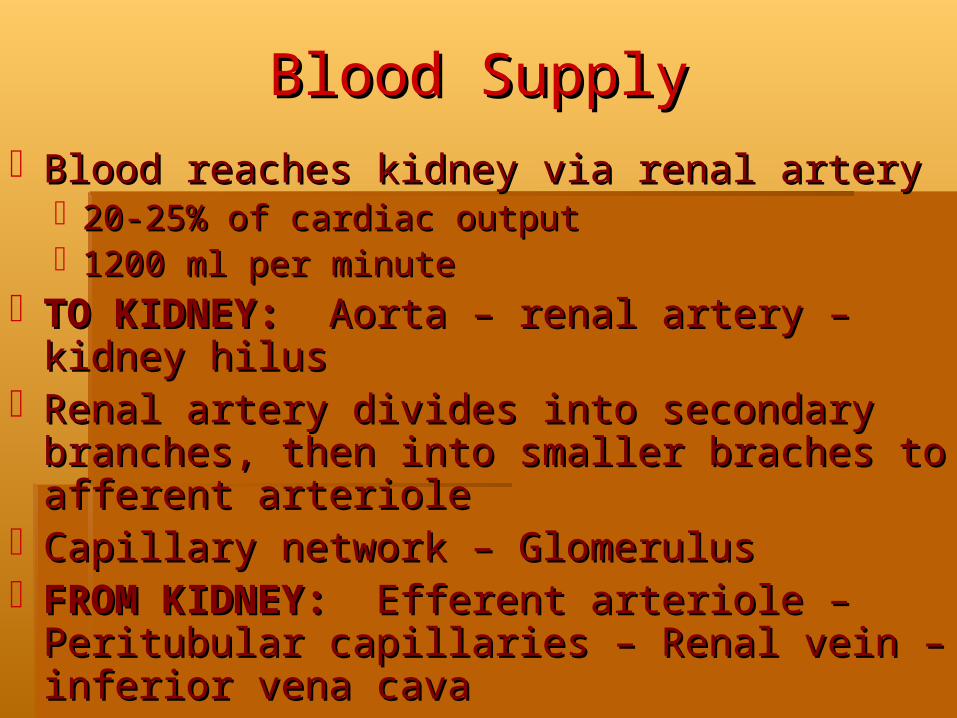
Blood SupplyBlood SupplyBlood reaches kidney via renal arteryBlood reaches kidney via renal artery
20-25% of cardiac output20-25% of cardiac output1200 ml per minute1200 ml per minute
TO KIDNEY:TO KIDNEY: Aorta – renal artery – kidney Aorta – renal artery – kidney hilus hilus
Renal artery divides into secondary branches, Renal artery divides into secondary branches, then into smaller braches to afferent arteriolethen into smaller braches to afferent arteriole
Capillary network – GlomerulusCapillary network – GlomerulusFROM KIDNEY:FROM KIDNEY: Efferent arteriole – Efferent arteriole –
Peritubular capillaries – Renal vein – inferior Peritubular capillaries – Renal vein – inferior vena cavavena cava

Nephron FunctionNephron FunctionPhysiology of Urine Physiology of Urine
FormationFormation
Normal glomerular function-Normal glomerular function- Urine formation Urine formation starts at glomerulus where blood is filteredstarts at glomerulus where blood is filtered
GFR-(Glomerular filtration rateGFR-(Glomerular filtration rate)- amt of blood )- amt of blood filtered by glomeruli in a given timefiltered by glomeruli in a given time
Normal GFR-Normal GFR- 125ml/minute, however only 1 ml 125ml/minute, however only 1 ml per minute becomes urine, most is reabsorbedper minute becomes urine, most is reabsorbed

Nephron FunctionNephron Function

Renal FunctionRenal Function

Other Kidney FunctionsOther Kidney FunctionsHormone ProductionHormone Production
ErythropoietinErythropoietinIn response to hypoxia & decreased renal blood flowIn response to hypoxia & decreased renal blood flowStimulates RBC production in the bone marrowStimulates RBC production in the bone marrowDeficiencies lead to anemia in renal failureDeficiencies lead to anemia in renal failure
ReninReninReleased from juxtaglomerular apparatus of the nephronReleased from juxtaglomerular apparatus of the nephronIn response to < arterial BP, renal ischemia, > NA+ In response to < arterial BP, renal ischemia, > NA+
concentrationconcentrationSplits angiotensinogen into angiotensin I = angiotensin IISplits angiotensinogen into angiotensin I = angiotensin IIStimulates aldosterone from the adrenal cortex = water + Stimulates aldosterone from the adrenal cortex = water +
NA+ retention + peripheral vasoconstrictionNA+ retention + peripheral vasoconstriction

Renin-angiotensin-Renin-angiotensin-aldosterone Systemaldosterone System

Other Kidney FunctionsOther Kidney FunctionsHormone ProductionHormone Production
Prostaglandins (PGs)Prostaglandins (PGs) Kidney medullaKidney medullaVasodilating action = increases renal blood flow Vasodilating action = increases renal blood flow
and promotes NA+ excretionand promotes NA+ excretionCounteracts the vasoconstrictor effect of Counteracts the vasoconstrictor effect of
angiotensin and norepinephrineangiotensin and norepinephrineLowers arterial BP by decreasing systemic vascular Lowers arterial BP by decreasing systemic vascular
resistanceresistanceActive metabolite of Vitamin DActive metabolite of Vitamin D – second step – second step
in activating Vitamin D after action of ultraviolet in activating Vitamin D after action of ultraviolet radiation on cholesterol in the skinradiation on cholesterol in the skin

Nursing ProcessNursing ProcessAlterations in Urinary Alterations in Urinary
FunctionFunctionAssessmentAssessment: Patient history: Patient historyPhysical Assessment: Physical Assessment:
inspection, percussion, palpationinspection, percussion, palpationAssessment of UrineAssessment of Urine: color, clarity, odor: color, clarity, odorUrine testing & specimen collectionUrine testing & specimen collection
Urinalysis, C&S, Composite urine collection Urinalysis, C&S, Composite urine collection Creatinine Clearance – 85-135ml/minCreatinine Clearance – 85-135ml/min
Diagnostic testsDiagnostic tests: KUB (kidney, ureter, bladder) : KUB (kidney, ureter, bladder) renal ultrasound, renal CT scanrenal ultrasound, renal CT scan
Invasive:Invasive: IVP, Cystoscopy, arteriogram, IVP, Cystoscopy, arteriogram, urodymanicsurodymanics

Upper Upper Urinary Tract InfectionsUrinary Tract Infections
Acute PyelonephritisAcute PyelonephritisChronic PyelonephritisChronic PyelonephritisAcute GlomerulonephritisAcute GlomerulonephritisAcute Poststreptococcal Acute Poststreptococcal
GlomerulonephritisGlomerulonephritisChronic GlomerulonephritisChronic Glomerulonephritis

Acute PyelonephritisAcute Pyelonephritis
Inflammation of the renal parenchyma and Inflammation of the renal parenchyma and collecting systemcollecting system
Most common cause: Most common cause: bacterial bacterial (E.coli, (E.coli, Proteus, Klebsiella, Enterobacter species)Proteus, Klebsiella, Enterobacter species)
Pre-existing factor: vesicoureteral refluxPre-existing factor: vesicoureteral refluxCommonly begins in the renal medulla and Commonly begins in the renal medulla and
spreads to the adjacent cortexspreads to the adjacent cortexRecurring episodes may lead to chronic Recurring episodes may lead to chronic
pyelonephritispyelonephritisUrosepsisUrosepsis: bacteriuria and bacteremia: bacteriuria and bacteremia
Acute PyelonephritisAcute Pyelonephritis
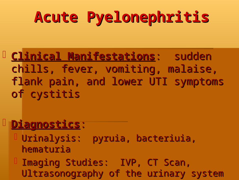
Clinical ManifestationsClinical Manifestations: sudden chills, fever, : sudden chills, fever, vomiting, malaise, flank pain, and lower UTI vomiting, malaise, flank pain, and lower UTI symptoms of cystitissymptoms of cystitis
DiagnosticsDiagnostics::Urinalysis: pyruia, bacteriuia, hematuriaUrinalysis: pyruia, bacteriuia, hematuriaImaging Studies: IVP, CT Scan, Ultrasonography Imaging Studies: IVP, CT Scan, Ultrasonography
of the urinary systemof the urinary system

Acute PyelonephritisAcute PyelonephritisMedical Management for Mild Symptoms:Medical Management for Mild Symptoms:
Short hosp stay for IV antibiotic or OP oral Short hosp stay for IV antibiotic or OP oral antibioticsantibioticsEmpiric broad spectrum (Ampicillin / Vancomycin) Empiric broad spectrum (Ampicillin / Vancomycin)
combined with aminoglycoside combined with aminoglycoside Change to sensitivity-guided therapy when culture results Change to sensitivity-guided therapy when culture results
are available for 14-21 daysare available for 14-21 daysSulfa—Bactrim / Cipro / FloxinSulfa—Bactrim / Cipro / Floxin
Adequate fluid intakeAdequate fluid intakeNonsteroidal antiinflammatory drugsNonsteroidal antiinflammatory drugsAntipyretic drugsAntipyretic drugsUrinary analgesics – PyridiumUrinary analgesics – PyridiumFollow-up culturesFollow-up cultures & imaging studies & imaging studies
Relapse may occur – treated with 6-week course of antibiotics Relapse may occur – treated with 6-week course of antibiotics Antibiotic prophylaxisAntibiotic prophylaxis
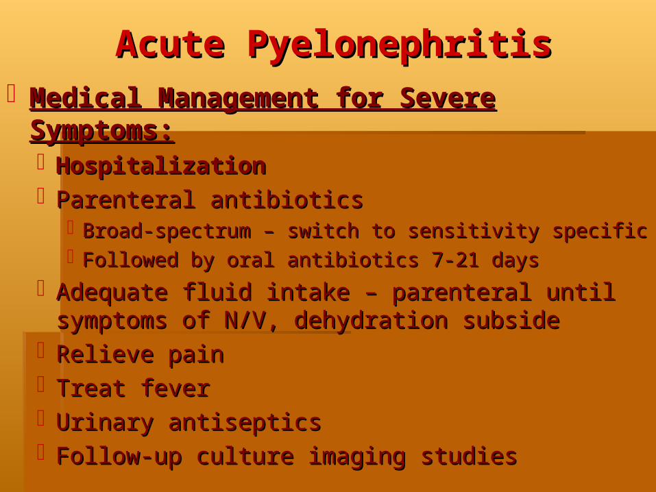
Acute PyelonephritisAcute PyelonephritisMedical Management for Severe Symptoms:Medical Management for Severe Symptoms:
HospitalizationHospitalizationParenteral antibioticsParenteral antibiotics
Broad-spectrum – switch to sensitivity specificBroad-spectrum – switch to sensitivity specificFollowed by oral antibiotics 7-21 daysFollowed by oral antibiotics 7-21 days
Adequate fluid intake – parenteral until symptoms Adequate fluid intake – parenteral until symptoms of N/V, dehydration subsideof N/V, dehydration subside
Relieve painRelieve painTreat feverTreat feverUrinary antisepticsUrinary antisepticsFollow-up culture imaging studiesFollow-up culture imaging studies

Chronic PyelonephritisChronic PyelonephritisTerm used to describe a kidney that has Term used to describe a kidney that has
lost function due to scarring and fibrosislost function due to scarring and fibrosisResult of chronic upper urinary tract infectionsResult of chronic upper urinary tract infectionsOther namesOther names: interstitial nephritis, chronic : interstitial nephritis, chronic
atrophic pyelonephritis, reflux nephropathyatrophic pyelonephritis, reflux nephropathy
Level of renal function depends onLevel of renal function depends on: : whether one or both kidneys are affectedwhether one or both kidneys are affectedmagnitude of scarringmagnitude of scarringthe presence of co-existing infectionthe presence of co-existing infection
Progresses to end-stage renal disease Progresses to end-stage renal disease when both kidneys are affectedwhen both kidneys are affected

Acute GlomerulonephritisAcute GlomerulonephritisImmunologic process resulting in inflammation of Immunologic process resulting in inflammation of
the glomerulithe glomeruliUsually affects both kidneys equallyUsually affects both kidneys equallyTubular, interstitial, and vascular changes occurTubular, interstitial, and vascular changes occur
Etiology:Etiology:Two types:Two types:
Antibodies have specificity for antigens within the glomerular Antibodies have specificity for antigens within the glomerular basement membrane (GBM) – produce autoantibodies – to one’s basement membrane (GBM) – produce autoantibodies – to one’s own tissue -- mechanism unknownown tissue -- mechanism unknown
Antibodies react with circulating nonglomerular antigens and are Antibodies react with circulating nonglomerular antigens and are randomly deposited as immune complexes along the GBMrandomly deposited as immune complexes along the GBM
End result: glomerular injury as a result of inflammationEnd result: glomerular injury as a result of inflammation

Acute GlomerulonephritisAcute GlomerulonephritisClinical ManifestationsClinical Manifestations::
Varying degrees of hematuriaVarying degrees of hematuriaVarying degrees of urinary excretion of WBC and castsVarying degrees of urinary excretion of WBC and castsProteinuriaProteinuriaElevated BUN and Creatinine and AlbuminElevated BUN and Creatinine and Albumin+ renal biopsy+ renal biopsy
Medical Management:Medical Management:RestRestSodium and fluid restrictionSodium and fluid restrictionDiureticsDiureticsAntihypertensive therapyAntihypertensive therapyDecreased dietary proteinDecreased dietary protein

GlomerulonephritisGlomerulonephritis

Chronic GlomerulonephritisChronic GlomerulonephritisSyndromeSyndrome – end-stage glomerular inflammatory – end-stage glomerular inflammatory
diseasediseaseProteinuria, hematuria, slow development of uremic Proteinuria, hematuria, slow development of uremic
syndrome = syndrome = decreased renal functiondecreased renal functionSlow course toward renal failure over a few to as Slow course toward renal failure over a few to as
many as 30 yearsmany as 30 yearsOften found coincidentally with abnormal UA or Often found coincidentally with abnormal UA or
elevated blood pressureelevated blood pressureConfirmed with ultrasound and CT scan – Renal BxConfirmed with ultrasound and CT scan – Renal BxMedical ManagementMedical Management: :
Treat HTNTreat HTNTreat UTIsTreat UTIsProtein and Phosphate restrictionProtein and Phosphate restriction

Acute Poststreptococcal Acute Poststreptococcal GlomerulonephritisGlomerulonephritis
Most common in children & young adultsMost common in children & young adults5-21 days after a streptococcal sore throat or 5-21 days after a streptococcal sore throat or
impetigoimpetigoNephrotoxic strains of group A B-hemolytic Nephrotoxic strains of group A B-hemolytic
streptococcistreptococciAntibodies are produced to the strept antigenAntibodies are produced to the strept antigenUnknown mechanism – the antigen-antibody Unknown mechanism – the antigen-antibody
complexes are deposited in the glomeruli – complexes are deposited in the glomeruli – leads to = decreased glomerular filtration & leads to = decreased glomerular filtration & inflammation inflammation
Acute Poststreptococcal Acute Poststreptococcal GlomerulonephritisGlomerulonephritis
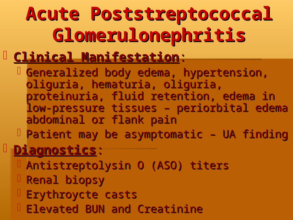
Clinical ManifestationClinical Manifestation::Generalized body edema, hypertension, oliguria, Generalized body edema, hypertension, oliguria,
hematuria, oliguria, proteinuria, fluid retention, hematuria, oliguria, proteinuria, fluid retention, edema in low-pressure tissues – periorbital edema edema in low-pressure tissues – periorbital edema abdominal or flank painabdominal or flank pain
Patient may be asymptomatic – UA findingPatient may be asymptomatic – UA findingDiagnosticsDiagnostics::
Antistreptolysin O (ASO) titersAntistreptolysin O (ASO) titersRenal biopsyRenal biopsyErythroycte casts Erythroycte casts Elevated BUN and CreatinineElevated BUN and Creatinine

Acute Poststreptococcal Acute Poststreptococcal GlomerulonephritisGlomerulonephritis
Medical ManagementMedical Management::Rest until signs of glomerular inflammation subside Rest until signs of glomerular inflammation subside
(proteinuria & hematuria)(proteinuria & hematuria)Treat hypertensionTreat hypertensionRestrict sodium & fluid intakeRestrict sodium & fluid intakeAntibiotics only if streptococcal infection is still Antibiotics only if streptococcal infection is still
presentpresent
Prevention:Prevention: Early diagnosis & treatment of Early diagnosis & treatment of sore throats and skin lesions; good personal sore throats and skin lesions; good personal hygiene, patient adherence to antibiotic therapyhygiene, patient adherence to antibiotic therapy
Renal ConditionsRenal Conditions
Polycystic KidneyPolycystic Kidney
Renal Artery StenosisRenal Artery Stenosis
Renal TuberculosisRenal Tuberculosis
HIV—associated NephropathyHIV—associated Nephropathy
Nephrotic SyndromeNephrotic Syndrome

Polycystic KidneyPolycystic KidneyOne of the most common genetic diseasesOne of the most common genetic diseasesTwo forms:Two forms:
Childhood manifestationChildhood manifestation: rare autosomal : rare autosomal recessive disorder with rapid progressionrecessive disorder with rapid progression
Adult manifestationAdult manifestation: autosomal dominant : autosomal dominant disorder – latent – 30-40 years of agedisorder – latent – 30-40 years of age
Involves both kidneysInvolves both kidneysCortex & medulla are filled with thin-walled Cortex & medulla are filled with thin-walled
cysts that are several mm – cm in diametercysts that are several mm – cm in diameterCysts enlarge – contain blood and pus - destroy Cysts enlarge – contain blood and pus - destroy
surrounding tissuesurrounding tissue

Polycystic KidneyPolycystic Kidney
Clinical Manifestation:Clinical Manifestation:Symptoms appear when the cysts begin to enlargeSymptoms appear when the cysts begin to enlargeAbdominal and/or flank painAbdominal and/or flank painPalpable enlarged kidneysPalpable enlarged kidneysHematuriaHematuriaUTIUTIHypertensionHypertension
Diagnosis:Diagnosis:Family History, IVP, ultrasound, CT scanFamily History, IVP, ultrasound, CT scan
Usually progresses to end-stage renal Usually progresses to end-stage renal failurefailure

Renal Artery StenosisRenal Artery StenosisPartial occlusion of one or both renal arteriesPartial occlusion of one or both renal arteries
Atherosclerotic narrowing or fibromuscular Atherosclerotic narrowing or fibromuscular hyperplasiahyperplasia
1-2% of hypertension1-2% of hypertension
Diagnosis: Renal arteriogramDiagnosis: Renal arteriogramTherapy Goal:Therapy Goal:
Control hypertensionControl hypertensionRestore kidney perfusionRestore kidney perfusion
Percutaneous transluminal renal angioplastyPercutaneous transluminal renal angioplastySurgical revascularization (splenic artery or aorta)Surgical revascularization (splenic artery or aorta)

Renal Artery StenosisRenal Artery Stenosis
Renal TuberculosisRenal TuberculosisRarely a primary lesionRarely a primary lesion
Onset 5-8 years after primary pulmonary TBOnset 5-8 years after primary pulmonary TB
Initially asymptomaticInitially asymptomaticLow grade fever, when infection descends to bladder: Low grade fever, when infection descends to bladder:
polyuria, dysuria, epididymitis in menpolyuria, dysuria, epididymitis in menDiagnosis:Diagnosis: TB in Urine; IVP TB in Urine; IVPLong Term: scarring of renal parenchyma & ureteral Long Term: scarring of renal parenchyma & ureteral
stricturesstricturesEarlier the treatment – less likely renal failure will Earlier the treatment – less likely renal failure will
occuroccurFive drugs: Isoniazid (INH), rifampin, pyrazinamide, Five drugs: Isoniazid (INH), rifampin, pyrazinamide,
streptomycin, ethambutol streptomycin, ethambutol
HIV—associated HIV—associated NephropathyNephropathy
Range from mild fluid & electrolyte abnormalities to Range from mild fluid & electrolyte abnormalities to progressive renal impairment and renal failureprogressive renal impairment and renal failure
10% incidence – highest among IV drug users10% incidence – highest among IV drug usersClinical Manifestations:Clinical Manifestations:
Proteinuria & nephrotic syndromeProteinuria & nephrotic syndromeProgressive azotemia, enlarged kidney, rapid progression Progressive azotemia, enlarged kidney, rapid progression
to end-stage renal failureto end-stage renal failureAcute renal failure: most commonly seen in patients with Acute renal failure: most commonly seen in patients with
AIDS who is critically ill with HIV-related infection or AIDS who is critically ill with HIV-related infection or malignancymalignancy
Treatment: Depends on treatment of primary Treatment: Depends on treatment of primary disease - Dialysisdisease - Dialysis

Nephrotic SyndromeNephrotic Syndrome
Decreased urine outputDecreased urine outputProteinuriaProteinuriaVolume overload Volume overload CHFCHFDysrhythmiasDysrhythmiasN/VN/VUremic frostUremic frostAnemiaAnemia

Vascular & Vascular & TubularTubular Pathogenesis Pathogenesis
Nephrotic SyndromeNephrotic Syndrome
Increase in nitrogen waste in bloodIncrease in nitrogen waste in blood
Fluid and electrolyte disturbanceFluid and electrolyte disturbance
Treatment either conservative or Treatment either conservative or aggressiveaggressive

Renal Disease Renal Disease Assessment - LabsAssessment - Labs
Elevated BUNElevated BUNElevated creatinineElevated creatinineElevated potassiumElevated potassiumElevated phosphateElevated phosphateDecreased calcium Decreased calcium Decreased HCO3 and pHDecreased HCO3 and pH
Renal Disease TreatmentRenal Disease Treatment
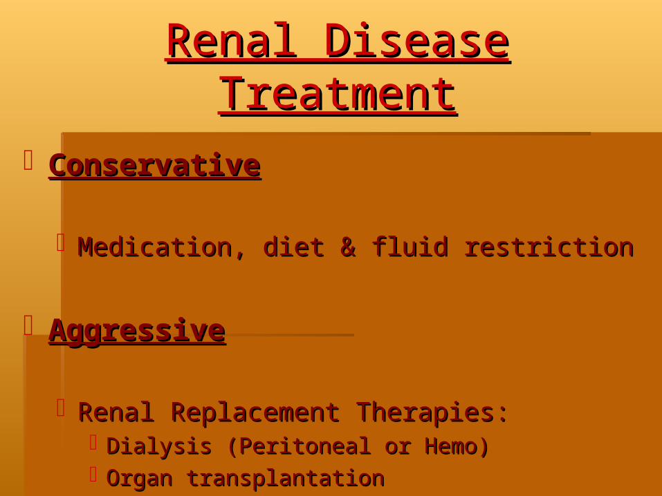
ConservativeConservative
Medication, diet & fluid restrictionMedication, diet & fluid restriction
AggressiveAggressive
Renal Replacement Therapies: Renal Replacement Therapies: Dialysis (Peritoneal or Hemo) Dialysis (Peritoneal or Hemo) Organ transplantationOrgan transplantation

Acute Renal FailureAcute Renal Failure

4+ Pitting Edema4+ Pitting Edema

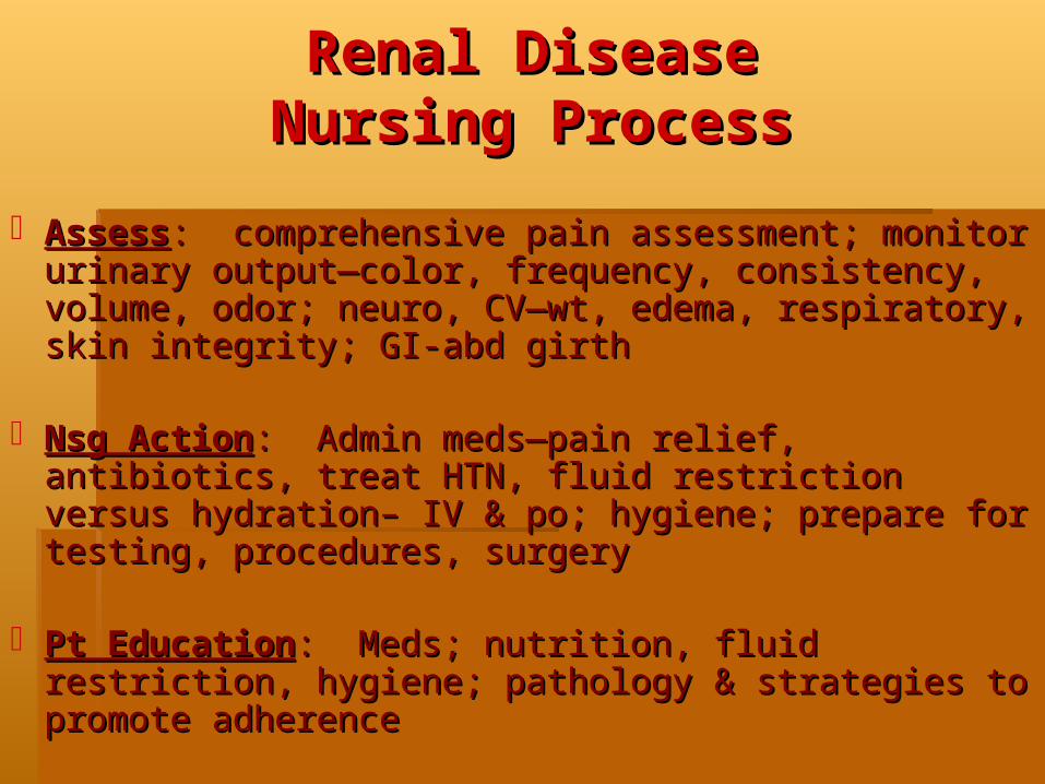
Renal DiseaseRenal DiseaseNursing ProcessNursing Process
AssessAssess: comprehensive pain assessment; monitor : comprehensive pain assessment; monitor urinary output—color, frequency, consistency, volume, urinary output—color, frequency, consistency, volume, odor; neuro, CV—wt, edema, respiratory, skin odor; neuro, CV—wt, edema, respiratory, skin integrity; GI-abd girthintegrity; GI-abd girth
Nsg ActionNsg Action: Admin meds—pain relief, antibiotics, : Admin meds—pain relief, antibiotics, treat HTN, fluid restriction versus hydration– IV & po; treat HTN, fluid restriction versus hydration– IV & po; hygiene; prepare for testing, procedures, surgeryhygiene; prepare for testing, procedures, surgery
Pt EducationPt Education: Meds; nutrition, fluid restriction, : Meds; nutrition, fluid restriction, hygiene; pathology & strategies to promote adherencehygiene; pathology & strategies to promote adherence

Peritoneal DialysisPeritoneal Dialysis

HemodialysisHemodialysis

Renal TransplantRenal Transplant
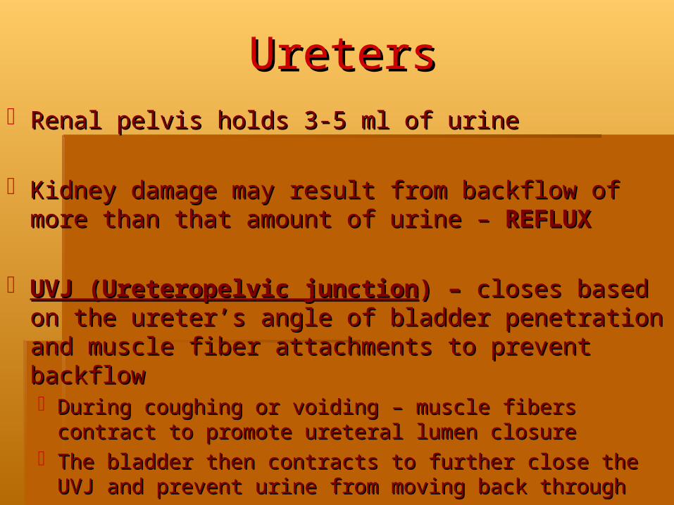
UretersUretersRenal pelvis holds 3-5 ml of urineRenal pelvis holds 3-5 ml of urine
Kidney damage may result from backflow of more Kidney damage may result from backflow of more than that amount of urine – than that amount of urine – REFLUXREFLUX
UVJ (Ureteropelvic junctionUVJ (Ureteropelvic junction) – ) – closes based on the closes based on the ureter’s angle of bladder penetration and muscle fiber ureter’s angle of bladder penetration and muscle fiber attachments to prevent backflowattachments to prevent backflow During coughing or voiding – muscle fibers contract to During coughing or voiding – muscle fibers contract to
promote ureteral lumen closurepromote ureteral lumen closureThe bladder then contracts to further close the UVJ and The bladder then contracts to further close the UVJ and
prevent urine from moving back throughprevent urine from moving back through

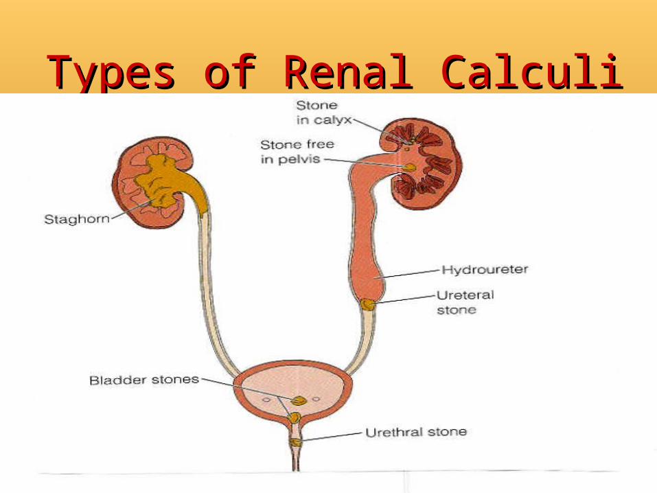
NephrolithiasisNephrolithiasis500,000 people in US annually500,000 people in US annually20-55 years of age20-55 years of ageMore common in men than women More common in men than women
Except for struvite stones associated with UTIExcept for struvite stones associated with UTINo single theory can account for stone formationNo single theory can account for stone formation
Urinary pH, solute load, urinary stasis, urinary infection with Urinary pH, solute load, urinary stasis, urinary infection with urea-splitting bacteriaurea-splitting bacteria
Five major categories:Five major categories:Calcium phosphateCalcium phosphateCalcium oxalateCalcium oxalateUric acidUric acidCystineCystineStruviteStruvite

Risk Factors for the Risk Factors for the Development of Renal CalculiDevelopment of Renal Calculi
MetabolicMetabolic:: Increased urine levels of calcium, oxaluric acid, uric acid, citric acidIncreased urine levels of calcium, oxaluric acid, uric acid, citric acid
ClimateClimate:: Warm climates cause increase fluid loss, low urine volume, and Warm climates cause increase fluid loss, low urine volume, and
increased solute concentration in urineincreased solute concentration in urine Diet:Diet:
Proteins that increase uric acid excretionProteins that increase uric acid excretion Excessive amounts of tea or fruit juices that elevate urinary oxalate levelExcessive amounts of tea or fruit juices that elevate urinary oxalate level Large intake of calcium and oxalateLarge intake of calcium and oxalate Low fluid intakeLow fluid intake
Genetic FactorsGenetic Factors:: Family history of stone formation, cystinuria, gout, renal acidosisFamily history of stone formation, cystinuria, gout, renal acidosis
LifestyleLifestyle:: Sedentary occupation, immobilitySedentary occupation, immobility
Types of Renal CalculiTypes of Renal Calculi

Renal CalculiRenal CalculiClinical ManifestationClinical Manifestation::
Abdominal or flank painAbdominal or flank painHematuriaHematuria““Renal Colic” – passing into the ureterRenal Colic” – passing into the ureterNausea & vomitingNausea & vomitingChills, feverChills, fever
DiagnosisDiagnosis::UA, Urine C&S, IVP, retrograde pyelogram, UA, Urine C&S, IVP, retrograde pyelogram,
ultrasound, cystoscopyultrasound, cystoscopyRenal function: BUN, Serum Creatinine Renal function: BUN, Serum Creatinine

Renal CalculiRenal CalculiMedical ManagementMedical Management: :
Acute: treat pain, infection, obstructionAcute: treat pain, infection, obstructionNarcotics, for fluids—IV and po, strain urineNarcotics, for fluids—IV and po, strain urine
Evaluate cause of stone formation: history, stone Evaluate cause of stone formation: history, stone analysisanalysis
Adequate hydration, dietary NA+ restriction, dietary Adequate hydration, dietary NA+ restriction, dietary changes, medicationchanges, medication
Treatment of struvite stones: control of infectionTreatment of struvite stones: control of infection

Renal CalculiRenal CalculiRemovalRemoval
Indications for Endourologic, lithotripsy or open Indications for Endourologic, lithotripsy or open surgical stone removal:surgical stone removal:Stones too large for spontaneous passageStones too large for spontaneous passageStones associated with bacteriuria or symptomatic Stones associated with bacteriuria or symptomatic
infectioninfectionStones causing impaired renal functionStones causing impaired renal functionStones causing persistent pain, nausea, or ileusStones causing persistent pain, nausea, or ileusInability of patient to be treated medicallyInability of patient to be treated medicallyPatient with one kidneyPatient with one kidney

Renal CalculiRenal CalculiRemovalRemoval
Endourological ProceduresEndourological ProceduresCystoscopy – remove stones from bladderCystoscopy – remove stones from bladderCystolitholapaxy – cysto with lithotrite (stone Cystolitholapaxy – cysto with lithotrite (stone
crusher) – then flushed out of bladdercrusher) – then flushed out of bladderCystoscopic lithotripsy – cysto with pulverize stonesCystoscopic lithotripsy – cysto with pulverize stonesFlexible ureteroscopes: remove stones from ureter, Flexible ureteroscopes: remove stones from ureter,
kidney pelvis – may be used with ultrasound, kidney pelvis – may be used with ultrasound, electrohydraulic, or laser lithotripsyelectrohydraulic, or laser lithotripsy
Percutaneous nephrolithotomy -- nephrostomy tube Percutaneous nephrolithotomy -- nephrostomy tube left in place for a period of timeleft in place for a period of time

Percutaneous NephrostomyPercutaneous Nephrostomy
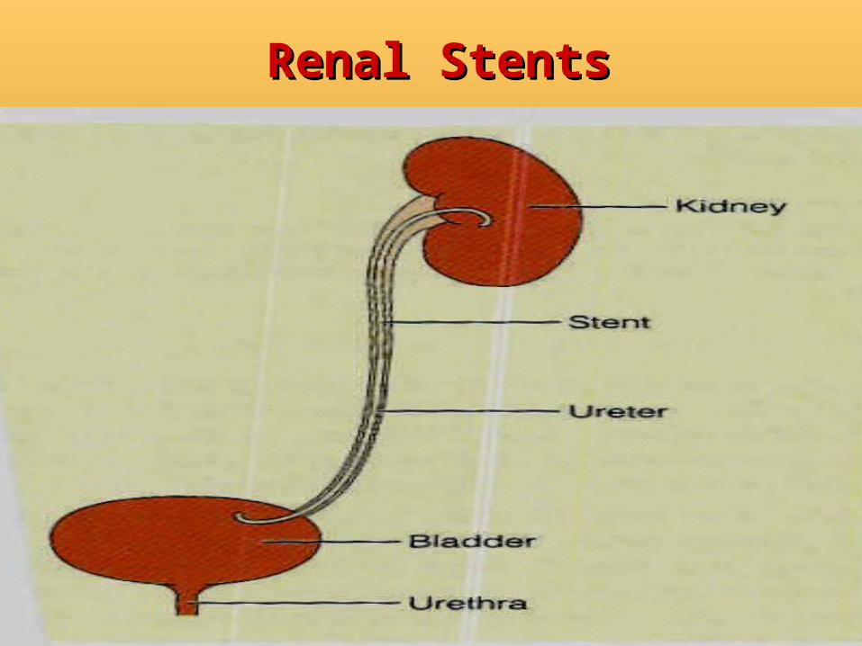
Renal StentsRenal Stents

Incisions for Kidney Incisions for Kidney SurgerySurgery

Renal CalculiRenal CalculiRemovalRemoval
Invasive LithotripsyInvasive LithotripsyPercutaneous ultrasonic lithotripsy – via Percutaneous ultrasonic lithotripsy – via
percutaneous nephroscopepercutaneous nephroscopeElectrohydraulic lithotripsy – percutaneousElectrohydraulic lithotripsy – percutaneousLaser lithotripsy probes – lower ureteral and large Laser lithotripsy probes – lower ureteral and large
bladder stonesbladder stonesNon-invasive - Extracorporeal shock-wave Non-invasive - Extracorporeal shock-wave
lithotripsylithotripsyPatient is anesthetizedPatient is anesthetizedHigh-energy acoustic shock waves shatter stone High-energy acoustic shock waves shatter stone
without damaging surrounding tissuewithout damaging surrounding tissue
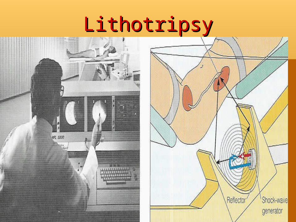
LithotripsyLithotripsy

Renal CalculiRenal CalculiNursing DiagnosesNursing Diagnoses
Acute painAcute painAnxiety r/t uncertain outcomeAnxiety r/t uncertain outcomeIneffective therapeutic regimen managementIneffective therapeutic regimen managementImpaired urinary eliminationImpaired urinary eliminationRisk for infectionRisk for infection

Renal CalculiRenal CalculiNursing ManagementNursing Management
AssessAssess: Pain—guarding, pain scale, occurrence—: Pain—guarding, pain scale, occurrence—colic versus ongoing, tenderness on palpation; colic versus ongoing, tenderness on palpation; History: recent/chronic UTI, immobility, gout, History: recent/chronic UTI, immobility, gout, hyperparathyroidism, prostatic hyperplasia; family hyperparathyroidism, prostatic hyperplasia; family history of calculi; urine output; oliguria, hematuria; labshistory of calculi; urine output; oliguria, hematuria; labs—BUN, CR, UA, Urine C&S, Increased uric acid, —BUN, CR, UA, Urine C&S, Increased uric acid, calciumcalcium
ActionAction: Relieve pain; Treat UTI; Admin meds; Force : Relieve pain; Treat UTI; Admin meds; Force fluids PO - >2L/day; Maintain IV patency; strain urine; fluids PO - >2L/day; Maintain IV patency; strain urine; position of comfortposition of comfort
Pt EducationPt Education: Rationale for treatment; Measures to : Rationale for treatment; Measures to prevent future recurrence (once calculi origin is prevent future recurrence (once calculi origin is determined)—dietary restrictions (purine, calcium, determined)—dietary restrictions (purine, calcium, oxalatesoxalates

Renal CalculiRenal CalculiNutritional TherapyNutritional Therapy
Foods high in purine, calcium, or oxalateFoods high in purine, calcium, or oxalate::PurinePurine::
High: Sardines, herring, mussels, liver, kidney, goose, High: Sardines, herring, mussels, liver, kidney, goose, venison, meat soups sweetbreadsvenison, meat soups sweetbreads
Moderate: Chicken, salmon, crab, veal, mutton, bacon, Moderate: Chicken, salmon, crab, veal, mutton, bacon, pork, beef, hampork, beef, ham
CalciumCalcium: milk, cheese, ice cream, yogurt, sauces : milk, cheese, ice cream, yogurt, sauces containing milk, all beans (except green beans), containing milk, all beans (except green beans), lentils, fish with fine bones (sardines, kippers herring, lentils, fish with fine bones (sardines, kippers herring, salmon); dried fruits, nuts, chocolate, cocoa, Ovaltinesalmon); dried fruits, nuts, chocolate, cocoa, Ovaltine
OxalateOxalate: spinach, rhubarb, asparagus, cabbage, : spinach, rhubarb, asparagus, cabbage, tomatoes, beets, nuts, celery, parsley, runner beans, tomatoes, beets, nuts, celery, parsley, runner beans, chocolate, cocoa, instant coffee, Ovaltine, tea; chocolate, cocoa, instant coffee, Ovaltine, tea; Worcestershire sauceWorcestershire sauce