interference dissociation in the presence of dual ... · interference dissociation in the presence...
TRANSCRIPT

Interference dissociation in the presence of dualatrioventricular nodal physiologyEmilio L. Garcia, MD, Robert Kim, MD, Steve S. Hsu, MD, FHRS, John N. Catanzaro, MD, FACC
From the University of Florida Health Science Center, Jacksonville, Florida.
IntroductionDual atrioventricular nodal physiology (DAVNP) is presentin 10% to 35% of the general population and is known toregress with aging.1 It usually manifests as a critical AHjump on electrophysiologic testing or on surface electro-cardiography as a change in PR interval with subsequenttachycardia. Preferential choice of pathway engagement(slow vs fast) is dependent upon refractory periods of thepathways and conduction velocity. We present a raremanifestation of DAVNP on surface electrocardiographywith alternating engagement and disengagement of the 2pathways masquerading as AV dissociation.
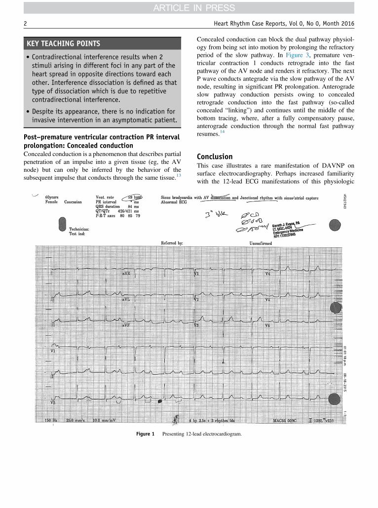
Case reportA 60-year-old woman with no significant past medicalhistory, in her usual state of health, presented to herophthalmologist’s office for a scheduled procedure ofcataract surgery. Prior to the procedure an electrocardiogram(ECG) (Figure 1) was performed, owing to an irregularpulse. The ECG was interpreted as abnormal by herphysician and subsequently led to deferral of her procedure.She was referred to our hospital, where she was admitted forfurther assessment of her abnormal ECG. The patient deniedany previous symptoms of palpitations, syncopal episodes,chest pain, or lightheadedness. She does not take anymedications and her physical examination including hervital signs were all within normal limits.
Figure 1 demonstrates her presenting ECG at her oph-thalmologist’s office upon the ophthalmologist’s noticingher pulse to be “irregular.” Upon admission, alternating or“grouped beating” and intervals of progressive PR short-ening were noted on telemetry. There was absence ofsymptom rhythm correlation. Transthoracic echocardiogra-phy demonstrated absence of structural heart disease andnormal left ventricular ejection fraction. Exercise treadmilltesting demonstrated shortening of her PR interval,
KEYWORDS Interference dissociation; Dual AV nodal physiology; PR alter-nans (Heart Rhythm Case Reports 2016;0:1–4)
Address reprint requests and correspondence: Dr John N. Catanzaro,University of Florida Health Science Center, 655W. 8th St, Jacksonville, FL32202. E-mail address: [email protected].
2214-0271 B 2016 Heart Rhythm Society. Published by Elsevier Inc. This is an o(http://creativecommons.org/licenses/by-nc-nd/4.0/).
chronotropic competence, and achievement of her maximumpredicted heart rate.
Upon return to her room a continuous rhythm strip wasperformed (Figure 2), which again illustrated progressive PRinterval shortening, as previously seen on telemetry. Closeinspection of a repeat ECG (Figure 3) demonstrates abruptshortening and lengthening of the PR interval (“PR alter-nans”) with engagement and disengagement of the slow and/or fast pathway. Her QRS complex was narrow, suggestingbrisk infranodal conduction. Her ECG normalized prior todischarge with 1:1 conduction down the fast pathway. Giventhat she was asymptomatic, she was followed as anoutpatient.
DiscussionDual AV nodal physiology indicates the presence of 2distinct electrophysiologic pathways with different conduc-tion velocities and refractory periods. The shorter PR intervalrepresents conduction over the fast pathway and the longerPR interval represents conduction over the slow pathway.The shift in conduction from fast to slow pathway can occurspontaneously or can be provoked or terminated by an atrialpremature complex, atrial tachycardia, interpolated junc-tional premature complexes, or a ventricular prematurecomplex. Findings compatible with simultaneous conductionalong 2 pathways in response to a ventricular prematurecomplex were noted in our patient’s ECG (Figure 3),indicating presence of underlying dual AV nodal physiology.
Dual AV nodal physiology can manifest itself as normalsinus rhythm, spontaneous shortening or lengthening of thePR interval persisting for varying periods of time, PRinterval alternans,2–7 PR interval alternans with Wenckebachsequence of the slowly and rapidly conducting pathways,and conduction along both pathways in response to a singlesinus impulse.8–12 In the presence of sinus rhythm thepresenting ECG (Figure 1) and continuous rhythm strip(Figure 2) illustrate an unusual presentation masquerading asdual AV nodal physiology. These 2 ECGs show interferencedissociation with progressive PR interval shortening inparallel with the influence of autonomic activity acceleratingthe heart rate. This causes the P waves to approach the QRSand allows a ventricular capture, which establishes restora-tion of normal sinus rhythm.
pen access article under the CC BY-NC-ND licensehttp://dx.doi.org/10.1016/j.hrcr.2016.08.017
KEY TEACHING POINTS
� Contradirectional interference results when 2stimuli arising in different foci in any part of theheart spread in opposite directions toward eachother. Interference dissociation is defined as thattype of dissociation which is due to repetitivecontradirectional interference.
� Despite its appearance, there is no indication forinvasive intervention in an asymptomatic patient.
Heart Rhythm Case Reports, Vol 0, No 0, Month 20162
Post–premature ventricular contraction PR intervalprolongation: Concealed conductionConcealed conduction is a phenomenon that describes partialpenetration of an impulse into a given tissue (eg, the AVnode) but can only be inferred by the behavior of thesubsequent impulse that conducts through the same tissue.13
Figure 1 Presenting 12-le
Concealed conduction can block the dual pathway physiol-ogy from being set into motion by prolonging the refractoryperiod of the slow pathway. In Figure 3, premature ven-tricular contraction 1 conducts retrograde into the fastpathway of the AV node and renders it refractory. The nextP wave conducts antegrade via the slow pathway of the AVnode, resulting in significant PR prolongation. Anterogradeslow pathway conduction persists owing to concealedretrograde conduction into the fast pathway (so-calledconcealed “linking”) and continues until the middle of thebottom tracing, where, after a fully compensatory pause,anterograde conduction through the normal fast pathwayresumes.14
ConclusionThis case illustrates a rare manifestation of DAVNP onsurface electrocardiography. Perhaps increased familiaritywith the 12-lead ECG manifestations of this physiologic
ad electrocardiogram.
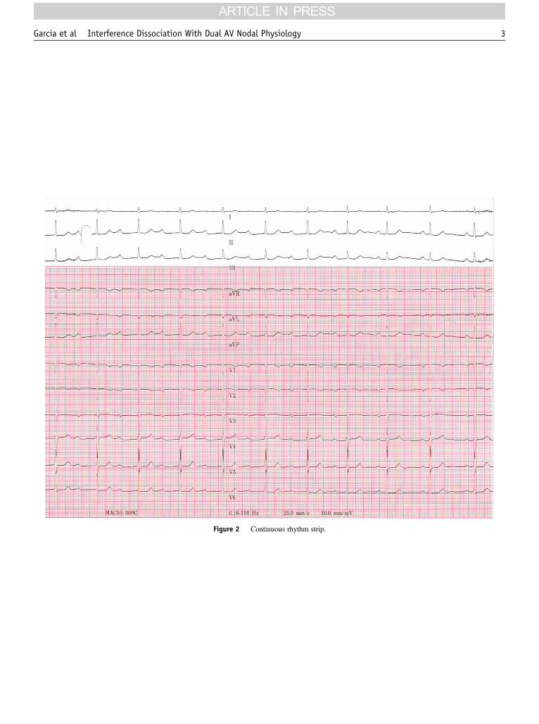
Figure 2 Continuous rhythm strip.
3Garcia et al Interference Dissociation With Dual AV Nodal Physiology

Figure 3 Fast pathway initially disengaged by a premature ventricular contraction (PVC1), subsequently engaging the slow pathway with the same prematureventricular contraction (PVC1), and reengaging the fast pathway once again (PVC2). Slow pathway conduction demarcated by long PR interval marked bybrackets [ ] with transition to fast pathway conduction indicated by asterisks *.
Heart Rhythm Case Reports, Vol 0, No 0, Month 20164
phenomenon may alter a differential diagnosis to preventunnecessary admissions or pacemaker implantation.
References1. Mani BC, Pavri BB. Dual atrioventricular nodal pathways physiology: a review
of relevant anatomy, electrophysiology, and electrocardiographic manifestationsfor the physician-in-training. Indian Pacing Electrophysiol J 2014;14(1):12–25.
2. Schamroth L, PerlmanMM. Periodic variation in AV conduction: a study in differentialdual AV pathway conduction and refractoriness. J Electrocardiol 1973;6:81–84.
3. Katz L, Pick A. Clinical electrocardiography: the arrhythmias. Philadelphia: Lea& Febiger; 1956:567–568.
4. Pick A, Langendorf R. Interpretation of complex arrhythmias. Philadelphia: Lea& Febiger; 1979:227, 231
5. Mamlin JJ, Fisch C, Sustained AV. conduction delay due to interpolatedventricular premature systole. Am J Cardiol 1965;16:765–766.
6. Kinoshita S, Kawasaki T, Fujiwara S, Okimori K. Periodic variation in AVconduction time: mechanism of initiation, maintenance and termination ofperiods of long PR intervals. Am J Cardiol 1984;53:1288–1291.
7. Surawicz B, Fisch C. Cardiac alternans: diverse mechanisms and clinicalmanifestations. J Am Coll Cardiol 1992;20:483–499.9.
8. Fisch C. Electrocardiography of Arrhythmias. Philadelphia: Lea & Febiger; 1990:393.
9. Zipes DP. Specific arrhythmias: diagnosis and treatment. In: Braunwald E, ed.Heart Disease. Philadelphia: Saunders; 1992:689.
10. Lee KW, Badhwar N, Scheinman MM. Supraventricular tachycardia – part I.Curr Probl Cardiol 2008;33:467–546.
11. Lin LJ, Lin JL, Lai LP, Chen JH, Tseng YZ, Lien WP. Effects of pharmacologicalautonomic blockade on dual atrioventricular nodal pathways physiology inpatients with slow-fast atrioventricular nodal reentrant tachycardia. Pacing ClinElectrophysiol 1998;21:1375–1379.
12. Belhassen B, Fish R, Glikson M, Glick A, Eldar M, Laniado S, Viskin S.Noninvasive diagnosis of dual AV node physiology in patients with AV nodalreentrant tachycardia by administration of adenosine-5'-triphosphate during sinusrhythm. Circulation 1998;98:47–53.
13. Denes P, Wu D, Dhingra R, et al. Dual atrioventricular nodal pathways. Acommon electrophysiological response. Br Heart J 1975;37:1069–1076.
14. Fisch C J Am Coll Cardiolol. 1989;Nov 1;14(5):1127-38.