interesting cases from the pediatric er - texas children's ... · •started on ceftriaxone...
TRANSCRIPT

INTERESTING CASES FROM THE PEDIATRIC ERKimberly Childers, RN, MS, CPNP-AC/PC
Texas Children’s Emergency Center - Nurse Practitioner
Baylor College of Medicine – Clinical Instructor
Vanessa Thomas, MD
Pediatric Emergency Medicine Fellow
Baylor College of Medicine/Texas Children's Hospital
CASE 1: “OW MY ARM!”
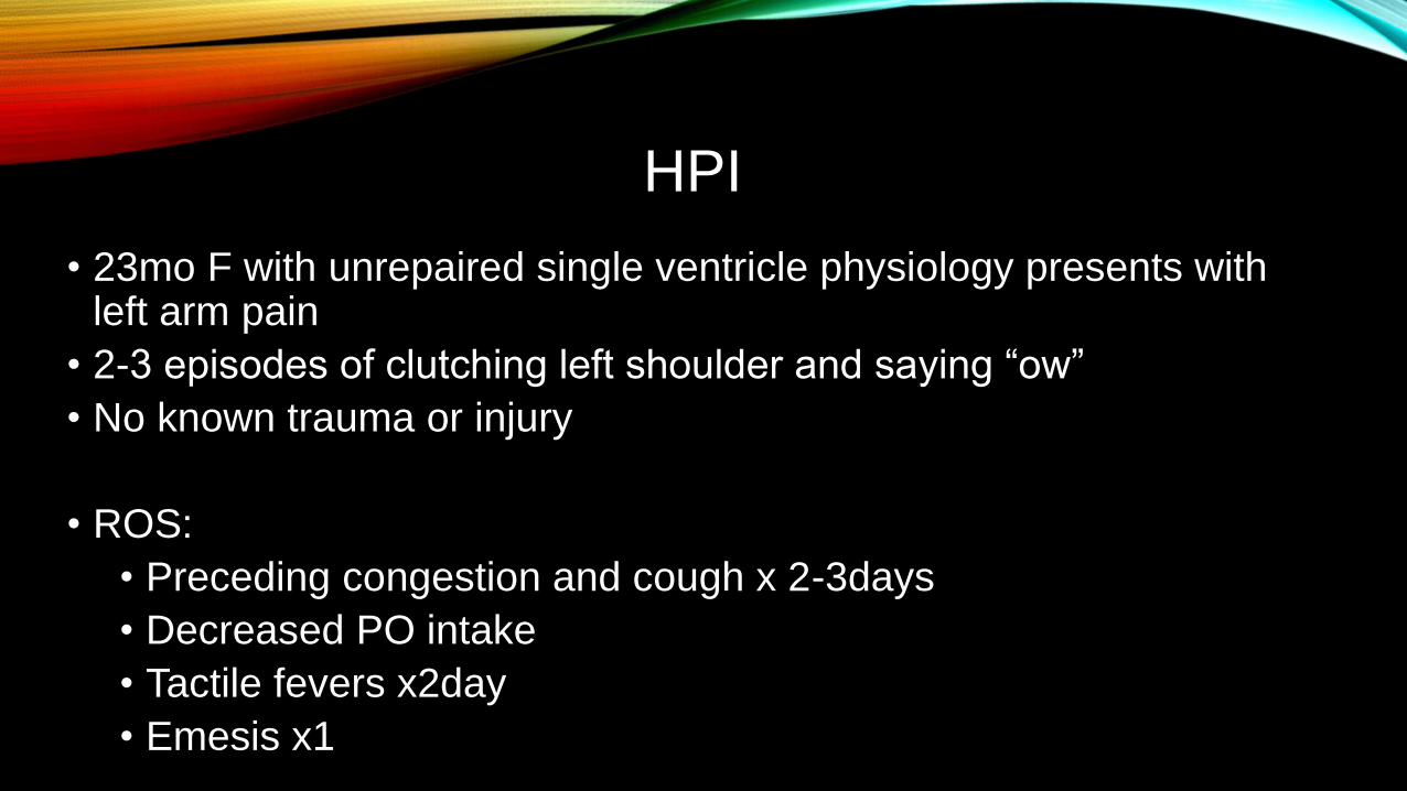
HPI
• 23mo F with unrepaired single ventricle physiology presents with left arm pain
• 2-3 episodes of clutching left shoulder and saying “ow”
• No known trauma or injury
• ROS:
• Preceding congestion and cough x 2-3days
• Decreased PO intake
• Tactile fevers x2day
• Emesis x1

HPI
• PMH: Single ventricle: common atrium, AV septal defect with dominant RV and DORV with moderate pulmonary stenosis
• PSH: No cardiac surgeries
• Meds: None
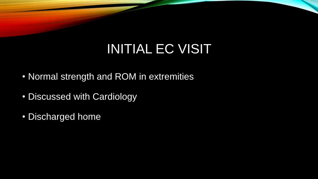
INITIAL EC VISIT
• Normal strength and ROM in extremities
• Discussed with Cardiology
• Discharged home

RETURN EC VISIT
• Continued emesis, fever, poor PO
• New left leg pain and refusal to bear weight
• Generally fussy
• Exam:
• Refuses to bear weight on left leg
• No obvious joint tenderness or pain
• Full ROM left leg
• Normal strength
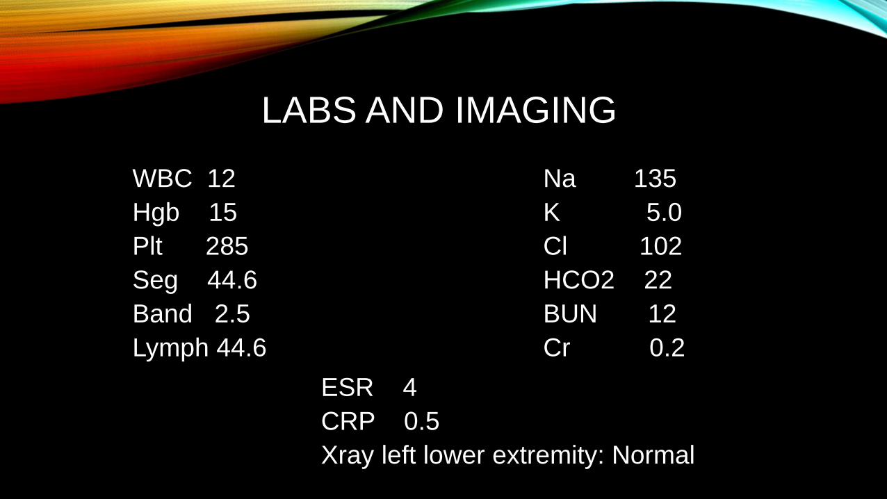
LABS AND IMAGING
WBC 12
Hgb 15
Plt 285
Seg 44.6
Band 2.5
Lymph 44.6
Na 135
K 5.0
Cl 102
HCO2 22
BUN 12
Cr 0.2
ESR 4
CRP 0.5
Xray left lower extremity: Normal


DIFFERENTIAL DIAGNOSIS
• Toxic/transient synovitis with URI
• Myalgias with viral illness
• Septic arthritis
• Fracture
• Myocardial infarction
• Ischemic stroke
• Seizure followed by Todd’s paralysis

HOSPITAL COURSE
• Admitted for fever and refusal to bear weight
• Started having seizure like episodes
• Subsequent left arm and leg weakness

HOSPITAL COURSE
• CT head: large parenchymal hematoma R frontal lobe and left posterior thalamic lesion concerning for hemorrhagic stroke
• Received antiepileptics and antibiotics
• No initial anticoagulation due to hemorrhage
• Stroke workup: thrombophilia risk factors (elevated factor 8 and fibrinogen)
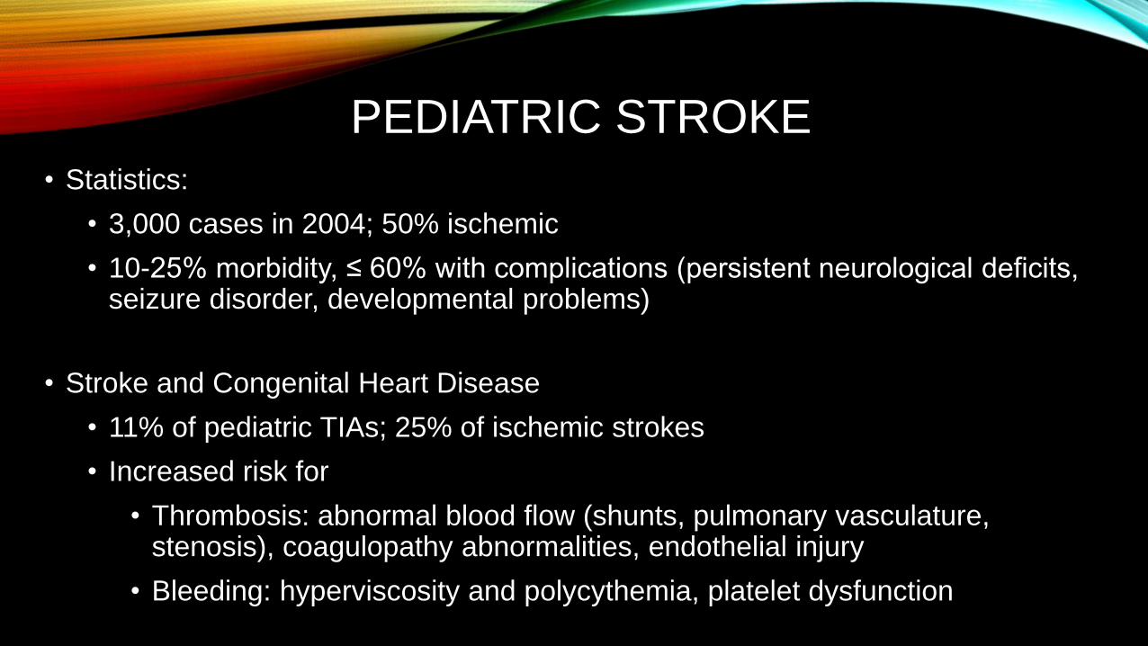
PEDIATRIC STROKE
• Statistics:
• 3,000 cases in 2004; 50% ischemic
• 10-25% morbidity, ≤ 60% with complications (persistent neurological deficits, seizure disorder, developmental problems)
• Stroke and Congenital Heart Disease
• 11% of pediatric TIAs; 25% of ischemic strokes
• Increased risk for
• Thrombosis: abnormal blood flow (shunts, pulmonary vasculature, stenosis), coagulopathy abnormalities, endothelial injury
• Bleeding: hyperviscosity and polycythemia, platelet dysfunction

CLINICAL DIAGNOSIS
• Symptoms:
• Acute ischemic stroke: focal neurologic deficit (hemiplegia 94% of cases)
• Hemorrhagic strokes: headaches, altered mental status, or vomiting
• Seizures common with both (50% of strokes)
• Age based:
• Neonates: seizures, apnea, lethargy
• Toddlers: increased crying/sleepiness, irritability, feeding difficulty, vomiting, sepsis-like with cold extremities
• Older children: focal neurologic symptoms

TREATMENT
• Safety and efficacy of thrombolysis for acute stroke in children have not been established (American Heart Association)
• Perinatal stroke: ventricular drainage for hydrocephalus caused by IVH, vitamin K, platelets
• Sickle Cell: hydration and exchange transfusion
• Vasculitis: Corticosteroids and cytotoxic agents

CASE 2: “ONE RING TO RULE THEM ALL…”
Special thanks to Joseph Allen, MD

PRESENTATION
16 year old male who presents to the office after dropping a can of vegetables on the 3rd finger. Findings include:
• Small laceration (1/2 cm) to the dorsal surface without active bleeding
• Swelling at the PIP joint
• Ring that cannot be easily removed with gentle traction

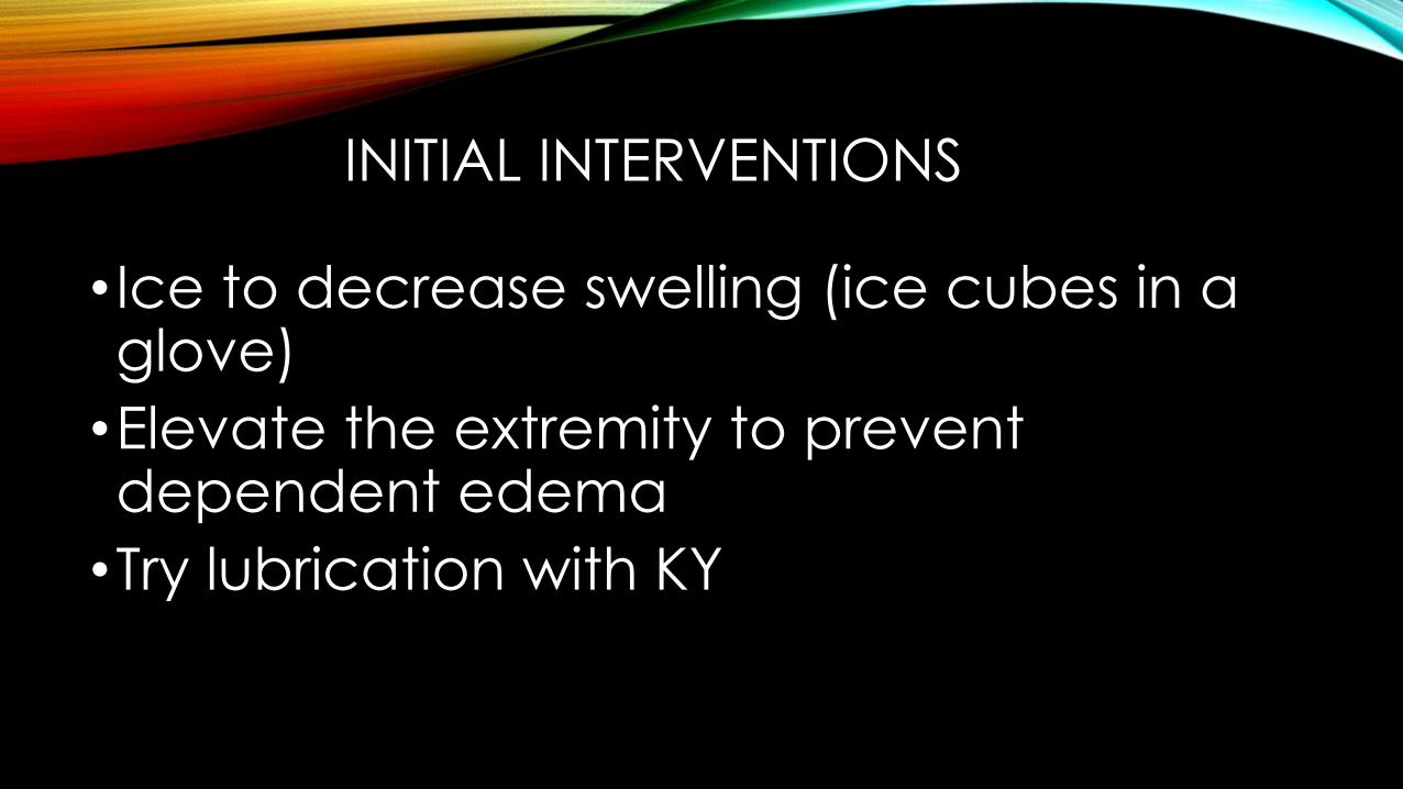
INITIAL INTERVENTIONS
• Ice to decrease swelling (ice cubes in a glove)
•Elevate the extremity to prevent dependent edema
•Try lubrication with KY
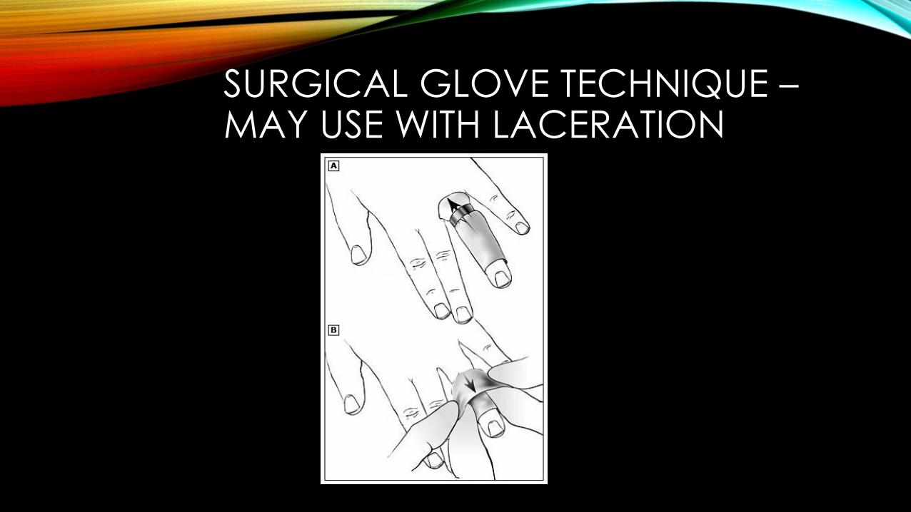
SURGICAL GLOVE TECHNIQUE –MAY USE WITH LACERATION

ELASTIC TAPE METHOD – MAY USE WITH LACERATION
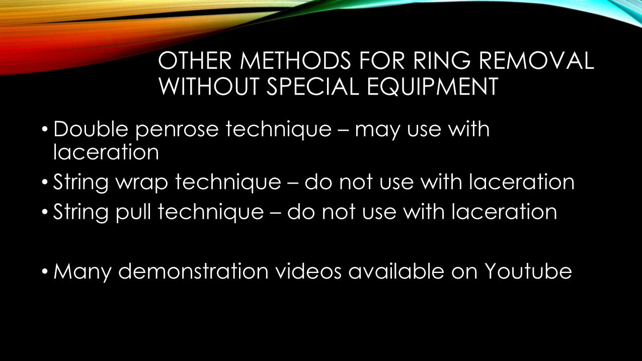
OTHER METHODS FOR RING REMOVAL WITHOUT SPECIAL EQUIPMENT
• Double penrose technique – may use with laceration
• String wrap technique – do not use with laceration
• String pull technique – do not use with laceration
• Many demonstration videos available on Youtube

AFTER MULTIPLE ATTEMPTS, YOU ARE UNABLE TO REMOVE THE RING AND THE PATIENT SEEKS CARE AT THE ER.


REMOVAL ATTEMPTS CONTINUE…
The ER staff attempts to remove the ring and break two ring cutters…
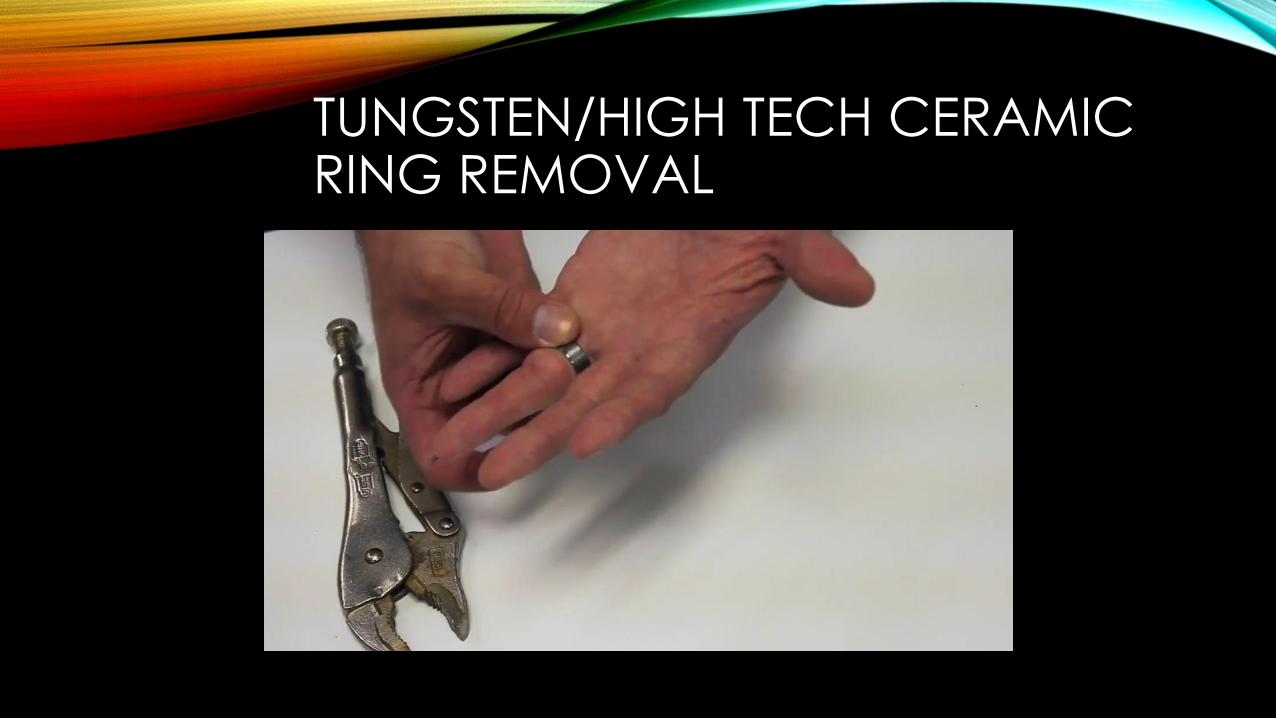
TUNGSTEN/HIGH TECH CERAMIC RING REMOVAL
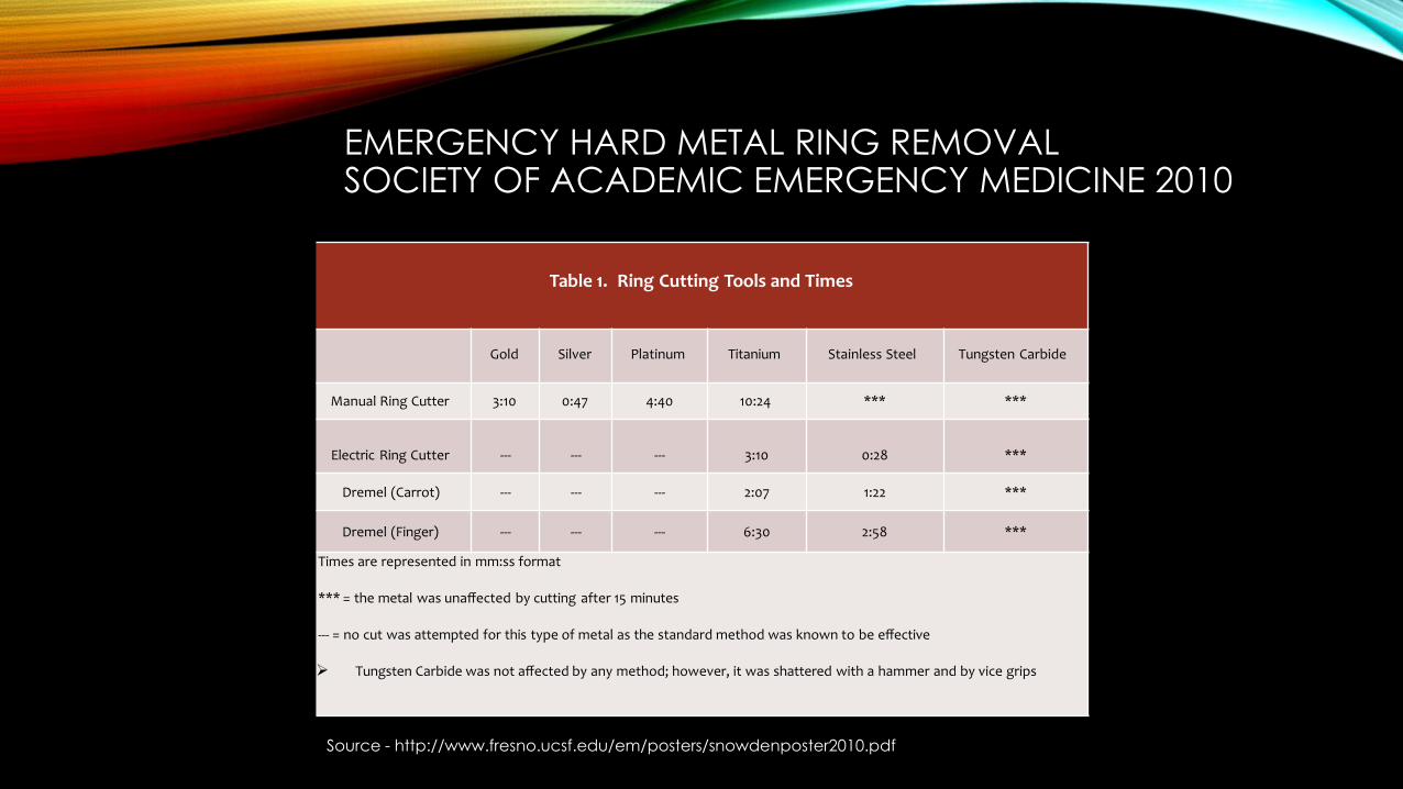
EMERGENCY HARD METAL RING REMOVALSOCIETY OF ACADEMIC EMERGENCY MEDICINE 2010
Table 1. Ring Cutting Tools and Times
Gold Silver Platinum Titanium Stainless Steel Tungsten Carbide
Manual Ring Cutter 3:10 0:47 4:40 10:24 *** ***
Electric Ring Cutter --- --- --- 3:10 0:28 ***
Dremel (Carrot) --- --- --- 2:07 1:22 ***
Dremel (Finger) --- --- --- 6:30 2:58 ***
Times are represented in mm:ss format
*** = the metal was unaffected by cutting after 15 minutes
--- = no cut was attempted for this type of metal as the standard method was known to be effective
Tungsten Carbide was not affected by any method; however, it was shattered with a hammer and by vice grips
Source - http://www.fresno.ucsf.edu/em/posters/snowdenposter2010.pdf
POST REMOVAL CONSIDERATIONS
• Wound care
• Tetanus prophylaxis
• Consider x-rays for possible fracture
• Prevention
QUESTIONS?

CASE 3: NEVERENDING FEVER
HPI
• 3 year old girl with 15 days of daily fever (102-104oF)
• Improving cough and congestion
• Recently treated with amoxicillin for acute otitis media finished 5 days ago
• Otalgia resolved with antibiotics but not fever
• 1 lb weight loss since ill
PMH/SH
• “Many” malaria flairs in past
• Born in Burundi
• Moved to US 1.5 years ago, no travel since then
• Negative PPD
• Immunizations: on catch up schedule
• ROS: Weight loss, decreased appetite, decreased UOP
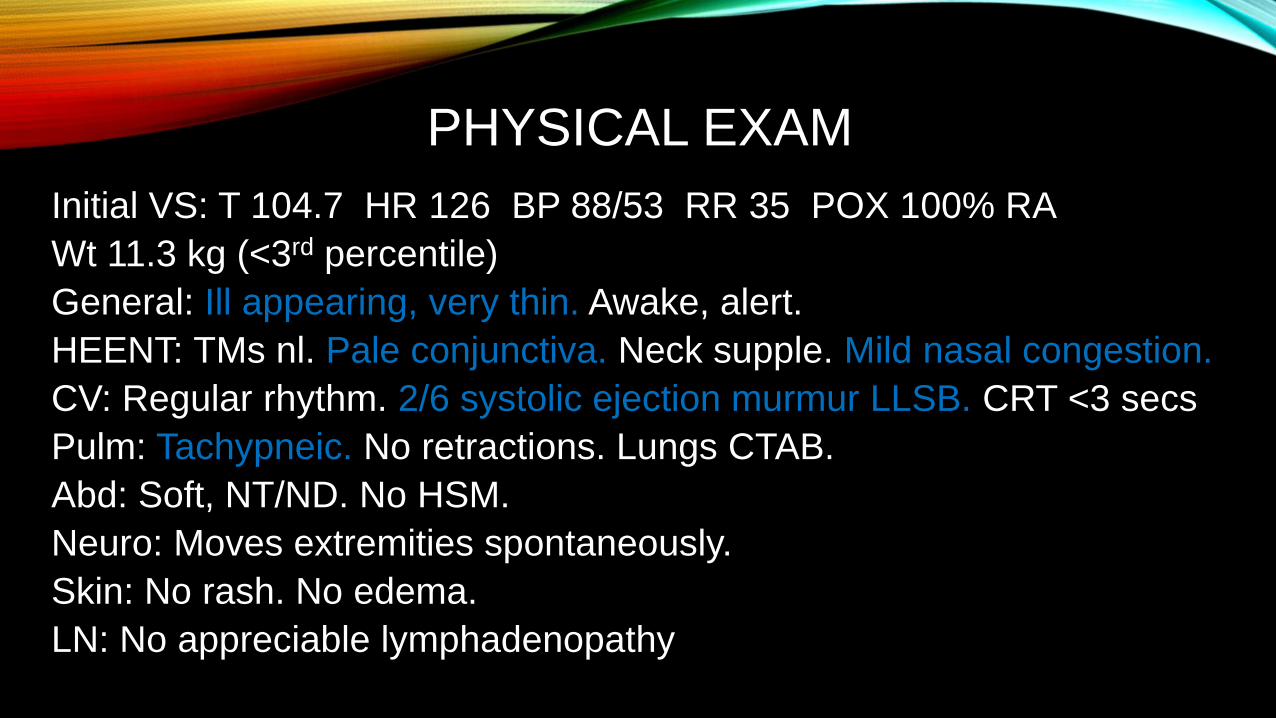
PHYSICAL EXAM
Initial VS: T 104.7 HR 126 BP 88/53 RR 35 POX 100% RA
Wt 11.3 kg (<3rd percentile)
General: Ill appearing, very thin. Awake, alert.
HEENT: TMs nl. Pale conjunctiva. Neck supple. Mild nasal congestion.
CV: Regular rhythm. 2/6 systolic ejection murmur LLSB. CRT <3 secs
Pulm: Tachypneic. No retractions. Lungs CTAB.
Abd: Soft, NT/ND. No HSM.
Neuro: Moves extremities spontaneously.
Skin: No rash. No edema.
LN: No appreciable lymphadenopathy

RESULTSWBC 6 Seg 25%, Band 21%, Lymph 50%, Mono 4%, Eos 0%
Hgb 9.2 MCV 73, ANC 2800
Plt 109
ESR 81
CRP 7.3
CXR: Enlarged cardiac silhouette
Mg 2
Phos 4.3
AST 91
ALT 55
Alk phos 143
GGT 86
Alb 3.1
Bili 0.1
Na 135
K 4.2
Cl 97
CO2 25
BUN 13
Cr 0.25
Glucose 101
Ca 8.6
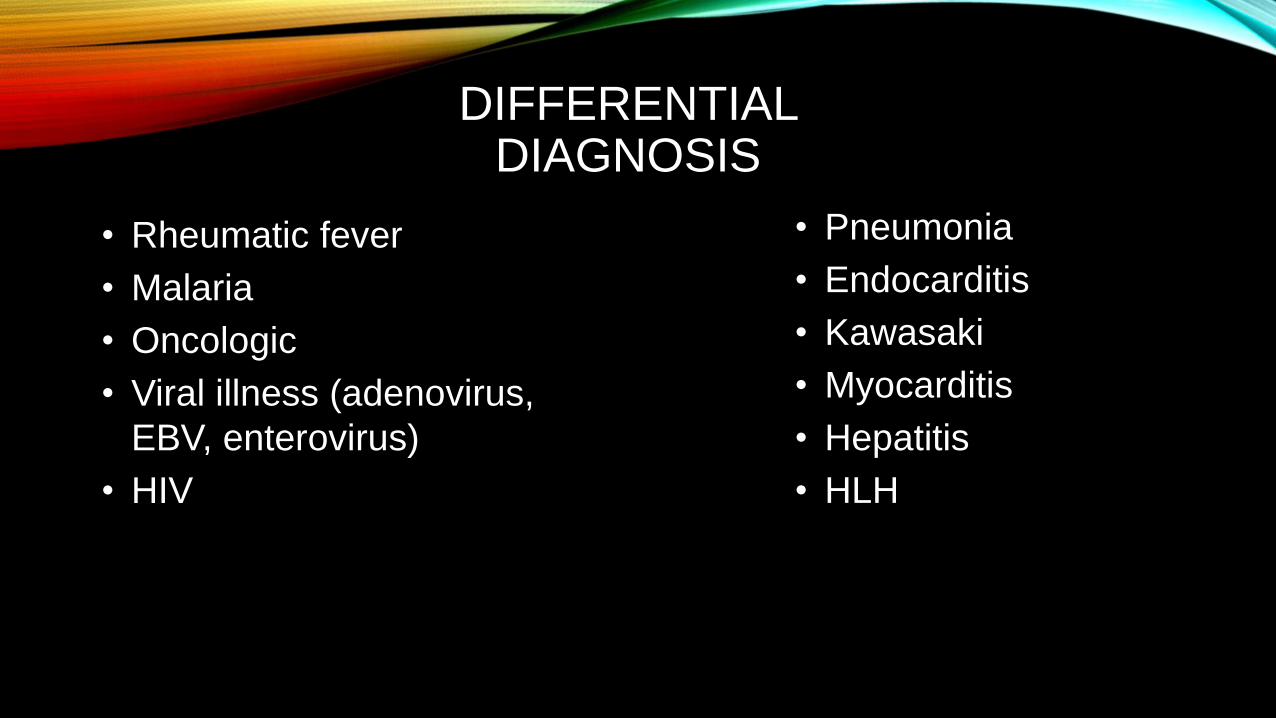
DIFFERENTIAL DIAGNOSIS
• Rheumatic fever
• Malaria
• Oncologic
• Viral illness (adenovirus,
EBV, enterovirus)
• HIV
• Pneumonia
• Endocarditis
• Kawasaki
• Myocarditis
• Hepatitis
• HLH
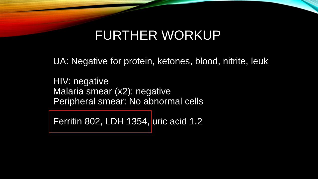
FURTHER WORKUP
UA: Negative for protein, ketones, blood, nitrite, leuk
HIV: negativeMalaria smear (x2): negativePeripheral smear: No abnormal cells
Ferritin 802, LDH 1354, uric acid 1.2

INFECTIOUS DISEASES
• EBV and CMV: past EBV infection
• Parvo neg
• Bartonella neg
• Rickettsial panel neg
• Typhus fever ab neg
• Flu, RSV, adeno, entero,
HMPV, paraflu neg
• Hepatitis panel: wnl
• Quantiferon neg
• Brucella neg
• Stool: O&P and culture negative
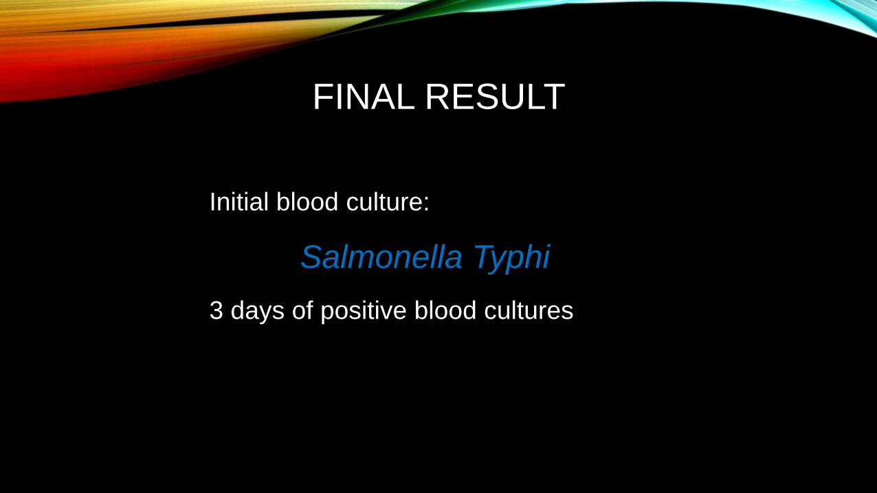
FINAL RESULT
Initial blood culture:
Salmonella Typhi
3 days of positive blood cultures
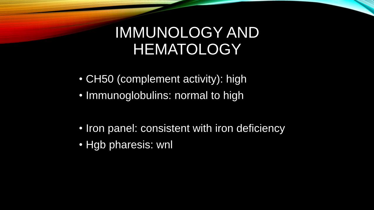
IMMUNOLOGY AND HEMATOLOGY
• CH50 (complement activity): high
• Immunoglobulins: normal to high
• Iron panel: consistent with iron deficiency
• Hgb pharesis: wnl

HOSPITAL COURSE
• Started on ceftriaxone with positive blood culture, received 14 days IV
• Developed abdominal pain and distension on HD #8, Xrayshowed constipation
• Fever persisted until HD #13
• CRP and ESR downtrended
• Discharged home with boost supplement for weight and iron supplement for anemia


TYPHOID FEVER
• Etiology: Caused by Salmonella typhi serotype
• Incidence:
• 21 million cases annually
• 222,000 typhoid-related deaths annually
• Mostly Asia and Africa
• Transmission: Contaminated food or water
• Incubation period:
• Average 7-14 days
• Range 3-60 days

TYPHOID FEVER
• Early Symptoms: 1st week
• Fever, headache, malaise, anorexia, lethargy
• Relative bradycardia
• Later Symptoms: 2nd week
• Hepatomegaly, splenomegaly
• Skin: dactylitis, rose spots
• Abdominal pain: can cause ileus, intestinal perforation, cholecystitis
• More severe:
• Persistent bacteremia
• Meningitis, encephalopathy
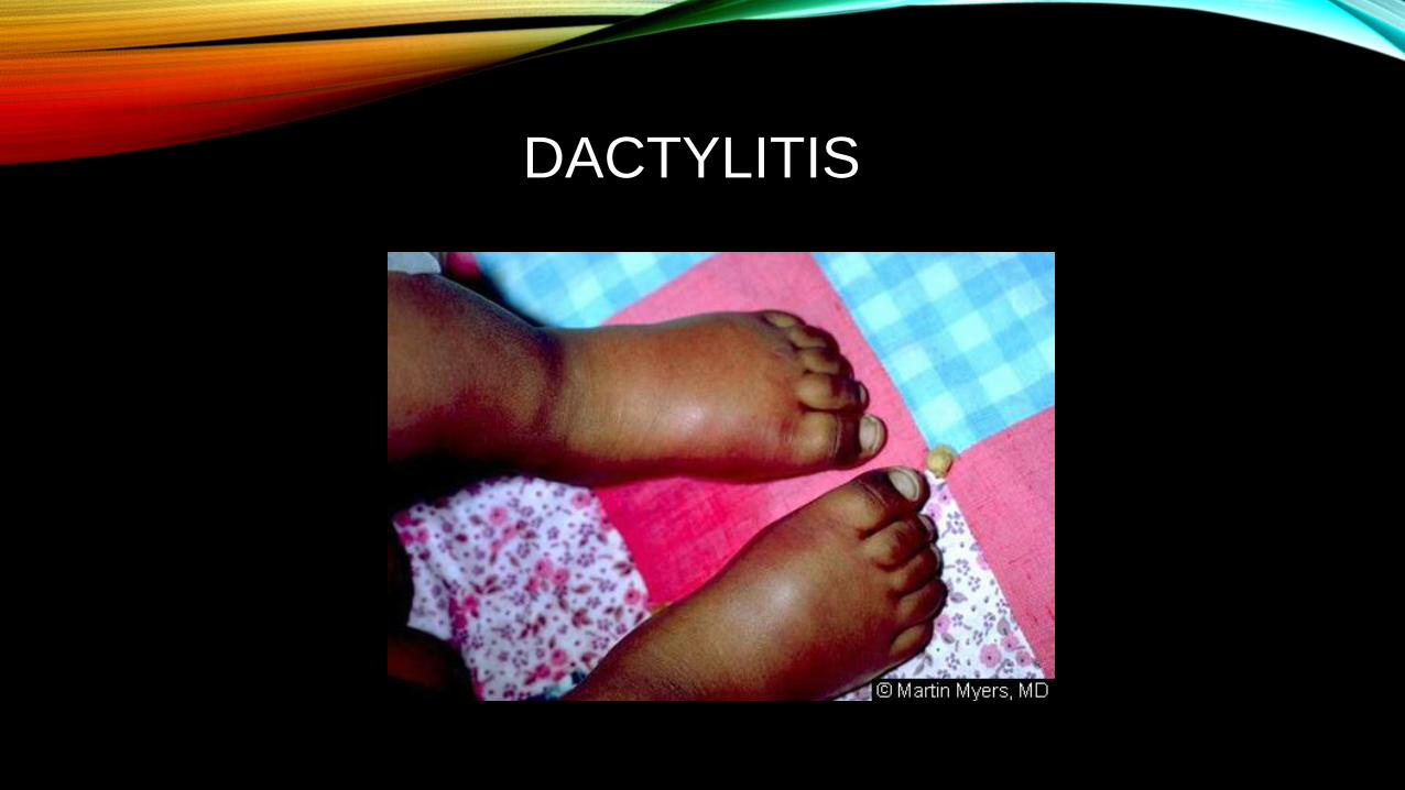
DACTYLITIS

ROSE SPOTS

TYPHOID FEVER
• Lab abnormalities:
• Anemia
• Leukopenia or leukocytosis (if later in illness, associated with intestinal perforation)
• Abnormal liver function
• Diagnosis:
• Blood culture (positive in 40-80% of patients)
• Serologies: not useful in endemic areas (may represent previous infection)

TYPHOID FEVER
• Treatment
• With antibiotics, mortality rates decreased from 15% to <1%
• Options: fluoroquinolones, 3rd generation cephalosporins, or azithromycin
• Resource limited areas: chloramphenicol, ampicillin, Bactrim
• Uncomplicated disease: oral antibiotics
• Severe/complicated disease:
• Empiric with ceftriaxone 10-14 days
• Consider steroids
• Prevention: Typhoid vaccine
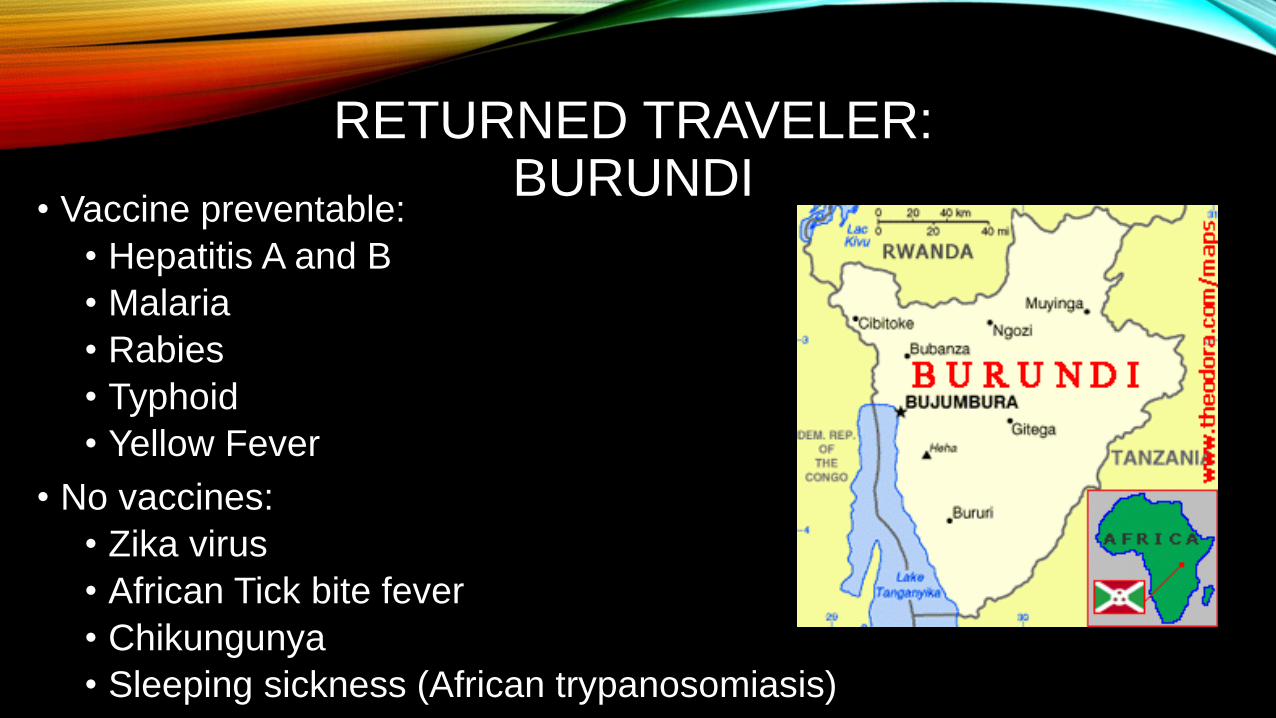
RETURNED TRAVELER: BURUNDI
• Vaccine preventable:
• Hepatitis A and B
• Malaria
• Rabies
• Typhoid
• Yellow Fever
• No vaccines:
• Zika virus
• African Tick bite fever
• Chikungunya
• Sleeping sickness (African trypanosomiasis)
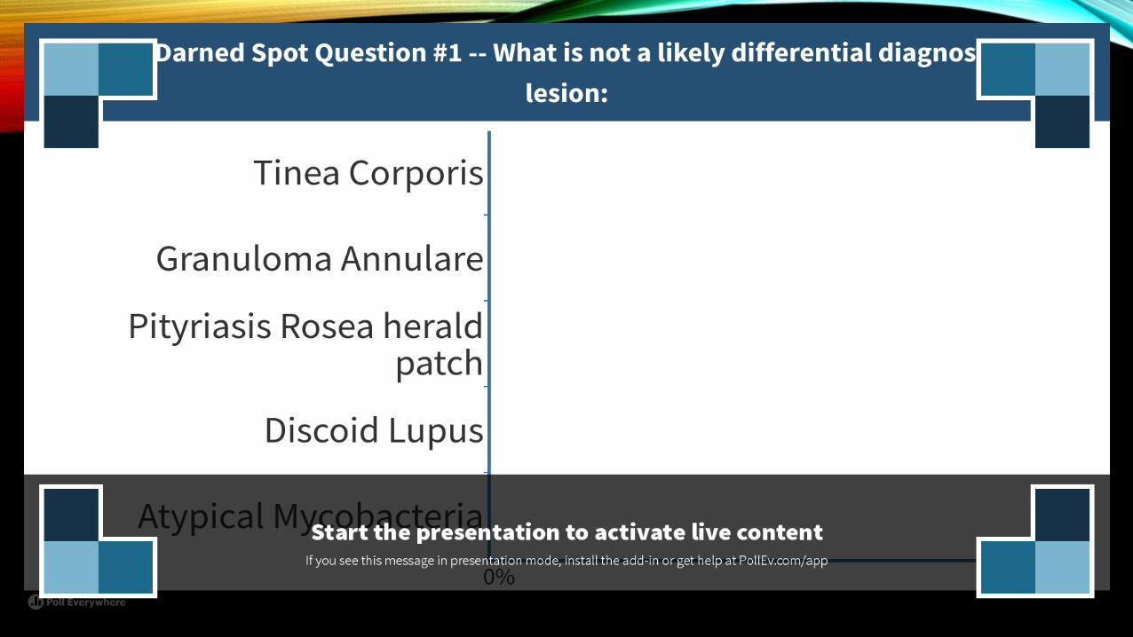
CASE 4: “OUT, OUT DARNED SPOT”
PRESENTATION
A previously healthy 8 year old girl with a lesion to the side of the face for 9 months. Lesion was unresponsive to hydrocortisone and clotrimazole after being seen by the pcp for multiple visits. Parents note that the lesion began as a small pink papule and grew in size. Increased itching to the area over the last 3 days.
No other new or constitutional symptoms.


ER EVALUATION
• Family had dermatology appointment scheduled at LBJ, but family wanted to be seen at TCH.
• Differential included discoid lupus versus partially treated tinea corporis
• Fungal culture obtained and sent. Discussed with family that it was a very slow growing test.
• Prescribed topical ketoconazole
• Discharged home with dermatology appointment in 3 days

DERMATOLOGY EVALUATION
• Visit #1 Suspect discoid lupus
• Punch biopsy needed for confirmation
• Patient was very tearful during discussion of biopsy so recommend biopsy in OR
• 11 days later parents declined biopsy due to significant skin improvement
• Trial of topical clobetasol prescribed
• Visit #2 referred to rheumatology and ophthalmology
• Told derm that she was being followed by Harris Co. hepatology
• Labs sent and prescribed Plaquenil/hydrochloroquine orally

BACK TO THE ER
• 2 months of initial ER visit – returned for vomiting/fever/LUQ pain
• Received (2) 20 ml/kg NS boluses for tachycardia/SIRS and ceftriaxone
• Abnormal labs including elevated liver enzymes, ANC=960, lipase=586
• +SSA and ANA 1:320
• Admitted to rheumatology for 6 days for pancreatitis
• 6 days stay with clinical improvement
• Discharged home on Plaquenil and steroids

RHEUMATOLOGY CLINIC
• Completed ophthalmology appointment with normal exam/no uveitis
• Wean steroids, continue Plaquenil and PPI
• Begin Imuran/Azathioprine
• Continue dermatology follow up


LAST FOLLOW UP
Seen in dermatology clinic and doing well.
Questions?
CASE 5: APNEA, JAUNDICE, AND
VOMITING…OH MY!!
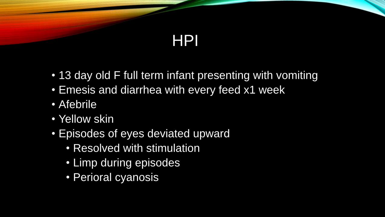
HPI
• 13 day old F full term infant presenting with vomiting
• Emesis and diarrhea with every feed x1 week
• Afebrile
• Yellow skin
• Episodes of eyes deviated upward
• Resolved with stimulation
• Limp during episodes
• Perioral cyanosis

PMH
• 40W6D by C-section due failure to progress
• Episode NBNB emesis on DOL 1:
• NICU admission
• AXR dilated air loops
• Contrast enema normal
• Meconium plug
• ROS: Negative for fever, cough, congestion
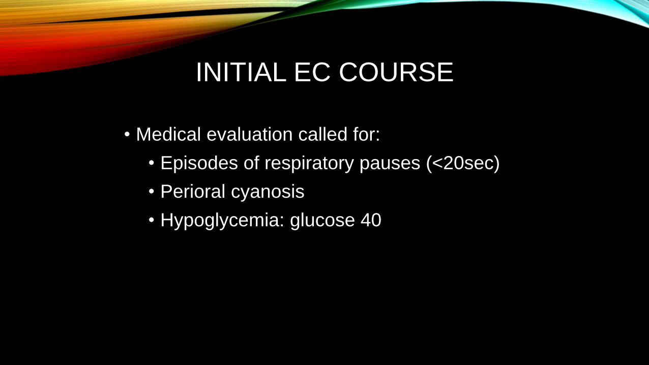
INITIAL EC COURSE
• Medical evaluation called for:
• Episodes of respiratory pauses (<20sec)
• Perioral cyanosis
• Hypoglycemia: glucose 40

PHYSICAL EXAM
Initial VS: T 98.4 HR 142 BP 85/52 RR 36 POX 70-100% Wt 3.8kg
General: Sleepy but arousable with stimulation.
HEENT: AFOSF. PERRLA. Mild scleral icterus. Neck supple. No nasal congestion.
CV: RRR. No murmurs. 2+ brachial/femoral pulses.
Pulm: Intermittent pauses of 10sec each. One with 5 secs of perioral cyanosis with sats 70%. Lungs CTAB.
Abd: Mild distension, nontender. Decreased bowel sounds. No HSM.
Neuro: Decreased tone all extremities. Moves extremities spontaneously.
Skin: Mild jaundice
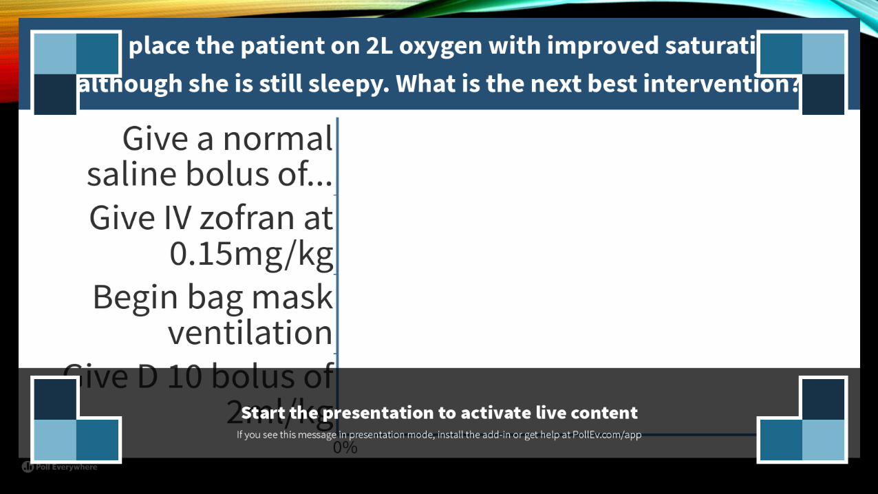

EC COURSE
• Received D10 bolus of 2 ml/kg
• More awake
• Then episode of emesis followed by apnea and cyanosis
• Patient intubated for airway protection and apnea


DIFFERENTIAL DIAGNOSIS
• Sepsis
• Metabolic disorder
• Pyloric stenosis
• Intestinal obstruction
• Liver disease
• Viral gastroenteritis
• UTI
• NAT
• Congenital heart disease
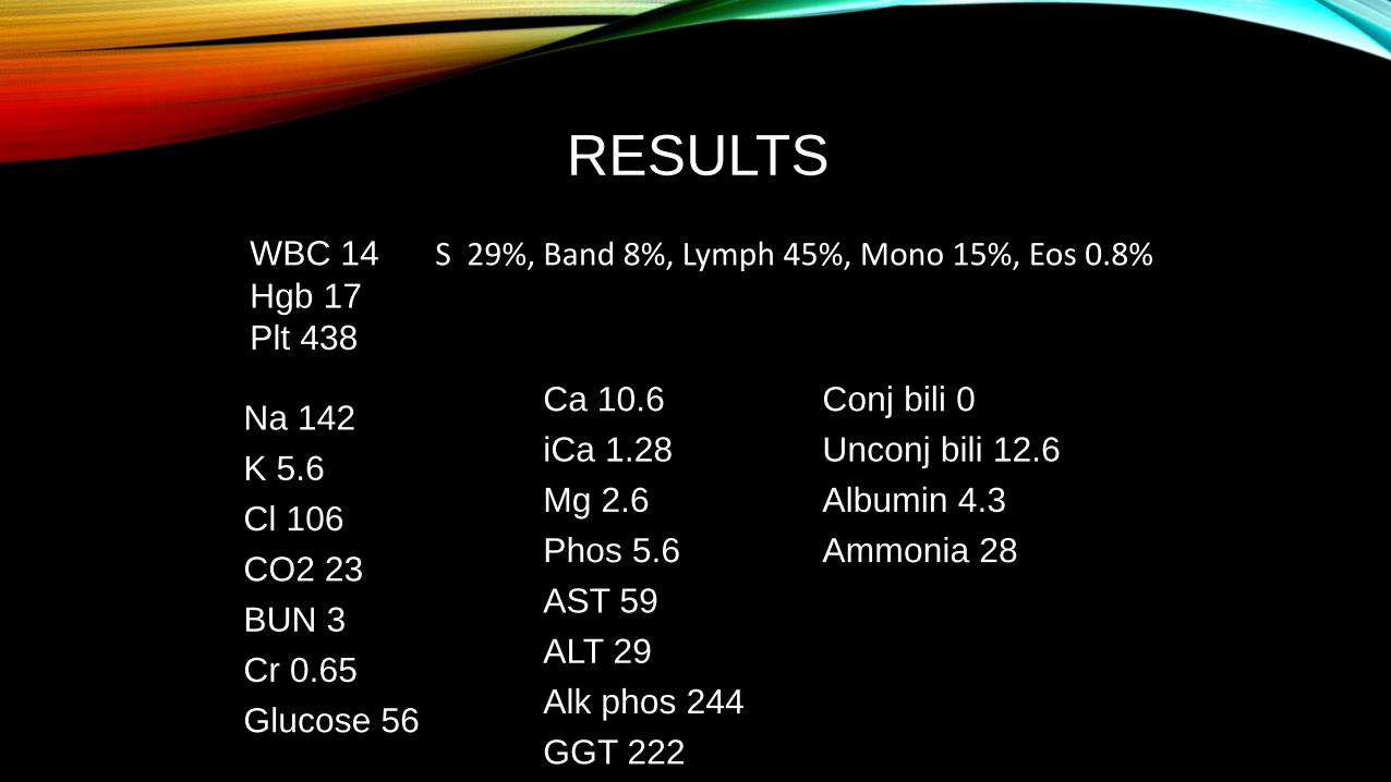
RESULTS
WBC 14 S 29%, Band 8%, Lymph 45%, Mono 15%, Eos 0.8%Hgb 17
Plt 438
Ca 10.6
iCa 1.28
Mg 2.6
Phos 5.6
AST 59
ALT 29
Alk phos 244
GGT 222
Conj bili 0
Unconj bili 12.6
Albumin 4.3
Ammonia 28
Na 142
K 5.6
Cl 106
CO2 23
BUN 3
Cr 0.65
Glucose 56
RESULTS
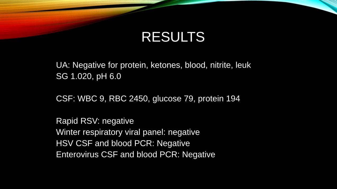
UA: Negative for protein, ketones, blood, nitrite, leuk
SG 1.020, pH 6.0
CSF: WBC 9, RBC 2450, glucose 79, protein 194
Rapid RSV: negative
Winter respiratory viral panel: negative
HSV CSF and blood PCR: Negative
Enterovirus CSF and blood PCR: Negative
RESULTS
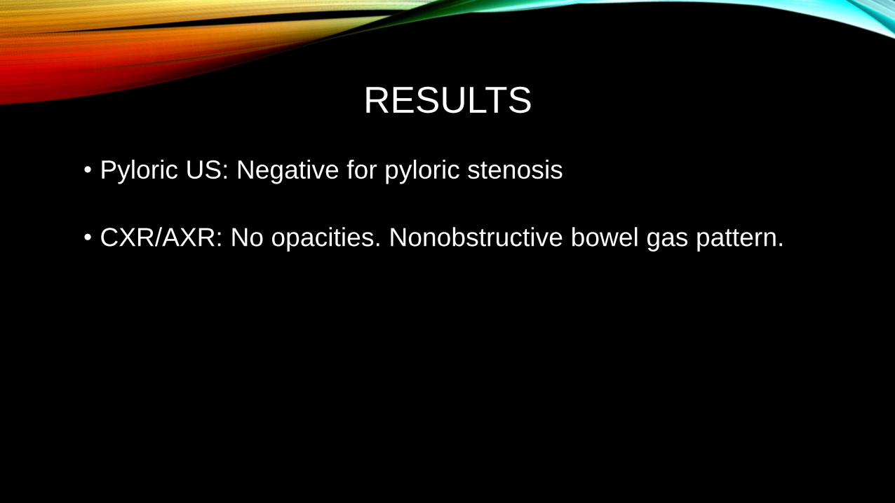
• Pyloric US: Negative for pyloric stenosis
• CXR/AXR: No opacities. Nonobstructive bowel gas pattern.
RESULTS
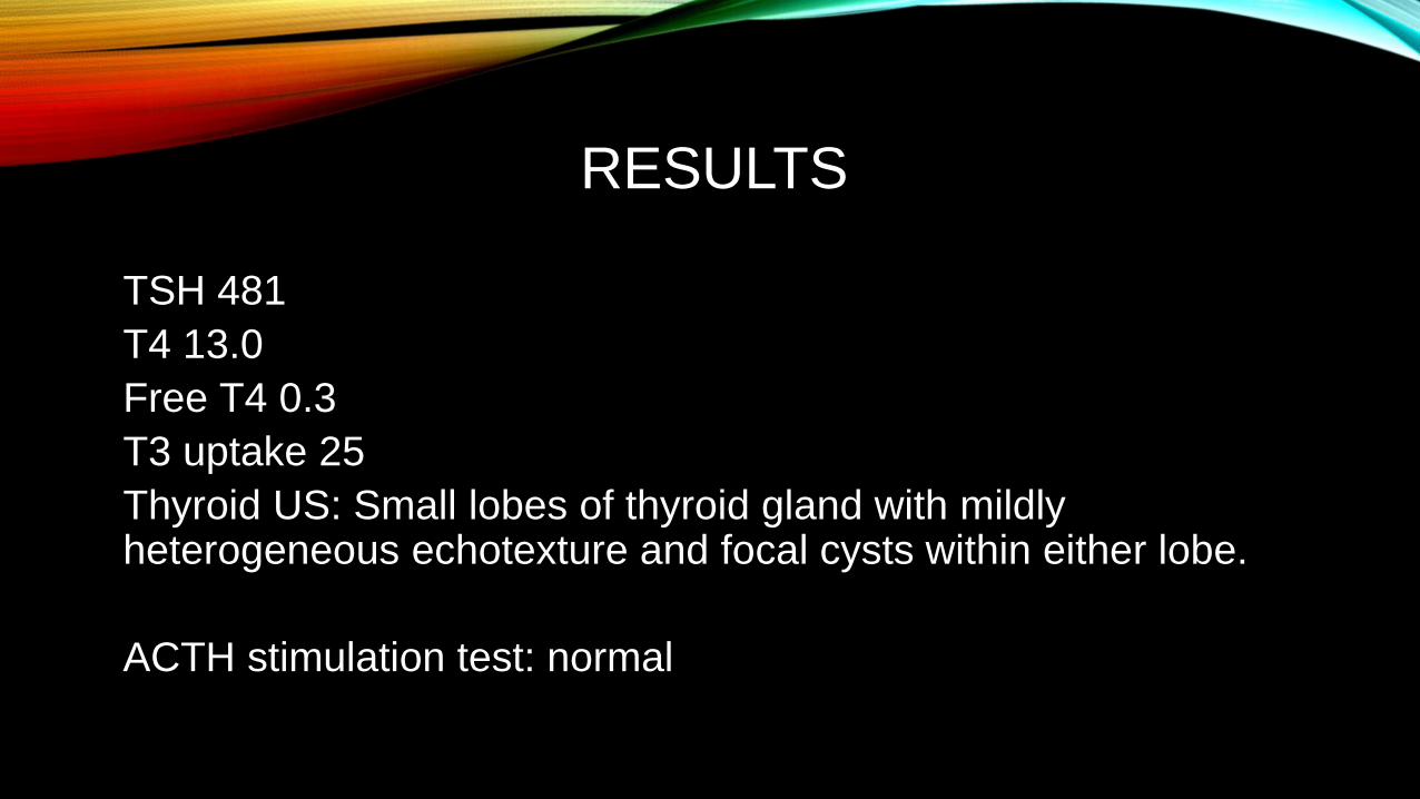
TSH 481
T4 13.0
Free T4 0.3
T3 uptake 25
Thyroid US: Small lobes of thyroid gland with mildly heterogeneous echotexture and focal cysts within either lobe.
ACTH stimulation test: normal
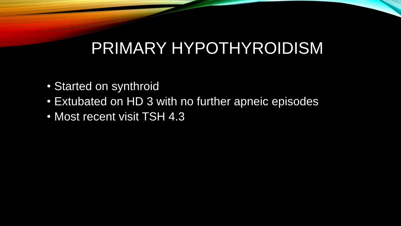
PRIMARY HYPOTHYROIDISM
• Started on synthroid
• Extubated on HD 3 with no further apneic episodes
• Most recent visit TSH 4.3
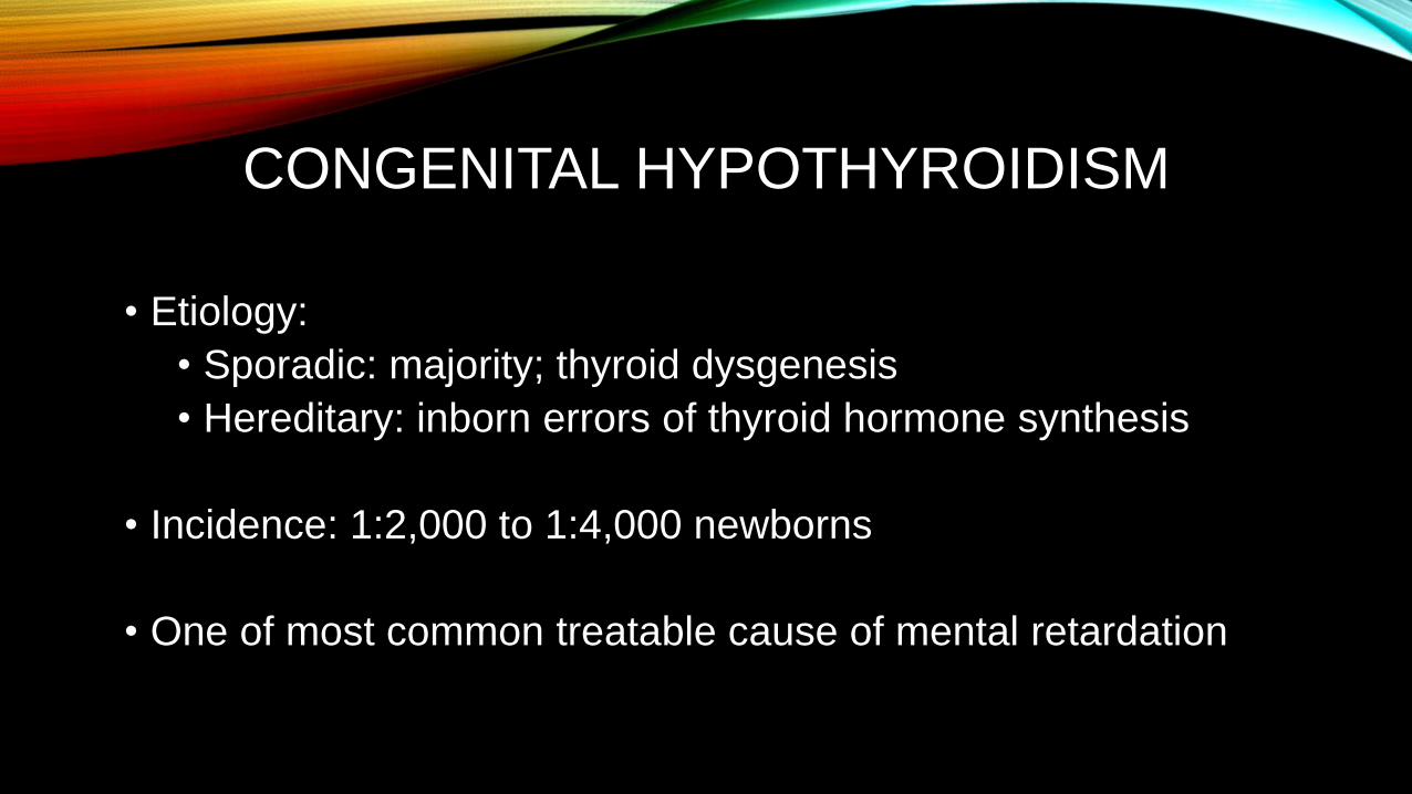
CONGENITAL HYPOTHYROIDISM
• Etiology:
• Sporadic: majority; thyroid dysgenesis
• Hereditary: inborn errors of thyroid hormone synthesis
• Incidence: 1:2,000 to 1:4,000 newborns
• One of most common treatable cause of mental retardation
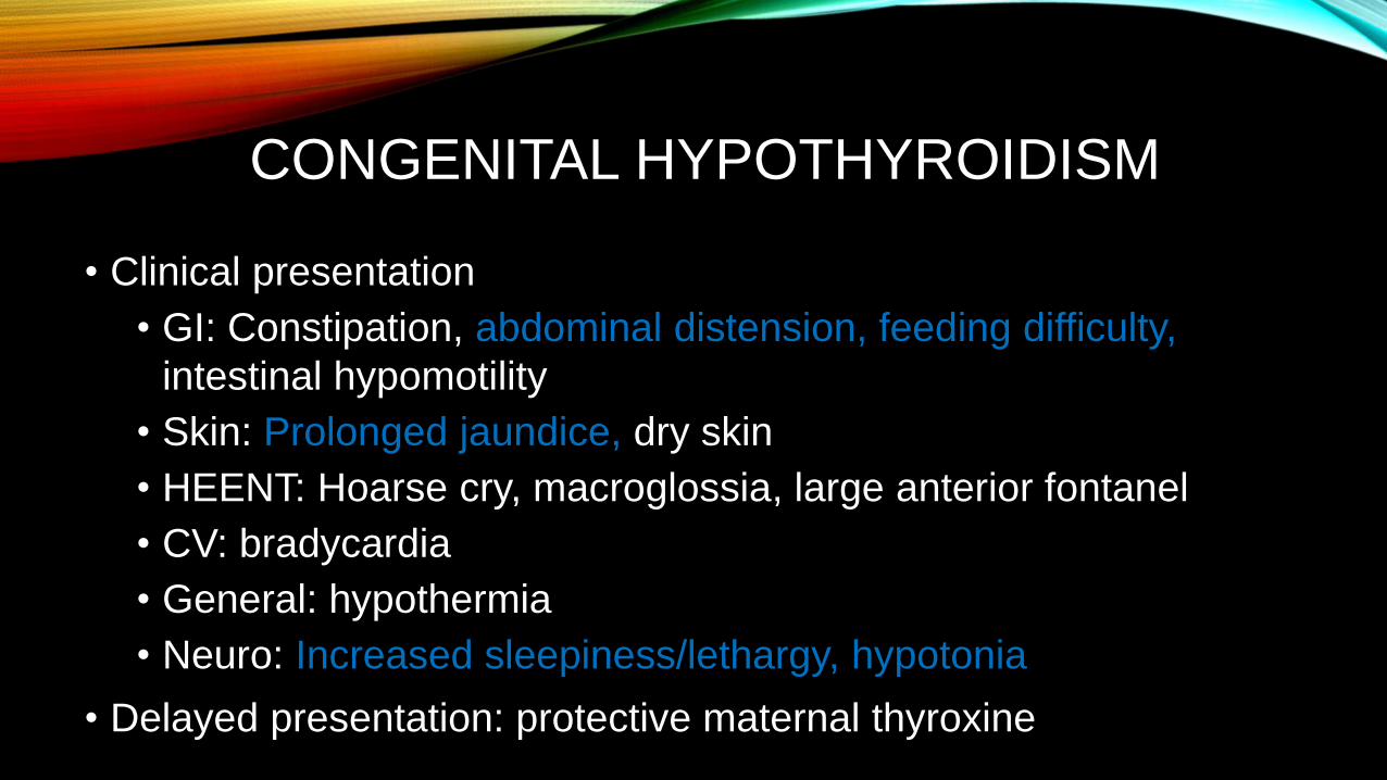
CONGENITAL HYPOTHYROIDISM
• Clinical presentation
• GI: Constipation, abdominal distension, feeding difficulty,
intestinal hypomotility
• Skin: Prolonged jaundice, dry skin
• HEENT: Hoarse cry, macroglossia, large anterior fontanel
• CV: bradycardia
• General: hypothermia
• Neuro: Increased sleepiness/lethargy, hypotonia
• Delayed presentation: protective maternal thyroxine

APNEA AND HYPOTHYROIDISM
• Sleep study: 43% infants with central apnea
• Resolve after treatment with synthroid
• Respiratory distress/failure with congenital hypothyroidism
CONGENITAL HYPOTHYROIDISM
• Diagnosis:
• Newborn screening: T4 and/or TSH
• Primary: low T4, high TSH
• Central: low T4, low/normal TSH
• Thyroid US
• Treatment:
• Early treatment, higher IQ levels
• Oral levothyroxine

CASE 6: “EYE” SEE YOU

PRESENTATION
5 year old previously healthy female with left eye redness intermittently for one week complaining of:
• Itching
• Mild crusting
• Photophobia
• Tearing
• photophobia
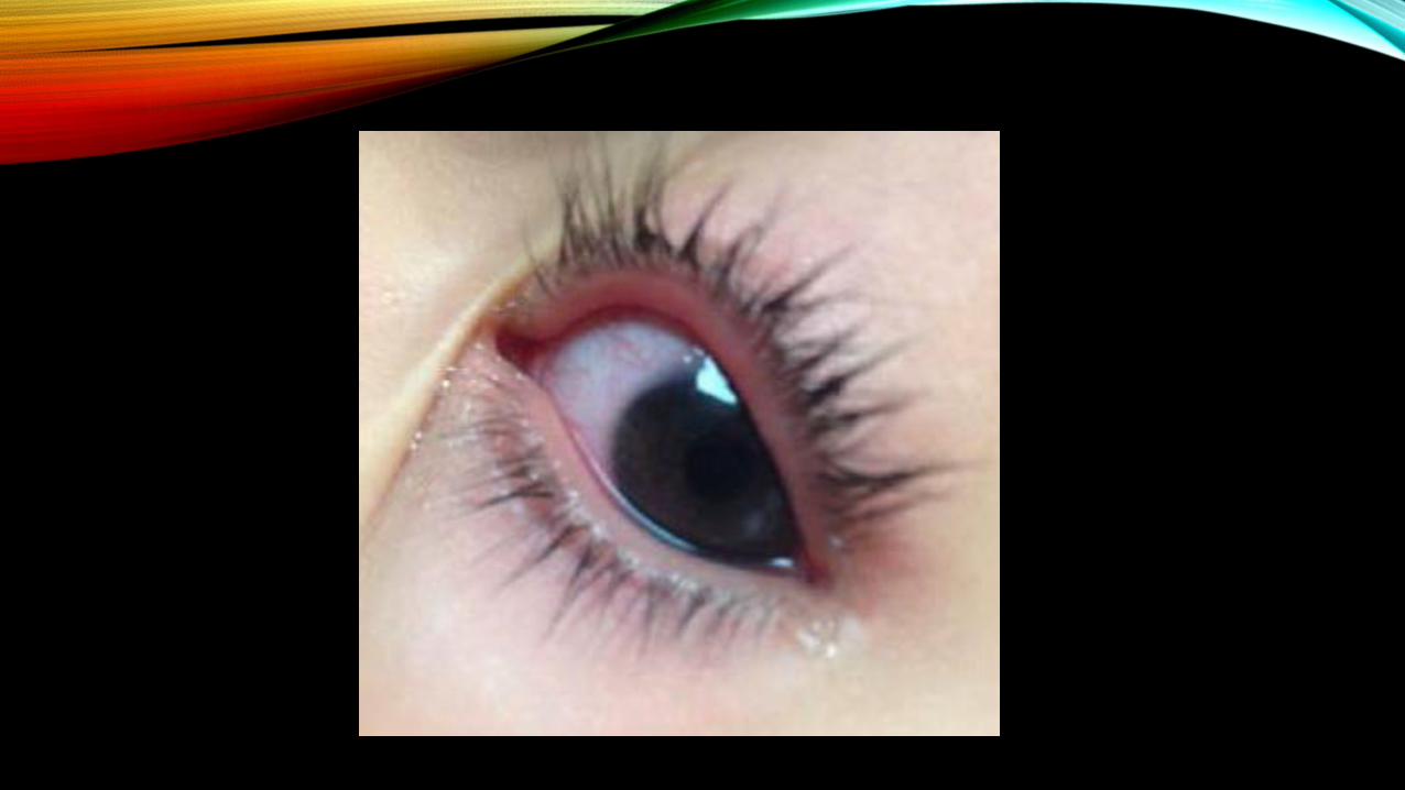

ER EVALUATION
• No uptake on fluorescein exam
• Decreased visual acuity
• Small white spot at 4 o’clock
• Erythema left conjunctiva and sclera
• Hyperemia and edema conjunctiva palpebrae

OPTHALMOLOGY CLINIC
• Per mom, patient has had eye redness on and off for the past year about 3 episodes.
• history of styes
• had a sty about 6 mos ago that recently popped about a month ago
• moderate blepharitis both eyes
• corneal scars both eyes
PHLYCTENULAR KERATITIS
• occurs primarily in children from 6 months to 16 years old
• higher prevalence in females and higher incidence during spring
• corneal lesions typically may have more severe pain and photophobia
• can lead to ulceration, scarring and mild to moderate vision loss
• Although rare, corneal perforation is possible as well

HYPERSENSITIVITY REACTION TO BACTERIAL ANTIGENS
• primarily staphylococcal
• TB
• Chlamydia and other bacterial agents
• Risk factors for S. aureus exposure include chronic blepharitis and suppurative keratitis.


PRESCRIBED TREATMENT
• Maxitrol eye drops 4x/day left eye only
• Recommend daily lid scrubs with warm compresses at least 3-4x/day
• Recommend start of Erythromycin eye ointment at bedtime
• Blepharitis: Start frequent warm compresses for treatment, baby shampoo scrubs for prevention

“RICE SOCK COMPRESSES”

FOLLOW UP
• Called by eye clinic for follow up
• Didn’t show