interactive report why do we sleep? - iitkhome.iitk.ac.in/~osegu/sleep3.pdf · interactive report...
TRANSCRIPT
Brain Research 886 (2000) 208–223www.elsevier.com/ locate /bres
Interactive report1Why do we sleep?
a,b , c*Terrence J. Sejnowski , Alain DestexheaHoward Hughes Medical Institute and the Salk Institute, 10010 North Torrey Pines Road, La Jolla, CA 92037, USA
bDepartment of Biology, University of California at San Diego, La Jolla, CA 92093, USAcUnite de Neurosciences Integratives et Computationnelles, CNRS, UPR-2191, Avenue de la Terrasse, 91198 Gif-sur-Yvette, France
Accepted 8 October 2000
Abstract
Slow-wave sleep consists in slowly recurring waves that are associated with a large-scale spatio-temporal synchrony across neocortex.These slow-wave complexes alternate with brief episodes of fast oscillations, similar to the sustained fast oscillations that occur during thewake state. We propose that alternating fast and slow waves consolidate information acquired previously during wakefulness. Slow-wavesleep would thus begin with spindle oscillations that open molecular gates to plasticity, then proceed by iteratively ‘recalling’ and‘storing’ information primed in neural assemblies. This scenario provides a biophysical mechanism consistent with the growing evidencethat sleep serves to consolidate memories. 2000 Elsevier Science B.V. All rights reserved.
Keywords: Slow-wave sleep; Spindle oscillation; Spatio-temporal synchrony; Synaptic plasticity; Memory consolidation; Computational model; Rapid eyemovement sleep
1. Introduction activity that occurs during sleep directly enters conscious-ness.
On average, a third of our lives pass by in sleep. New methods have, however, been developed to eaves-Although body movements are largely suppressed during drop on this ongoing activity and computational models ofsleep, resulting in reduced external behavior, the internal sleep states have been mathematically analyzed and simu-activity of the brain has a richness that defies explanation. lated with digital computers. This approach is still in itsAt the onset of sleep, brief episodes of 7–14 Hz synchron- infancy, but it may someday allow us to better understandized spindling occurs in the thalamus and cortex, produc- the purpose of the extensive activity that occurs in sleepinging large-scale spatio-temporal coherence throughout the brains.forebrain. During sleep the low-amplitude, high-frequency Before approaching the behavioral consequences ofactivity in the neocortex characteristic of the awake state is sleep, we must first understand the patterns of electricalreplaced with high-amplitude, low-frequency rhythms activity and the biochemical states of neurons that occur in[117]; the cortex alternates between periods of slow-wave the brain during sleep. A phenomenological description ofsleep in the 2–4 Hz range and episodes of rapid eye sleep states will provide a firm foundation for generatingmovement (REM) sleep, characterized by sharp waves of hypotheses for the functions of sleep that can be ex-activity in the pons, the thalamus and the occipital cortex. perimentally tested. This summary is based on two recent
The widespread activity that occurs in the brain during reviews, i.e. Refs. [100,117].sleep has a purpose; however, there is still no consensus on The next section focuses on the biophysical aspects ofwhat that might be. Activity in the sleeping brain is largely slow-wave sleep oscillations, including the influence ofhidden from us because very little content of the brain thalamic-generated oscillations on cortical cells and the
analysis of the spatial and temporal distribution of neuro-nal activity during slow-wave sleep. A scenario is intro-
1Published on the World Wide Web on 7 November 2000. duced for the steps that may occur leading to the consoli-*Corresponding author. The Salk Institute, Computational Neurobiol-
dation of recent memories, including the conditions lead-ogy Laboratory, 10010 North Torrey Pines Road, La Jolla, CA 92037,ing to the opening of calcium-mediated biochemicalUSA. Tel.: 11-858-453-4100; fax: 11-858-587-0417.
E-mail address: [email protected] (T.J. Sejnowski). pathways triggering gene expression and a ‘recall-store’
0006-8993/00/$ – see front matter 2000 Elsevier Science B.V. All rights reserved.PI I : S0006-8993( 00 )03007-9

T.J. Sejnowski, A. Destexhe / Brain Research 886 (2000) 208 –223 209
iteration during slow-wave sleep. This summary is based In simulations of thalamocortical cells oscillating in theon two recent articles, i.e. Refs. [30,42]. bursting mode at delta frequency, depolarizing cortical
Recent behavioral and physiological experiments will be inputs are easily able to reset the cell to a new phase of itsreviewed supporting the hypothesis that memory consoli- rhythm [84].dation may occur during sleep. Finally, some computation- Thalamic synchronization can also be induced byal issues are examined arising from recent attempts to scale stimulating cortical foci that are not directly connected toup neural network learning algorithms to multilayered the thalamic nuclei where the recordings are performed;architectures. this recruitment of thalamic cells may be achieved through
Some concrete suggestions are made for how the the reticular thalamic nucleus, which receives collaterals ofdifferent sleep rhythms could contribute to memory con- layer VI corticothalamic cells and thalamic neurons thatsolidation [39,105,106]. project to the cortex [8]; the reticular cells are exclusively
inhibitory and project back to the thalamus (but not to thecerebral cortex) and also innervate other cells of the
2. Brain rhythms during sleep reticular thalamic nucleus. The reticular nucleus is unique-ly positioned to influence the flow of information between
The thalamus and cerebral cortex are intimately linked the thalamus and cerebral cortex.by means of reciprocal projections [72]. The thalamus is Delta waves are not present throughout the entire brain.the major gateway for the flow of information toward the For example, during slow-wave sleep, the hippocampuscerebral cortex and is the first station at which incoming undergoes brief, 50–100 ms sharp waves, which are highlysignals can be modulated during sleep. This modulation synchronized events throughout the hippocampus [21].shifts the brain from an alert, aroused state, open to signals Sharp waves, also called large irregular amplitude (LIA)from the outside world, to the closed state of sleep. The activity, are often associated with brief periods of fastelectroencephalogram (EEG) during early stage of quies- oscillations (100–200 Hz) called ‘ripples’ which synchron-cent sleep is associated with spindle waves, which occur at ize action potentials among hippocampal neurons [133].a frequency of 7–14 Hz; as sleep deepens, delta waveswith slower frequencies (1–4 Hz) appear on the EEG[117]. Even slower rhythms occur during slow-wave sleep 2.2. Spindle oscillationsthat group delta waves and spindles [3,29,90].
EEG spindles are characteristic of brain electrical2.1. Delta oscillations synchronization at sleep onset, an electrographic landmark
for the transition from waking to sleep that is associatedDelta waves (1–4 Hz) were initially shown to arise with loss of perceptual awareness. Spindle oscillations
between cortical layers II to III and V [116]. Intracellular consist of 7–14 Hz waxing-and-waning field potentials,recordings in vivo and in vitro indicate that the thalamus is grouped in sequences that last for 1–3 s and recur oncealso involved in the generation of this rhythm [117]. A every 3–10 s. These oscillations are generated in thedelta-frequency rhythm can be generated in single cells by thalamus as the result of synaptic interactions and intrinsicthe interplay of two intrinsic currents of thalamocortical membrane properties of inhibitory neurons of the reticularneurons: the hyperpolarization-activated cation current (I ) thalamic nucleus and excitatory thalamocortical, and theirh
21and the transient low-threshold Ca current (I ). A wide interaction with cortical pyramidal neurons [117].T
variety of other ionic currents with different voltage In intracellular recordings from reticular anddependencies and kinetics of activation and inactivation thalamocortical cells as well as from computational model-contribute to the shaping of the amplitude and time-course ing, these two neuronal classes display a mirror imageof each burst of action potentials, as revealed both through during spindles [117]. In reticular cells, rhythmic (7–14
21biological experiments and computational modeling Hz) bursts are generated by low-threshold Ca spikes and[83,89]. are superimposed on a slowly rising and decaying de-
The hyperpolarization of thalamocortical cells is a polarizing envelope. The bursts of reticular cells inhibitcritical factor for the interplay between I and I that large numbers of the amocortical cells through theirh T
generates delta oscillation [117]. At the normal resting divergent GABAergic axons, leading to the appearance oflevel in vivo, I is inactivated, but a hyperpolarization of rhythmic inhibitory postsynaptic potentials (IPSPs) inT
10 mV can lead to spontaneous, self-sustained delta thalamocortical neurons. Some of these IPSPs result in21oscillation. The dependence of delta oscillation upon enough removal of inactivation of the low-threshold Ca
21membrane hyperpolarization can also be demonstrated in current to be followed by a rebound Ca spike andsimulations of thalamic neurons based on Hodgkin–Hux- associated burst of action potentials. These periodic burstsley-like kinetic models of the ionic currents [89]. in thalamocortical cells converge onto reticular neurons
Corticothalamic volleys potentiate and synchronize the and close the loop for rhythmic oscillation. A simpledelta oscillation of simultaneously recorded thalamic cells. model consisting of a thalamocortical cell reciprocally
210 T.J. Sejnowski, A. Destexhe / Brain Research 886 (2000) 208 –223
interacting with a reticular cell already demonstrates the cortex [31,32]. Modeling studies have shown how theessential features of spindling [43]. corticothalamic projections organize the global coherence
The waxing and waning of the spindling in this two- of thalamic oscillations [41].neuron model is controlled by the intracellular calcium During spindling and slow-wave sleep, the thalamuslevel in the thalamocortical neuron, which increases with excites the cortex with patterns of activity that are more
21each Ca spike; calcium binding to the I channels spatially and temporally coherent than would be normallyh
change their voltage dependence and eventually terminate encountered in the awake state. Depolarizing pulses of21the spindle [40], a prediction verified experimentally [81]. Ca that enter thalamic and cortical neurons may in-
Thalamic reticular neurons are involved in the genesis of fluence enzyme cascades and regulate gene expression,synchronized thalamocortical oscillations, which depend in homeostatically adjusting the balance of ionic currents andpart on their complex bursting properties. A high density regulatory mechanisms. This widespread activity could beof low-threshold calcium current (I ) has to be present in used to reorganize cortical networks following learning inT
the dendrites of these cells compared to the soma to the awake state [131], as discussed below.reproduce the firing pattern characteristics found ex-perimentally in vitro and in vivo [47]. 2.3. Arousal
Isolation of the reticular nucleus from the rest of thethalamus and cerebral cortex abolishes spindle oscillations Electrical activation of certain brainstem and hypo-in thalamocortical systems, but the deafferented reticular thalamic regions, including the so-called reticular activat-thalamic nucleus can generate oscillations at spindle ing system, causes a variety of neurotransmitters includingfrequencies [118]. Axonal and, in some species dendroden- acetylcholine (ACh), norepinephrine (NE), serotonin (5-dritic, interconnections between reticular cells may allow HT), histamine (HA), and glutamate to be released thoughthe coupling and interaction of these endogenous oscil- diffuse ascending axonal arborizations. These neuro-lators, thereby generating oscillations in an isolated nu- modulators mimic arousal by suppressing sleep spindles,cleus. Models of simplified reticular thalamic neurons with delta waves, and slow cellular rhythms, and by replacingfull connectivity and slow mutual inhibition exhibit syn- these low-frequency oscillations to activity similar to thatchronous oscillatory activity, but the frequency is below of the awake attentive animal. In cortical pyramidalthe range of the spindling rhythm [45,128]. An array of neurons, ACh, NE, 5-HT, HA, and glutamate can reduce
1model reticular neurons with fast inhibition between three distinct K currents, thereby resulting in a markedlylocally-connected neurons exhibits 8–10 Hz oscillations in enhanced responsiveness to depolarizing inputs andthe local field potential in the model (based on the average changes in neuronal firing mode [88]. Adenosine, and
1membrane potential for a cluster of nearby neurons) that GABA can reduce excitability by increasing membrane Kwax and wane similar to experimental observations [45]. conductance.
Spindling has been observed in thalamic slice prepara- These neurotransmitter systems abolish the low-fre-tions [127]. However, when the reticular cells were quency rhythms in thalamocortical systems during wakingisolated from the thalamocortical cells, spindling was and REM sleep, as well as promote more tonic activity orabolished. Models of the thalamus suggest that a larger and the appearance of fast oscillations. The changes in firingmore intact collection of reticular thalamic cells may be between sleep and arousal in thalamic neurons are accom-needed to generate spindle waves autonomously. Another plished by depolarization of the membrane potential by 5
21possible reason is that the presence of neuromodulators in to 20 mV, which inactivates the low-threshold Ca currentvivo keep the resting levels of reticular cells more depolar- and therefore inhibits burst firing. These results have beenized than in vitro; in the model, the oscillations in the simulated in models of thalamocortical and reticularreticular network are abolished at resting levels that are too neurons [40,89].hyperpolarized [46]. The spindling observed in thalamicslices exhibits traveling waves [76]. Many properties of 2.4. Rapid eye movement (REM) sleepthese traveling waves can be modeled by a one-dimension-al chain of thalamocortical and reticular neurons recip- REM sleep is characterized by an abolition of low-rocally connected within local neighborhoods [44,57]. frequency oscillations and an increase in cellular excitabili-
In the cortex, synchrony between cortical cells separate ty, although motor output is markedly inhibited [68]. In theby 1 cm was insensitive to cutting the horizontal intracorti- thalamocortical system, REM sleep and wakefulness havecal connections between them, suggesting that cor- equivalent electrophysiological signatures [80]. Dreamticothalamic projections have a powerful impact in reports are common upon arousal from REM sleep, but cansynchronizing distant part of cortex and thalamus through also occur during slow-wave sleep. Despite great interest,corticothalamic loops. The overall spindle activity pattern there is no generally accepted function for dreams or, forin the thalamus is, in turn, organized by the cortex. The that matter, for the sleep state itself. It is of interest thatspatiotemporal properties of synchronized thalamic spindle during REM sleep, the activity in the hippocampus, whichoscillations became disorganized after removal of the forms reciprocal connections with the neocortex, displays a
T.J. Sejnowski, A. Destexhe / Brain Research 886 (2000) 208 –223 211
strong, low-frequency rhythm in the theta range, 6–8 Hz, and second, that these oscillations are also present duringwhich is entrained by inhibitory inputs from the septal sleep-like activity patterns; this goes beyond the conven-nuclei, which primarily project to the inhibitory inter- tional view that fast oscillations are present only duringneurons in the hippocampus. brain-activated states. Intracortical coherency of fast oscil-
In the hippocampus there is a population of excitatory lations is coupled with synchronized fast rhythms inprincipal cells with recurrent connections that strongly corticothalamic circuits. The coherence of the fast rhythmsinteract with inhibitory interneurons. Paradoxically, in- is short range whereas low-frequency sleep rhythms ex-creasing an external inhibitory drive onto the inhibitory hibit synchronization on a larger spatial scale (see below).interneurons, without directly affecting any other part of The diversity of cortical cells and their complex interac-the network, can in some circumstances cause the inter- tions make it difficult to model cortical networks with theneurons to increase their firing rates [125]. For this to same confidence that thalamic networks have beenoccur, recurrent connections among the excitatory cells modeled. It is not, however, difficult to generate oscillatoryhave to be strong enough to make the excitatory network activity in the 20–80 Hz range with networks of simplifiedunstable when feedback inhibition is removed. When there neurons [100]. These models have revealed the need tois a periodically varying input, such as the input to the regulate the tendency of recurrent networks to oscillate.hippocampus from the septal nucleus, there should be a The excitability of neurons can be controlled by inhibition;systematic relationship between the phase shift and depth however, inhibition is also an efficient mechanism forof modulation for each interneuron. This prediction was synchronizing large populations of pyramidal neuronstested and confirmed by recordings from interneurons in because of voltage-dependent mechanisms in their somatathe CA1 region of the rat hippocampus in vivo [125]. and the strategic location of inhibitory boutons on the
The two major sleep phases, slow-wave and REM sleep, somata and the initial segments of axons, where actionare mirrored by two awake states in the hippocampus of potentials are initiated [82]. Networks of reciprocallyrats and probably in other mammals: the first is a state of interconnected interneurons can produce gamma oscilla-low-frequency theta oscillation that occurs during active tions when the interneurons fire doublets [124]. Realisticexploration, which resembles the state of the brain during simulations of cortical neurons demonstrate that sparseREM sleep, and a second awake state consisting of sharp excitatory connectivity between distant populations ofwaves that occurs during rest, which resembles the activity neurons can produce synchronization within one or twoin the hippocampus characteristic of slow-wave sleep. cycles, but only if the long-range connections are made on
inhibitory as well as excitatory neurons [20].2.5. Fast oscillations Thalamocortical oscillations are characterized by distinct
spatiotemporal patterns of activity, which suggests thatChanges in the activity pattern generated by cortical these oscillations may play distinct roles. We turn to the
neurons and circuits are less stereotyped than those of biophysical characterization of these oscillations in orderthalamic cells and circuits, although some common fea- to explore possible roles.tures exist. The low-frequency oscillations of the corticalEEG disappear upon arousal and are replaced by higherfrequency rhythms in the 20–80 Hz frequency range, 3. Biophysical aspects of sleep oscillationswhich includes beta and gamma frequency bands. As in thethalamus, these alterations in cortical activity take place, at To investigate the possible role of oscillations, weleast in part, by depolarization of pyramidal cells, pre- studied the effects of thalamic input on neocortical pyrami-
1sumably through the reduction of specialized K conduct- dal neurons based on intracellular recordings in vivo andances by ACh, NE and other neuromodulators [88,114]. computational models [30]. The data and model suggest
The fast oscillations in the EEG occur during some that spindle oscillations are highly effective at inducing21behaviors such as immobility during hunting and focused Ca entry in the dendrites of pyramidal neurons, which
attention to stimuli during complex sensory or motor tasks. might prime the neuron for biochemical events that laterNeurons throughout the nervous system (for example, could lead to permanent changes in the network. In theretina, lateral geniculate thalamic nucleus, and cortex) have second part, we examine the spatiotemporal distribution ofthe ability to generate repetitive trains of action potentials the patterns of activity associated with different events ofin the frequency range 20–80 Hz, although the synchroni- wake and sleep states [42]. These results lead to a scenariozation of this activity into behaviorally relevant subgroups in which slow-wave sleep oscillations serve to reorganizeof widely spaced neurons has only been demonstrated in the cortical network (see details in Ref. [39]).the cerebral cortex [58].
Fast (30–40 Hz) spontaneous oscillations have been 3.1. Characterization of the effect of thalamic inputs inobserved in intracortical, corticothalamic, and in- neocortical pyramidal neuronstrathalamic networks [42,115], demonstrating, first, thatfast oscillations are in phase throughout the cortical depth, Highly synchronized spindle oscillations are generated

212 T.J. Sejnowski, A. Destexhe / Brain Research 886 (2000) 208 –223
by intrathalamic and thalamocortical loops in which the EPSPs do indeed occur in pyramidal cell dendrites, theyrebound bursts of thalamocortical (TC) cells are central. are not apparent in somatic recordings because of strongEPSP/IPSP sequences often follow synchronized thalamic feedforward inhibition?inputs in neocortical pyramidal neurons during sleep The possibility was explored with computational modelsspindles (Fig. 1A1–A2). The dendrites of neocortical and intracellular recordings in vivo [30]. Intracellularlypyramidal cells therefore receive highly synchronized and recorded pyramidal neurons in the suprasylvian gyrus ofpowerful excitatory inputs from the thalamus. However, cats under barbiturate anesthesia showed that spindledespite the potentially powerful nature of these synchron- oscillations in the EEG are paralleled with EPSP/IPSPized thalamic inputs, pyramidal neurons have a relatively sequences in cortical neurons (Fig. 1A1–A2). Theselow rate of discharge during spindle oscillations [51,112], sequences were indistinguishable from that obtained fromas shown in Fig. 1A. Could it be that although strong thalamic stimulation, suggesting that spindle-related IPSPs
Fig. 1. Evidence that spindle oscillations evoke calcium entry in pyramidal neurons. (A) In vivo intracellular recordings in suprasylvian cortical neuronsduring spindle oscillations under barbiturate anesthesia. (A1) simultaneous recording of intracellular and extracellular activity. (A2) each cycle of thespindle oscillation corresponds to EPSP/IPSP sequences in the recorded pyramidal neuron. (A3) dual intracellular recording in which one of the neuron(middle trace) was recorded with chloride-filled pipettes. In this case, the EPSP/IPSP sequence transformed into a powerful burst of action potentials. (B)Computational models of thalamic inputs in pyramidal neurons. (B1) morphology used in the simulations. A layer V pyramidal neuron recordedintracellularly in A was filled and its morphology was integrated into simulations. Simulations of thalamic inputs in layers I, IV and VI (gray areas) weredirectly compared to the experimental recordings of thalamic inputs in that cell. (B2) simulated EPSP/IPSP sequences and bursts following inversion of thechloride reversal potential. The model could match experiments only if both excitatory and inhibitory conductances were strong. (B3) calcium transients inthe dendrites of the model following thalamic inputs. The membrane potential at the soma (top trace) consisted in an EPSP/IPSP sequence. However,representing the profile of calcium concentration (bottom trace) along a path from soma (left) to distal apical dendrite (*) shows important calciumtransients consequent to strong dendritic depolarization. Figure modified from Ref. [30] (see for details).
T.J. Sejnowski, A. Destexhe / Brain Research 886 (2000) 208 –223 213
were triggered by thalamic inputs. To further characterize pyramidal neurons: the dendrites are dominated by excitat-the IPSP component, dual intracellular recordings were ory synapses while the soma essentially receives inhibitoryperformed in which one cell was recorded using a K–Cl synapses [37]. This imbalance necessarily implies that thefilled pipette. Cells recorded with chloride pipettes fired dendrites of pyramidal neurons must experience strongbursts of 4–7 action potentials at 100 Hz with spike depolarization, but this depolarization is not visible at theinactivation, in phase with spindle waves (Fig. 1A3). somatic level due to the proximal inhibition. Thus the
Computational models were designed based on these model suggests that during spindling, cortical pyramidalexperiments. Neurons were impaled for intracellular re- cells receive strong dendritic excitation in parallel withcording, stained with Neurobiotin and reconstructed using strong inhibition around the soma, preventing the cell froma computerized tracing system, as shown in Fig. 1B1. The firing.model of thalamic EPSP/IPSP sequences was based on It thus seems that, during sleep spindles, the synchron-three sets of constraints: (a) The relative density of ized bursts of high-frequency action potentials in thalamicexcitatory and inhibitory synapses in different regions of neurons generate in the cortex ideal conditions for stronglythe cell; (b) The fraction of these synapses activated by a depolarizing their dendrites but keeping the cell fromgiven stimulus; and (c) The kinetics of the different types firing. Simulations indicate that this type of input is well-of receptors involved (see details in Ref. [30]). Thalamic suited to triggering calcium entry into dendrites (Fig. 1B3).axons end preferentially in cortical layers I, IV and VI This is consistent with in vitro studies showing that
21[64,72]. Based on these anatomical data, the thalamocorti- dendritic depolarization is accompanied by Ca entrycal synapses are distributed 15% in layer I (more than 800 [134]. The proximal inhibition would allow a massive
21m above the soma), 60% in layer IV (from 50 to 200 m Ca entry into the dendrites of pyramidal neurons withoutabove the soma), and 25% in layer VI (below 200 m of the producing excessive firing in the cortical network.soma); there were no excitatory synapses in any other Various calcium-dependent mechanisms are involved inlayers in the model. A schematic of this distribution is synaptic plasticity (reviewed in Ref. [56]). Thus, calciumshown in Fig. 1B1. entry during sleep may serve to prime the synapses for
This model was used to estimate the excitatory and permanent changes. In particular, CaMKII, an enzyme thatinhibitory conductances underlying spindle-related EPSP/ is abundant at synapses and is implicated in synapticIPSP sequences. To reproduce the strong burst discharges plasticity of excitatory synapses in the cortex and else-
21observed with chloride-filled pipettes, it was necessary to where [108,129], is not only sensitive to Ca but also to21include relatively strong GABA -mediated conductances. the frequency of Ca spikes [38]. It is possible that sleepA
Under these conditions, the reversed IPSPs could explain spindles provide a selective signal to efficiently activatethe genesis of these strong bursts. However, to reproduce CaMKII in the dendrites of cortical pyramidal neurons.
21the relatively moderate discharge under normal conditions, Another possible consequence of massive Ca entry init was also necessary to assume significant excitatory pyramidal neurons is calcium-dependent gene expression,conductances. Only under these conditions was it possible which is also frequency sensitive [60,79] in the delta rangeto reproduce both moderate control discharges and bursts of frequencies (1–4 Hz). Calcium that enters dendritesin the presence of chloride (Fig. 1A2). The exact values of during spindles may accumulate in the endoplasmic re-the conductances, the kinetics of synaptic receptors and the ticulum, which forms a continuous compartment within thedistribution of synapses can be found in Ref. [30]. neuron continuous with the nucleus. Calcium-stimulated
This combination of models with in vivo intracellular calcium release from the endoplasmic reticulum duringrecordings strongly suggests that during spindling, the delta waves may then deliver the calcium signal to thestrength of thalamic input on neocortical pyramidal cells nucleus [11].
21should be stronger than that indicated by the low firing The repeated Ca entry may activate a molecularrates of these cells. The model suggests that the thalamic ‘gate’, opening the door to gene expression. This possi-inputs on neocortical pyramidal cells could evoke powerful bility is based on the observation that repeated high-glutamatergic conductances in parallel with strong frequency stimuli, but not isolated stimuli, activate proteinGABAergic conductances. It is conceivable that this strong kinase A (PKA), an enzyme implicated in long-lastingGABAergic component prevents the cell from firing by synaptic changes and long-term memory [2]. The proposedshunting the EPSPs to prevent avalanches of excitatory mechanism is that PKA acts like a ‘gate’ by inhibitingdischarges. This conclusion is in agreement with direct phosphoprotein phosphatases, which themselves exert ameasurements of conductances during visual inputs in tonic inhibition over biochemical cascades leading to genecortical neurons that also revealed large GABAergic expression [2,15,16] (for a model, see Ref. [12]). Theconductances [19]. evidence for this hypothetical mechanism is supported by
The synaptic conductances evoked by thalamic inputs in observations that activation of PKA alone does not inducecortical pyramidal neurons are more than a simple shunt of synaptic changes, but blocking PKA suppresses long-termthe EPSPs by IPSPs. Morphological data indicate that synaptic changes in the hippocampus [53].excitation and inhibition are not evenly distributed in All the necessary enzymes for this mechanism are
214 T.J. Sejnowski, A. Destexhe / Brain Research 886 (2000) 208 –223
located at or near the active zone of the synapse coherence are similar to the fast oscillations during wake-[26,48,75,93]. fulness (Fig. 2B, right panel). This similarity applied to the
Similar mechanisms could occur during spindling. It relation between these fast oscillations with unit dis-21may be that the massive Ca entry during spindles, charges, which showed that the depth-negative EEG
repeated at approx. 10 Hz frequency, provides the con- components are correlated with an increased probability ofditions needed to open a molecular gate, for example unit firing (Fig. 2C, right panels).mediated by PKA. Sleep spindles could therefore provide a Therefore, these brief episodes are electrophysiological-physiological signal similar to the repeated tetanus used to ly indistinguishable from the ‘sustained’ fast oscillations ofinduce long-term synaptic changes in slices. It could also awake and REM sleep.provide a ‘priming’ signal, opening a gate that allows The observation that highly coherent slow-wave patternspermanent changes to subsequent inputs following the alternate with brief episodes of low-coherence fast oscilla-sleep spindles (see below). tions can be interpreted in several ways. Slow waves and
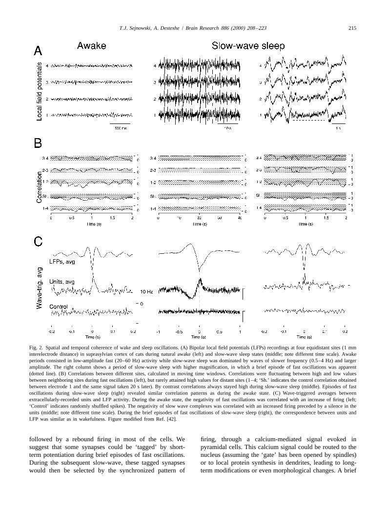
fast oscillations might represent different states of respon-3.2. Spatiotemporal structure of slow-wave sleep siveness of cortical neurons and different receptive fieldoscillations properties [34,50,63,132]. We favor another interpretation
[42,39], in which slow-wave sleep is viewed as a cyclic,Spindles generally appear during the early phase of iterating process leading to memory consolidation. Brief
slow-wave sleep, which is later dominated by other types episodes of processing similar to the awake state (episodesof oscillation, such as slow-waves (delta waves at 1–4 Hz of fast oscillations) alternate with highly synchronizedor slow oscillations of ,1 Hz). We analyzed the spatiotem- network events (slow-waves). As shown above, during theporal coherence of these types of oscillations in the rebound of the network (depth negativity of the slowsuprasylvian gyrus of cats during natural awake and sleep wave), cortical cells should receive strong EPSPs followedstates [42]. We found that low-amplitude fast oscillations by IPSPs, which is likely to trigger a massive calciumof wakefulness (Fig. 2A, left panel) are characterized by entry in the dendrites. Slow-waves may therefore be part ofrelatively low spatiotemporal coherence, in agreement with a process for establishing permanent changes in theprevious findings [49,59,113]. This is shown in the correla- network through calcium-dependent mechanisms. Slow-tions, which fluctuated in both space and time, only wave sleep in this scenario could then be part of ‘recall’reaching high values occasionally and only for neighboring and ‘store’ sequences, in which the fast oscillations couldsites (Fig. 2B, left panel). reflect recalled events experienced previously, which are
Analyzing the correlations between extracellularly-re- stored in the network through the synchronized firing thatcorded units and local EEG revealed that units fired occurs during the slow-wave complexes in the EEG.tonically, and wave-triggered averages showed that unitfiring was correlated with the depth-EEG negativity (Fig. 3.3. A biophysically-based hypothesis for network2C, left panel). Similar characteristics were found for fast reorganization during sleeposcillations during REM sleep (see details in Ref. [42]).
In contrast, slow oscillations are remarkably coherent The global picture that emerges from the studies sum-across several millimeters in cortex (Fig. 2A, middle marized above is that two types of sleep oscillation,panel). Consistent with this, correlations during slow spindles and slow waves, may have complementary rolesoscillations stay high across wide regions of the cortex in network reorganization. At sleep onset, the thalamus(Fig. 2B, middle panel). Remarkably, slow-wave complex- enters a burst mode and generates synchronized spindlees are correlated with a concerted decreased/ increased oscillations. During these oscillations, the high-frequencyfiring in single units (Fig. 2C, middle panel). This shows bursts of thalamic relay cells occur synchronously in thethat these high-amplitude EEG waves reflect a remarkably thalamus and therefore provide unusually strong excitat-synchronized network event consisting of a generalized ory / inhibitory inputs in cortical pyramidal neurons. Thesilence followed by an increased firing, or network ‘re- repetition of these inputs at approx. 10 Hz is particularlybound’. Similar conclusions were drawn for delta waves in efficient to trigger periodic calcium entry in corticalvarious preparations [7,25,54], for spontaneous slow oscil- dendrites and activate intracellular mechanisms, such aslations under anesthesia [28,123] as well as in cortical CaMKII or molecular gates. This process could serve toslices [102]. open the door between synaptic activation and gene
Perhaps the most interesting observation was that slow- expression, so that pyramidal neurons are ready to producewaves also contain a myriad of brief episodes of low- permanent changes in response to some specific synapticamplitude fast oscillations that are nested within slow- events that need to be consolidated.wave complexes (Fig. 2A, right panel). This corroborates As sleep deepens, slow-waves such as delta oscillationsprevious observations in anesthetized animals [63,132] or progressively dominate the EEG. A slow-wave complex isduring natural sleep [115]. Focusing specifically on these a remarkably synchronized network event, in which abrief episodes showed that their spatial and temporal concerted period of silence is seen across the network,
T.J. Sejnowski, A. Destexhe / Brain Research 886 (2000) 208 –223 215
Fig. 2. Spatial and temporal coherence of wake and sleep oscillations. (A) Bipolar local field potentials (LFPs) recordings at four equidistant sites (1 mminterelectrode distance) in suprasylvian cortex of cats during natural awake (left) and slow-wave sleep states (middle; note different time scale). Awakeperiods consisted in low-amplitude fast (20–60 Hz) activity while slow-wave sleep was dominated by waves of slower frequency (0.5–4 Hz) and largeramplitude. The right column shows a period of slow-wave sleep with higher magnification, in which a brief episode of fast oscillations was apparent(dotted line). (B) Correlations between different sites, calculated in moving time windows. Correlations were fluctuating between high and low valuesbetween neighboring sites during fast oscillations (left), but rarely attained high values for distant sites (1–4; ‘Sh.’ indicates the control correlation obtainedbetween electrode 1 and the same signal taken 20 s later). By contrast correlations always stayed high during slow-wave sleep (middle). Episodes of fastoscillations during slow-wave sleep (right) revealed similar correlation patterns as during the awake state. (C) Wave-triggered averages betweenextracellularly-recorded units and LFP activity. During the awake state, the negativity of fast oscillations was correlated with an increase of firing (left;‘Control’ indicates randomly shuffled spikes). The negativity of slow wave complexes was correlated with an increased firing preceded by a silence in theunits (middle; note different time scale). During the brief episodes of fast oscillations of slow-wave sleep (right), the correspondence between units andLFP was similar as in wakefulness. Figure modified from Ref. [42].
followed by a rebound firing in most of the cells. We firing, through a calcium-mediated signal evoked insuggest that some synapses could be ‘tagged’ by short- pyramidal cells. This calcium signal could be routed to theterm potentiation during brief episodes of fast oscillations. nucleus (assuming the ‘gate’ has been opened by spindles)During the subsequent slow-wave, these tagged synapses or to local protein synthesis in dendrites, leading to long-would then be selected by the synchronized pattern of term modifications or even morphological changes. A brief

216 T.J. Sejnowski, A. Destexhe / Brain Research 886 (2000) 208 –223
episode of fast oscillations that follows next would then Changes occur in the correlations between hippocampalpotentiate or tag another subset of synapses; these tagged place cells in freely moving rats as a consequence ofsynapses would be selected by the next slow-wave for learning a new environment [131]. In these experiments,permanent changes, and the cycle repeats itself several neurons that had neighboring place fields and fired togetherhundred times during the slow-wave sleep episode. during exploration of an environment became more highly
Alternating slow-wave complexes with brief episodes of correlated during subsequent sleep episodes in comparisonfast oscillations during slow-wave sleep could thus result with activity during preceding sleep episodes. The corre-in the permanent formation of small sets of strongly lated firing of neurons in the hippocampus during sleepinteracting neurons. This type of interactions might pro- may be a ‘played back’ version of newly acquired ex-vide a biophysical mechanism for the consolidation of periences to the neocortex through feedback projectionsmemory traces in cerebral cortex. This theme is explored [22,27,86,87,107]. Thus, the neocortex during the wakein more detail in the next section. state provides the hippocampal formation with a detailed
description of the days events; during sleep, the inputsfrom the hippocampus recreates some version of these
4. Sleep and memory consolidation events in the neocortex, where permanent memory repre-sentations are gradually formed over repeated episodes of
There is growing evidence that memory consolidation sleep.occurs during sleep [22–24,120,119]. However, not much Cortical representations of objects and events are widelyis known about how the different phases of sleep contrib- distributed in cerebral cortex; for example, the representa-ute to consolidation or what biophysical mechanisms are tion of the shape of a violin might be stored in the visualinvolved. Our emerging understanding of the events that cortex, the sounds made by a violin in the auditory cortex,take place in the thalamocortical system during sleep how it is grasped in the parietal cortex, and how it issuggest that sleep spindle oscillations might have a promi- played in the motor cortex [35]. Problems arise when newnent role in gating mechanisms for plasticity in pyramidal experiences and objects must be integrated with existingneurons, and in a subsequent phase of sleep the alternation information that is widely distributed. Learning algorithmsof slow waves and fast oscillations could provide the designed for artificial neural networks that use suchsubstrate for consolidation. This mechanism might involve distributed representations can suffer from ‘catastrophica interactions between the hippocampus and the neocortex. interference’ when new information is stored in the sameWe review here evidence for this scenario and discuss neural circuits as previously stored information [87].some consequences that raise new issues and explore new Therefore, the brain must solve two problems duringhypotheses. learning: where to make the changes needed to create new
links between existing memories, and how to make4.1. Hippocampus, neocortex and retrograde amnesia changes that are compatible with previously stored
memories.Behavioral experiments on retrograde amnesia have
shown that lesions limited to the hippocampus and sur- 4.2. Computational models of sleeprounding regions produce memory impairment in mon-keys; only recent memories are impaired while remote During REM sleep the brain is as metabolically active asmemories remain intact [6,136]. These experiments sug- in the awake state. What is all this metabolic activity beinggest that the hippocampal formation is required for mem- used for? One intriguing possibility is that informationory storage for only a limited time period after learning. As acquired by the brain during the day is being recalledtime passes, its role in memory diminishes, and a more during sleep [68]. Neural network models of associativepermanent memory gradually develops, probably in memory included such a ‘sleep phase’ to calibrate theneocortex, that is independent of the hippocampal forma- storage of memories acquired by Hebbian mechanismstion [18,73,87,110,136]. The hippocampus may not be the [4,33,70]. Another related idea is that sleep is used tosite of storage for the information before it is consolidated, consolidate memories of recent facts and events (reviewedbut may instead facilitate the associative links between in Ref. [106]).information stored in different parts of the cerebral cortex In one computational explanation for memory consolida-and other parts of the brain. tion, brain activity in the neocortex representing previous
Until recently, the processes that may occur in the cortex events during waking is reactivated through feedback fromduring consolidation could only be inferred indirectly from the hippocampal formation. Before consolidation, the lacklesion experiments, but recordings from freely moving rats of direct connections between distant brain areas preventduring awake and sleep states corroborate the idea that the parts of the memory trace to reactivate other parts thathippocampus and the neocortex exchange information represent a unique episode. During sleep, indirect con-during sleep states [131,107,120]. Neurons in the rat nections are formed within the neocortex that allow thehippocampus respond to places in the environment [96]. memory traces in different parts of the brain to become
T.J. Sejnowski, A. Destexhe / Brain Research 886 (2000) 208 –223 217
re-excited without the need of the hippocampus [5,87]. tribution for the world. There is also a goal-directedAfter learning, activity in the hippocampus is no longer reinforcement learning system in the brain that involvesneeded to reactive a memory, and in the process, the subcortical as well as cortical structures [95]. Unsuper-elements of the specific memory have been integrated into vised awake–sleep learning and other forms of learningthe general knowledge store by virtue of repeated reactiva- could work together, biasing, shifting and adapting corticaltions. This type of model might be called the completion representations to insure survival in complex and uncertainmodel for memory consolidation since the purpose of sleep environments.in this model is to improve associative pattern completion The two computational explanations offered above forin a sparsely connected system of networks in the neocor- memory consolidation during sleep are neither mutuallytex. exclusive nor exhaustive. They nonetheless have the virtue
In another theoretical approach to memory consolida- of allowing specific predictions to be made for some of thetion, the cortex stores probability densities for sensory puzzling physiological phenomenon summarized above.states in a hierarchy of layers; that is, the higher areas of For the completion hypothesis, feedback projections fromthe cortex encode higher-order statistical regularities in the higher cortical areas to lower ones are used during sleepsensory inputs and in the absence of sensory input can for the purpose of reactivating assemblies of neurons thatgenerate activity in earlier stages of processing with the had previously been used to represent specific episodicsame statistics using feedback connections. By generating memories. In this case, plasticity should occur toideal input patterns, the feedforward connections respon- strengthen connections that encourage the same patterns tosible for recognition can be accurately trained to improve reoccur in the future. In the case of the generativeprocessing at earlier stages [66]. Conversely, when the hypothesis, ideal patterns of activity are instantiated inbrain is awake, the sensory inputs drive the feedforward lower cortical areas for the purpose of altering the feedfor-system, during which the weights on the feedback con- ward connections.nections can be altered, thereby improving the generativemodel. This two-phase process produces an internal, 4.3. Interactions between the hippocampus and thehierarchical representation of experiences that are econ- neocortexomical. The feedback connections in this model are used togenerate prototypical input patterns. The learning mecha- During the transition from wakefulness to sleep, thenisms needed are biologically possible since, unlike previ- cerebral cortex becomes less responsive to external sensoryous learning algorithms for hierarchical networks that inputs and less concerned with actively gathering infor-required a detailed teacher and error backpropagation, this mation. The thalamus, which during the wake state relaysawake–sleep model only depends on locally available sensory information from the periphery to the cortex,signals and there is no teacher other than the sensory becomes less of a relay and more of a mirror, as feedbackexperience. connection from the cortex to the thalamus become
The awake–sleep learning algorithm attempts to capture capable of entraining thalamic neurons through synchro-the statistics of sensory inputs with an internal code that is nous bursting. In a sense, during sleep, the cortex nocapable of representing component features that are com- longer listens to the outside world, but rather to itself.mon to many objects. Because these statistical components Feedback connections from the cortex to the thalamusare not apparent without comparing many sensory ex- become as important as thalamocortical ones and infor-periences, the training process is gradual, in the sense that mation can flow in both directions.only small changes are made during any one awake–sleep Theta oscillations occur in the hippocampus when ancycle. Although the feedback connections are not used animal explores its environment and also during REMduring the awake or feed forward phase of the algorithm, it sleep, when the cortex is in a state that resembles activeis possible to view them as representing a prior probability exploration. During this state the hippocampus is primeddistribution on complex brain states. Thus, if sensory input for receiving and retaining information from the neocortex:is locally ambiguous, it may be possible for the feedback the inputs to the hippocampus report on the detailed stateconnections from higher levels in the hierarchy help of the cortex while the animal is actively exploring thedisambiguate them [65,78]. world and the synapses in the hippocampus are particularly
The awake–sleep model is limited to a passive, un- susceptible to changes in efficacy [71]. In contrast, duringsupervised form of learning that is entirely driven by the slow-wave sleep, the hippocampus bombards the cortexstatistics of sensory states. Not all sensory inputs are with activity rather than the sensory world. Recently storedequally important, and some tasks might require special information in the hippocampus may be played back to therepresentations. It would be easy to add attentional mecha- neocortex during slow-wave sleep, but in combinationsnisms that would modulate the learning rate with signifi- that may not have occurred simultaneous during the daycance of the stimulus. There may also be biases in cortical and at a rate that is much faster [98]. This information is arepresentations at birth that are specified during develop- distillation of recent sensory impressions and cortical statesment, which could incorporate a prior probability dis- that are activated during REM sleep. The information
218 T.J. Sejnowski, A. Destexhe / Brain Research 886 (2000) 208 –223
stored in the hippocampus is probably not a literal copy of based on mechanisms for synaptic plasticity that areinformation stored in the neocortex, which has a much synapse specific and driven by local signals. In particular,larger capacity, but rather is an abstraction of that in- Donald Hebb in 1949 [62] proposed a Neurophysiologicalformation, a pointer that is capable of reactivating that Postulate for how synaptic strengths could be adjusted byinformation in the neocortex through feedback connection an activity-dependent mechanism:from the hippocampus.
Feedback from the hippocampus to the neocortex takes ‘‘When an axon of cell A is near enough to excite cellthe form of sharp waves, brief bursts of activity that occur B and repeatedly or persistently takes part in firing it,at intervals of 0.3 to 3 Hz [21]. Not much is known about some growth process or metabolic change takes placethe mechanisms that initiate sharp waves in the hippocam- in one or both cells such that A’s efficiency, as one ofpus or elsewhere (see Ref. [97]). It is thought that the cells firing B, is increased.’’ (Hebb, 1949, p. 62)spontaneous activity in hippocampal neurons and associ-ated neurons in the hippocampal formation ignites an The traditional interpretation of this postulate is thatassembly of reciprocally connected neurons to discharge in synaptic plasticity is based on coincidences between theless than 100 ms. The temporary associations between release of neurotransmitter from a presynaptic terminal andneurons formed during the day, perhaps at different times, the depolarization of the postsynaptic dendrite. Evidencemay be recapitulated during the brief bursts of activity that for a coincidence detection mechanism has been found inthen can imprint traces of these associations on the the hippocampus, where long-term potentiation (LTP),neocortex, which is in a receptive state during slow-wave discovered there in 1973 by Tim Bliss and Terje Lomo,activity. was shown to be Hebbian in the sense of temporal
When the neocortex switches from slow-wave sleep to coincidence [14,74]. LTP of synapses on hippocampalREM sleep, characterized by high-frequency activity, the neurons can be elicited by pairing synaptic input withhippocampus switches from sharp wave activity to a theta strongly depolarizing current [130], when neither alonerhythm. During REM, the cortex may activate recently produces a long lasting change, consistent with thisformed associations between neurons, which may lead to interpretation. Furthermore, the induction of LTP at somechanges in the connection strengths of neuron in the synapses is controlled by the NMDA receptor, whichhippocampus, while it is in a theta state. This cycle of requires both binding of glutamate and depolarization toreciprocal activation and reactivation occurs repeatedly allow entry of calcium into the cell. Another issue is thatduring sleep. increases in the strength of a synapse from random
In addition to the prominent slow-wave activity in the coincidences will end inexorably in saturation [104]. Hebbcortex during sleep, there is also a high-frequency oscilla- suggested that unused synapses might decay, and a form oftion of 40 Hz during REM in the neocortex, similar to that long-term depression (LTD) induced by low frequencywhich occurs during attentive waking states. At even activity might provide such decay from spontaneoushigher frequencies, ‘ripples’ of action potentials in the activity in the cortex, and a long train of 1 Hz does in fact100–200 Hz range fire in synchrony during sharp waves in induce LTD in hippocampal neurons [9]. However, ifthe hippocampus. The extent of synchrony between the synaptic strengths are to encode long-term memories it isfiring of neurons in these high-frequency states is more important to have a mechanism for LTD as specific as thatspatially localized than during the low-frequency oscilla- for LTP.tions. Inhibitory neurons in the thalamus and the cortex are Synapses between pairs of cells can be directly ex-of particular important in producing synchrony and in amined with dual intracellular recordings in cortical slices.controlling the spatial extent of the coherent populations. In an experiment designed to test the importance of
Synchrony and other network properties could be ex- relative timing of the presynaptic release of neurotrans-ploited for controlling the flow of information between mitter and the postsynaptic activity to LTP, Markram et al.brain areas and for deciding where to store important [85] paired stimulation of cell A either 10 ms before orinformation. Synchronization can enhance the signal-to- after spike initiation in cell B. They found reliable LTPnoise ratio of a message [67,101] but it can also reduce the when the presynaptic stimulus preceded the postsynapticamount of information that can be encoded [100] since spike, but, remarkably, there was LTD when the presynap-perfectly synchronous firing in a pool of neurons signals a tic stimulus immediately followed the postsynaptic spike.single event. Thus, perfectly synchronous oscillation would Similar results have been found for hippocampal neuronscorrespond to a carrier wave upon which deviations can be grown in culture [13,36], between retinal axons andused to code information [122]. neurons in the optic tectum of frogs [135], and in the
electrical line organ of weakly electric fish [10] — this is4.4. Temporally asymmetric hebbian plasticity different from the others in that it is of opposite polarity
(presynaptic release before the postsynaptic spike causesThe discussion so far has focused on global states of LTD). Thus, this temporally asymmetry in synaptic plas-
thalamocortical assemblies, but long-term memory may be ticity is widespread in cerebellar as well as cortical
T.J. Sejnowski, A. Destexhe / Brain Research 886 (2000) 208 –223 219
coincidence. Thus, the importance of temporal order isimplicit in Hebb’s formulation. If cell A produces anexcitatory event just before cell B fires a spike then it islikely to have contributed. Hebb did not specify whatshould happen if cell A fires just after cell B, butweakening is consistent with causality since it is thenunlikely for cell A to have caused cell B to fire. Thetemporally asymmetric learning rule may be more ‘Heb-bian’ than the earlier coincidence version (see also Refs.[77,111]).
For the spike at the soma to influence synapses on distaldendrites, there must be a flow of information from thesoma toward the dendrites, which violates the principle ofdynamic polarization. This reverse flow of informationcould not occur without active currents in dendrites, whichwe now know support exactly the sort of backpropagatingaction potentials in pyramidal neurons required by thestrict form of Hebb’s postulate. Detailed simulations of
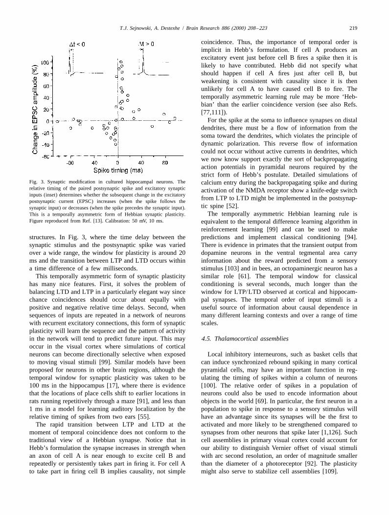
Fig. 3. Synaptic modification in cultured hippocampal neurons. The calcium entry during the backpropagating spike and duringrelative timing of the paired postsynaptic spike and excitatory synaptic activation of the NMDA receptor show a knife-edge switchinputs (inset) determines whether the subsequent change in the excitatory
from LTP to LTD might be implemented in the postsynap-postsynaptic current (EPSC) increases (when the spike follows thetic spine [52].synaptic input) or decreases (when the spike precedes the synaptic input).
The temporally asymmetric Hebbian learning rule isThis is a temporally asymmetric form of Hebbian synaptic plasticity.Figure reproduced from Ref. [13]. Calibration: 50 mV, 10 ms. equivalent to the temporal difference learning algorithm in
reinforcement learning [99] and can be used to makestructures. In Fig. 3, where the time delay between the predictions and implement classical conditioning [94].synaptic stimulus and the postsynaptic spike was varied There is evidence in primates that the transient output fromover a wide range, the window for plasticity is around 20 dopamine neurons in the ventral tegmental area carryms and the transition between LTP and LTD occurs within information about the reward predicted from a sensorya time difference of a few milliseconds. stimulus [103] and in bees, an octopaminergic neuron has a
This temporally asymmetric form of synaptic plasticity similar role [61]. The temporal window for classicalhas many nice features. First, it solves the problem of conditioning is several seconds, much longer than thebalancing LTD and LTP in a particularly elegant way since window for LTP/LTD observed at cortical and hippocam-chance coincidences should occur about equally with pal synapses. The temporal order of input stimuli is apositive and negative relative time delays. Second, when useful source of information about causal dependence insequences of inputs are repeated in a network of neurons many different learning contexts and over a range of timewith recurrent excitatory connections, this form of synaptic scales.plasticity will learn the sequence and the pattern of activityin the network will tend to predict future input. This may 4.5. Thalamocortical assembliesoccur in the visual cortex where simulations of corticalneurons can become directionally selective when exposed Local inhibitory interneurons, such as basket cells thatto moving visual stimuli [99]. Similar models have been can induce synchronized rebound spiking in many corticalproposed for neurons in other brain regions, although the pyramidal cells, may have an important function in reg-temporal window for synaptic plasticity was taken to be ulating the timing of spikes within a column of neurons100 ms in the hippocampus [17], where there is evidence [100]. The relative order of spikes in a population ofthat the locations of place cells shift to earlier locations in neurons could also be used to encode information aboutrats running repetitively through a maze [91], and less than objects in the world [69]. In particular, the first neuron in a1 ms in a model for learning auditory localization by the population to spike in response to a sensory stimulus willrelative timing of spikes from two ears [55]. have an advantage since its synapses will be the first to
The rapid transition between LTP and LTD at the activated and more likely to be strengthened compared tomoment of temporal coincidence does not conform to the synapses from other neurons that spike later [1,126]. Suchtraditional view of a Hebbian synapse. Notice that in cell assemblies in primary visual cortex could account forHebb’s formulation the synapse increases in strength when our ability to distinguish Vernier offset of visual stimulian axon of cell A is near enough to excite cell B and with arc second resolution, an order of magnitude smallerrepeatedly or persistently takes part in firing it. For cell A than the diameter of a photoreceptor [92]. The plasticityto take part in firing cell B implies causality, not simple might also serve to stabilize cell assemblies [109].
220 T.J. Sejnowski, A. Destexhe / Brain Research 886 (2000) 208 –223
We can now be more specific about what a cell cortical assemblies through feedback connections. Theassembly might be and how it might provide insight into synapses between these assemblies could then becomethe role of sleep oscillations in the consolidation of cortical primed through thalamocortical spindles [107]. The assem-memory traces. Consider first the recruitment of neurons in blies identified from both bottom up and the top downvisual cortex in response to a flashed visual stimulus in the sources can be further strengthened through the molecularawake state. Stimulus specificity of single neurons is mechanisms outlined earlier.already present in the first few spikes of a response [121].If the most selective neurons came to threshold first, theycould recruit neurons through local connectivity within a 5. Conclusionscolumn. Inhibitory neurons would also be recruited, whichwould limit the total size of the assembly that represents Spindle oscillations appear in the EEG during the earlythe stimulus in the column. At the same time, temporally stages of slow-wave sleep. We have shown that duringasymmetric Hebbian plasticity would strengthen the se- these oscillations, cortical pyramidal neurons are bom-quence of neurons responding to the stimulus. Noise in the barded by unusually powerful excitatory inputs in thesystem would produce a different order of firing each dendrites in parallel with strong inhibitory inputs aroundpresentation, which would result in reciprocal connection the soma [30]. We suggest here that this pattern of
21among the neurons most selective for the repeated excitation–inhibition provokes a massive Ca entry that21stimulus. This defines a cortical assembly by construction. specifically activates Ca -dependent molecular gates in
Sensory experience may not produce a sufficiently the spindling cells. Spindle oscillations could thereforestrong tetanization of cortical synapses to produce LTP, open the door to subsequent long-term changes in corticalparticularly in a primary sensory areas where the threshold networks.for plasticity is set high, but it may be sufficient to prime As sleep deepens, slow waves progressively dominatethe synapses in the assembly that are later consolidated the EEG. These slow-wave complexes alternate with briefduring slow-wave sleep. It would be dangerous to change episodes of fast oscillations having similar properties asthe strengths of synapses in a feedforward system during the sustained fast oscillations that occur during wakeful-the processing itself. It is safer to statistically sample the ness [42]. We propose that these brief periods represent ainputs over a long time interval and wait until the cortex is recall of information acquired previously during wakeful-in no longer processing sensory information before ir- ness, which are subsequently stored by highly synchron-reversible changes are made to the network. This may be ized events that appear as slow waves in the EEG. Slow-the reason why the relay of sensory information through wave sleep would thus begin by spindle oscillations thatthe thalamus is reduced during sleep. open molecular gates to plasticity, then proceed by itera-
LTP through temporally asymmetric Hebbian plasticity tively ‘recalling’ and ‘storing’ information primed independs on the relative timing of the presynaptic and neural assemblies.postsynaptic spikes, the frequency of pairing, and the total Although speculative, this scenario is consistent withnumber of paired spikes. Thalamocortical sleep oscillations what is currently known about the biophysical mechanismsprovide conditions that allow the relative phase of neurons of sleep oscillations (see details in Ref. [39]). It is alsoin an assembly to be consistently maintained over many consistent with the growing evidence that sleep serves topairing. This is a consequence of the spatio-temporal consolidate memories, as well as with models that requirecoherence observed during spindling and slow-wave sleep. a ‘sleep’ phase for the long-term learning of generativeThis mechanism could be used to strengthen connections representations [66]. The key insight is that slow-wavebetween neurons in an assembly if the neurons in an sleep is a specific state in which information is consoli-
21assembly could be identified. If too many neurons are dated by activating Ca -mediated intracellular cascades in21activated then assemblies that share neurons will interfere pyramidal neurons. Implementing such a massive Ca
with each other and lose their identity. entry and network reorganization must necessarily takeOne way to select a neural assembly in the cortex during time and be performed during a state in which normal
sleep is a bottom up approach that depends on identifying processing such as sensory processing — should not occur;assemblies of neurons interconnected by recently primed this may ultimately be the primary reason why we need tosynapses. This takes advantage of the hyperpolarized sleep.periods in slow wave complexes during which the fre-quency of minis can be used to selecting neurons that havethe highest density of synapses that were recently primed Acknowledgementsby short-term potentiation [123]. A second way to select aneural assembly, a top down approach, is appropriate for We are grateful to Yves Fregnac for helpful commentsthe highest levels of cortical representation that receive on this manuscript, as well as to Diego Contreras andinputs from the hippocampal formation. Here, inputs Mircea Steriade for extensive discussions. Research sup-arising from sharp waves in the hippocampus select ported by CNRS, NIH, MRC of Canada, and HHMI.
T.J. Sejnowski, A. Destexhe / Brain Research 886 (2000) 208 –223 221
F.H. Gage, Nucleus basalis and thalamic control of neocorticalReferencesactivity in the freely moving rat, J. Neurosci. 8 (1988) 4007–4026.
[26] D.W. Carr, R.E. Stofko-Hahn, I.D. Fraser, R.D. Cone, J.D. Scott,[1] L.F. Abbott, S. Song, Temporally asymmetric Hebbian learning, Localization of the cAMP-dependent protein kinase to the post-
spike timing and neuronal response variability, Adv. Neural Infor- synaptic densities by A-kinase anchoring proteins. Characterizationmat. Processing Syst. 11 (1999) 69–75. of AKAP 79, J. Biol. Chem. 267 (1992) 16816–16823.
[2] T. Abel, P.V. Nguyen, M. Barad, T.A.S. Deuel, E.R. Kandel, R. [27] J.J. Chrobak, G. Buzsaki, Selective activation of deep layer (V–VI)Bourtchouladze, Genetic demonstration of a role for PKA in the late retrohippocampal cortical neurons during hippocampal sharp wavesphase of LTP and in hippocampus-based long-term memory, Cell 88 in the behaving rat, J. Neurosci. 14 (1994) 6160–6170.(1997) 615–626.
[28] D. Contreras, M. Steriade, Cellular bases of EEG slow rhythms: a[3] P. Achermann, A. Borbely, Low-frequency (,1 Hz) oscillations in study of dynamic corticothalamic relationships, J. Neurosci. 15
the human sleep EEG, Neuroscience 81 (1997) 213–222. (1995) 604–622.[4] D.H. Ackley, G.E. Hinton, T.J. Sejnowski, A learning algorithm for [29] D. Contreras, M. Steriade, Synchronization of low-frequency
Boltzmann Machines, Cognit. Sci. 9 (1985) 147–169. rhythms in corticothalamic networks, Neuroscience 76 (1997) 11–[5] P. Alvarez, L.R. Squire, Memory consolidation and the medial 24.
temporal lobe: a simple network model, Proc. Natl. Acad. Sci. USA [30] D. Contreras, A. Destexhe, M. Steriade, Intracellular and computa-91 (1994) 7041–7045. tional characterization of the intracortical inhibitory control of
[6] P. Alvarez, S. Zola-Morgan, L.R. Squire, Damage limited to the synchronized thalamic inputs in vivo, J. Neuro-physiol. 78 (1997)hippocampal region produces long-lasting memory impairment in 335–350.monkeys, J. Neurosci. 15 (1995) 3796–3807. [31] D. Contreras, A. Destexhe, T.J. Sejnowski, M. Steriade, Control of
[7] C.J. Ball, P. Gloor, N. Schaul, The cortical electromicrophysiology spatiotemporal coherence of a thalamic oscillation by cor-of pathological delta waves in the electroencephalogram of cats, ticothalamic feedback, Science 274 (1996) 771–774.Electroencephalogr. Clin. Neurophysiol. 43 (1977) 346–361. [32] D. Contreras, A. Destexhe, T.J. Sejnowski, M. Steriade, Spatiotem-
[8] M. Bazhenov, I. Timofeev, M. Steriade, T.J. Sejnowski, Computa- poral patterns of spindle oscillations in cortex and thalamus, J.tional models of thalamocortical augmenting responses, J. Neurosci. Neurosci. 17 (1997) 1179–1196.18 (1998) 6444–6465. [33] F. Crick, G. Mitchison, The function of dream sleep, Nature 304
[9] M.F. Bear, W.C. Abraham, Long-term depression in hippocampus, (1983) 111–114.Annu. Rev. Neurosci. 19 (1996) 437–462. [34] S.J. Cruikshank, N.M. Weinberger, Receptive-field plasticity in the
[10] C.C. Bell, V.Z. Han, Y. Sugawara, K. Grant, Synaptic plasticity in a adult auditory cortex induced by Hebbian covariance, J. Neurosci.cerebellum-like structure depends on temporal order, Nature 387 16 (1996) 861–875.(1997) 278–281. [35] A.R. Damasio, D. Tranel, Nouns and verbs are retrieved with
[11] M.J. Berridge, Neuronal calcium signaling, Neuron 21 (1998) 13– differently distributed neural systems, Proc. Natl. Acad. Sci. USA 9026. (1993) 4957–4960.
[12] U.S. Bhalla, R. Iyengar, Emergent properties of networks of [36] D. Debanne, B.H. Gahwiler, S.M. Thompson, Long-term synapticbiological signaling pathways, Science 283 (1999) 381–387. plasticity between pairs of individual CA3 pyramidal cells in rat
[13] G. Bi, M. Poo, Activity-induced synaptic modifications in hip- hippocampal slice cultures, J. Physiol. 507 (1998) 237–247.pocampal culture: dependence on spike timing, synaptic strength ˜[37] J. DeFelipe, I. Farinas, The pyramidal neuron of the cerebral cortex:and cell type, J. Neurosci. 18 (1998) 10464–10472. morphological and chemical characteristics of the synaptic inputs,
[14] T.V. Bliss, G.L. Collingridge, A synaptic model of memory: long- Prog. Neurobiol. 39 (1992) 563–607.term potentiation in the hippocampus, Nature 361 (1993) 31–39. [38] P. De Koninck, H. Schulman, Sensitivity of CaM kinase II to the
21[15] R.D. Blitzer, T. Wong, R. Nouranifar, R. Iyengar, E.M. Landau, frequency of Ca oscillations, Science 279 (1998) 227–230.Postsynaptic cAMP pathway gates early LTP in hippocampal CA1 [39] A. Destexhe, T.J. Sejnowski, The Thalamocortical Assembly,region, Neuron 15 (1995) 1403–1414. Oxford University Press, Oxford, UK, 2001, in press.
[16] R.D. Blitzer, J.H. Connor, G.P. Brown, T. Wong, S. Shenolikar, R. [40] A. Destexhe, A. Babloyantz, T.J. Sejnowski, Ionic mechanisms forIyengar, E.M. Landau, Gating of CaMKII by cAMP-regulated intrinsic slow oscillations in thalamic relay neurons, Biophys. J. 65protein phosphatase activity during LTP, Science 280 (1998) 1940– (1993) 1538–1552.1942. [41] A. Destexhe, D. Contreras, M. Steriade, Mechanisms underlying the
[17] K.I. Blum, L.F. Abbott, A model of spatial map formation in the synchronizing action of corticothalamic feedback through inhibitionhippocampus of the rat, Neural Comput. 8 (1996) 85–93. of thalamic relay cells, J. Neurophysiol. 79 (1998) 999–1016.
[18] B. Bontempi, C. Laurent-Demir, C. Destrade, R. Jaffard, Time- [42] A. Destexhe, D. Contreras, M. Steriade, Spatiotemporal analysis ofdependent reorganization of brain circuitry underlying long-term local field potentials and unit discharges in cat cerebral cortexmemory storage, Nature 400 (1999) 671–675. during natural wake and sleep states, J. Neurosci. 19 (1999) 4595–
[19] L.J. Borg-Graham, C. Monier, Y. Fregnac, Visual input evokes 4608.transient and strong shunting inhibition in visual cortical neurons, [43] A. Destexhe, D.A. McCormick, T.J. Sejnowski, A model for 8–10Nature 393 (1998) 369–373. Hz spindling in interconnected thalamic relay and reticularis neu-
[20] P. Bush, T.J. Sejnowski, Inhibition synchronizes sparsely connected rons, Biophys. J. 65 (1993) 2474–2478.cortical neurons within and between columns in realistic network [44] A. Destexhe, T. Bal, D.A. McCormick, T.J. Sejnowski, Ionicmodels, J. Comput. Neurosci. 3 (1996) 91–110. mechanisms underlying synchronized oscillations and propagating
[21] G. Buzsaki, Hippocampal sharp waves: their origin and significance, waves in a model of ferret thalamic slices, J. Neurophysiol. 76Brain Res. 398 (1986) 242–252. (1996) 2049–2070.
[22] G. Buzsaki, Two-stage model of memory trace formation, a role for [45] A. Destexhe, D. Contreras, T.J. Sejnowski, M. Steriade, A model of‘noisy’ brain states, Neuroscience 31 (1989) 551–570. spindle rhythmicity in the isolated thalamic reticular nucleus, J.
[23] G. Buzsaki, The hippocampo–neocortical dialogue, Cerebral Cortex Neurophysiol. 72 (1994) 803–818.6 (1996) 81–92. [46] A. Destexhe, D. Contreras, T.J. Sejnowski, M. Steriade, Modeling
[24] G. Buzsaki, Memory consolidation during sleep: a neurophysiologi- the control of reticular thalamic oscillations by neuromodulators,cal perspective, J. Sleep Res. 7 (Suppl. 1) (1998) 17–23. NeuroReport 5 (1994) 2217–2220.
[25] G. Buzsaki, R.G. Bickford, G. Ponomareff, L.J. Thal, R. Mandel, [47] A. Destexhe, D. Contreras, M. Steriade, T.J. Sejnowski, J.R.

222 T.J. Sejnowski, A. Destexhe / Brain Research 886 (2000) 208 –223
Huguenard, In vivo, in vitro and computational analysis of dendritic [72] E.G. Jones, The Thalamus, Plenum Press, New York, 1985.calcium currents in thalamic reticular neurons, J. Neurosci. 16 [73] N. Kapur, D.J. Brooks, Temporally-specific retrograde amnesia in(1996) 169–185. two cases of discrete bilateral hippocampal pathology, Hippocampus
[48] A. Dosemeci, T.S. Reese, Inhibition of endogenous phosphatase in a 9 (1999) 247–254.postsynaptic density fraction allows extensive phosphorylation of [74] S.R. Kelso, A.H. Ganong, T.H. Brown, Hebbian synapses inthe major postsynaptic density protein, J. Neurochem. 61 (1993) hippocampus, Proc. Natl. Acad. Sci. USA 83 (1986) 5326–5330.550–555. [75] M.B. Kennedy, M.K. Bennett, N.E. Erondu, Biochemical and
[49] R. Eckhorn, R. Bauer, W. Jordan, M. Brosch, W. Kruse, M. Munk, immunochemical evidence that the ‘major postsynaptic densityH.J. Reitboek, Coherent oscillations: a mechanism of feature linking protein’ is a subunit of a calmodulin-dependent protein kinase, Proc.in the visual cortex? Multiple electrode and correlation analyses in Natl. Acad. Sci. USA 80 (1983) 7357–7361.the cat, Biol. Cybernetics 60 (1988) 121–130. [76] U. Kim, T. Bal, D.A. McCormick, Spindle waves are propagating
[50] J.M. Edeline, Y. Manunta, E. Hennevin, Auditory thalamus neurons synchronized oscillations in the ferret LGNd in vitro, J. Neuro-during sleep: changes in frequency selectivity, threshold, and physiol. 74 (1995) 1301–1323.receptive field size, J. Neurophysiol. 84 (2000) 934–952.
[77] W.B. Levy, O. Stewart, Temporal contiguity requirements for long-[51] E.V. Evarts, Temporal patterns of discharge of pyramidal tract
term associative potentiation /depression in the hippocampus,neurons during sleep and waking in the monkey, J. Neurophysiol. 27
Neuroscience 8 (1983) 791–797.(1964) 152–171.
[78] M.S. Lewicki, T.J. Sejnowski, Bayesian unsupervised learning of[52] K.M. Franks, T.M. Bartol, M. Poo, T.J. Sejnowski, High spatial andhigher order structure, Adv. Neural Informat. Processing Syst. 9temporal resolution estimates of calcium dynamics in dendritic(1997) 529–535.spines using MCELL simulations, Soc. Neurosci. Abstracts 26
[79] W. Li, J. Llopis, M. Whitney, G. Zlokarnik, R.Y. Tsien, Cell-(2000) 1122.21permanent caged InsP3 ester shows that Ca spike frequency can[53] U. Frey, Y.Y. Huang, E.R. Kandel, Effects of cAMP simulate a late
optimize gene expression, Nature 392 (1998) 936–941.phase of LTP in hippocampal CA1 neurons, Science 260 (1993)[80] R.R. Llinas, D. Pare, Of dreaming and wakefulness, Neuroscience1661–1664.
44 (1991) 521–535.[54] J.D. Frost, P.R. Kellaway, A. Gol, Single-unit discharges in isolated[81] A. Luthi, D.A. McCormick, Periodicity of thalamic synchronizedcerebral cortex, Exp. Neurol. 14 (1966) 305–316.
21oscillations: the role of Ca -mediated upregulation of Ih, Neuron[55] W. Gerstner, R. Kempter, J.L. van Hemmen, H. Wagner, A neural20 (1998) 553–563.learning rule for sub-millisecond temporal coding, Nature 383
[82] W.W. Lytton, T.J. Sejnowski, Simulations of cortical pyramidal(1996) 76–78.neurons synchronized by inhibitory interneurons, J. Neurophysiol.[56] A. Ghosh, M.E. Greenberg, Calcium signaling in neurons: molecular66 (1991) 1059–1079.mechanisms and cellular consequences, Science 268 (1995) 239–
[83] W.W. Lytton, T.J. Sejnowski, Computer model of ethosuximide’s247.[57] D. Golomb, X.J. Wang, J. Rinzel, Propagation of spindle waves in a effect on a thalamic cell, Ann. Neurol. 32 (1992) 131–139.
thalamic slice model, J. Neurophysiol. 75 (1996) 750–769. [84] W.W. Lytton, A. Destexhe, T.J. Sejnowski, Control of slow oscilla-[58] C. Gray, Synchronous oscillations in neuronal systems: Mechanisms tions in the thalamo-cortical neuron: a computer model, Neuro-
and functions, J. Comput. Neurosci. 1 (1994) 11–38. science 70 (1996) 673–684.[59] C.M. Gray, W. Singer, Stimulus-specific neuronal oscillations in [85] H. Markram, J. Lubke, M. Frotscher, B. Sakmann, Regulation of
orientation columns of cat visual cortex, Proc. Natl. Acad. Sci. USA synaptic efficacy by coincidence of postsynaptic APs and EPSPs,86 (1989) 1698–1702. Science 275 (1997) 213–215.
[60] X. Gu, N.C. Spitzer, Distinct aspects of neuronal differentiation [86] D. Marr, Simple memory: A theory for the archicortex, Philos.21encoded by frequency of spontaneous Ca transients, Nature 375 Trans. R. Soc. (Lond.) 262 (1971) 23–81.
(1995) 784–787. [87] J.L. McClelland, B.L. McNaughton, R.C. O’Reilly, Why there are[61] M. Hammer, R. Menzel, Learning and memory in the honeybee, J. complementary learning systems in the hippocampus and neocortex:
Neurosci. 15 (1995) 1617–1630. insights from the successes and failures of connectionist models of[62] D.O. Hebb, Organization of Behavior: A Neuropsychological learning and memory, Psychol. Rev. 102 (1995) 419–457.
Theory, Wiley, New York, 1949. [88] D.A. McCormick, Neurotransmitter actions in the thalamus and[63] S. Herculano-Houzel, M.H. Munk, S. Neuenschwander, W. Singer, cerebral cortex and their role in neuromodulation of thalamocortical
Precisely synchronized oscillatory firing patterns require elec- activity, Prog. Neurobiol. 39 (1992) 337–388.troencephalographic activation, J. Neurosci. 19 (1999) 3992–4010. [89] D.A. McCormick, J.R. Huguenard, A model of the electrophysio-
[64] M. Herkenham, Laminar organization of thalamic projections to the logical properties of thalamocortical relay neurons, J. Neurophysiol.rat neocortex, Science 207 (1980) 532–535. 68 (1992) 1384–1400.
[65] G.E. Hinton, Z. Ghahramani, Generative models for discovering [90] M. McKeown, C. Humphries, P. Acherman, A. Borbely, T.J.sparse distributed representations, Philos. Trans. R. Soc. Lond. Ser. Sejnowski, Low frequency interactions in the human sleep EEG,B 352 (1997) 1177–1190. Sleep Res. 7 (Suppl. 1) (1998) 48–56.
[66] G.E. Hinton, P. Dayan, B.J. Frey, R.M. Neal, The ‘wake–sleep’ [91] M.R. Mehta, C.A. Barnes, B.L. McNaughton, Experience-depen-algorithm for unsupervised neural networks, Science 268 (1995) dent, asymmetric expansion of hippocampal place fields, Proc. Natl.1158–1161. Acad. Sci. USA 94 (1997) 8918–8921.
ˆ[67] N. Ho, A. Destexhe, Synaptic background activity enhances the [92] D.A. Miller, S.W. Zucker, Computing with self-excitatory cliques: Aresponsiveness of neocortical pyramidal neurons, J. Neurophysiol. model and an application to hyperacuity-scale computation in visual84 (2000) 1488–1496. cortex, Neural Comput. 11 (1999) 21–66.
[68] J.A. Hobson, The Dreaming Brain, Basic Books, New York, 1988. [93] N. Mons, A. Harry, P. Dubourg, R.T. Premont, R. Iyengar, D.M.[69] J.J. Hopfield, Pattern recognition computation using action potential Cooper, Immunohisto-chemical localization of adenylyl cyclase in
timing for stimulus representation, Nature 376 (1995) 33–36. rat brain indicates a highly selective concentration at synapses, Proc.[70] J.J. Hopfield, D.I. Feinstein, R.G. Palmer, ‘Unlearning’ has a Natl. Acad. Sci. USA 92 (1995) 8473–8477.
stabilizing effect in collective memories, Nature 304 (1983) 158– [94] P.R. Montague, T.J. Sejnowski, The predictive brain: temporal159. coincidence and temporal order in synaptic learning mechanisms,
[71] P.T. Huerta, J.E. Lisman, Synaptic plasticity during the cholinergic Learning Memory 1 (1994) 1–33.theta-frequency oscillation in vitro, Hippocampus 6 (1996) 58–61. [95] P.R. Montague, P. Dayan, T.J. Sejnowski, A framework for
T.J. Sejnowski, A. Destexhe / Brain Research 886 (2000) 208 –223 223
mesencephalic dopamine systems based on predictive Hebbian oscillations in the sleeping and aroused brain, Science 262 (1993)learning, J. Neurosci. 16 (1996) 1936–1947. 679–685.
ˆ[96] J. O’Keefe, L. Nadel, The Hippocampus as a Cognitive Map, [118] M. Steriade, L. Domich, G. Oakson, M. Deschenes, The deaf-Clarendon Press, Oxford, UK, 1978. ferented reticular thalamic nucleus generates spindle rhythmicity, J.
[97] D. Pare, J. Dong, H. Gaudreau, Amygdalo-entorhinal relations and Neurophysiol. 57 (1987) 260–273.their reflection in the hippocampal formation: generation of sharp [119] R. Stickgold, D. Whidbee, B. Schirmer, V. Patel, J.A. Hobson,sleep potentials, J. Neurosci. 15 (1995) 2482–2503. Visual discrimination task improvement: A multi-step process
[98] Y.L. Qin, B.L. McNaughton, W.E. Skaggs, C.A. Barnes, Memory occurring during sleep, J. Cognit. Neurosci. 12 (2000) 246–254.reprocessing in cortico–cortical and hippocampocortical neuronal [120] G.R. Sutherland, B. McNaughton, Memory trace reactivation inensembles, Philos. Trans. R. Soc. Lond. Ser. B 352 (1997) 1525– hippocampal and neo-cortical neuronal ensembles, Curr. Opin.1533. Neurobiol. 10 (2000) 180–186.
[99] R.P.N. Rao, T.J. Sejnowski, Predictive sequence learning in re- [121] S.J. Thorpe, J. Gautrais, Rapid visual processing using spikecurrent neocortical circuits, Adv. Neural Informat. Processing Syst. asynchrony, Adv. Neural Informat. Processing Syst. 9 (1997)12 (2000) 164–170. 901–907.
[100] R. Ritz, T.J. Sejnowski, Synchronous oscillatory activity in sensory [122] P.H.E. Tiesinga, J.-M. Fellous, J.G. Jose, T.J. Sejnowski, Opticalsystems: New vistas on mechanisms, Curr. Opin. Neurobiol. 7 information transfer in synchronized neocortical neurons, Neuro-(1997) 536–546. computing, in press.
[101] E. Salinas, T.J. Sejnowski, Impact of correlated synaptic input on [123] I. Timofeev, F. Grenier, M. Bazhenov, T.J. Sejnowski, M. Steriade,output firing rate and variability in simple neuronal models, J. Origin of slow cortical oscillations in deafferented cortical slabs.Neurosci. 20 (2000) 6193–6209. Cerebral Cortex 10 (2000) in press.
[102] M.V. Sanchez Vives, D.A. McCormick, Cellular and network [124] R.D. Traub, M.A. Whittington, I.M. Stanford, J.G. Jefferys, Amechanisms of rhythmic recurrent activity in neocortex, Nat. mechanism for generation of long-range synchronous fast oscilla-Neurosci. 3 (2000) 1027–1034. tions in the cortex, Nature 383 (1996) 621–624.
[103] W. Schultz, P. Dayan, P.R. Montague, A neural substrate of [125] M.V. Tsodyks, W.E. Skaggs, T.J. Sejnowski, B.L. McNaughton,prediction and reward, Science 275 (1997) 1593–1599. Paradoxical effects of external modulation of inhibitory inter-
[104] T.J. Sejnowski, Storing covariance with nonlinearly interacting neurons, J. Neurosci. 17 (1997) 4382–4388.neurons, J. Math. Biol. 4 (1977) 203–211. [126] R. Van Rullen, J. Gautrais, A. Delorme, S. Thorpe, Face processing
[105] T.J. Sejnowski, Sleep and memory, Curr. Biol. 5 (1995) 832–834. using one spike per neurone, Biosystems 48 (1998) 229–239.[106] T.J. Sejnowski, The computational neuroethology of sleep, in: N. [127] M. von Krosigk, T. Bal, D.A. McCormick, Cellular mechanisms of
Elsner, R. Wehner (Eds.), New Neuroethology on the Move, Vol. 1, a synchronized oscillation in the thalamus, Science 261 (1993)Georg Theime Verlag, Stuttgart, Germany, 1998, pp. 127–144. 361–364.
[107] A.G. Siapas, M.A. Wilson, Coordinated interactions between [128] X.J. Wang, J. Rinzel, Spindle rhythmicity in the reticularis thalamihippocampal ripples and cortical spindles during slow-wave sleep, nucleus — synchronization among inhibitory neurons, Neuro-Neuron 21 (1998) 1123–1128. science 53 (1993) 899–904.
[108] T.R. Soderling, Calcium/calmodulin-dependent protein kinase II: [129] G.Y. Wu, H.T. Cline, Stabilization of dendritic arbor structure inrole in learning and memory, Mol. Cell. Biochem. 127–128 (1993) vivo by CaMKII, Science 279 (1998) 222–226.93–101. [130] H. Wigstrom, B. Gustafsson, Y.Y. Huang, W.C. Abraham, Hip-
[109] S. Song, K.D. Miller, L.F. Abbott, Competitive Hebbian learning pocampal long-term potentiation is induced by pairing singlethrough spike-timing-dependent synaptic plasticity, Nat. Neurosci. afferent volleys with intracellularly injected depolarizing current3 (2000) 919–926. pulses, Acta Physiol. Scand. 126 (1986) 317–319.
[110] L.R. Squire, S. Zola-Morgan, The medial temporal lobe memory [131] M.A. Wilson, B.L. McNaughton, Reactivation of hippocampalsystem, Science 253 (1991) 1380–1386. ensemble memories during sleep, Science 265 (1994) 676–679.
[111] G. Stent, A physiological mechanism for Hebb’s postulate of [132] F. Worgotter, K. Suder, Y. Zhao, N. Kerscher, U.T. Eysel, K.learning, Proc. Natl. Acad. Sci. USA 70 (1973) 997–1001. Funke, State-dependent receptive-field restructuring in the visual
[112] M. Steriade, Cortical long-axoned cells and putative interneurons cortex, Nature 396 (1998) 165–168.during the sleep–waking cycle, Behav. Brain Sci. 3 (1978) 465– [133] A. Ylinen, A. Bragin, Z. Nadasdy, G. Jando, I. Szabo, A. Sik, G.514. Buzsaki, Sharp wave-associated high-frequency oscillation (200
[113] M. Steriade, F. Amzica, Intracortical and corticothalamic Hz) in the intact hippocampus: network and intracellular mecha-coherency of fast spontaneous oscillations, Proc. Natl. Acad. Sci. nisms, J. Neurosci. 15 (1995) 30–46.USA 93 (1996) 2533–2538. [134] R. Yuste, D.W. Tank, Dendritic integration in mammalian neurons,
[114] M. Steriade, R.W. McCarley, Brainstem Control of Wakefulness a century after Cajal, Neuron 16 (1996) 701–716.and Sleep, Plenum Press, New York, 1990. [135] L.I. Zhang, H.W. Tao, C.E. Holt, W.A. Harris, M. Poo, A critical
[115] M. Steriade, F. Amzica, D. Contreras, Synchronization of fast window for cooperation and competition among developing re-(30–40 Hz) spontaneous cortical rhythms during brain arousal, J. tinotectal synapses, Nature 395 (1998) 37–44.Neurosci. 16 (1996) 392–417. [136] S.M. Zola-Morgan, L.R. Squire, The primate hippocampal forma-
[116] M. Steriade, E.G. Jones, R.R. Llinas, Thalamic Oscillations and tion: evidence for a time-limited role in memory storage, ScienceSignalling, Wiley, New York, 1990. 250 (1990) 288–290.
[117] M. Steriade, D.A. McCormick, T.J. Sejnowski, Thalamocortical