interactive report parvalbumin-positive projection …songbird/pubs/pdfs/wild et al 2001.pdf ·...
TRANSCRIPT
Brain Research 917 (2001) 235–252www.bres-interactive.com
Interactive report
Parvalbumin-positive projection neurons characterise the vocalqpremotor pathway in male, but not female, zebra finches
a , a b*J. Martin Wild , Matthew N. Williams, , Roderick A. SuthersaDivision of Anatomy, School of Medical and Health Sciences, University of Auckland, Auckland 92019, New Zealand
bSection of Physiology, Medical Sciences, Indiana University, Bloomington, IN 47405, USA
Accepted 11 June 2001
Abstract
Parvalbumin (PV) and calbindin (CB) immunoreactivities were assessed in nucleus robustus archistriatalis (RA) of male and femalezebra finches, together with retrograde labelling of RA neurons. The results of double and triple labelling experiments suggested that, inmales, moderately and faintly PV-positive neurons were projection neurons, but that all intensely PV-positive cells were not. The latter,which are presumably interneurons, were also intensely CB-positive, and may correspond to the GABAergic inhibitory interneuronsidentified by others. In addition, the complete RA pathway and its terminal fields in the respiratory–vocal nuclei of the brainstem werestrongly PV-positive. In female zebra finches, which do not sing, no evidence was found that PV-positive RA cells were projectionneurons, yet the pattern of projections of RA neurons, as determined by anterograde transport of biotinylated dextran amine, was verysimilar to that of RA in males. Moreover, in females, RA neurons retrogradely labelled from injections of cholera toxin B-chain into thetracheosyringeal nucleus (XIIts) were abundant and included, in the lateral part of the nucleus, a population of cells that were as large asthose in the male RA. Parvalbumin immunoreactivity was also present in RA and its projections in males of several other songbird species(northern cardinal, brown headed cowbird, canary) and in the female cardinal, which sings to some extent, but the labelling was not asintense as that in male zebra finches. 2001 Elsevier Science B.V. All rights reserved.
Theme: Neural basis of behavior
Topic: Neuroethology
Keywords: Calcium binding protein; Vocalization; Bird
1. Introduction [20,23,31,32,36]. RA is thus intimately concerned with thecontrol of vocal production via its influence on syringeal
In oscine songbirds, such as zebra finches and canaries, and respiratory muscles. The coordination of the activity ofthere is a set of interconnected nuclei in the forebrain that these functionally related but disparate muscle groupsis dedicated to the learning and production of song during singing may be effected via an inhibitory RA[2,20,21]. The output from this circuit is a spherical interneuron that has extensive dendritic aborizations withinnucleus in the caudal telencephalon called robustus archis- the nucleus [27].triatalis (RA). This nucleus is remarkable for a variety of Another remarkable feature of RA in zebra finches andreasons, not least of which is the fact that it has long canaries is that it is sexually differentiated, being muchdescending projections that terminate directly on vocal larger in the adult male, who sings, than in the adultmotor neurons (tracheosyringeal nucleus or XIIts) and on female, who sings much less or not at all [12,15,16,19].premotor respiratory nuclei in the medulla The diameter of RA somata in the adult male zebra finch
can be more than twice that of female RA somata [9], andmale RA neurons also have larger dendritic fields [8]; yetqPublished on the World Wide Web on 25 September 2001.RA neurons in the female are known to project upon XIIts,*Corresponding author.as they do in the male, albeit providing a smaller terminalE-mail addresses: [email protected] (J. Martin Wild),
[email protected] (J. Martin Wild). field [9]. In this last study, however, retrograde confirma-
0006-8993/01/$ – see front matter 2001 Elsevier Science B.V. All rights reserved.PI I : S0006-8993( 01 )02938-9
236 J. Martin Wild et al. / Brain Research 917 (2001) 235 –252
tion of the origin of the projection to XIIts in the female 2. Materials and methodswas based on a single case, which, until recently [12], wasunique in the literature on this subject. Other projections of 2.1. SubjectsRA in females, such as those to the intercollicular region,have been described as somewhat different from those of Thirty-eight songbirds were used in this study: sixteenRA in males [9], and no information is available on female male and sixteen female zebra finches (Taeniopygia gut-RA projections to other targets identified in males, such as tata), one male canary (Serinus canaria), two female andthe respiratory premotor nuclei, which are presumably one male northern cardinals (Cardinalis cardinalis), oneinvolved in calling, as well as in singing [9,32]. female and one male brown-headed cowbird (Melothrus
A third feature of RA, again in the male, is that many of ater). The exact ages of the birds were unknown, but allits neurons are positive for the calcium binding protein were adult, species-specific song having been recorded orparvalbumin [3], an immunoreactivity that has a develop- monitored in the male zebra finches, male and femalemental pattern reflecting that of the nucleus as a whole [4]. northern cardinals, male canary and male cowbird. TheThe presence of these PV-positive neurons, and of fewer cowbirds and cardinals were wild caught, the other speciescalbindin-D -positive neurons (CB-positive neurons were obtained from commercial suppliers.28K
[4]), in RA is curious, however, because it is not knownwhether they are projection neurons or have axons that 2.2. Anaesthesiaremain within the confines of the nucleus, i.e. interneurons.Inhibitory interneurons in RA of adult male zebra finches For all surgical procedures, the animals were anaesthet-have been suggested by immunostaining for GABA ised either by intramuscular injections of a mixture of[10,24] and have been demonstrated by a combination of ketamine (50 mg/kg) and xylazine (20 mg/kg) or byintracellular electrophysiology and staining. Spiro et al. inhaled isofluorane gas carried in oxygen. The procedures[27] showed that neurons with fast action potentials and were carried out in accordance with the guidelines stipu-steep current–frequency relationships had small somata lated by the University of Auckland Animal Ethics Com-
2(|136 mm ) with thin aspiny processes that extended mittee, and every effort was made to minimise the numbersthroughout large parts of the nucleus. These neurons were of experimental animals used.found to be distinctly similar in morphology to RAneurons stained for glutamic acid decarboxylase (GAD), 2.3. Injection proceduressupporting the idea that they were inhibitory interneurons[27]. But since the PV-positive neurons thus far demon- All injections in either RA or XIIts were made using astrated in RA have been reported to have somewhat larger combination of stereotaxis [28] and electrophysiologicalsomata (12–17 mm diameter in adult male zebra finch [3]), recording. The head skin was reflected from the midline, ait is not clear whether they are likely to include the burr hole made in the skull either over the caudal telen-inhibitory interneurons identified by Spiro et al. [27] and cephalon or over the cerebellum, and the dura incised. ASakaguchi et al. [24]. On the other hand, if the PV-positive tungsten microelectrode (Frederick Haer; 3–5 MV) wasneurons originate long descending projections, then that used to record multiunit activity (an indifferent electrodewould be unusual, since parvalbumin is not generally was inserted in a neck muscle) as it was passed eitherassociated with such neurons (e.g. [11]). through the archistriatum and into RA, or through the
In the present study, we sought to determine whether cerebellum and into XIIts. The dorsal border of RA wasPV-positive and CB-positive neurons are present in RA of identified by a sudden increase in multiunit activityfemale as well as male songbirds, and whether or not these followed by a characteristic ‘bursty’ pattern of dischargeneurons are projection neurons. This involved labelling RA through the depth of the nucleus [32]. RA in females wasneurons by retrograde transport from XIIts and double or more difficult to locate because of its small size. XIIts wastriple labelling for parvalbumin and calbindin. We also identified by its characteristic respiratory rhythm in phaseassessed the presence of parvalbumin immunoreactivity with expiration [33].within the whole vocal premotor pathway, and compared Following identification of the nuclei, a glass mi-this with the RA pathway defined by anterograde tracing cropipette loaded with one of the tracers replaced themethods, in both males and females. The main focus of the recording electrode and injections were made either ion-study was on adult zebra finches, in which sexual differen- tophoretically (2–4 mA positive current, 7 s on, 7 s off fortiation of brain and vocal behavior is pronounced, but a a total time of 10–20 min) or using air pressure via asmall number of birds of other songbird species (cowbird, picospritzer (General Valve, Fairfield, NJ, USA).canary, northern cardinal) were included to assess the The tracers used were: biotinylated dextran aminegenerality of the parvalbumin immunoreactivity of the RA (BDA), dextran tetramethylrhodamine (Fluoro-ruby) andpathway and to extend the findings to species in which the dextran fluorescein (Fluoro-emerald), (each 10 K M ,w
female sings, albeit less than the male. lysine fixable; Molecular Probes), all 10% solutions in
J. Martin Wild et al. / Brain Research 917 (2001) 235 –252 237
phosphate buffered saline (PBS); or Cholera toxin B-chain horse serum (or rabbit serum for sections stained only for(CTB, List Biological Laboratories), 1% in PBS. CTB). All other incubations were for 1–2 h duration.
Unilateral injections of BDA were made in RA of five Between incubations, all sections were thoroughly washedfemale and two male zebra finches, one female northern in PBS. For all immunochemical procedures outlinedcardinal, and one male canary. Injections of one of the below, the antibodies and ancillary agents used wereother tracers were made in XIIts of five female and nine carefully chosen and checked for cross reactivity betweenmale zebra finches. The XIIts injections were either antibodies and/or epitopes.unilateral or bilateral. If bilateral, different tracers wereused to deposit different tracers on each side, e.g. CTB on 2.6.1. BDA reactivityone side and Fluoro-ruby on the other. Sections were incubated with streptavidin peroxidase
medium (1:1000, Molecular Probes), followed by incuba-2.4. Lesions tions in a 3,39-diaminobenzidine (DAB) medium (0.025%
DAB with 0.005% H O and 0.002% CoCl in PBS). This2 2 2Unilateral electrolytic lesions were made in RA in two resulted in a black opaque reaction product.
male zebra finches and one male canary. Stainless steel 00insect pins, insulated except at their tips, were used to first
2.6.2. CTB immunoreactivitylocate RA by recording multiunit activity, and then to
Sections were incubated overnight in a goat anti-CTBproduce a lesion by passing 0.25 mA anodal current for 30
medium (1:30 000, List Biological Laboratories), then withs. In one of the zebra finches and in the canary, BDA was
a biotinylated rabbit anti-goat antibody (1:200, Sigma).injected into RA on the opposite side of the brain. This
This was followed by incubations with streptavidin per-was done to ensure that the unilateral lesioning did not in
oxidase and DAB (as above). In some cases, no CoCl was2some way interfere with the projections of the contralateraladded to the DAB medium, which produced a brown
RA and its associated PV-immunoreactivity.reaction product.
2.5. Perfusion and sectioning2.6.3. Parvalbumin immunoreactivity
Sections were incubated overnight in a monoclonal anti-All birds were allowed to survive from 2 to 7 daysparvalbumin antibody medium (1:10 000 for opaque DABdepending on the tracer injected. Generally, in the after-staining, or 1:1000 for fluorescence microscopy. Thisnoon, they were deeply anaesthetised with an overdose ofantibody was raised against carp parvalbumin: Sigma, orketamine and xylazine and perfused through the heart withSWANT 235, Basel, Switzerland), followed by asaline, followed by fixative (4% paraformaldehyde in 0.1biotinylated horse anti-mouse antibody (1:200, Vector);M sodium phosphate buffer, pH 7.4). The brains werethen by incubations in either streptavidin peroxidaseremoved from the skull and postfixed in the same fixativefollowed by DAB (as described above), or a variety offor 5–15 h. After cryoprotection in 30% sucrose in PBSfluorescent streptavidin complexes (1:200, i.e. conjugatedfor 15–48 h, the brains were sectioned at 35 mm in eitherwith either fluorescein, rhodamine, or Alexa 448, 546 orthe coronal or sagittal planes on a freezing microtome. The568 depending on whether green or red fluorescence wassections were collected serially in four series, one or morerequired; all Molecular Probes products). These fluorescentof which from each case was processed to reveal one orlabels were used primarily in Multiple labelling protocols.more of the tracers and, sometimes also parvalbumin
and/or calbindin immunoreactivity. For some controlstudies, sections through RA were cut at 10 mm and 2.6.4. Calbindin immunoreactivitycollected in two serially adjacent series. One series was Two different anti-calbindin antibodies were used (bothprocessed for parvalbumin (Section 2.6.3) and the other for raised against chicken calbindin): For staining sectionscalbindin immunoreactivity (Section 2.6.4). with opaque DAB reaction product, some sections were
incubated with a monoclonal antibody (1:10 000, Sigma),2.6. Processing and immunochemistry then with a biotinylated horse anti-mouse antibody, fol-
lowed by streptavidin peroxidase and DAB (as above forTo improve penetrability of the immunochemical and parvalbumin). Other sections destined for multiple label-
histochemical agents, and to block non-specific peroxidase ling fluorescence microscopy were incubated with anreactivity, most sections were pretreated with 50% aqueous antibody raised in rabbit (1:500, Emson, Cambridge, UK)methanol containing 1% H O for 10–20 min. All im- [14,25]), then with a biotinylated donkey anti-rabbit2 2
munochemical reactions were carried out on free-floating (1:200, Amersham) antibody, and the immunoreactivitysections at room temperature in PBS containing 0.4% was finally revealed using the variety of the streptavidinTriton X-100. All primary antibody incubations were complexes described above, as well as streptavidin Alexaovernight in duration, and all media contained normal 350 (Molecular Probes).
238 J. Martin Wild et al. / Brain Research 917 (2001) 235 –252
2.7. Multiple labelling protocols was not the case. However, in order to ensure that cross-reactivity was not a potential problem in the interpretation
For the assessment of the presence of retrogradely of the double labelling of cells with the mouse anti-labelled neurons and parvalbumin and/or calbindin im- parvalbumin and rabbit anti-calbindin antibodies, adjacentmunoreactivity within RA, some of the procedures de- 10-mm frozen sections through the RA of a male zebrascribed above were combined using either black-brown finch were incubated either with the anti-parvalbumin oropaque staining techniques, or by double or triple-coloured the anti-calbindin antibody, at the same dilutions as givenfluorescence techniques. above for 35-mm thick sections. Parvalbumin was visual-
CTB (black)-parvalbumin (brown) opaque staining was ised (green) indirectly by using the appropriate biotinylatedgenerally achieved by incubation of sections in a cocktail secondary antibody and streptavidin fluorescein, whereasof the goat anti-CTB (1:30 000) and the mouse anti- calbindin was visualised (red) using the direct secondaryparvalbumin (1:10 000) primary antibodies (as above). The goat anti rabbit Alexa 546 antibody.CTB was stained black using the biotinylated rabbit anti- These double and triple labelling procedures enabled thegoat second antibody, streptavidin peroxidase and the DAB distributions and possible co-localisation of parvalbuminCoCl (black) procedure. Parvalbumin immunoreactivity and calbindin and retrogradely labelled neurones in RA to2
was revealed and rendered brown using the biotinylated be assessed with both bright field and fluorescence micro-horse anti-mouse second antibody, streptavidin peroxidase scopy.DAB (brown) procedure (as above). Sections to be examined for transported BDA were
CTB (red)-Parvalbumin (green) fluorescent staining was mounted on lightly subbed slides, air dried, dehydrated ingenerally achieved by incubating sections in a cocktail of alcohol, cleared in xylenes, and coverslipped with DePeX.the anti-CTB (1:30 000) and anti-parvalbumin (1:1000) One series was left uncounterstained, and one was counter-antibodies (described above). CTB was then stained red stained with cresyl violet. These sections were viewed inwith a direct secondary, donkey anti-goat Alexa 546 bright field and the projections were drawn using aantibody (1:200, Molecular Probes). Parvalbumin immuno- macroprojector and camera lucida. Sections to be ex-reactivity was next stained green, indirectly by incubations amined and photographed in the fluorescence microscopewith the biotinylated horse anti-mouse second antibody were mounted on slides and coverslipped either with(1:200), followed by streptavidin fluorescein (1:200). Citifluor (Agar Aids) or Prolong (Molecular Probes) an-
Rhodamine dextran (red) and parvalbumin (green) tifade agents. In some cases, the coverslips were sub-fluorescent staining was achieved by incubating sections sequently removed and the section counterstained withthat contained retrogradely labelled red fluorescent RA cresyl violet.neurons with the mouse anti-parvalbumin primary antibody Colour 35-mm photographic records were made of the(1:1000); then by incubations with the biotinylated horse same field of some of the multiply fluorescent sections,anti-mouse secondary antibody (1:200), and finally strep- and of adjacent 10-mm sections in the cross-reactivitytavidin fluorescein (1:200). control case, in order to map the distributions of singly,
Rhodamine dextran (red), parvalbumin (green) and doubly or triply labelled cells. Comparisons of the dis-calbindin (blue) fluorescent staining was achieved by tributions of the different labels was achieved by scanningincubating sections that contained retrogradely labelled red sets of three 35-mm slides (of red, green and bluefluorescent RA neurones in a cocktail of the mouse anti- fluorescence) of the same field, or of two 35-mm slides (ofparvalbumin (1:1000) and rabbit anti-calbindin (1:500) red and green fluorescence) of adjacent sections in theantibodies. The parvalbumin was stained green using a cross-reactivity control case, into PhotoShop (Adobe) withdirect secondary goat anti-mouse Alexa 488 antibody a Microtek ArtixScan 4000t slide scanner. These images(1:200, Molecular Probes). Calbindin was subsequently were then carefully superimposed using various landmarksstained for by incubating sections with a biotinylated (e.g. blood vessels, see Fig. 4G–J), and combined to showdonkey anti-rabbit secondary antibody (1:200), followed two of the colours in the same image. Single and combinedby incubation with streptavidin Alexa 350 (1:100 Molecu- images were adjusted for evenness of illumination, bright-lar Probes). ness, contrast and colour balance. Cell sizes were mea-
Negative controls involved the omission of one or more sured using a 403 objective and drawing tube.of the primary antibodies, which always resulted in theabsence of labelling. Internal control for multiple labellingof parvalbumin, calbindin and retrogradely labelled neuro- 3. Resultsnes was provided by the presence, in the same section, ofdifferent telencephalic and brainstem neurons that were 3.1. RA in the zebra finchsingly fluorescent, i.e. that showed only red, green or bluefluorescence. If cross reactivity between the various pro- In Nissl-stained sagittal sections, the adult female RAcedures had been a problem, most cells would be expected forms an elipsoid or ovoid nucleus within the caudalto be fluorescent for more than one of the colours, which archistriatum (Fig. 1A; see also [16]). In coronal sections it

J. Martin Wild et al. / Brain Research 917 (2001) 235 –252 239
Fig. 1. (A) Photomicrograph of Nissl stained parasagittal section through RA of female zebra finch; rostral to the left. (B) Photomicrograph of Nisslstained frontal section through RA of female zebra finch; medial to the left. (C) Photomicrograph of Nissl stained frontal section through RA of male zebrafinch (outlined with arrow heads); medial to the left. (D) Neurons in RA of a female zebra finch retrogradely labelled with CTB from an injection centeredon XIIts, with spread to the medial part of nucleus retroambigualis; medial to the left (compare D with B). (E) Higher power view of the boxed area in themedial part of RA shown in (D). Note the cytoplasmic concentration of the CTB. (F) Higher power view of the boxed area in the lateral part of RA shownin (D). Compare the sizes of neurons in (E) and (F) and note the difference in packing density of labelled neurons. (G) Nissl counterstained version of (E).Note the many small, densely packed, basophilic cells between the retrogradely labelled cells (compare E and G). (H) Nissl counterstained version of (F).Note the smaller number and more dispersed nature of the small basophilic cells than in (G). LAD, lamina archstriatalis dorsalis; calibration bars5100 mm;that shown in (H) applies also to (E, F and G).
240 J. Martin Wild et al. / Brain Research 917 (2001) 235 –252
J. Martin Wild et al. / Brain Research 917 (2001) 235 –252 241
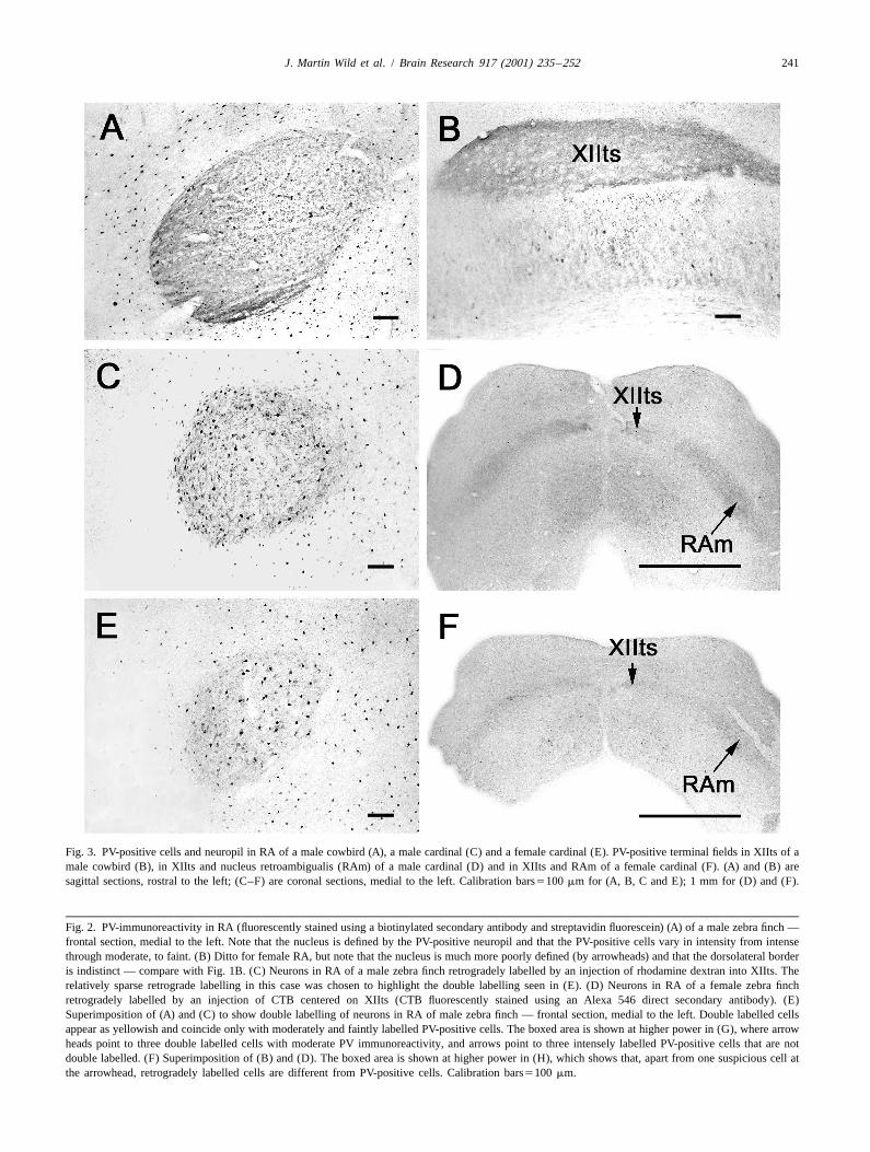
Fig. 3. PV-positive cells and neuropil in RA of a male cowbird (A), a male cardinal (C) and a female cardinal (E). PV-positive terminal fields in XIIts of amale cowbird (B), in XIIts and nucleus retroambigualis (RAm) of a male cardinal (D) and in XIIts and RAm of a female cardinal (F). (A) and (B) aresagittal sections, rostral to the left; (C–F) are coronal sections, medial to the left. Calibration bars5100 mm for (A, B, C and E); 1 mm for (D) and (F).
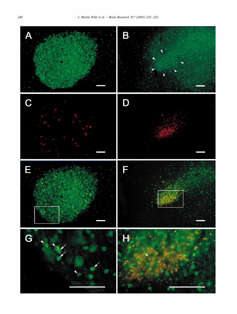
Fig. 2. PV-immunoreactivity in RA (fluorescently stained using a biotinylated secondary antibody and streptavidin fluorescein) (A) of a male zebra finch —frontal section, medial to the left. Note that the nucleus is defined by the PV-positive neuropil and that the PV-positive cells vary in intensity from intensethrough moderate, to faint. (B) Ditto for female RA, but note that the nucleus is much more poorly defined (by arrowheads) and that the dorsolateral borderis indistinct — compare with Fig. 1B. (C) Neurons in RA of a male zebra finch retrogradely labelled by an injection of rhodamine dextran into XIIts. Therelatively sparse retrograde labelling in this case was chosen to highlight the double labelling seen in (E). (D) Neurons in RA of a female zebra finchretrogradely labelled by an injection of CTB centered on XIIts (CTB fluorescently stained using an Alexa 546 direct secondary antibody). (E)Superimposition of (A) and (C) to show double labelling of neurons in RA of male zebra finch — frontal section, medial to the left. Double labelled cellsappear as yellowish and coincide only with moderately and faintly labelled PV-positive cells. The boxed area is shown at higher power in (G), where arrowheads point to three double labelled cells with moderate PV immunoreactivity, and arrows point to three intensely labelled PV-positive cells that are notdouble labelled. (F) Superimposition of (B) and (D). The boxed area is shown at higher power in (H), which shows that, apart from one suspicious cell atthe arrowhead, retrogradely labelled cells are different from PV-positive cells. Calibration bars5100 mm.
242 J. Martin Wild et al. / Brain Research 917 (2001) 235 –252
forms more of a slender arc, convex medially, often with a the clear border of this neuropil coincides with the borderpoorly defined dorsolateral border (Fig. 1B; see also of the nucleus as defined in Nissl stained material (Fig.[12,19]). The nucleus is typically half the width of the 2A). In the female zebra finch, the border of the PV-adult male RA (see Fig. 1C) and the individual cells are positive neuropil is much less clear (Fig. 2B), corre-more tightly packed than in the male, especially in medial sponding with the less well defined border of RA asparts of the nucleus, where there is a preponderance of defined in Nissl stained material (compare Fig. 1B). RA issmall cells (|5–10 mm mean somal diameter [16]). The also PV-positive in male brown-headed cowbirds and malesmallest of these (|5 mm) tend to be basophilic (Fig. 1G), northern cardinals (Fig. 3A and C). It is also relativelyare never retrogradely labelled from XIIts injections, and well defined by PV-positive neuropil in the female northernare probably glia ([9] and see below). Interspersed between cardinal (Fig. 3E), which sings to some extent.the glia, and often approximated by them, are small In both males and females, a substantial contribution toneurons (|8–10 mm) that have a pale-staining cytoplasm. the PV-positive neuropil in RA is made by the processes ofAs one moves more laterally through the female RA, PV-positive cell bodies, which vary both in intensity and inhowever, the cell size tends to become more variable, and size (Fig. 2A,B,G and H). Although their intensity variesthere is an increasing number of larger cells that are as continuously from very weak to very strong, the PV-large as those in the male RA (12–20 mm, Fig. 1D and F). positive cells may be conveniently categorized for the
RA may also be defined on the basis of retrograde purposes of description as intensely labelled, moderatelylabelling from target nuclei (Figs. 1D, 2C and D, and 4A labelled and faintly labelled. These categories, however, doand B). However, in the female, if different populations of not also sort for cell size, for there was a range of sizes inRA neurons have different projection targets, as appears to each category. The intensely labelled cells measured in thebe the case in the male [30], and has also been suggested order of 10–13 mm (smallest somal diameter), comparedin the female [12], then a total definition of RA based on with the slightly larger, moderately labelled cells (12–15retrograde labelling can only be achieved if all target mm). Weakly labelled PV-positive cells could not benuclei are injected. In the present study, only XIIts was a measured with certainty due to the poor definition of theirtarget nucleus for injection, although some of the larger boundaries. PV-positive cell bodies of various intensitiesinjections also included parts of the adjacent nucleus were also present in RA of cowbirds and northern cardi-retroambigualis (RAm), due to spread of tracer from the nals (Fig. 3).injection center. Retrograde labelling in RA of one of these In the archistriatum outside RA in zebra finches, thecases (female zebra finch) is shown in Fig. 1D. In Nissl PV-positive cell bodies also varied in intensity and size,counterstained sections (Fig. 1G and H) it was clear that and were less concentrated than in RA (Fig. 2A and B). Inthe retrogradely labelled cells were different from and the male, the intensely labelled archistriatal cells weregenerally larger than the small, basophilic RA cells generally smaller than those in RA, whereas in the femaledescribed above, supporting the suggestion that these latter the reverse tended to be true. Thus, PV-positive cells in thesmall cells are glia [9]. The neurons retrogradely labelled female RA were generally smaller than those in the malewith CTB formed a mediolateral size and packing density RA (compare Fig. 2G and H).gradient, with the smaller ones (8–10 mm in diameter)medially and more densely packed, and the larger ones 3.2. Are the RA projection neurons also PV-positive?(12–20 mm) laterally and more dispersed (Fig. 1D–H).
RA can also be defined on the basis of its densely Double labelling for both retrogradely transported tracerlabelled PV-positive neuropil [3,4]. In the male zebra finch, and parvalbumin was carried out in a series of male and
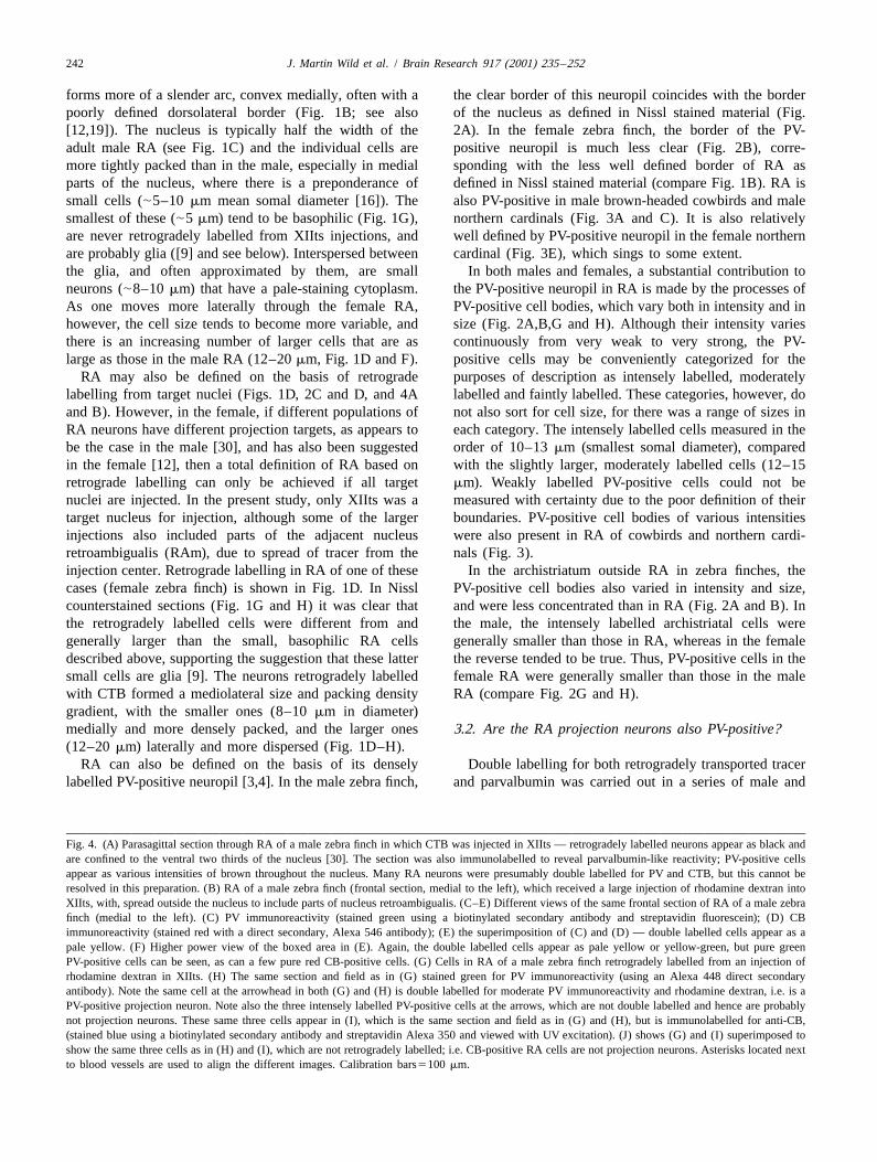
Fig. 4. (A) Parasagittal section through RA of a male zebra finch in which CTB was injected in XIIts — retrogradely labelled neurons appear as black andare confined to the ventral two thirds of the nucleus [30]. The section was also immunolabelled to reveal parvalbumin-like reactivity; PV-positive cellsappear as various intensities of brown throughout the nucleus. Many RA neurons were presumably double labelled for PV and CTB, but this cannot beresolved in this preparation. (B) RA of a male zebra finch (frontal section, medial to the left), which received a large injection of rhodamine dextran intoXIIts, with, spread outside the nucleus to include parts of nucleus retroambigualis. (C–E) Different views of the same frontal section of RA of a male zebrafinch (medial to the left). (C) PV immunoreactivity (stained green using a biotinylated secondary antibody and streptavidin fluorescein); (D) CBimmunoreactivity (stained red with a direct secondary, Alexa 546 antibody); (E) the superimposition of (C) and (D) — double labelled cells appear as apale yellow. (F) Higher power view of the boxed area in (E). Again, the double labelled cells appear as pale yellow or yellow-green, but pure greenPV-positive cells can be seen, as can a few pure red CB-positive cells. (G) Cells in RA of a male zebra finch retrogradely labelled from an injection ofrhodamine dextran in XIIts. (H) The same section and field as in (G) stained green for PV immunoreactivity (using an Alexa 448 direct secondaryantibody). Note the same cell at the arrowhead in both (G) and (H) is double labelled for moderate PV immunoreactivity and rhodamine dextran, i.e. is aPV-positive projection neuron. Note also the three intensely labelled PV-positive cells at the arrows, which are not double labelled and hence are probablynot projection neurons. These same three cells appear in (I), which is the same section and field as in (G) and (H), but is immunolabelled for anti-CB,(stained blue using a biotinylated secondary antibody and streptavidin Alexa 350 and viewed with UV excitation). (J) shows (G) and (I) superimposed toshow the same three cells as in (H) and (I), which are not retrogradely labelled; i.e. CB-positive RA cells are not projection neurons. Asterisks located nextto blood vessels are used to align the different images. Calibration bars5100 mm.

J. Martin Wild et al. / Brain Research 917 (2001) 235 –252 243
244 J. Martin Wild et al. / Brain Research 917 (2001) 235 –252
female zebra finches. Fig. 4A shows RA in a male zebra labelling of 10-mm sections through RA with either anti-finch that received an injection of CTB in XIIts; the PV or anti-CB alone. Fig. 5 shows clearly, that when twosection was subsequently stained for anti-PV. Retrogradely such adjacent sections are accurately superimposed, thoselabelled neurons appear black and are largely confined to RA cells that were cut in two or three during sectioning,the ventral two thirds of the nucleus, as expected on the and were intensely labelled for anti-PV, were also intenselybasis of previous studies of RA topography with respect to labelled for anti-CB. Fig. 5 also confirms that retrogradeits target nuclei [30]. The PV-positive cell bodies appear as labelling that resulted from a XIIts injection of rhodaminevarious shades of brown and are distributed throughout all dextran, was confined to RA cells that were moderately orparts of the nucleus. While there is a strong suggestion that faintly labelled for anti-PV. These cells were alwaysmany projection neurons are PV-positive in this material, calbindin negative.the data remain equivocal because of the inability to In the female zebra finch, double labelling for anti-PVresolve black and brown in the same cell. In fluorescent and anti-CB showed that some intensely labelled PV-material, double labelling of RA cells with retrogradely positive cells were also intensely positive for anti-CB (notlabelled tracer and anti-PV was clearly present in males, shown). However, there were many fewer of these doublybut only with respect to the moderately and some faintly labelled cells, owing to the relative sparseness of CB-PV-positive cell types (Fig. 2A,C,E and G). Intensely positive neurons in the female RA. Triple labelling was notlabelled PV-positive cell bodies were never retrogradely performed in the female.labelled (Figs. 2G and 4G and H), and no retrogradely Outside RA, in the neostriatum, there were also manylabelled cell body was ever intensely positive for parval- cells double labelled for anti-PV and anti-CB, but therebumin. In the female RA, retrogradely labelled RA cells were also many PV-positive cells that were not CB-posi-were numerous following XIIts injections of CTB (Figs. tive, providing additional internal controls for cross reac-1D, 2D) but none of them was definitely PV-positive (Fig. tivity in the same section that contained RA.2B,D,F and H).
3.4. RA pathway is PV-positive in male but not female3.3. Further identification of the intensely labelled PV- zebra finchespositive RA neurons
In males, the whole of the RA pathway, including theDouble labelling for parvalbumin and calbindin was tracts and terminal fields, were intensely PV-positive (Fig.
carried out both in sections from birds not receiving 6A,C,E–G). The medial part of the occipitomesencephalicinjections of retrograde tracer, and from birds that received tract (OM), which carries the RA axons out of theinjections of rhodamine dextran in XIIts. In the male zebra hemisphere, was positive (Fig. 7B), as was the rest of thefinch, every intensely labelled PV-positive cell in RA was tract as it courses through the diencephalon and brainstemalso intensely positive for anti-CB (Fig. 4C–F), and none (Fig. 6E). PV-positive terminal fields were present in allof such doubly labelled cells were retrogradely labelled the nuclei known to receive projections from RA in male(Fig. 4G–J). As in other cases described above, some zebra finches and canaries [32,36, Fig. 8, present study],moderately or faintly labelled PV-positive cells were also viz. the dorsomedial nucleus of the intercollicular complexretrogradely labelled (Fig. 4G and H), suggesting that (DM; Fig. 7C), the nucleus infra-olivaris superior (IOS),these were projection neurons. But we observed no RA the ventrolateral nucleus of the rostral medulla (RVL),cells that were doubly labelled for anti-CB and retrograde nucleus parambigualis (PAm), nucleus retroambigualistracer (Fig. 4 J), i.e. CB-positive RA cells do not appear to (RAm; Fig. 6C), and XIIts and SH (Figs. 6F and 7D). Thebe projection neurons. Since moderately or faintly labelled densest immunolabelling was in the suprahypoglossal areaPV-positive and CB-positive RA cells formed different (SH) where there were peri-somatic concentrations aroundpopulations, which could be seen in the same section that XIIts and RAm cell bodies (Fig. 6G). In contrast, inalso contained cells intensely labelled with both anti-PV females these tracts and terminal fields were largely absentand anti-CB (Fig. 4F), an internal control for cross (Fig. 6B and D). Occasionally, faint labelling of the OMreactivity was provided. In addition, cross reactivity as an tract was present in the lower medulla, but no terminalinterpretation of double labelling with anti-PV and anti-CB fields were present in either RAm or XIIts which occur atantibodies in the cocktail procedure (see Multiple labelling these levels (Fig. 6D). Note, parenthetically, the absoluteprotocols) was ruled out on the basis of the results of size difference between the male and female medullary
Fig. 5. (B) and (C) PV (stained green using a biotinylated secondary antibody and streptavidin fluorescein) and CB immunoreactivity (stained red using anAlexa 546 direct secondary antibody), respectively, in adjacent 10-mm thick frontal sections through the ventral region of RA. (A) Superimposition of (B)and (C) to show some cells that were stained with both anti-PV and anti-CB antibodies, separately applied to each section (e.g. up arrows). Arrowheadsdirected leftwards point to cells that were intensely positive only for PV; while arrowheads directed rightwards point to cells that were only CB positive.The boxed area in B shows one cell that was retrogradely labelled by an injection of rhodamine dextran into XIIts — note the particulate nature of thelabel, in contrast to the generally solid labelling of PV and CB positive cells. Calibration bar550 mm.
J. Martin Wild et al. / Brain Research 917 (2001) 235 –252 245
246 J. Martin Wild et al. / Brain Research 917 (2001) 235 –252
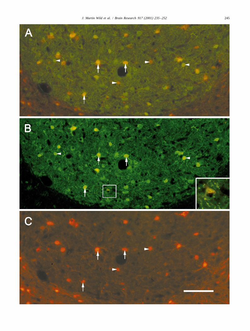
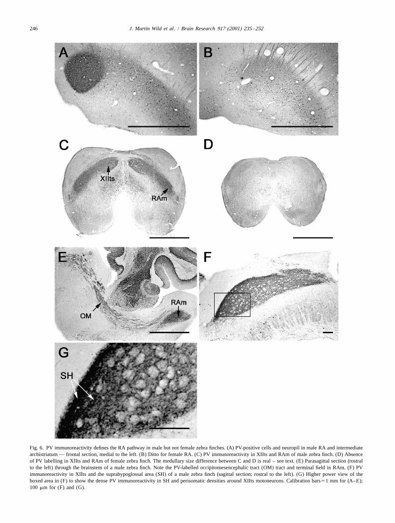
Fig. 6. PV immunoreactivity defines the RA pathway in male but not female zebra finches. (A) PV-positive cells and neuropil in male RA and intermediatearchistriatum — frontal section, medial to the left. (B) Ditto for female RA. (C) PV immunoreactivity in XIIts and RAm of male zebra finch. (D) Absenceof PV labelling in XIIts and RAm of female zebra finch. The medullary size difference between C and D is real – see text. (E) Parasagittal section (rostralto the left) through the brainstem of a male zebra finch. Note the PV-labelled occipitomesencephalic tract (OM) tract and terminal field in RAm. (F) PVimmunoreactivity in XIIts and the suprahypoglossal area (SH) of a male zebra finch (sagittal section; rostral to the left). (G) Higher power view of theboxed area in (F) to show the dense PV immunoreactivity in SH and perisomatic densities around XIIts motoneurons. Calibration bars51 mm for (A–E);100 mm for (F) and (G).
J. Martin Wild et al. / Brain Research 917 (2001) 235 –252 247
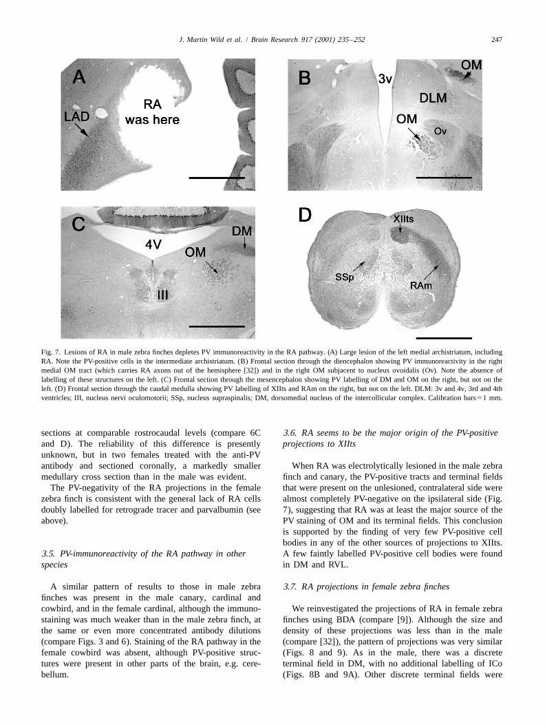
Fig. 7. Lesions of RA in male zebra finches depletes PV immunoreactivity in the RA pathway. (A) Large lesion of the left medial archistriatum, includingRA. Note the PV-positive cells in the intermediate archistriatum. (B) Frontal section through the diencephalon showing PV immunoreactivity in the rightmedial OM tract (which carries RA axons out of the hemisphere [32]) and in the right OM subjacent to nucleus ovoidalis (Ov). Note the absence oflabelling of these structures on the left. (C) Frontal section through the mesencephalon showing PV labelling of DM and OM on the right, but not on theleft. (D) Frontal section through the caudal medulla showing PV labelling of XIIts and RAm on the right, but not on the left. DLM: 3v and 4v, 3rd and 4thventricles; III, nucleus nervi oculomotorii; SSp, nucleus supraspinalis; DM, dorsomedial nucleus of the intercollicular complex. Calibration bars51 mm.
sections at comparable rostrocaudal levels (compare 6C 3.6. RA seems to be the major origin of the PV-positiveand D). The reliability of this difference is presently projections to XIItsunknown, but in two females treated with the anti-PVantibody and sectioned coronally, a markedly smaller When RA was electrolytically lesioned in the male zebramedullary cross section than in the male was evident. finch and canary, the PV-positive tracts and terminal fields
The PV-negativity of the RA projections in the female that were present on the unlesioned, contralateral side werezebra finch is consistent with the general lack of RA cells almost completely PV-negative on the ipsilateral side (Fig.doubly labelled for retrograde tracer and parvalbumin (see 7), suggesting that RA was at least the major source of theabove). PV staining of OM and its terminal fields. This conclusion
is supported by the finding of very few PV-positive cellbodies in any of the other sources of projections to XIIts.
3.5. PV-immunoreactivity of the RA pathway in other A few faintly labelled PV-positive cell bodies were foundspecies in DM and RVL.
A similar pattern of results to those in male zebra 3.7. RA projections in female zebra finchesfinches was present in the male canary, cardinal andcowbird, and in the female cardinal, although the immuno- We reinvestigated the projections of RA in female zebrastaining was much weaker than in the male zebra finch, at finches using BDA (compare [9]). Although the size andthe same or even more concentrated antibody dilutions density of these projections was less than in the male(compare Figs. 3 and 6). Staining of the RA pathway in the (compare [32]), the pattern of projections was very similarfemale cowbird was absent, although PV-positive struc- (Figs. 8 and 9). As in the male, there was a discretetures were present in other parts of the brain, e.g. cere- terminal field in DM, with no additional labelling of ICobellum. (Figs. 8B and 9A). Other discrete terminal fields were
248J.
Martin
Wild
etal.
/B
rainR
esearch917
(2001)235
–252
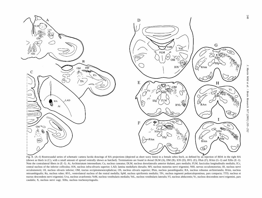
Fig. 8. (A–I) Rostrocaudal series of schematic camera lucida drawings of RA projections (depicted as short wavy lines) in a female zebra finch, as defined by an injection of BDA in the right RA(shown as black in (C), with a small amount of spread ventrally shown as hatched). Terminations are found in dorsal DLM (A), DM (B), IOS (D), RVL (E), PAm (F), RAm (G–I) and XIIts (E–I).Note the contralateral fibers in (E–I). Ai, Archistriatum intermedium; Cu, nucleus cuneatus; DLM, nucleus dorsolateralis anterior thalami, pars medialis; FLM, fasciculus longitudinalis medialis; ICc,central nucleus of the inferior colliculus; IOS, nucleus infra-olivaris superior; LAD, lamina medullaris dorsalis; MV, nucleus motorius nervi trigemini; NIII, nervus occulomotorius; III, nucleus nerviocculomotorii; OI, nucleus olivaris inferior; OM, tractus occipitomesencephalicus; OS, nucleus olivaris superior; PAm, nucleus parambigualis; RA, nucleus robustus archistriatalis; RAm, nucleusretroambigualis; Ru, nucleus ruber; RVL, ventrolateral nucleus of the rostral medulla; SpM, nucleus spiriformis medialis; TPc, nucleus tegmenti pedunculopontinus, pars compacta; TTD, nucleus ettractus descendens nervi trigemini; Uva, nucleus uvaeformis; VeM, nucleus vestibularis medialis; VeL, nucleus vestibularis lateralis; VI, nucleus abducentis; Vc, nucleus descendens nervi trigemini, parscaudalis; X, nucleus nervi vagi; XIIts, nucleus tracheosyringealis.
J. Martin Wild et al. / Brain Research 917 (2001) 235 –252 249
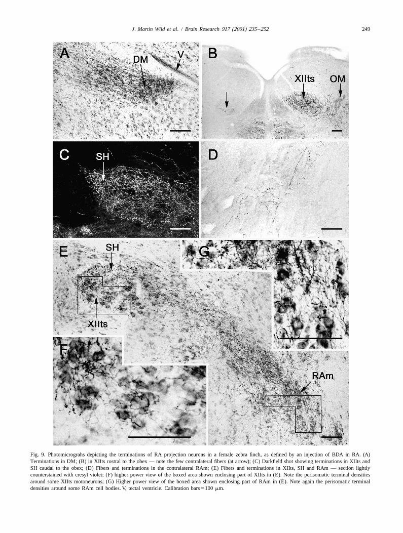
Fig. 9. Photomicrograhs depicting the terminations of RA projection neurons in a female zebra finch, as defined by an injection of BDA in RA. (A)Terminations in DM; (B) in XIIts rostral to the obex — note the few contralateral fibers (at arrow); (C) Darkfield shot showing terminations in XIIts andSH caudal to the obex; (D) Fibers and terminations in the contralateral RAm; (E) Fibers and terminations in XIIts, SH and RAm — section lightlycounterstained with cresyl violet; (F) higher power view of the boxed area shown enclosing part of XIIts in (E). Note the perisomatic terminal densitiesaround some XIIts motoneurons; (G) Higher power view of the boxed area shown enclosing part of RAm in (E). Note again the perisomatic terminaldensities around some RAm cell bodies. V, tectal ventricle. Calibration bars5100 mm.
250 J. Martin Wild et al. / Brain Research 917 (2001) 235 –252
present in IOS, RVL, PAm, RAm, XIIts and the sup- forebrain circuit involving the nucleus 1MAN [29], andrahypoglossal area (SH), where the terminations were there was specific labelling of DM in the midbrain. Thisdensest (Fig. 9C). Moreover, the terminations in XIIts and latter is new evidence, since Gurney [9] described diffusethe respiratory premotor nuclei (e.g. RAm) included many projections that encompassed both DM and the surround-that appeared to be axosomatic (Fig. 9F and G). There ing intercollicular nucleus (ICo) in the female. This resultwere also a few terminations in the dorsolateral part of was probably due to spread of his tritiated amino acidsDLM, as in the male (Fig. 8A; see also [32]). Curiously, in outside RA to archistriatal regions that are now known, inthe best case, there were more contralateral projections in the male, to project to regions of ICo that surround DMthe medulla than in any male thus far observed (Fig. 9B [17,35]. In the rest of the brainstem there were substantialand D; (compare [36]). A very similar pattern of ipsilateral projections to IOS, to RVL and to both PAm and RAm, asRA projections was present in the female cardinal, but the well as to XIIts and the suprahypoglossal area (SH). As inRA injection in this single case was only partially on the male, the great majority of these projections weretarget, thereby revealing only a portion of the descending ipsilateral, and there was strong evidence suggesting thattract. the terminations on XIIts and RAm cells were frequently
axosomatic. There was also good evidence for sparsecontralateral labelling of XIIts and RAm, even more so
4. Discussion than in males (Fig. 8D; (compare [36]).The question is, what function do these RA projections
In songbird species in which the female sings little or perform in the non-singing female zebra finch? As Simp-not at all, sexual differentiation of the brain during early son and Vicario [26] asked with respect to the female HVCdevelopment results in a female RA that is much reduced and RA, are the projections simply a remnant of ain size compared with that in the male [1,9,12,15,16]. developmental program that is only fully expressed inHowever, in the female zebra finch the number of neurons males, or do they have some other, as yet unexplainedreported to be present in the RA of adult birds is raison d’etre? Perhaps they are involved in calling, whichapparently quite variable, as witnessed by the figures of presumably requires the activation of abdominal expiratory6950 (range 6000–7600) given by Gurney [9], 2900 (range muscles, at the least, and, if the call is acoustically2400–3700) given by Konishi and Akutagawa [16], and modulated, then syringeal activation would be required in|8000 given by Johnson and Sellix [12]. In the respective addition. However, Simpson and Vicario [26] showed thatstudies, these figures compare with the proportionally less the ‘long’ or ‘distance’ call of the female zebra finch,variable counts of 16 400, 13 600 and |12 000 for num- which is characteristically longer than that of the long callbers of neurons in the adult male RA. In the present study, of the male [38,39], has a lower fundamental frequencythis variability in the numbers of neurons in the adult than that of the male and lacks certain learned featuresfemale RA was supported by the findings of apparently such as a fast frequency modulation, was largely unaffect-different overall sizes of RA in different birds, although ed either by cutting the tracheosyringeal nerves bilaterally,neuron density was not measured. Only about 22% of RA or by lesioning RA or HVc. Simpson and Vicario [26]neurons in the adult female zebra finch are projection concluded, therefore, that their ‘‘data suggest that theneurons having XIIts as their target, compared with more HVC–RA–nXIIts–syrinx pathway is essential for pro-than twice as many in the male [12]. duction of the learned vocalizations made by male zebra
The maximum somal diameter of neurons in the adult finches but is not needed for production of the unlearnedfemale RA has been reported to be about 8 mm [9,16], but female call’’ (p. 1555).a variation in cell density or size across the nucleus has not Whatever its function in the female, the RA pathway ispreviously been noted. In the present study, both Nissl also different from that of the male with respect to itsstaining and retrograde labelling of RA neurons showed almost total lack of parvalbumin immunoreactivity, whichthat cells in more lateral parts of the female nucleus, as is correlated with the lack of cell bodies in RA that wereseen in frontal sections, were less densely packed than in double labelled for anti-PV and retrograde label. Thus,more medial parts of the nucleus and generally approxi- while there are many cell bodies in the female RA thatmated the sizes of male RA neurons (12–20 mm). This stain positively for anti-PV, none of them was positivelyobservation appears to be supported by the findings of identified as doubly labelled by injections of retrogradeJohnson and Sellix [12; see their Fig. 5C], but its signifi- tracer in XIIts. In contrast, in the male RA, all retrogradelycance is unclear. labelled XIIts-projecting neurons were also stained faintly
Nevertheless, it is clear from the present results that in or moderately for anti-PV, but not all PV-positive cellthe adult female, RA has a substantial projection to XIIts bodies were also retrogradely labelled. There are twoand all the other respiratory–vocal nuclei that have been reasons relevant to the last part of this statement, oneidentified in the male [32]. From rostral to caudal, there technical and the other substantive. In the case depicted inwas sparse labelling of the dorsolateral part of DLM, Fig. 2, relatively few RA neurons were retrogradelywhich in the male is the origin of a feedback loop to a labelled from the XIIts injection, so the presence of many
J. Martin Wild et al. / Brain Research 917 (2001) 235 –252 251
PV-positive RA cell bodies that were not also retrogradely of parvalbumin and calbindin would be conducive to theirlabelled in the same sections is not surprising. However, in postulated role of mediating the tonic to phasic transitionsother cases such as those depicted in Figs. 4A and 4B, in firing patterns during singing, when HVc afferentwhere many more RA neurons were retrogradely labelled, activity is transformed into the phasic bursts that character-there were still many PV-positive cells that were not ise RA activity [27,37].double labelled. Some of these were faintly or moderately There are few instances in the CNS where both parval-labelled for PV, reflecting the subtotal nature of the bumin and calbindin co-localise in the same neurons,retrograde labelling, even in these cases; but others were notable exceptions being cerebellar Purkinje cells ofintensely labelled, and these intensely labelled PV-positive mammals and birds ([4, unpublished observations 7]),cells were never retrogradely labelled in any of the cases. which are also GABAergic, and rat dorsal root ganglia [6].Thus, in the male, but not in the female zebra finch, the In the present study in the adult male zebra finch, allXIIts projecting RA neurons are characterised by their intensely labelled PV-positive cells were doubly labelledfaint-to-moderate PV immunoreactivity, and this reactivity for anti-CB, whereas the moderately labelled PV-positivealso characterises the whole pathway and terminal fields. and CB-positive cells formed separate populations. In aHowever, such is the density of the terminations of RA developmental study of PV and CB immunoreactivity inaxons in RAm, XIIts and particularly SH, that the PV the zebra finch, Braun et al. [4] showed that PV- andimmunostaining in these structures appears intense, rather CB-positive cells in RA had different developmentalthan faint or moderate. The RA lesion data strongly histories and differed markedly in numbers, the CB-posi-suggest that the PV staining of these fields derives from tive cells being relatively sparse and generally surroundedRA and not from other sources of afferent nuclei. by CB-negative neuropil. We also found that CB-positive
The intensely labelled PV and CB cell bodies in the RA cells and neuropil to be much less dense than themale RA do not project to XIIts, and since they are not PV-positive RA cells and neuropil. However, the source ofconfined to a particular RA region, such as the dorsal ‘cap’ the dense PV-positive RA neuropil might not be totallythat projects to nuclei other than XIIts (e.g. DM and the intrinsic to the nucleus; some of it could result from therespiratory premotor nuclei [12,31,32,36], it seems likely projections of HVc and/or lMAN cells, some of which arethat they are not projection neurons at all. Their soma size also PV-positive [3,4, Wild, unpublished observations].is similar to that of the GABAergic RA neurons identified As mentioned in the Introduction, parvalbumin is notby others [10,24,27], which could suggest that the in- usually associated with long projection neurons, the vasttensely PV- and CB-positive RA cells also contain GABA. majority of PV-positive neurons in the CNS being inter-As suggested by Spiro et al. [24], such inhibitory inter- neurons [7]. However, Preuss and Kaas [22] identified aneurons would be strategically placed to link and coordi- population of cells in layer V of M1 (Betz cells) and partsnate the activity of otherwise unconnected groups of RA of somatosensory cortex in primates (Macaca and Galago).projection neurons that might innervate functionally differ- It is of related interest that RA is located in a part of theent motor (syringeal) and premotor (respiratory) neurons in avian archistriatum that has been likened, on the basis ofthe medulla. GABA-containing cells have also been iden- its long descending projections to a variety of brainstemtified in RA of the female zebra finch [24], and these, too, sensory and motor nuclei, to layers V–VI of somatosen-could be coincident with the intensely PV- and CB-positive sorimotor cortex of mammals ([20,40]; see alsocells identified in the female RA in the present study, but [32,34,35]). The presence of PV-positive neurons in RAwhether such cells also have an inhibitory function in the and surrounding intermediate archistriatum is thus con-non-singing female is not known. sistent with the findings of Preuss and Kaas [22] of
Given the postulated general role of calcium binding PV-positive neurons in layer V of somatic cortex inproteins in intracellular calcium buffering [7], the potential primates.co-localization of high levels of parvalbumin and calbindin The PV immunostaining of the RA pathway in the malein these GABAergic interneurons of the male RA would be canary, cardinal and cowbird was never as strong as it wasconsistent with their intrinsic physiology of fast action in the zebra finch, it was weakest in the female cardinal,potentials that fire at very high frequencies in response to and was absent in the female cowbird. The reasons for thedepolarizing currents, as in other systems [13,18,27]. More weaker staining in males of other species are not clear. Allspecifically, Caillard et al. [5] have suggested that in such were adult, established singers, and the perfusion andhigh frequency GABAergic interneurons (such as cerebel- immunohistochemical protocols were similar to those usedlar stellate cells synapsing on Purkinje cells), synaptic in the zebra finches; in fact, lower final dilutions oftransmission during bursts could be maintained by parval- antibody were also used to try and overcome this differ-bumin quickly reducing in the presynaptic terminal re- ence, but to no avail. However, the fact some staining wassidual calcium associated with the preceding action po- detectable in the female cardinal, which is known to singtential, and by preventing cumulative facilitation so that to some extent, and not in the female zebra finch orsynaptic strength is maintained at resting levels. In the cowbird, which do not sing, could suggest that parval-GABAergic interneurons of the songbird RA, high levels bumin immunoreactivity of RA projection neurons might
252 J. Martin Wild et al. / Brain Research 917 (2001) 235 –252
[17] C.V. Mello, G.E. Vates, S. Okuhata, F. Nottebohm, Descendingbe correlated with the ability to sing and not with gender.auditory pathways in the adult male zebra finch (TaeniopygiaBut this suggestion must remain speculative until furtherguttata), J. Comp. Neurol. 395 (1998) 137–160.
tests are carried out on a range of other species in which[18] N.P. Morris, S.J. Harris, Z. Henderson, Parvalbumin-immuno-
the female sings. reactive, fast-spiking neurons in the medial septum/diagonal bandcomplex of the rat: intracellular recordings in vitro, Neuroscience 92(1999) 589–600.
[19] F. Nottebohm, A.P. Arnold, Sexual dimorphism in vocal controlAcknowledgementsareas of the songbird brain, Science 194 (1976) 211–213.
[20] F. Nottebohm, T.M. Stokes, C.M. Leonard, Central control of songSupported by NIH Grant RO1 NS29467-07 in the canary, Serinus canaria, J. Comp. Neurol. 165 (1976) 457–
486.[21] F. Nottebohm, D.B. Kelley, J.A. Paton, Connections of vocal control
nuclei in the canary telencephalon, J. Comp. Neurol. 207 (1982)References344–357.
[22] T.M. Preuss, J.H. Kaas, Parvalbumin-like immunoreactivity of layer[1] S.W. Bottjer, E.A. Meisner, A.P. Arnold, Changes in neuronal
V pyramidal cells in the motor and somatosensory cortex of adultnumber, density and size account for increases in volume of song-
primates, Brain Res. 712 (1996) 353–357.control nuclei during song development in zebra finches, Neurosci.
[23] H. Reinke, J.M. Wild, Identification and connections of inspiratoryLett. 67 (1987) 263–268.
premotor neurons in songbirds and budgerigar, J. Comp. Neurol.[2] S.W. Bottjer, K.A. Halsema, S.A. Brown, E.A. Meisner, Axonal
391 (1998) 147–163.connections of a forebrain nucleus involved with vocal learning in
[24] H. Sakaguchi, Sex differences in the developmental changes ofzebra finches, J. Comp. Neurol. 279 (1989) 312–326.
GABAergic neurons in zebra finch song control nuclei, Exp. Brain[3] K. Braun, H. Scheich, M. Schachner, C.W. Heizman, Distribution of
Res. 108 (1996) 62–68.parvalbumin, cytochrome oxidase activity and 14C-2-deoxyglucose
[25] A. Seto-Oshima, P.C. Emson, E. Lawson, C.Q. Mountjoy, L.H.uptake in the brain of the zebra finch. I. Auditory and vocal motorCarasco, Loss of matrix calcium-binding protein-containing neuronssystems, Cell Tissue Res. 240 (1985) 101–115.in Huntington’s disease, Lancet 1 (1988) 1252–1255.[4] K. Braun, H. Scheich, C.W. Heizman, W. Hunziker, Parvalbumin and
[26] B. Simpson, D.S. Vicario, Brain pathways for learned and unlearnedcalbindin-D28K immunoereactivity as developmental markers ofvocalizations differ in zebra finches, J. Neurosci. 10 (1990) 1541–auditory and vocal motor nuclei of the zebra finch, Neuroscience 401556.(1991) 853–869.
[27] J.E. Spiro, M.B. Dalva, R. Mooney, Long-range inhibition within[5] O. Caillard, H. Moreno, B. Schwaller, I. Llano, M.R. Celio, A.the zebra finch song nucleus RA can coordinate the firing ofMarty, Role of the calcium-binding protein parvalbumin in short-multiple projection neurons, J. Neurophysiol. 81 (1999) 3007–3020.term synaptic plasticity, Proc. Natl. Acad. Sci. USA 97 (2000)
[28] T.M. Stokes, C.M. Leonard, F. Nottebohm, The telencephalon,13372–13377.diencephalon, and mesencephalon of the canary, Serinus canaria, in[6] P.A. Carr, T. Yamamoto, G. Karmy, K.G. Baimbridge, J.I. Nagy,stereotaxic coordinates, J. Comp. Neurol. 156 (1974) 337–374.Parvalbumin is highly colocalized with calbindin D28k and rarely
[29] G.E. Vates, D.S. Vicario, F. Nottebohm, Reafferent thalamo–corticalwith calcitonin gene-related peptide in dorsal root ganglia neuronsloops in the song system of oscine songbirds, J. Comp. Neurol. 380of rat, Brain Res. 497 (1989) 163–170.(1997) 275–290.[7] M.R. Celio, Calbindin D-28 k and parvalbumin in the rat nervous
[30] D.S. Vicario, Organization of the Zebra finch song control system:system, Neuroscience 35 (1990) 375–475.II. Functional organization of outputs from nucleus Robustus[8] T.J. DeVoogd, F. Nottebohm, Sex differences in dendritic morpholo-archistriatalis, J. Comp. Neurol. 309 (1991) 486–494.gy of a song control nucleus in the canary: a quantitative Golgi
[31] D.S. Vicario, A new brain stem pathway for vocal control in thestudy, J. Comp. Neurol. 196 (1981) 309–316.Zebra finch song system, NeuroReport 4 (1993) 983–986.[9] M. Gurney, Hormonal control of cell form and number in the zebra
[32] J.M. Wild, Descending projections of the songbird nucleus robustusfinch song system, J. Neurosci. 1 (1981) 658–673.archistriatalis, J. Comp. Neurol. 338 (1993) 225–241.[10] W. Grisham, A.P. Arnold, Distribution of GABA-like immuno-
[33] J.M. Wild, The avian nucleus retroambigualis: a nucleus forreactivity in the song system of the zebra finch, Brain Res. 651breathing, singing and calling, Brain Res. 606 (1993) 119–124.(1994) 115–122.
[34] J.M. Wild, S.M. Farabaugh, Organization of afferent and efferent[11] P.R. Hof, I.I. Glezer, F. Conde, R.A. Flagg, M.B. Rubin, E.A.projections of nucleus basalis prosencephali in a passerineNimchinsky, D.M. Vogt Weisenhorn, J. Chem. Neuroanat. 16 (1999)(Taeniopygia guttata), J. Comp. Neurol. 365 (1996) 306–328.77–116.
[35] J.M. Wild, M.N. Williams, Rostral wulst in passerine birds. I. Origin,[12] F. Johnson, M. Sellix, Reorganization of a telencephalic motorcourse and terminations of an avian pyramidal tract, J. Comp.region during sexual differentiation and vocal learning in zebraNeurol. 416 (2000) 429–450.finches, Dev. Brain Res. 121 (2000) 253–263.
[36] J.M. Wild, M.N. Williams, R.A. Suthers, Neural pathways for[13] Y. Kawaguchi, H. Katsumura, T. Kosaka, C.W. Heizmann, K. Hama,bilateral vocal control in songbirds, J. Comp. Neurol. 423 (2000)Fast spiking cells in rat hippocampus (CA region) contain the1
413–426.calcium-binding protein parvalbumin, Brain Res. 416 (1987) 369–[37] A.C. Yu, D. Margoliash, Temporal hierarchical control of singing in374.
birds, Science 273 (1996) 1871–1875.[14] H. Kiyama, A. Seto-Oshima, P.C. Emson, Calbindin D as a28K
[38] R. Zann, Structural variation in the zebra finch distance call, Z.marker for the degeneration of the striatonigral pathway in Hunting-Tierpsychol. 66 (1984) 328–345.toin’s disease. Brain Res. (1990) 209–214.
[39] R. Zann, Ontogeny of the zebra finch distance call: I. Effects of[15] M. Konishi, M. Gurney, Sexual differentiation of brain and be-cross-fostering to bengalese finches, Z. Tierpsychol. 68 (1985)havior. Trends in Neurosci. January (1982) 20–23.1–23.[16] M. Konishi, E. Akutagawa, Neuronal growth, atrophy and death in a
sexually dimorphic song nucleus in the zebra finch, Nature 315 [40] H. Zeier, H.J. Karten, The archistriatum of the pigeon: organization(1985) 145–147. of afferent and efferent connections, Brain Res. 31 (1971) 313–326.