interaction of cellular proteins with the 59 end of ...jvi.asm.org/content/74/18/8558.full.pdf ·...
TRANSCRIPT
JOURNAL OF VIROLOGY,0022-538X/00/$04.0010
Sept. 2000, p. 8558–8562 Vol. 74, No. 18
Copyright © 2000, American Society for Microbiology. All Rights Reserved.
Interaction of Cellular Proteins with the 59 End ofNorwalk Virus Genomic RNA
ANA LORENA GUTIERREZ-ESCOLANO,1* ZAMIRATH URIBE BRITO,1 ROSA M. DEL ANGEL,1 AND XI JIANG2
Departamento de Patologıa Experimental, Centro de Investigacion y de Estudios Avanzados del IPN, Mexico City,Mexico,1 and Center for Pediatric Research, Children’s Hospital for the King’s Daughters,
Eastern Virginia Medical School, Norfolk, Virginia2
Received 29 February 2000/Accepted 20 June 2000
The lack of a susceptible cell line and an animal model for Norwalk virus (NV) infection has prompted thedevelopment of alternative strategies to generate in vitro RNAs that approximate the authentic viral genome.This approach has allowed the study of viral RNA replication and gene expression. In this study, using mobilityshift and cross-linking assays, we detected several cellular proteins from HeLa and CaCo-2 cell extracts thatbind to, and form stable complexes with, the first 110 nucleotides of the 5* end of NV genomic RNA, a regionpreviously predicted to form a double stem-loop structure. These proteins had molecular weights similar tothose of the HeLa cellular proteins that bind to the internal ribosomal entry site of poliovirus RNA. HeLaproteins La, PCBP-2, and PTB, which are important for poliovirus translation, and hnRNP L, which is possiblyimplicated in hepatitis C virus translation, interact with NV RNA. These protein-RNA interactions are likelyto play a role in NV translation and/or replication.
Norwalk virus (NV) is the prototype strain of human calici-viruses and has been implicated in outbreaks of nonbacterialacute gastroenteritis in the U.S. and many other countries (2,15, 30). The virus is small (27 to 35 nm in diameter), round,nonenveloped, and with an amorphous surface structure (29,40). The virion contains a 7.7-kb single-stranded, positive RNAgenome; the RNA is polyadenylated and attaches with a VPgat its 59 end (6, 10). Genome sequence analysis has revealedthree open reading frames (ORFs). ORF 1 encodes a polypro-tein that is processed into nonstructural proteins required forvirus replication and has sequence homology to picornavirus2C helicase, 3C protease, and 3D RNA-dependent RNA poly-merase (12). ORF 2 encodes the viral capsid protein, and ORF3 encodes a small basic protein with an unknown function (27).NV also produces a 2.3-kb subgenomic RNA containing ORFs2 and 3, each of them having a strong AUG initiation codon,suggesting that they may be expressed independently (27).
The conserved sequence identified at the 59 end of thegenomic and subgenomic RNAs of NV suggests that it mightbe important for virus replication; 23 (88%) of the first 26nucleotides (nt) of the two RNAs are identical (20). Thiselement is also present in other caliciviruses, including therabbit hemorrhagic disease virus and the feline calicivirus (9,20, 34, 42). Sequence analysis of the NV RNA predicted adouble stem-loop structure at the 59 end of the genomic (nt 1to 110) and subgenomic (nt 5280 to 5356) RNAs (27). Asimilar double stem loop was also predicted upstream of ORF3 (nt 6848 to 6941). However, the role of these predictedstructures in viral RNA replication remains unknown.
Highly conserved secondary RNA structures are known tobe present at the 59 and 39 ends or in the internal regions of thegenomes of picornaviruses, hepatitis C virus, dengue virus,Japanese encephalitis virus, and simian hemorrhagic fever vi-rus (7, 8, 11, 13, 22, 24, 28, 34, 38, 43, 47). Studies of viral RNA
interaction with cellular proteins have identified several ele-ments in the viral RNAs that are important for viral replication(1, 3, 13). RNA-protein complexes are formed when authenticviral RNA or in vitro synthesized viral RNA transcripts areincubated with cell extracts. These complexes are involved inviral RNA replication and translation (1, 7, 8, 13, 17, 19, 22, 23,24, 25, 28, 33, 43, 47, 48).
The absence of a permissive cell line and a susceptible ani-mal model for NV infection has made it difficult to study thebiology of the virus. The successful cloning and sequencing ofthe NV genome and other human calicivirus genomes hasallowed much progress in our knowledge of gene coding strat-egies, genomic organization, viral RNA replication, and geneexpression (20, 26, 27, 34). However, in the case of NV little isknown about the mechanisms of viral replication. In this study,we performed binding experiments of in vitro synthesized NVRNA and HeLa and CaCo-2 cell extracts. Our results demon-strate that the 59 end of the NV genome contains elements thatbind specifically to different cellular proteins, some of whichinclude HeLa proteins, such as La, hnRNPL, PTB, andPCBP-2, that are known to be involved in the poliovirus inter-nal ribosomal entry site (IRES)-associated translation (3, 16,17, 19, 21, 28, 34, 35, 36) and hepatitis C virus translation (18, 23).
MATERIALS AND METHODS
Cells. HeLa cells were grown in Dulbecco’s minimal essential medium sup-plemented with 10% newborn calf serum, 5,000 U of penicillin, and 5 mg ofstreptomycin. CaCo-2 cells (a human colon adenocarcinoma cell line) weregrown in Dulbecco’s minimal essential medium containing 0.11% glutamine,0.02% sodium pyruvate, 0.47% NaCl, 13 nonessential amino acids, 5,000 U ofpenicillin, 5 mg of streptomycin, and 10% fetal bovine serum. Both cell lines weregrown in a 5% CO2 incubator at 37°C. The culture medium was changed everyother day until the cells reached confluency.
Subcloning of the 5* end of NV genomic cDNA. A cDNA clone of nt 1 to 1810of the NV genome was constructed from the cDNA clone 5030 (27) by PCRusing a 59 primer containing the first 12 nt of the NV genome that were missedin cDNA 5030 and a 39 primer containing the sequence around the EcoRV siteat nt 1810 of the NV genome. To facilitate synthesis of RNA using this cDNA asa template, a bacteriophage T7 RNA promoter sequence and a MluI site wereincluded in the 59 primer. The PCR-amplified 1.8-kb cDNA was subsequentlycloned into a pGEM-T vector (Promega).
In vitro transcription of 5* NV genomic RNAs. NV genomic RNAs containingnt 1 to 191 were prepared by in vitro transcription using T7 RNA polymerase
* Corresponding author. Mailing address: Departamento de Pato-logıa Experimental, Centro de Investigacion y de Estudios Avanzadosdel IPN, Av. IPN 2508, Col. San Pedro Zacatenco, Mexico, D.F. C.P.07360, Mexico. Phone: (52)5 747-3800, ext. 5647. Fax: (52)5 747-7107.E-mail: [email protected].
8558
on June 15, 2018 by guesthttp://jvi.asm
.org/D
ownloaded from
with the NV 1.8-kb cDNA predigested with MluI and HaeII as a template,following a method previously described (38). After the transcription reaction,the DNA template was removed by treating the samples with DNase RQ1(Promega) in the presence of RNase inhibitors (Promega). Unincorporatednucleotides in the reaction mixture were removed by gel filtration. For synthesisof radiolabeled RNA transcripts, [a-32P]UTP (Dupont) was included in thetranscription reaction.
Two additional RNAs containing nt 1 to 110 and 111 to 191 of the NV genomewere produced by in vitro transcription using T7 RNA polymerase from the twoPCR-amplified cDNAs containing the respective regions. The PCR was per-formed using the NV 1.8-kb cDNA as the template. The 59 primers of the twopairs used in the PCR also contained the bacteriophage T7 promoter sequence.The PCR was performed for 35 cycles of 94°C for 1 min, 56°C for 1 min, and 72°Cfor 30 s, using a Perkin-Elmer Cetus DNA thermocycler. The resulting PCRproducts were purified by a QIAquiq gel extraction G-50 kit (Qiagen) beforethey were used as templates for RNA synthesis.
Preparation of HeLa and CaCo-2 cell extracts. HeLa and CaCo-2 cells werewashed twice with cold phosphate-buffered saline and once with 5 volumes ofwashing buffer (10 mM HEPES [pH 7.9], 1.5 mM MgCl2, 10 mM KCl). After thefinal wash, the cells were resuspended in 2 cell volumes of washing buffer. HeLacells were homogenized with 20 strokes and CaCo-2 cells were homogenized with60 strokes in a glass Dounce homogenizer. The cell homogenates were centri-fuged at 10,000 rpm for 30 min in a Sorvall GSA rotor. The supernatant, calledS10 extract, was divided into aliquots, and the concentration of proteins in eachextract was determined by the Bradford assay (5).
Mobility shift electrophoresis assay. Variable amounts (4 to 20 mg) of S10extracts from HeLa or CaCo-2 cells were preincubated for 15 min at 4°C withequal amounts of tRNA in a buffer containing 10 mM HEPES (pH 7.4), 0.1 mMEDTA, 0.2 mM dithiothreitol, 8 mM MgCl2, 4 mM spermidine, 3 mM ATP, 2mM GTP, and 10% (vol/vol) glycerol in a final volume of 10 ml. For each RNAbinding experiment, 1 3 106 cpm of 32P-labeled NV RNA was added to thereaction mixture and incubated for 15 min at 4°C. Before loading the gels, a finalincubation with 20 units of RNase A and 20 mg of RNase T1 was performed for15 min at room temperature. RNA-protein complexes were analyzed by electro-phoresis on a 6% polyacrylamide (acrylamide-bisacrylamide, 80:1) gel in 0.53TBE buffer (90 mM Tris, 64.6 mM boric acid, 2.5 mM EDTA [pH 8.3]) run at 20mA for 4 h. Gels were dried and autoradiographed. To determine the stability ofthe RNA-protein complexes, different concentrations of KCl were included inthe preincubation reaction. For competition experiments, unlabeled RNAs wereadded to the preincubation reaction mixture.
Mobility supershift electrophoresis assay. Twelve micrograms of monoclonalantibodies to HeLa PCBP-2 protein were incubated with 12 mg of S10 extracts.The antigen-antibody reaction was allowed to proceed for 30 min on ice beforeaddition of labeled RNA. The antibody-RNA-protein supercomplex was furtherprocessed under the same conditions described for the RNA-protein complex.The monoclonal antibodies were generously provided by B. Semler, University ofCalifornia, Irvine.
UV cross-linking of RNA-protein complex. UV cross-linking of RNA-proteincomplexes was performed using a method described previously (16, 17) in areaction mixture containing 200 pmol of a-32P-labeled RNA and 60 mg of S10extract or 500 ng of the recombinant polypyrimidine tract-binding (PTB) protein.The recombinant PTB protein was kindly provided by Monica DeNova, Centrode Investigacion y de Estudios Avanzados del IPN, Mexico City, Mexico. Thesequence of the human PTB was obtained from plasmid PTB-pRC/CMV (kindlyprovided by Stanley Lemon, University of North Carolina). Following cross-
linking, the samples were fractionated in 10% sodium dodecyl sulfate (SDS)-polyacrylamide gels. The gels were fixed, dried, and autoradiographed.
Immunoprecipitation of La-RNA and hnRNP-L-RNA complexes. After UVcross-linking of CaCo-2 cytoplasmic extracts to nt 1 to 110 of NV RNA andRNase treatment were done as described above, the samples were incubated with10 ml of protein G-Sepharose 4B beads for 2 h at 4°C and centrifuged at 12,000rpm for 5 min in a microcentrifuge. Supernatants were incubated overnight at4°C with monoclonal anti-La or anti-hnRNP-L antibodies (kindly provided by N.Sonenberg, McGill University, Montreal, Quebec, Canada, and G. Dreyfuss,University of Pennsylvania School of Medicine, Philadelphia, respectively) over-night at 4°C. The immunocomplexes were immobilized on protein G-Sepharose4B beads saturated with 2% bovine serum albumin for another 2 h at 4°C.Unbound materials were washed five times with NETS buffer (50 mM Tris-HCl[pH 7.4], 5 mM EDTA, 1 mM dithiothreitol, 100 mM NaCl, 0.05% NonidetP-40). Bound proteins were analyzed by SDS-polyacrylamide gel electrophoresis(PAGE) followed by autoradiography. Parallel reaction mixtures were made withan unrelated antiactin monoclonal antibody as a control.
RESULTS
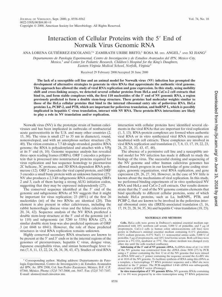
Interaction of the 5* end of NV RNAs with cell extracts. Todetermine whether the 59 end of the NV genome was recog-nized by cellular proteins, 32P-labeled RNA transcripts from nt1 to 191 (including the predicted double stem loop of nt 1 to110) were incubated with the S10 extract from HeLa cells. Amajor RNA-protein complex was observed following electro-phoresis (Fig. 1). Similar results were obtained when CaCo-2extracts were used (data not shown). The migration of thecomplex was retarded when increasing concentrations of S10extracts were used (Fig. 1A, lanes 2 to 4). Electrophoresisperformed following RNase treatment showed the formationof three complexes with different migration patterns in bothHeLa cells (Fig. 1B, lane 2) and CaCo-2 cells (lane 3). Com-plexes II and III revealed stronger RNA labeling than complexI in HeLa S10 extracts, while complexes I and II showedstronger signals in CaCo-2 extracts.
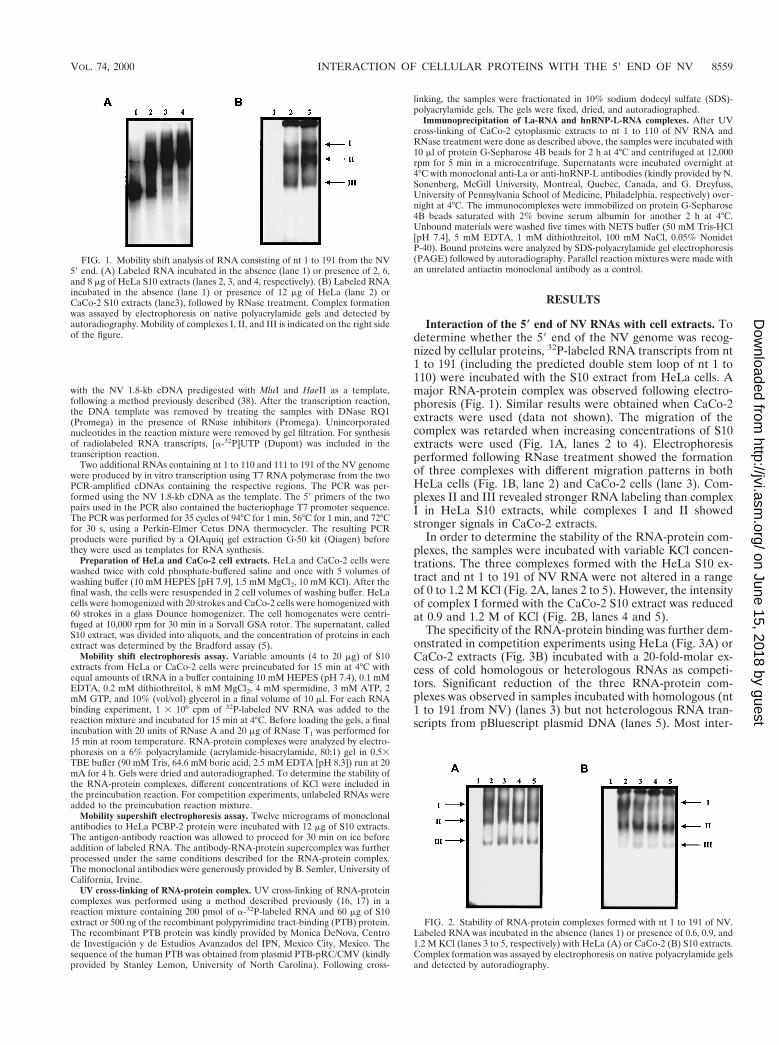
In order to determine the stability of the RNA-protein com-plexes, the samples were incubated with variable KCl concen-trations. The three complexes formed with the HeLa S10 ex-tract and nt 1 to 191 of NV RNA were not altered in a rangeof 0 to 1.2 M KCl (Fig. 2A, lanes 2 to 5). However, the intensityof complex I formed with the CaCo-2 S10 extract was reducedat 0.9 and 1.2 M of KCl (Fig. 2B, lanes 4 and 5).
The specificity of the RNA-protein binding was further dem-onstrated in competition experiments using HeLa (Fig. 3A) orCaCo-2 extracts (Fig. 3B) incubated with a 20-fold-molar ex-cess of cold homologous or heterologous RNAs as competi-tors. Significant reduction of the three RNA-protein com-plexes was observed in samples incubated with homologous (nt1 to 191 from NV) (lanes 3) but not heterologous RNA tran-scripts from pBluescript plasmid DNA (lanes 5). Most inter-
FIG. 1. Mobility shift analysis of RNA consisting of nt 1 to 191 from the NV59 end. (A) Labeled RNA incubated in the absence (lane 1) or presence of 2, 6,and 8 mg of HeLa S10 extracts (lanes 2, 3, and 4, respectively). (B) Labeled RNAincubated in the absence (lane 1) or presence of 12 mg of HeLa (lane 2) orCaCo-2 S10 extracts (lane3), followed by RNase treatment. Complex formationwas assayed by electrophoresis on native polyacrylamide gels and detected byautoradiography. Mobility of complexes I, II, and III is indicated on the right sideof the figure.
FIG. 2. Stability of RNA-protein complexes formed with nt 1 to 191 of NV.Labeled RNA was incubated in the absence (lanes 1) or presence of 0.6, 0.9, and1.2 M KCl (lanes 3 to 5, respectively) with HeLa (A) or CaCo-2 (B) S10 extracts.Complex formation was assayed by electrophoresis on native polyacrylamide gelsand detected by autoradiography.
VOL. 74, 2000 INTERACTION OF CELLULAR PROTEINS WITH THE 59 END OF NV 8559
on June 15, 2018 by guesthttp://jvi.asm
.org/D
ownloaded from
estingly, a heterologous RNA (nt 275 to 628) from poliovirus(PV) efficiently competed with the NV RNA in the formationof complexes (lanes 4), suggesting that both viruses could sharethe same cellular machinery for replication.
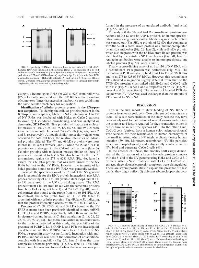
Identification of cellular proteins present in the RNA-pro-tein complexes. To identify the cellular proteins present in theRNA-protein complexes, labeled RNA containing nt 1 to 191of NV RNA was incubated with HeLa or CaCo-2 extracts,followed by UV-induced cross-linking, and was analyzed ondenaturing SDS-PAGE. Nine proteins with apparent molecu-lar masses of 110, 97, 85, 80, 75, 68, 60, 52, and 39 kDa wereidentified from both HeLa and CaCo-2 cells (Fig. 4A, lanes 2,and 3, respectively). Although similar molecular weights wereobserved for both cell lines, the relative intensities of individ-ual proteins differed. The 97- and 68-kDa proteins were moreintense in HeLa cell extracts (lane 2), while the 75- and 39-kDaproteins were stronger in the CaCo-2 cell extracts (lane 3).Cellular proteins with molecular masses similar to those ofproteins cross-linked to NV RNA also bound to the PV 59untranslated region (nt 275 to 628) RNA (Fig. 4A, lane 1),except for a 60-kDa protein that was cross-linked to the NVRNA but not to the PV RNA. However, the intensity of la-beled proteins bound to the PV RNA was generally weaker.
To locate the specific region of the 59 end of the NV genomethat is responsible for the RNA-protein interactions, two RNAprobes consisting of nt 1 to 110 (double stem loop) and nt 111to 191 were used in the UV cross-linking assays. The RNAprobe from nt 1 to 110 cross-linked with the same nine proteinsfrom both HeLa (Fig. 4B, lane 1) and CaCo-2 (Fig. 4B, lane 2)cell extracts that bound to the probe from nt 1 to 191 (Fig. 4A).In contrast, the RNA probe from nt 111 to 191 failed tocross-link with any cellular proteins (Fig. 4B, lane 3), indicatingthat the protein interaction occurs within nt 1 to 110 of NV.
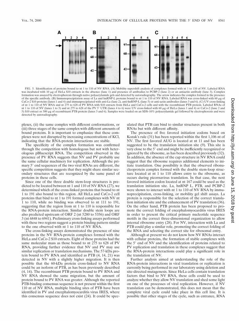
Proteins of 97, 68, 57/60, 52, and 39 kDa bound to the PVIRES element have been previously identified as unr, hnRNPL, PTB, La, and PCBP2, respectively. All of them are involvedin picornavirus and hepatitis C virus translation (3, 18, 21, 22,23, 24, 28, 35, 36, 44). Due to the similarities in molecular massto the proteins detected in this study, the possibility of thepresence of PCBP-2, La, hnRNP-L, and PTB was investigated.To determine whether PCBP-2 binds to nt 1 to 110 of NVRNA, a supershift assay was performed. Incubation with anti-PCBP-2 antibodies resulted in the formation of an additionalcomplex with less mobility (Fig. 5A, lane 2) than the threecomplexes observed previously (Fig. 5A, lane 1). This addi-tional complex was not formed when the reaction was per-
formed in the presence of an unrelated antibody (anti-actin)(Fig. 5A, lane 3).
To analyze if the 52- and 68-kDa cross-linked proteins cor-respond to the La and hnRNP L proteins, an immunoprecipi-tation assay using monoclonal antibodies against each proteinwas carried out (Fig. 5B). A protein of 52 kDa that comigrateswith the 52-kDa cross-linked protein was immunoprecipitatedby anti-La antibodies (Fig. 5B, lane 2), while a 68-kDa protein,which also migrates with the 68-kDa cross-linked protein, wasidentified by the anti-hnRNP L antibodies (Fig. 5B, lane 5).Antiactin antibodies were unable to immunoprecipitate anylabeled proteins (Fig. 5B, lanes 3 and 6).
Finally, a cross-linking assay of nt 1 to 110 of NV RNA witha recombinant PTB protein was performed (Fig. 5C). Therecombinant PTB was able to bind to nt 1 to 110 of NV RNAsand to nt 275 to 628 of PV RNAs. However, this recombinantPTB showed a migration slightly different from that of the57/60-kDa proteins cross-linked with HeLa and CaCo-2 cellswith NV (Fig. 5C, lanes 1 and 2, respectively) or PV (Fig. 5C,lanes 4 and 5, respectively). The amount of labeled PTB de-tected when PV RNA was used was larger than the amount ofPTB bound to NV RNA.
DISCUSSION
This is the first report to show binding of NV RNA toproteins from eukaryotic cells. Two different cell extracts wereused. HeLa cells were included in the study because they havebeen widely used for cultivation of several viruses and containthe proteins and factors required for their translation either incell culture or in cell-free systems (45). On the other hand,CaCo-2 cells (derived from a human colon adenocarcinoma)were selected for their resemblance to human enterocytes ofthe small intestine, where NV might replicate during humaninfection (39, 40). Moreover, recombinant NV-like particles,which are morphologically and antigenically similar to nativeNV, bind and penetrate CaCo-2 cells (46).
In the absence of RNase, the mobility shift assays demon-strated the formation of a large ribonucleoprotein complexwith the 59 end of the NV genome using HeLa and CaCo-2 S10extracts. After RNase treatment with HeLa or CaCo-2 S10extracts, three ribonucleoprotein complexes were distinguished.There are several possibilities to explain the presence of thesebands: they might reflect (i) different ribonucleoprotein com-
FIG. 3. Specificity of RNA-protein complexes formed with nt 1 to 191 of NV.Labeled RNA was incubated in the absence (lanes 2) or presence of a 20-fold-molar excess of a homologous (lanes 3) or heterologous competitor consisting ofpoliovirus nt 275 to 628 RNA (lanes 4) or pBluescript RNA (lanes 5). Free RNAwas loaded on lanes 1. HeLa S10 extracts (A) and CaCo-2 S10 extracts (B) areshown. Complex formation was assayed by electrophoresis through native poly-acrylamide gels and detected by autoradiography.
FIG. 4. UV-induced cross-linking of HeLa and CaCo-2 S10 extracts to la-beled RNAs from nt 1 to 191, 1 to 110, and 111 to 191 of NV. (A) Labeled RNAof nt 1 to 191 of NV (lanes 2 and 3) and nt 275 to 628 of the PV 59 untranslatedregion (lane 1) were UV cross-linked with 60 mg of HeLa extracts (lanes 1 and2) or CaCo-2 S10 extracts (lane 3). (B) Labeled RNA consisting of nt 1 to 110(lanes 1 and 2) and nt 111 to 191 (lane 3) were UV cross-linked with 60 mg ofHeLa extracts (lane1) or CaCo-2 S10 extracts (lanes 2 and 3). Proteins wereseparated by SDS–12.5% PAGE and detected by autoradiography. Numbers tothe right of panels indicate molecular masses in kilodaltons.
8560 GUTIERREZ-ESCOLANO ET AL. J. VIROL.
on June 15, 2018 by guesthttp://jvi.asm
.org/D
ownloaded from
plexes, (ii) the same complex with different conformations, or(iii) three stages of the same complex with different amounts ofbound proteins. It is important to emphasize that these com-plexes were not disrupted by increasing concentrations of KCl,indicating that the RNA-protein interactions are stable.
The specificity of the complex formation was confirmedthrough the competition with homologous but not with heter-ologous pBluescript RNA. The competition observed in thepresence of PV RNA suggests that NV and PV probably usethe same cellular machinery for replication. Although the pri-mary 59 end sequences of the two viral RNAs are different, thespecific competition suggests that they might share similar sec-ondary structures that are recognized by the same panel ofproteins in these cells.
Since one of the three double stem-loop structures is pre-dicted to be located between nt 1 and 110 of NV RNA (27), wedetermined which of the cross-linked proteins that bound to nt1 to 191 also bound to this region. All HeLa and CaCo-2 cellproteins that bind to nt 1 to 191 formed complexes with NV nt1 to 110, while no binding was observed to nt 111 to 191,suggesting that the region from nt 1 to 110 is responsible forthe RNA-protein interaction. Similar double stem loops arealso predicted upstream of ORF 2 (nt 5280 to 5356) and ORF3 (nt 6848 to 6941). Preliminary cross-linking assays performedwith these two regions suggest a protein binding pattern similarto the one observed with nt 1 to 110 of NV RNA.
The cross-linking assays demonstrated the presence of nineproteins in the NV RNA-protein complexes formed with theHeLa and CaCo-2 S10 extracts. Eight of these proteins had thesame molecular mass as those bound to nt 275 to 628 of PVRNA, providing further evidence that NV and PV may usesimilar replication or translation mechanisms. The 57-kDa pro-tein bound to PV RNA and identified as PTB (4, 14, 21) wasdetected in NV with a slightly higher migration. It is thenpossible that the 60-kDa protein cross-linked to NV RNAcould be an isoform of PTB, as has been previously described(4, 14). The recombinant PTB protein bound to PV RNA andNV RNA showed the same migration, but the amount ofprotein bound to PV RNA was greater. Although the reportedPTB-binding consensus sequence is not present within the first110 nt of NV RNA, multiple binding sites of PTB have beenidentified in PV and encephalomyocarditis virus IRES, wherethis consensus sequence does not exist (24). It could be spec-
ulated that PTB can bind to similar structures present in bothRNAs but with different affinity.
The presence of five favored initiation codons based onKozak’s rule (31) has been reported within the first 1,106 nt ofNV. The first favored AUG is located at nt 11 and has beensuggested to be the translation initiation site (9). This site isvery close to the 59 end and might be inefficiently recognized orignored by the ribosome, as has been described previously (32).In addition, the absence of the cap structure in NV RNA couldsuggest that the ribosome requires additional elements to ini-tiate translation. One possibility is that the observed ribonu-cleoprotein complex formed with the double stem-loop struc-ture located at nt 1 to 110 allows entry to the ribosome, asoccurs during picornavirus translation. In that case, the nextfavored initiation codon located at nt 167 could be used as thetranslation initiation site. La, hnRNP L, PTB, and PCBP-2were shown to interact with nt 1 to 110 of NV RNA by immu-noprecipitation, cross-linking, or mobility gel shift assays. Laprotein is responsible for the selection of the correct transla-tion initiation site and the enhancement of PV translation (36).On the other hand, PTB protein has been proposed to pro-mote the correct folding of encephalomyocarditis virus IRESin order to present the critical primary nucleotide sequencemotifs in the correct three-dimensional organization to allowinternal ribosome entry (28). For NV, interaction with La andPTB could play a similar role, promoting the correct folding ofthe RNA and selecting the correct site for ribosomal entry.
Although at present we do not know how NV RNAs interactwith cellular proteins, the formation of stable complexes withthe 59 end of NV and the identification of proteins related toPV replication and translation in these complexes suggest thatthe RNA-protein interactions could play a significant role inthe translation of NV.
Further analysis aimed at understanding the role of theRNA-protein interactions in viral translation or replication iscurrently being performed in our laboratory using deletion andsite-directed mutagenesis. Since HeLa cells contain translationfactors that bind to NV RNA, these cells could be used toanalyze whether they allow NV translation and shed some lighton one of the processes of viral replication. However, if NVtranslation can be demonstrated, this does not mean that thecomplete viral cycle could take place in this cell line. It ispossible that other stages of the cycle, such as entrance, RNA
FIG. 5. Identification of proteins bound to nt 1 to 110 of NV RNA. (A) Mobility supershift analysis of complexes formed with nt 1 to 110 of NV. Labeled RNAwas incubated with 10 mg of HeLa S10 extracts in the absence (lane 1) and presence of antibodies to PCBP-2 (lane 2) or an antiactin antibody (lane 3). Complexformation was assayed by electrophoresis through native polyacrylamide gels and detected by autoradiography. The arrow indicates the complex formed in the presenceof the specific antibody. (B) Immunoprecipitation assay of La and hnRNP-L proteins bound to nt 1 to 110 of NV RNA. Labeled RNA was cross-linked with 60 mg ofCaCo-2 S10 proteins (lanes 1 and 4) and immunprecipitated with anti-La (lane 2), anti-hnRNP-L (lane 5) or anti-actin antibodies (lanes 3 and 6). (C) UV cross-linkingof nt 1 to 110 of NV RNA and nt 275 to 628 of PV RNA with S10 extracts from HeLa and CaCo-2 cells and with the recombinant PTB protein. Labeled RNAs ofnt 1 to 110 of NV (lanes 1 to 3) and nt 275 to 628 of the PV 59 UTR (lanes 4 to 6) were UV cross-linked with 60 mg of HeLa (lanes 1 and 4) or CaCo-2 (lane 2 and5) S10 extract or 500 mg of recombinant PTB protein (lanes 3 and 6). Samples were loaded on an SDS–10% polyacrylamide gel followed by electrophoresis and weredetected by autoradiography.
VOL. 74, 2000 INTERACTION OF CELLULAR PROTEINS WITH THE 59 END OF NV 8561
on June 15, 2018 by guesthttp://jvi.asm
.org/D
ownloaded from
replication, or assembly, are blocked in these cells, thus ex-plaining their nonpermissive nature to NV.
ACKNOWLEDGMENTS
We thank Fernando Medina for the cell cultures, Jaime Escobar fortechnical assistance, and Monica DeNova for the recombinant PTBprotein. We gratefully acknowledge Stanley Lemon for providing thePTB sequence, B. Semler (University of California, Irvine) for theanti-PCBP2 antibodies, N. Sonenberg (McGill University, Montreal,Quebec, Canada) for the anti-La antibodies, and G. Dreyfuss (Uni-versity of Pennsylvania School of Medicine, Philadelphia) for the anti-hnRNP-L antibodies. We also thank Martha Espinosa-Cantellano forcritical comments on the manuscript.
This work was supported by grants from the Consejo Nacional deCiencia y Tecnologıa.
REFERENCES1. Andino, R., G. E. Rieckhof, P. L. Achacoso, and D. Baltimore. 1993. Polio-
virus RNA synthesis utilizes an RNP complex formed around the 59 end ofviral RNA. EMBO J. 12:3587–3598.
2. Blacklow, N. R., and H. B. Greenberg. 1991. Viral gastroenteritis. N. Engl.J. Med. 325:252–264.
3. Blyn, L. B., J. S. Towner, B. L. Semler, and E. Ehrenfeld. 1997. Requirementof poly(rC) binding protein 2 for translation of poliovirus RNA. J. Virol.71:6243–6246.
4. Bothwell, A. L. M., D. W. Ballard, W. M. Philbrick, G. Lindwall, S. E. Maher,M. M. Bridgett, S. F. Jamison, and M. A. Garcia-Blanco. 1991. Purinepolypyrimidine tract binding protein. J. Biol. Chem. 25:24657–24663.
5. Bradford, M. M. 1976. A rapid and sensitive method for the quantitation ofmicrogram quantities of protein utilizing the principle of protein-dye bind-ing. Anal. Biochem. 72:248–254.
6. Burroughs, J. N., and F. Brown. 1978. Presence of a covalently linked proteinon calicivirus RNA. J. Gen. Virol. 41:443–446.
7. Chang, K. H., E. A. Brown, and S. M. Lemon. 1993. Cell type-specificproteins which interact with the 59 nontranslated region of hepatitis A virusRNA. J. Virol. 67:6716–6725.
8. Chen, C. J., M. D. Kuo, L. J. Chen, S. L. Hsu, Y. M. Wang, and J. H. Lin.1997. RNA-protein interactions: involvement of NS3, NS5, and 39 codingregions of Japanese encephalitis virus genomic RNA. J. Virol. 71:3466–3473.
9. Clarke, I. N., and P. R. Lambden. 1997. The molecular biology of calicivi-ruses. J. Gen. Virol. 78:291–301.
10. Dunham, D. M., X. Jiang, T. Berke, A. W. Smith, and D. O. Matson. 1998.Genomic mapping of a calicivirus VPg. Arch. Virol. 143:2421–2430.
11. Ehrenfeld, E., and B. Semler. 1995. Anatomy of the poliovirus internalribosome entry site. Curr. Top. Microbiol. Immunol. 203:65–83.
12. Ehresmann, D. W., and F. L. Schaffer. 1977. RNA synthesized in calicivirus-infected cells is atypical of picornaviruses. J. Virol. 22:572–576.
13. Gamarnik, A. V., and R. Andino. 1998. Switch from translation to RNAreplication in a positive-stranded RNA virus. Genes Dev. 12:2293–2304.
14. Gil, A., P. A. Sharp, S. F. Jamison, and M. A. Garcia-Blanco. 1991. Char-acterization of cDNAs encoding the polypyrimidine tract binding protein.Genes Dev. 5:1224–1236.
15. Greenberg, H. B., J. Valdesuso, A. Z. Kapikian, R. M. Chanock, R. G. Wyatt,W. Szmuness, J. Larrick, J. Kaplan, H. Gilman, and D. A. Sack. 1979.Prevalence of antibodies to the Norwalk virus in various countries. Infect.Immun. 26:270–273.
16. Gutierrez-Escolano, A. L., and R. M. del Angel. 1996. Nuclear proteins bindto poliovirus 59 untranslated region. Arch. Med. Res. 27:413–419.
17. Gutierrez-Escolano, A. L., M. A. Denova, V. R. Racaniello, and R. M. delAngel. 1997. Attenuating mutations in the poliovirus 59 untranslated regionalter its interaction with polypyrimidine tract-binding protein. J. Virol. 71:3826–3833.
18. Hahm, B., Y. K. Kim, J. H. Kim, T. Y. Kim, and S. K. Jang. 1998. Hetero-geneous nuclear ribonucleoprotein L interacts with the 39 border of theinternal ribosomal entry site of hepatitis C virus. J. Virol. 72:8782–8788.
19. Haller, A. A., and B. L. Semler. 1995. Stem-loop structure synergy in bindingcellular proteins to the 59 noncoding region of poliovirus RNA. Virology206:923–934.
20. Hardy, M. E., and M. K. Estes. 1996. Completion of the Norwalk virusgenome sequence. Virus Genes 12:287–290.
21. Hellen, C. U., G. W. Witherella, M. Schmid, H. S. Shin, T. V. Pestova, A. Gil,and E. Wimmer. 1993. A cytoplasmic 57-kDa protein that is required fortranslation of picornavirus RNA by internal ribosomal entry is identical tothe nuclear pyrimidine tract-binding protein. Proc. Natl. Acad. Sci. USA90:7642–7646.
22. Hunt, S. L., J. J. Hsuan, N. Totky, and R. Jackson. 1998. Unr, a cellularcytoplasmic RNA-binding protein with five cold-shock domains is requiredfor internal initiation of translation of human rhinovirus RNA. Genes Dev.13:437–448.
23. Hwang, Y. K., and M. A. Brinton. 1998. A 68-nucleotide sequence within the39 noncoding region of simian hemorrhagic fever virus negative-strand RNAbinds to four MA104 cell proteins. J. Virol. 72:4341–4351.
24. Ito, T., and M. Lai. 1999. An internal polypyrimidine-tract-binding site in thehepatitis C virus RNA attenuates translation, which is relieved by the 39untranslated sequence. Virology 254:288–296.
25. Jackson, R. J., and A. Kaminski. 1995. Internal initiation of translation ineukaryotes: the picornavirus paradigm and beyond. RNA 1:985–1000.
26. Jiang, X., M. Wang, D. Y. Graham, and M. K. Estes. 1992. Expression,self-assembly and antigenicity of the Norwalk virus capsid protein. J. Virol.66:6527–6532.
27. Jiang, X., M. Wang, K. Wang, and M. K. Estes. 1993. Sequence and genomicorganization of Norwalk virus. Virology 195:51–61.
28. Kaminski, A., S. L. Hunt, J. G. Patton, and R. J. Jackson. 1995. Directevidence that polypyrimidine tract-binding protein (PTB) is essential forinternal initiation of translation of encephalomyocarditis virus RNA. RNA1:924–938.
29. Kapikian, A. Z., R. G. Wyatt, R. Dolin, T. S. Thornhill, A. R. Kalika, andR. M. Chanock. 1972. Visualization by immune electron microscopy of a27-nm particle associated with acute infectious nonbacterial gastroenteritis.J. Virol. 10:1075–1081.
30. Kaplan, J. E., G. W. Gary, and R. C. Baron. 1982. Epidemiology of Norwalkgastroenteritis and the role of Norwalk virus in outbreaks of acute nonbac-terial gastroenteritis. Ann. Intern. Med. 96:756–761.
31. Kozak, M. 1997. Recognition of AUG and alternative initiator codons isaugmented by G in position 14 but is not generally affected by nucleotidesin positions 15 and 16. EMBO J. 16:2482–2492.
32. Kozak, M. 1991. A short leader sequence impairs the fidelity of initiation byeucaryotic ribosomes. Gene Expr. 1:111–115.
33. Lai, M. M. C. 1998. Cellular factors in the transcription and replication ofviral RNA genomes: a parallel to DNA-dependent RNA transcription. Vi-rology 244:1–12.
34. Lambden, P. R., B. Lui, and I. N. Clarke. 1995. A conserved sequence motifat the 59 terminus of the Southampton virus genome is characteristic of thecaliciviridae. Virus Genes 10:149–152.
35. Luz, N., and E. Beck. 1991. Interaction of a cellular 57-kilodalton proteinwith the internal translation initiation site of foot-and-mouth disease virus.J. Virol. 65:6486–6494.
36. Meerovitch, K., Y. V. Svitkin, H. S. Lee, F. Lejbkowics, D. J. Kenan, E. K. L.Chan, V. I. Agol, J. D. Keene, and N. Sonenberg. 1993. La autoantigenenhances and corrects aberrant translation of poliovirus RNA in reticulocytelysate. J. Virol. 67:3798–3807.
37. Palmenberg, A. C. 1989. Sequence alignments of picornaviral capsid pro-teins, p. 211–241. In B. L. Semler and E. Ehrenfeld (ed.), Molecular aspectsof picornavirus infection and detection. American Society for Microbiology,Washington, D.C.
38. Pelletier, J., G. Kaplan, V. R. Racaniello, and N. Sonenberg. 1988. Cap-independent translation of poliovirus mRNA is conferred by sequence ele-ments within the 59 noncoding region. Mol. Cell. Biol. 8:1103–1112.
39. Pinto, R. M., J. M. Diez, and A. Bosch. 1994. Use of the colonic carcinomacell line CaCo-2 for in vivo amplification and detection of enteric viruses.J. Med. Virol. 44:310–314.
40. Pinto, R. M., S. Robine-Leon, M. D. Appay, M. Kedinger, N. Triadou, E.Dussaulx, B. Lacroix, P. Simon-Assmann, K. Haffen, J. Fogh, and A.Zweibaum. 1983. Enterocyte-like differentiation and polarization of the hu-man colon carcinoma cell line Caco-2 in culture. Biol. Cell 47:323–330.
41. Prasad, B. V. V., R. Rothnagel, X. Jiang, and M. K. Estes. 1994. Three-dimensional structure of the baculovirus-expressed Norwalk virus capsid.J. Virol. 68:5117–5125.
42. Rinehart-Kim, J. E., W. M. Zhong, X. Jiang, A. W. Smith, and D. O. Matson.1998. Complete nucleotide sequence and genomic organization of a primatecalicivirus, Pan-1. Arch. Virol. 144:199–208.
43. Roehl, H. H., and B. L. Semler. 1995. Poliovirus infection enhances theformation of two ribonucleoprotein complexes at the 39 end of viral negative-strand RNA. J. Virol. 69:2954–2961.
44. Svitkine, Y. V., K. Meerovitch, H. S. Lee, J. N. Dholakia, D. J. Kenan, V. I.Agol, and N. Sonenberg. 1994. Internal initiation on poliovirus RNA: furthercharacterization of La function in poliovirus translation in vitro. J. Virol.68:1544–1550.
45. Villa-Komaroff, L., N. Gutteman, D. Baltimore, and H. F. Lodish. 1975.Complete translation of poliovirus RNA in a eukaryotic cell-free system.Proc. Natl. Acad. Sci. USA 72:4157–4161.
46. White, I. J., J. M. Ball, M. E. Hardy, T. N. Tanaka, N. Kitamoto, and M. K.Estes. 1996. Attachment and entry of recombinant Norwalk virus capsid tocultured human and animal cell lines. J. Virol. 70:6589–6597.
47. Witherell, G. W., A. Gil, and E. Wimmer. 1993. Interaction of polypyrimidinetract binding protein with the encephalomyocarditis virus mRNA internalribosomal entry site. Biochemistry 32:8268–8275.
48. Zeng, L., B. Falgout, and L. Markoff. 1998. Identification of specific nucle-otide sequences within the conserved 39-SL in the dengue type 2 virusgenome required for replication. J. Virol. 72:7510–7522.
8562 GUTIERREZ-ESCOLANO ET AL. J. VIROL.
on June 15, 2018 by guesthttp://jvi.asm
.org/D
ownloaded from