integrated algal bioprocess engineering for enhanced ... · integrated algal bioprocess engineering...
TRANSCRIPT

61RR: J Microbiol Biotechnol | Volume 6 | Issue 2 | June, 2017
Research & Reviews: Journal of Microbiology and Biotechnology e-ISSN:2320-3528p-ISSN:2347-2286
INTRODUCTION
Microalgae are photosynthetic microorganisms, able to rapidly generate biomass from solar energy and CO2 in water bodies and arable land, with efficiency 10 times greater than that of the terrestrial plants. Living not just in aquatic but also terrestrial ecosystem and harsh conditions, algae represent a large variety of species from a wide range of environmental conditions [1].
Autotrophs convert solar energy directly into organic molecules, but some algal species also grow as heterotrophs and/or mixotrophs on organic carbon, exhibiting several advantages over the autotrophic mode [2]. Table 1 shows the composition of microalgal cell wall and storage products which offer diverse spectrum of valuable products and environmental solutions such as energy sources (including jet fuel, aviation gas, biodiesel, gasoline, and bioethanol), pigments, food and nutritional compounds such as omega-3 fatty acids, pharmaceuticals, recombinant proteins, and vaccines, animal feeds, organic fertilizers, and biodegradable plastics [3-5]. There are advantages associated with algae such as much higher biomass productivity, the ability to consume harmful pollutants with minimal resource requirements and do not compete with food or agriculture for precious resources and land [6-8].
To date, algae have all the potential to play pivotal roles to remedy the energy, environment and food crisis prevailing in the
Integrated Algal Bioprocess Engineering for Enhanced Productivity of Lipid, Carbohydrate and High-value Bioactive Compounds
Mohd Azmuddin Abdullah1*, Syed Muhammad Usman Shah2,3, Sanaa Mahmoud Metwally Shanab4 and Hamdy El-Sayed Ahmad Ali5
1Institute of Marine Biotechnology, Universiti Malaysia Terengganu, 21030 Kuala Nerus, Terengganu, Malaysia2Department of Chemical Engineering, Universiti Teknologi PETRONAS, 32610 Seri Iskandar, Perak, Malaysia
3Department of Biosciences, COMSATS Institute of Information Technology, Park Road, 44000, Islamabad, Pakistan
4Department of Botany and Microbiology, Faculty of Science, Cairo University, 12613, Giza, Egypt5Department of Microbiology, National Center for Radiation Research and Technology (NCRRT),
Egyptian Atomic Energy Authority (EAEA), Egypt
Research Article
ABSTRACT
Algae are naturally the producers of lipids, carbohydrates and high-value bioactive biocompounds. These bioproducts accumulation tend to increase when algae are placed under environmental or nutritional stresses. Of great importance is to identify the engineering and molecular factors that could influence the triggering of constitutive products accumulation. This review explores issues and factors that could affect commercial-scale development for integrated microalgal bioprocess engineering especially in terms of different target products, engineering factors and the recovery of microalgae biomass and bioproducts as a source of renewable biofuels and high-value bioactive compounds.
Received date: 15/12/2016Accepted date: 27/06/2017Published date: 30/06/2017
*For Correspondence
Mohd Azmuddin Abdullah, Institute of Marine Biotechnology, Universiti Malaysia Terengganu, 21030 Kuala Nerus, Terengganu, Malaysia; Tel: +6096683104/3661.
E-mail: [email protected]; [email protected]
Keywords: Algae, Bioprocess Engineering, Lipid, Carbohydrate, Bioactive compounds

62
Research & Reviews: Journal of Microbiology and Biotechnology e-ISSN:2320-3528p-ISSN:2347-2286
RR: J Microbiol Biotechnol | Volume 6 | Issue 2 | June, 2017
world. Red algae, specifically, are important sources for many biologically active metabolites in comparison to other algal classes [3]. Species such as Cyanobacteria, Phormidium cebennse, Oscillatoria raciborskii, Scytonema burmanicum, Calothrix elenkinii, and Anabaena variabilis show anti-Human Immunodeficiency Virus-1 (HIV-1) activity, and tested positive for the presence of sulfolipids. Hydrocolloids, alginate, agar, and carrageenan produced from seaweeds are largely used as viscosity-modifying agents in foods and pharmaceuticals. Diatoms from a large and diverse group of unicellular eukaryotic algae, characterized by unique cell walls made of silica called a frustule [9], play a vital role in the ocean for CO2 fixation and O2 production. In addition to the uses as feeds for aquaculture and specialty oils such as omega-3 fatty acids, there has been interest in developing diatoms for nanotechnology application [10].
Fuel ethanol production in the United States has increased from 1.6 to 13.2 billion gallons from 2000 to 2010, consuming one third of the corn harvest. This has caused a significant increase in global grain prices [11], a situation similarly seen with biodiesel from edible vegetable oils such as soybeans, peanuts, rapeseeds and palm oil [12]. Microalgae therefore have emerged as among the most promising feedstocks for biofuels, not facing any of the food versus fuel issues. Algal cultivation for biofuel production in commercial scale appears not yet to be economical and sustainable. Improvements of the economics are possible with simultaneous production of specific high-value compounds and biofuels combined in a biorefinery concept [4-5,13]. Furthermore, as bulk commodities in industrial sectors as varied as pharmaceuticals, cosmetics, nutraceuticals, functional foods, and biofuels, readily available supply of algal extracts, fractions or pure compounds are of prime importance [14]. Algae culturing facilities could be located in aquatic environments, eliminating the utilization of arable land [15]. The qualities of the microalgal cells can be controlled using clean nutrient media for growth, thus avoiding the use of herbicides and pesticides, or any other toxic substances, and optimal conditions of the reactor systems.
In this review, an overview of different types of algal products, bioactivities, characteristics and production technologies are given with special emphasis on the production of lipids, carbohydrates and antioxidants and the strategies by the integrated reactor and downstream engineering considerations for enhanced productivity.
ALGAL BIOFACTORIES
Algae are biofactories for the production of a number of high-value compounds. Microalgal lipids contain the essential fatty acids eicosapentaenoic acid (EPA) and docosahexaenoic acid (DHA), and other high-value fatty acids (omega-3, γ-linolenic acid etc.) which may be absent in food crops. The neutral lipids, especially triacylglycerides (TAGs), are suitable for conversion to biodiesel (fatty acid methyl esters, FAME) [16]. Carbohydrates are the feedstock for the fermentation of sugars for the production of bioethanol [17]. Species such as diatoms are attractive for the discovery of novel metabolic pathways with range of novel biological processes presumably acquired during evolution [18], which may be absent in other commonly studied model organisms [19]. The high-value specific secondary metabolites include the pigments and vitamins. Algal proteins have high nutrition quality comparable to other referenced food proteins, because of good profile and high proportion of amino acids [20].
Lipids
Algae have a trigger which when put into stressful environments such as nutrient deprivation can alter the metabolic pathway towards hydrocarbon production (energy storage in the form of oils). The average lipid content of algae varies from 1-70% dry mass with oil levels of 20-50%, but the lipid may go as high as 60-90%, depending on strains and environments [21,22]. The oil or lipid from algae is extracted using solvents and turned into biodiesel through transesterification [15,23]. Although algae with 70% oil content exist, the growth system to maintain the culture at this level of oil content has not been developed that would make oil from algae an economical option [15]. Large-scale production is also hampered by the availability of few algal strains that can be selectively optimized for both high biomass productivity and high TAG content [24]. Some potential economical and model microalgae for high growth rate and productivity include Dunaliella salina, Chlorella sp., Chlamydomonas reinhardtii, Muriellopsis sp., Haematococcus pluvialis, Phaeodactylum tricornutum, Nannochloropsis sp. [25,26].
Carbohydrates
Carbohydrates are the major products derived from photosynthesis and the carbon fixation metabolism (the Calvin cycle) [27]. These are either accumulated in the plastids as reserve materials (e.g. starch), or become the main component of cell walls (e.g. cellulose, pectin, and sulfated polysaccharides). The composition and metabolism of carbohydrates (mainly starch and cellulose) in microalgae may differ significantly from species to species [28,29]. The carbohydrate or starch content of green microalgal species Chlorella, Dunaliella, Chlamydomonas and Scenedesmus have been reported to be 16-60% based on dry cell weight [29]. With 75% of algal complex carbohydrates hydrolyzable into a fermentable hexose monomer or 80% theoretical ethanol yield [30], there has been significant interest on microalgal utilization as an advanced energy feedstock for bioethanol production [31,32]. Absence of non-photosynthetic supporting structures (roots, stems or leaves) favors algal cultivation. Certain species can produce ethanol during dark-anaerobic fermentation and thus serve as a direct source for ethanol production. Macroalgae can also be harnessed while

63
Research & Reviews: Journal of Microbiology and Biotechnology e-ISSN:2320-3528p-ISSN:2347-2286
RR: J Microbiol Biotechnol | Volume 6 | Issue 2 | June, 2017
Oleaginous microalgae generate high starch/cellulose biomass waste after oil extraction, which can be hydrolyzed to generate sugary syrup as substrate for ethanol production [33]. It is easy to provide optimal levels or minimal nutrients for microalgae culturing from the well-mixed aqueous environment as compared to the soil. The starch can be converted directly into bioethanol under dark and anaerobic conditions. It is however of paramount importance to understand the fundamental metabolism of photosynthetic microalgal cells under dark conditions eventhough the bioethanol production rate and yield may be low [34,35]. Generally two methods are used fermentation (biochemical process) and gasification (thermo-chemical process) [13]. After oil extraction from the algal biomass, fermentation process ensues utilizing gluco-amylase, α-amylase and yeast, bacteria or fungi for fermenting sugars in algal residues into ethanol and carbon dioxide with used water that can be recycled [6].
Biobutanol and other higher alcohols from biomass feedstocks are known as advanced biofuels and may eventually replace bioethanol [36]. Butanol is now mainly produced by chemical synthesis using petroleum as the raw material [37]. Compared to ethanol, butanol not only has higher energy content and lower volatility, but also less hygroscopic, and mixes better with gasoline in any proportion. Biobutanol can also be produced from carbohydrate-based microalgae as an alternative fuel as it contains more energy and is less corrosive and water soluble [38], and may be well suited for use with the existing storage and distribution infrastructure of petroleum-based transportation fuels. However, biobutanol fermentation is much less efficient and less productive, with lower product titre and yield, attributable to severe inhibition of biobutanol on host cells [39].
Microalgal biomass can also be utilized to enhance gaseous biofuels such as methane and hydrogen through the anaerobic fermentation process [5,40]. The anaerobic digestion process can be made economically competitive by utilizing the microalgal residues remaining after the production of high grade biofuels such as bioethanol and biobutanol in an integrated biorefinery concept [5,40,41]. Methane production facilities therefore can be established to treat organic wastes produced from fermentation plants. Anaerobic digestion of algal biomass produces biogas with a high methane (over 60%) and low sulphur concentration which can reduce corrosion in the power generator [42]. Biohydrogen is another promising new energy carrier because it is cleaner and more efficient, particularly when used in fuel cells to directly generate electricity. Biohydrogen can be produced directly through the metabolic network of microalgae, but the efficiency is low. The starch-containing green alga Dunaliella tertiolecta and C. reinhardtii have been used successfully, achieving the H2 yields (based on starch) of 61% and 52%, respectively [43].
Table 1. Composition of microalgal cell wall and storage products [29,44,45].
Division Cell wall Storage products
Cyanophyta Lipopolysaccharides, CyanophyceanPeptidoglycan Starch
Chlorophyta Cellulose, hemicellulose Starch/lipidDinophyta Absence or contain few cellulose Starch
Cryptophyta Periplast StarchEuglenophyta Absence Paramylum/lipid
Rhodophyta Agar, carrageenan, cellulose,calcium carbonate Floridean starch
Heterokontophyta Naked or covered by scales orwith large quantities of silica Leucosin/lipid
Phaeophyta Alginic acid, Fucoidan, Ascophyllan LaminarinBacillariophyta
(Diatoms)Pectin, Hydrated silica, sulfated
polysaccharidesChyrsolaminarin, Lipid, Volutin granuls
(protein)
High-Value Bioactive Compounds
The three areas of research in aquatic natural products emerging in the last 3 decades are toxins, bioproducts and chemical ecology where more than 15000 novel compounds have been chemically determined. Marine compounds have wide diversity of molecular targets with marked selectivity and of pharmaceutical interest. Algae fall in this promising group to provide novel biochemically active substances [46-48]. However, the discovery of metabolites, already obtained from traditionally better-studied organisms, is expected to be at far lower rate in microalgae [49]. Secondary metabolism can be easily triggered by most forms of externally applied stress such as nutrient deficiency. The huge metabolic plasticity of microalgae may lead to dissimilar results depending on the physiological state, stressed or otherwise. The defence strategies developed could have resulted in significant level of structural-chemical diversity from different metabolic pathways to survive in a competitive environment, freshwater and marine [50,51]. These include pigments (chlorophylls, carotenoids and phycobilins), antioxidants, vitamins (β-carotenes), polysaccharides, triglycerides (fatty acids), and biomass [52].
One major potential application of microalgae is as nutraceuticals (functional foods) that can provide nutrition and

64
Research & Reviews: Journal of Microbiology and Biotechnology e-ISSN:2320-3528p-ISSN:2347-2286
RR: J Microbiol Biotechnol | Volume 6 | Issue 2 | June, 2017
pharmaceutical benefits to the body for prevention and treatment of diseases [53]. Products isolated or nutraceutical compound(s) purified from natural sources (plant or seaweed/algae) may be sold in medicinal forms not usually associated with food, but proven to have physiological benefits including promotion of healthy bones and teeth, weight reduction, provide protection against chronic diseases, improved immune system and brain development, healthier digestive system, blood pressure control, reduce cholesterol or cardiovascular risk events, antioxidant, antiviral, and anticancer properties [54,55]. Among potential nutraceuticals from microalgae include vitamins and minerals, polysaccharides, polyunsaturated fatty acids (PUFAs), phenolic and antioxidants, proteins, pigments/carotenoids, sterols and polysaccarides [56]. Examples include Haematococcus in powder or tablet form or astaxanthin extract.
Types
Fatty Acids and Glycerol: Fatty acids, saturated and unsaturated with one or more double bonds (mono/PUFAs), are important primary products for the production of proteins, nucleic acids and biomembranes. PUFAs play key roles in the cellular and tissue metabolism including the regulation of membrane fluidity, electron and oxygen transport as well as thermal adaptation. Stress environmental conditions affect fatty acids production and composition, and the reactive oxygen species (ROS) may induce lipid peroxidation which alters the biomembrane composition and function. Lipid and its fatty acids content affect the produced biodiesel characteristics, and exhibit different biological activities and allelopathic effects [57,58]. Glycerol accumulation in microalgae acts as osmoticum and regulates the osmotic pressure between cell and environment. Glycerol can be utilized to produce high value products related to the food, industrial or pharmaceutical sector [59], and it is also the by-product in lipid transesterification for biodiesel production.
Pigments: Pigments are colorful compounds which absorb and reflect certain wavelengths of visible light. In the photosynthetic system of microalgae, pigments which are the chlorophylls, phycobillins and carotenoids, act as light energy absorber. Chlorophylls, present in all higher-plants and photosynthetic algae and cyanobacteria, are greenish and lipid soluble, containing a porphyrin ring. Structurally, chlorophylls are the substituted tetraphurole with a centrally bound magnesium atom. The porphyrin tetraphurole is further esterified to a diterpene alcohol phytol to form chlorophyll [60]. In algae, there are four kinds of chlorophylls - the most important being chlorophyll a (Chl a) which absorbs most energy from wave length of violet-blue and orange-red light [61], and the chlorophylls b, c, d which are found in green, brown and red algae, respectively [62]. These are essential molecules for photosynthesis by passing their energized electrons onto molecules which will manufacture sugars. Phycobilins are bonded to water-soluble proteins known as phycobiliproteins. Phycobiliproteins present mainly in cyanobacteria and also in some red algae and cryptomonads as the principle photoreceptor, accessory or antenna for photosynthetic light by absorbing energy in the visible spectrum (450-650 nm) [63]. Phycobiliproteins are divided into phycocyanobilin (blue colored), phycoerythrobilin (red colored), phycouroblin (yellow colored) and phycobiviolin (purple colored).
Carotenoids are a group of over 700 natural lipid soluble pigments that are primarily produced within phytoplankton, plants, algae/seaweed and some fungal and bacterial species and highly present in fruits, vegetables, some seafoods and Mediterranean foods. These are hydrophobic accessory pigments with 40-carbon structures divided into two main groups the carotenes (non-oxygenated molecules) and the xanthophylls (oxygenated molecules). During photosynthesis, carotenoids play the role in light harvesting, photoprotection, superoxide (O2-) scavenging, excess energy dissipation and structure stabilization [64]. Carotenoids form pigment-protein complexes with peptide and are located mainly in the thylakoid membranes, chloroplasts or plastids. Some selected algal acetylenic carotenoids are shown in Figure 1. The main ones are astaxanthin, fucoxanthine, β-carotene and lutein. β-carotene (Figure 1a) is the primary carotenoid, while astaxanthin (Figure 1b) is the secondary carotenoid. Astaxanthin is a high-value algal carotenoid which has achieved commercial success. The molecule has two asymmetric carbons located at the 3 and 39 positions of the benzenoid rings on either end of the molecule (Figure 1b). Astaxanthin is ubiquitous especially in the marine environment, and known for the pinkish-red hue in the salmonids, shrimps lobsters and crayfish. Within algae and phytoplankton, astaxanthin is biosynthesized in the food chain, at the primary production level, which later may be consumed and ingested by zooplankton, insects or crustaceans [65]. Lutein is a primary carotenoid which involves in maintaining the structure and functioning of photosystems. Unlike the primary carotenoid β-carotene, or the secondary carotenoid astaxanthin, under stress conditions, lutein acts as a secondary carotenoid. The main species that are extensively studied on carotenoids are Dunaliella salina, Haematococcus pluvialis, Chlorella zofingiensis and Chlorella vulgaris due to their commercial potential in large scale cultures [66]. The accumulation of astaxanthin in Haematococcus pluvialis exceeds any other known source [67-69] and therefore is the most studied, but others such as Chlorella sp. [70], Chlorococcum sp. [71] or Scenedesmus sp. [72], are all potential producers of astaxanthin. The carotenoid lutein can be produced by Chlorella sp. [70,73], Muriellopsis sp. [74], Scenedesmus sp. [75] and Chlamydomonas sp. [76].

65
Research & Reviews: Journal of Microbiology and Biotechnology e-ISSN:2320-3528p-ISSN:2347-2286
RR: J Microbiol Biotechnol | Volume 6 | Issue 2 | June, 2017
Figure 1. Chemical structure of a) β-Carotene; b) astaxanthin; c) diadinochrome A; d) diadinochrome B; e) diatoxanthin; f) heteroxanthin (Modified from [77,78]).
Miscellaneous Compounds
Algae produce myriad of compounds with diverse structures and complexities. Among these are the polysaccharides, mycosporine-like amino acids (MAAs), halogenated compounds and polyhydroxyalkanoate (PHA). Polysaccharides display good antitumor, antiviral and immunostimulant activities [52,79,80], and can be used as agent for emulsion stabilization, as bioflocculants or thickening agent for alteration of water rheological characteristics and as heavy metal removal agents for treatment of polluted water. There are several eukaryotic microalgae like Chlorella sp., Porphyridium sp. [81], Rhodella sp. [82], Botryococcus sp. [83], Dunaliella sp. [84] and prokaryotic microalgae [79,85] that produce and excrete polysaccharides in relative high amounts ranging from about 0.5 g/L, up to as high as over 20 g/L as reported for Cyanobacterium cyanothece sp. 113 [86]. MAAs are a family of intracellular compounds involved in the protection of aquatic, marine and fresh water organisms against solar irradiation [87]. They are characterized by a cyclohexenone or cycohexenimine chromophore conjugated with one or two amino acids which present absorption maximum ranging from 310 to 360 nm [88]. Biosynthesis of MAAs is thought to occur via a branch of the shikimic acid pathway. Besides their role as a sunscreen in aquatic organisms, some MAAs can act as antioxidants [89] and provide protection against photo-oxidative stress induced by ROS.
Halogenated compounds, naturally produced by marine red and brown algae, are dispersed in different classes of primary and secondary metabolites including indoles, terpenes, acetogenins, phenols, fatty acids and volatile hydrocarbons. Derivatives of sesquiterpenes, polyhalogenated monoterpenes and halogenated fatty acids exhibit wide pharmacological activities including antibacterial and antitumor activities [90,91]. Polyhydroxyalkanoate (PHA) is a group of microbial (typically prokaryotic) carbon and

66
Research & Reviews: Journal of Microbiology and Biotechnology e-ISSN:2320-3528p-ISSN:2347-2286
RR: J Microbiol Biotechnol | Volume 6 | Issue 2 | June, 2017
energy storage materials and stress metabolites, which accumulate in response to unfavorable growth conditions. The simplest member of PHA is poly-β-hydroxybutyrate (PHB). PHB is a natural thermoplastic polyester, which has properties similar to petroleum-based plastics but with the advantage of complete biodegradability rendering it an interesting green alternative [92,93]. Τhe synthesis of PHA and PHB has been demonstrated by several cyanobacteria such as Spirulina sp. [92], Nostoc sp. [94], and Synechocystis sp. [95].
Biological Activities
Anti-oxidative and Anti-inflammatory activities: A balanced environment comprises of reactive oxygen species (ROS) and reactive nitrogen species (RNS) helping to promote regular cell health. ROS leads to the synthesis of appropriate amounts of signalling molecules during regular cell function which operate alongside the downstream products in the metabolic pathways. Inflammatory stimuli such as cytokines or pathogens increase the production of ROS and RNS dramatically which could damage the healthy cells. Singlet oxygen radicals (SOR) have been linked to the ageing process and to cancer, cardiovascular diseases, atherosclerosis, rheumatoid arthritis, muscular dystrophy, cataracts, and neurological disorders [78,96]. Redox homeostasis maintains the nucleophilic tone which accounts for a healthy physiological steady state. Both oxidant and antioxidant signaling are the main features of redox homeostasis. As the redox shift is rapidly reversed by feedback reactions, homeostasis is maintained by continuous signaling for the production and elimination of electrophiles and nucleophiles. Electrophiles and nucleophiles are not intrinsically harmful or protective, and redox homeostasis is an essential feature of both the response to challenges and subsequent feedback. While the balance between oxidants and nucleophiles is preserved in redox homeostasis, oxidative stress establishes a new radically altered redox steady state [97].
Chronic oxidative stress and inflammation may deplete natural (and dietary) antioxidants leading to an unbalanced redox homeostasis. Superoxide dismutase and glutathione peroxidase which are the natural enzymatic antioxidants could scavenge or quench the excess lipid- and water-soluble ROS and RNS, allowing signalling molecules and cellular pathways to upregulate the production of inflammatory mediators to operate normally towards homeostasis [78]. These anti-inflammatory activities can be attributed to conformational differences in the specific antioxidants in the cell membrane and mitochondrial intermembrane space. The precise transmembrane alignment in the lipid bilayer provides exposure of the hydrophilic ends of the antioxidant molecules to the internal cytoplasm and the aqueous outer environment of the cell (or the mitochondrial matrix and the intermembrane space of mitochondria), to facilitate electron transfer via the double bonds of the carbon scaffold of the antioxidant compounds [98]. The epidemiological, dietary, and in vivo animal model studies suggest that antioxidants are appropriate therapeutic option. Microalgae are rich in carotenoids which exhibit potent biological antioxidants that can absorb the excitation energy of SOR or ROS into their complex ringed chain and therefore suitable for therapeutic uses [96,99,100]. Carotenoids exhibit different biological activities [62], and due to their colors (yellow, orange, red), carotenoids are also used as natural colorants [101-103]. Among carotenoid compounds in microalgae are β-carotene in D. salina, astaxanthin, cantaxanthin and lutein in Haematococcus pluvialis [104], violaxanthin with two minor xanthophylls, antheraxanthin and zeaxanthin in C. ellipsoidea and lutein in C. vulgaris [105], and lutein in C. pyrenoidosa [106]. Transgenic mice fed with Chlorella sp. containing carotenoids such as β-carotene and lutein prevents progression in cognitive impairment to a significant extent [107]. Lutein is also recommended for cancer treatment and diseases related to retinal degeneration [108,109]. Astaxanthin provides a broad, “upstream” approach that quenches ROS/RNS or promotes free radical chain-breaking and reduces low-density lipoprotein oxidation and safe for human use [95,110].
Pigments with antioxidative activities are high-value compounds, classically function as food preservatives or additives, or as health promoting supplements [111], but are now considered in therapeutic, either ingested in native foods or as a part of formulated functional foods. Nutraceuticals and functional food ingredients may represent useful compounds, which are beneficial to vascular health to reduce the overall cardiovascular risk induced by dyslipidaemia and reduce the burden of the atherosclerosis process and coronary heart disease development [112,113]. These may act in parallel with statins drug administration or as adjuvants in case of failure or in situations where statins cannot be used. The mechanisms underlying such actions are not fully understood but may be related to reducing 7α-hydroxylase, increasing faecal excretion of cholesterol, decreasing 3-hydroxy-3-methylglutaryl-CoA reductase mRNA levels or reducing the secretion of very low-density lipoprotein [55]. Carotenoids are known to decrease the incidence and prevalence of cardiovascular events, most probably through the antioxidant action on free radicals or by acting as anti-inflammatory molecules (i.e., by modulating the lipoxygenase enzyme activity) [114]. Studies on Framingham Risk Score in patients suffering from metabolic syndrome and undergoing nutraceutical administration have supported the hypothesis about the use of nutraceuticals in primary cardiovascular prevention protocols to reduce the overall burden of cardiovascular disease morbidity and mortality [55,115]. Natural colorants such as Chlorophyll α and its derivative, pheophorbide Θ, possess antioxidant properties [62]. Phycobiliproteins can be utilized as natural colorants or fluorescent agents with antioxidants, anticancer, anti-inflammatory, neuroprotective and hepatoprotective pharmacological properties [58,116-118]. Phycocyanin which is extensively applied as colorant can be produced by Spirulina platensis; and phycoerythrin production as fluorescent agent by Porphyridium [116].
Sulphated polysaccharides composed of 10 sugars (primarily xylose, glucose, and galactose) in addition to glycoproteins and inorganic sulphate, from Porphyridium sp., have been reported to inhibit the spreading of immune cell recruitment towards inflammatory stimuli in vivo [119]. A soluble fraction isolated from Porphyridium strain UTEX 637 inhibits auto-oxidation of linoleic acid and other forms of oxidative damage in a dose-dependent manner. The antioxidant activity in vivo protects microalgae against ROS possibly by scavenging the free radicals and transporting them to the outer medium [81]. The crude extracts of Chlorella stigmatophora and Phaeodactylum tricornutum exhibit 25-30% anti-inflammatory capability of indomethacin, but the

67
Research & Reviews: Journal of Microbiology and Biotechnology e-ISSN:2320-3528p-ISSN:2347-2286
RR: J Microbiol Biotechnol | Volume 6 | Issue 2 | June, 2017
active components in the former have been found to be water-soluble [120], and the yields of 7.14% and 6.85%, respectively, purified from the extracts have been successfully proven, in vitro and in vivo, in delaying the hypersensitivity response with effects on the phagocytic activity [121].
Several species such as Dunaliella tertiolecta [122], Nannochloropsis oculata [123], Spirulina platensis [124], Tetraselmis suecica [125] and Euglena gracilis [126] produce vitamins (i.e. Vitamin E and C). Vitamins C, E and B12 produced by algae are reported to promote outgrowth effectively (neuroprotective activity) as well as radical scavenging ability. The products currently undergoing clinical testing show specific therapeutical targets which encompass ion channels, metabolic enzymes, microtubules, and DNA [127]. Table 2 shows the antioxidant activities of seven cyanobacterial species (Oscillatoria sp., Nostoc sp., Nostoc muscorum, Nostoc piscinale, Phormidium sp., Anabaena flos-aquae and Spirulina platensis) and three green microalgal species (Dictyochloropsis splendida, Chlorella sp. and Scenedesmus obliquus) based on β-Carotene-linoleic acid bleaching and ABTS (2,2'-azino-bis(3-ethylbenzothiazoline-6-sulphonic acid)) radical cation scavenging methods, using successive extraction by hexane, chloroform, ethyl acetate, ethanol (70%) and water. Using β-Carotene-linoleic acid bleaching method, the highest antioxidant activities were exhibited by Oscillatoria sp. hexane extract (97.7 ± 0.3%), S. obliquus chloroform extract (92.4 ± 0.3 %), S. platensis ethyl acetate extract (93.6 ± 0.2 %) and its water extract (90.1 ± 0.1 %), as well as Chlorella sp. ethanol (70%) extract (95.3 ± 0.1 %). These were comparable to the standard synthetic antioxidant, BHT (Butylated hydroxytoluene) (97.7 ± 0.3 %) and about four times more than that of the standard natural antioxidant, ascorbic acid (AscA) (25.5 ± 0.2 %). For ABTS radical cation scavenging method, the antioxidant activities of all extracts were however very low (8.6-20.1%) as compared to the BHT (98.0 ± 0.2%) and the AscA (99.45 ± 0.12 %). The antioxidant activities based on the DPPH (2,2-diphenyl-1-picrylhydrazyl) scavenging method reported earlier for all tested microalgal extracts have exhibited low to moderate antioxidant activities ranging between 26.3 ± 0.7 to 69.1 ± 0.4 % as compared to the BHT (85.8 ± 0.1 %) and AscA (94.6 ± 0.1 %) [5,128,129]. The β-Carotene-linoleic acid bleaching method has shown higher range of antioxidant activity (90.1-97.7%) than the DPPH scavenging method (26.3-69.1%) or ABTS radical cation scavenging method (8.6-20.1%). It is therefore of great importance to interpret any results on antioxidant activity based on which assay methods used [130-131].
Table 2. Antioxidant activities of different extracts of some microalgal species based on β-Carotene-linoleic acid bleaching and ABTS radical cation scavenging methods.
Antioxidant activity (%)Microalgal
species β- Carotene-linoleic acid bleaching method ABTS radical cation scavenging method
Hexane Chloroform Ethyl acetate
Ethanol (70%) Water Hexane Chloroform Ethyl
acetateEthanol (70%) Water
Oscillatoria sp. 97.7 ± 0.3 78.7 ± 0.1 75.4 ± 0.2 65.5 ± 0.1 72.2 ± 0.1 10.7 ± 0.2 9.7 ± 0.1 12.7 ± 0.6 12.4 ± 0.1 11.1 ± 0.3Nostoc sp. 84.5 ± 0.2 74.6 ± 0.3 83.1 ± 0.1 72.2 ± 0.1 75.9 ± 0.3 11.6 ± 0.1 11.9 ± 0.1 12.7 ± 0.1 10.7 ± 0.1 11.7 ± 0.3
N. muscorum 75.2 ± 0.1 67.4 ± 0.2 71.6 ± 0.1 68.3 ± 0.2 75.4 ± 0.3 16.9 ± 0.3 17.3 ± 0.1 17.5 ± 0.3 17.8 ± 0.2 17.3 ± 0.1N. piscinale 76.3 ± 0.1 74.4 ± 0.2 74.3 ± 0.1 76.1 ± 0.3 78.4 ± 0.2 12.1 ± 0.3 10.2 ± 0.2 12.3 ± 0.1 8.6 ± 0.2 11.4 ± 0.3Phormidium
sp. 73.7 ± 0.1 75.7 ± 0.1 81.1 ± 0.1 78.9 ± 0.2 75.4 ± 0.1 14.0 ± 0.1 14.0 ± 0.5 15.0 ± 0.2 13.1 ± 0.3 14.6 ± 0.3
A. flos-aquae 79.3 ± 0.3 74.8 ± 0.1 74.4 ± 0.1 88.4 ± 0.2 80.6 ± 0.2 12.9 ± 0.2 10.4 ± 0.2 10.9 ± 0.2 9.8 ± 0.1 11.3 ± 0.1S. platensis 73.3 ± 0.3 76.7 ± 0.1 93.6 ± 0.2 86.1 ± 0.2 90.1 ± 0.1 11.7 ± 0.2 20.1 ± 0.2 11.5 ± 0.1 12.8 ± 0.1 10.4 ± 0.1D. splendida 78.1 ± 0.2 73.4 ± 0.1 73.4 ± 0.3 77.4 ± 0.3 78.1 ± 0.1 9.8 ± 0.1 14.2 ± 0.2 12.0 ± 0.2 13.9 ± 0.2 12.1 ± 0.1Chlorella sp. 79.7 ± 0.1 79.7 ± 0.3 69.9 ± 0.1 95.3 ± 0.1 73.9 ± 0.2 10.9 ± 0.3 13.7 ± 0.1 14.9 ± 0.1 14.3 ± 0.1 13.8 ± 0.5S. obliquus 85.5 ± 0.2 92.4 ± 0.3 88.4 ± 0.3 87.8 ± 0.2 85.5 ± 0.3 17.4 ± 0.4 9.2 ± 0.3 11.1 ± 0.4 14.2 ± 0.1 11.4 ± 0.5
Standard antioxidant activity (%)AscA 25.5 ± 0.2 99.4 ± 0.1BHT 97.7 ± 0.3 98.0 ± 0.2
NB: Results were expressed as the mean values ± standard deviationsBHT: ButylhydroxytolueneAscA: Ascorbic acid
Anti-tumoral Activity
Quantitative carotenoid analysis of Euglena viridis suggests the presence of more than 86% acetylenic carotenoids, including monoacetylenic diatoxanthin (61%), diadinoxanthin (12% of which are rearranged to diadinochrome) and heteroxanthin (1%) (Figures 1c-1f). Porphyrin compounds derived from algae exhibit chemopreventive activity against carcinogenesis. Fucoxanthin (and fucoxanthinol) from diatoms, crysophytic and pheophytic algal species, which acts as accessory pigments, also show potent antioxidant, anti-inflammatory, anti-obesity, antidiabetic, anticancer, and antihypertensive activities and shown to improve plasma and hepatic lipid metabolism and blood glucose concentration [132,133]. Microalgal Tribonema aequala, Gonyostomum semen, Vacuolaria virescens (Raphidophyceae), and Pleurochloris meiringensis (Xanthophyceae) have significant amounts of these constitutive compounds [134]. The astaxanthin-rich alga Haematococcus pluvialis extracts have shown growth-inhibitory effects on human colon cancer cells [104]. H. pluvialis extracts (5-25 µg/ml) inhibit cell growth in a dose- and time-dependent manner,
68
Research & Reviews: Journal of Microbiology and Biotechnology e-ISSN:2320-3528p-ISSN:2347-2286
RR: J Microbiol Biotechnol | Volume 6 | Issue 2 | June, 2017
by arresting cell cycle progression and by promoting apoptosis. At 25 µg/ml, an increase of p53, p21 (WAF-1/CIP-1) and p27 expression (220%, 160%, 250%, respectively) was observed, concomitantly with a decrease of cyclin D1 expression (58%) and AKT phosphorylation (21%). A, strong up-regulation of apoptosis is observed with the changing ratio of Bax/Bcl-2 and Bcl-XL, and increasing phosphorylation of p38, JNK, and ERK1/2 by 160%, 242%, 280%, respectively. The growth-inhibitory effects are also observed in HT-29, LS-174, WiDr, SW-480 cells suggesting that H. pluvialis may protect from colon cancer [104]. Extracts of Chlorella vulgaris have shown activity against liver cancer in vitro and in vivo with inhibition of proliferation and increased apoptosis [135].
The putative role of astaxanthin in modulating cell growth is attributed to its ability to induce xenotoxic-metabolizing enzymes in the liver, to modulate the immune function and gap functional communication, and to regulate the intracellular redox status [136-141]. The cancer chemopreventive activity is thought to rely on the stabilization of DNA-topo cleavable complexes, which are intermediates in the catalytic cycle of those enzymes that eventually produce apoptosis. Topos is nuclear enzymes that regulate DNA topology which consist of Topo I and Topo II that differ in their functions and mechanisms of action. Topo I act by making a transient break in one DNA strand that allows the DNA helix to swivel and release torsional strain and, thus changes the linking number by steps of one. Topo II makes transient breaks in both strands of one DNA molecule, thus permitting the passage of another DNA duplex through the gap, and accordingly changes the linking number by steps of two. Both enzymes are crucial for cellular genetic processes such as DNA replication, transcription, recombination, and chromosome segregation during mitosis [142,143]. As synthetic astaxanthin is prohibitively expensive, the use of Haematococcus lacustris, H. pluvialis, Chlorococcum sp., and C. vulgaris as natural sources of astaxanthin has great potential for success [144].
Anti-microbial Activity
Multidrug-resistant Staphylococcus aureus (MRSA) strains have trigerred concern worldwide as they are not susceptible to most conventional antibiotics. The search and discovery of novel antibacterial compounds that follow distinct biochemical mechanisms of action, for eventual use in human patients, is becoming crucial and several marine microalgal species exhibit potent antimicrobial activities [78]. The chloroform extracts of Oscillatoria sp., Nostoc sp., Nostoc muscorum, Nostoc piscinale, Phormidium sp., Anabaena flos-aquae and Spirulina platensis have all shown antibacterial and antifungal activities [145]. The activity of P. tricornutum cell lysates against both gram-positive and gram-negative bacteria (including MRSA), even at micromole levels, is attributed to EPA, a PUFA [146]. PUFAs are essential components of higher eukaryotes. There is a growing awareness of the health benefits of PUFAs, such as γ-linolenic acid (GLA), arachidonic acid (ARA), docosahexaenoic acid (DHA) and EPA. Marine protists and dinoflagellates, such as species of Thraustochytrium, Schizochytrium and Crypthecodinium are the rich sources of DHA, whereas microalgae like Phaeodactylum and Monodus are good sources of EPA [147]. EPA, a compound synthesized de novo by diatoms is found chiefly as a polar lipid in structural cell components such as membranes [148] and may play a role in defence mechanism and cell responses to environmental triggers. Partially purified organic extract of Scenedesmus costatum exhibit activity against aquaculture bacteria because their fatty acids are longer than 10 carbon atoms in chain length which apparently induce lysis of bacterial protoplasts. Fatty acids, specifically palmitoleic and oleic acids, have been reported to have some antimicrobial activity, that lipids may kill microorganisms by leading to the disruption of the cellular membrane [149,150]. The ability of the fatty acids to interfere with bacterial growth and survival is known but the structure-function relationship studies suggest that the antimicrobial activity depends on both the chain length and the degree of unsaturation. Compounds such as cholesterol can antagonize antimicrobial features, that the the composition and concentration of free lipids should be taken into account [151]. The antialgal ability can be derived either from interference on chlorophyll and protein synthesis (as in Isochrysis galbana) or from changes in membrane permeability, coupled with dissociation of phycobilin assemblages on the thylakoid membranes that lead to leakage across the cell wall [152].
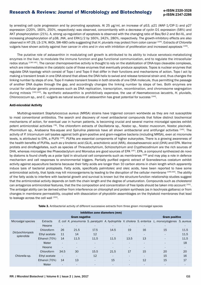
Table 3. Antibacterial activity of different successive extracts from three green microalgal species
Inhibition zone diameters (mm)Gram negative Gram positive
Microalgal species Extracts E. coli K. pneumonia S. paratyphi A. hydrophila V. cholera S. enterica L. monocytogenes S. aureus
Dictyochloropsis splendida
Hexane - - - - - - - -Chloroform 26 21.5 17.5 14.5 19 16 15 11.5
Ethyl acetate 11 14 12 - - - - 11.5Ethanol (70%) 14 11.5 11.5 11.5 13.5 13 - 11.5
Water - - - - - - - 18
Chlorella sp.
Hexane - - - - - - - -Chloroform 34.5 30 15.5 21.5 17 15 20 20
Ethyl acetate - - 12 - - - 15 16Ethanol (70%) 14 13 - 15 - 12 15 17
Water - - - - - - - -
69
Research & Reviews: Journal of Microbiology and Biotechnology e-ISSN:2320-3528p-ISSN:2347-2286
RR: J Microbiol Biotechnol | Volume 6 | Issue 2 | June, 2017
Scenedesmus obliquus
Hexane - - 21 24.5 23.5 18 13.5 13Chloroform 18.5 20.5 14 15.5 - 12.5 12 -
Ethyl acetate 16 - - - - - - -Ethanol (70%) 11 - - - - - - -
Water - - - 13 12 - 11 -
Amoxicillin Standard antibiotic - - - - - - 30 13.5
DMSO Control - - - - - - - -NB: Results are the means of diameter values ± standard deviation. (-): No activity DMSO: Dimethyl sulfoxide
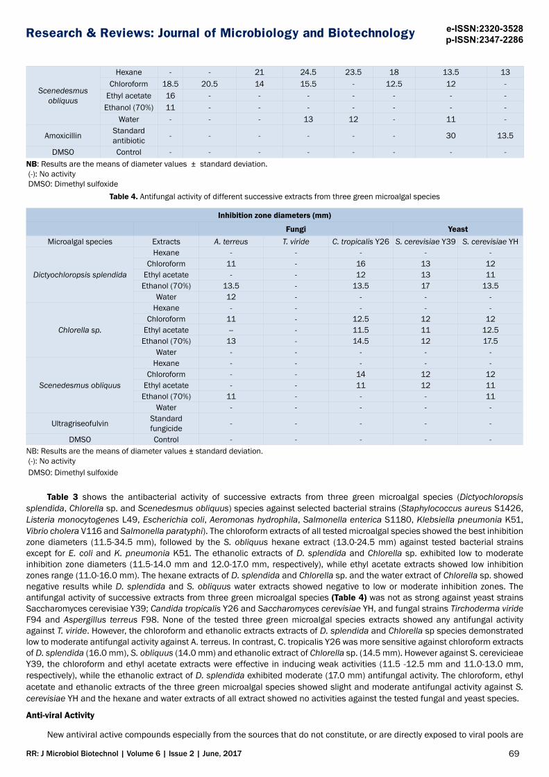
Table 4. Antifungal activity of different successive extracts from three green microalgal species
Inhibition zone diameters (mm)
Fungi YeastMicroalgal species Extracts A. terreus T. viride C. tropicalis Y26 S. cerevisiae Y39 S. cerevisiae YH
Dictyochloropsis splendida
Hexane - - - - -Chloroform 11 - 16 13 12
Ethyl acetate - - 12 13 11Ethanol (70%) 13.5 - 13.5 17 13.5
Water 12 - - - -
Chlorella sp.
Hexane - - - - -Chloroform 11 - 12.5 12 12
Ethyl acetate -- - 11.5 11 12.5Ethanol (70%) 13 - 14.5 12 17.5
Water - - - - -
Scenedesmus obliquus
Hexane - - - - -Chloroform - - 14 12 12
Ethyl acetate - - 11 12 11Ethanol (70%) 11 - - - 11
Water - - - - -
Ultragriseofulvin Standard fungicide - - - - -
DMSO Control - - - - -NB: Results are the means of diameter values ± standard deviation. (-): No activity DMSO: Dimethyl sulfoxide
Table 3 shows the antibacterial activity of successive extracts from three green microalgal species (Dictyochloropsis splendida, Chlorella sp. and Scenedesmus obliquus) species against selected bacterial strains (Staphylococcus aureus S1426, Listeria monocytogenes L49, Escherichia coli, Aeromonas hydrophila, Salmonella enterica S1180, Klebsiella pneumonia K51, Vibrio cholera V116 and Salmonella paratyphi). The chloroform extracts of all tested microalgal species showed the best inhibition zone diameters (11.5-34.5 mm), followed by the S. obliquus hexane extract (13.0-24.5 mm) against tested bacterial strains except for E. coli and K. pneumonia K51. The ethanolic extracts of D. splendida and Chlorella sp. exhibited low to moderate inhibition zone diameters (11.5-14.0 mm and 12.0-17.0 mm, respectively), while ethyl acetate extracts showed low inhibition zones range (11.0-16.0 mm). The hexane extracts of D. splendida and Chlorella sp. and the water extract of Chlorella sp. showed negative results while D. splendida and S. obliquus water extracts showed negative to low or moderate inhibition zones. The antifungal activity of successive extracts from three green microalgal species (Table 4) was not as strong against yeast strains Saccharomyces cerevisiae Y39; Candida tropicalis Y26 and Saccharomyces cerevisiae YH, and fungal strains Tirchoderma viride F94 and Aspergillus terreus F98. None of the tested three green microalgal species extracts showed any antifungal activity against T. viride. However, the chloroform and ethanolic extracts extracts of D. splendida and Chlorella sp species demonstrated low to moderate antifungal activity against A. terreus. In contrast, C. tropicalis Y26 was more sensitive against chloroform extracts of D. splendida (16.0 mm), S. obliquus (14.0 mm) and ethanolic extract of Chlorella sp. (14.5 mm). However against S. cerevicieae Y39, the chloroform and ethyl acetate extracts were effective in inducing weak activities (11.5 -12.5 mm and 11.0-13.0 mm, respectively), while the ethanolic extract of D. splendida exhibited moderate (17.0 mm) antifungal activity. The chloroform, ethyl acetate and ethanolic extracts of the three green microalgal species showed slight and moderate antifungal activity against S. cerevisiae YH and the hexane and water extracts of all extract showed no activities against the tested fungal and yeast species.
Anti-viral Activity
New antiviral active compounds especially from the sources that do not constitute, or are directly exposed to viral pools are

70
Research & Reviews: Journal of Microbiology and Biotechnology e-ISSN:2320-3528p-ISSN:2347-2286
RR: J Microbiol Biotechnol | Volume 6 | Issue 2 | June, 2017
of great necessity to address the drug-resistant mutations. Microalgae have consequently received more attention as a potential source of antiviral compounds. Currently, the antiviral drugs target only viral proteins that the discovery of small molecules that can specifically disrupt particular protein-protein interface is of keen interest in virology [78]. Sulphated polysaccharides have been shown to exhibit anti-viral activity against two enveloped rhabdoviruses: the Viral Hemorrhagic Septicemia Virus (VHSV) of salmonid fish and the African Swine Fever Virus (ASFV) [153]. Viral growth is generally divided into Stage I- adsorption and invasion of cells; Stage II- eclipse phase; and Stage III- maturity and release of virus particles. The Anti-Herpes Simplex Virus (HSV) factor from Dunaliella sp. extracts have been found to inactivate the initial viral function right after Stage I although the anti-HSV activity of the routinely applied antiviral compound acyclovir® is normally expressed at Stage II [154,155]. Sulphated exopolysaccharides from marine microalgae are claimed to interfere with Stage I of some enveloped viruses and offer unique advantages due to their antiviral spectrum such as against HSV and HIV-1 viruses [156]. Their inhibitory effect is due to the interaction with the positive charges on the virus or on the cell surface, thereby preventing virus penetration into the host cells [157,158]. In the case of HIV, they may also selectively inhibit reverse transcriptase which prevents the production of new viral particles after infection [159]. Several species of red microalgae containing highly sulphated polysaccharides with antiviral features consist mainly of xylose, glucose, and galactose and they are stable to extreme pH and temperature [160-162].
Anti-protozoal and Anti-plasmodial Activity
Trypanosomiasis is one of the most important parasitic diseases worldwide. The use of classical trypanocidal drugs has issues with undesirable side effects and low efficacy, making it pertinent to develop new drugs from natural products. Antiprotozoal algal extracts may be effective as preventive measures to control various protozoan diseases [163]. However, there is very limited research on marine algae focusing on antiprotozoal activity. One such study is the use of aqueous and organic extracts of Rhodophyta, Phaeophyta and Chlorophyta to evaluate the antiprotozoal activity in vitro against Trypanosoma cruzi trypomastigotes [164]. Another study use the ethanolic extracts of freshwater macrophytes Potamogeton perfoliatus, Ranunculus tricophyllus and Cladophora glomerata as well as marine macroalgae Dictyota dichotoma, Halopteris scoparia, Posidonia oceanica, Scinaia furcellata, Sargassum natans and Ulva lactuca for in vitro antiprotozoal activity against Trypanosoma brucei rhodesiense, Trypanosoma cruzi, Leishmania donovani and Plasmodium falciparum [165]. Several have shown promising anti-protozoan activities such as the crude seaweed extracts from green marine Cladophora rupestris, Codium fragile ssp. tomentosoides, Ulva intestinalis and Ulva lactuca against T. brucei rhodesiense, T. cruzi and L. donovani [166]; and the organic extracts of D. caribea, Lobophora variegata, Turbinaria turbinate Linnaeus, and Laurencia microcladia Kützing against T. cruzi trypomastigotes and Laurencia microcladia against Artemia salina and T. turbinate with high cytotoxicity [164].
Ethanol and ethyl acetate extracts of algae belonging to Chlorophyta, Heterokontophyta and Rhodophyta of algal exhibit antiplasmodial activities against P. falciparum (Erythrocytic stages), T. cruzi (Trypomastigotes) and L. donovani (Axenic amastigotes) [163]. The 7-dichloromethyl substituent in the organic extracts of endemic marine red alga Plocamium cornutum (Turner) Harvey show significantly higher antiplasmodial activity towards P. falciparum [167]; while out of four metabolites- sargaquinoic acid, sargahydroquinoic acid, sargaquinal and fucoxanthin isolated from marine algal Sargassum heterophyllum, fucoxanthin and sargaquinal show good antiplasmodial activity towards a chloroquine-sensitive strain of P. falciparum [168]. The red alga from genus Chondria, which produces cyclic polysulfides, terpenoids, amino acids and amines, have shown that the Domoic acid derivatives from C. armata not only exhbit larvicidal activity, but also the blood pressure lowering properties. Ethylacetate extract of Sargassum swartzii and Chondria dasyphylla further show larvicidal activities against larvae of malaria vector Anopheles stephensi with the mortality rate of 96 and 95%, respectively [169].
FACTORS AFFECTING ALGAL PRODUCTIVITY
The focus on renewable fuel sources has shifted the interest of large scale microalgal cultivation from the production of live feed (rotifers) in aquaculture and food additives towards lipid and carbohydrate accumulation and composition, waste remediation and bioenergy co-generation [4-5,40,170]. The establishment and set-up for microalgal biomass as a supplement to the primary feedstocks, should take the advantage of existing technologies and facilities (Figure 2). Both lipids and carbohydrates have reportedly been accumulated up to 60-65% of dry weight but with correspondingly lower biomass under stress conditions of temperature, salinity, light intensity and nutrients that enhancing productivity without compromising the cell growth rate is crucial [4-5,170,171]. Factors that have been the target of optimization are nitrogen depletion, temperature variation, Reactor configuration, osmatic stress and pH shift, CO2 supplement and irradiance [4,5].

71
Research & Reviews: Journal of Microbiology and Biotechnology e-ISSN:2320-3528p-ISSN:2347-2286
RR: J Microbiol Biotechnol | Volume 6 | Issue 2 | June, 2017
Figure 2. A conceptual process of ethanol production with the cultivation of starch-based microalgae (SSF - Simultaneous saccharification and fermentation), waste remediation and biogas co-generation (Adapted from [29]).
Reactor Configuration
Commercial plants may use one of the following technologies: -1) extensive ponds (lagoons); 2) raceway and circular ponds; 3) tubular photobioreactors; 4) fermentors (where algae are grown on organic substrates in the dark). The most common system is the large-scale outdoor cultivation in the form of artificial open pond or shallow raceway pond in which the suspension is mixed with a paddle wheel, as these are cheap to build and easy to operate and scale up [172]. The constraints include low productivity and biomass yield, high harvesting cost, water losses through evaporation, limited number of species which can be grown in ponds, vulnerability to contamination by other algae, grazers or bacteria, salinity and lower efficiency of CO2 use. The diurnal or climatic variations further make it difficult to control temperature fluctuations in open ponds [15]. The use of high ratio of inoculum to pond capacity and resilient microalgae strain that can grow in extreme culture conditions can minimize culture time (usually not more than 3-4 days) so as to minimize evaporation losses and contamination [173]. Other strategies include bicarbonate addition and raising pH to minimize Chlorella invasion of Spirulina culture; and ammonia as the N-source to suppress amoeba grazers [174].
Closed systems provide excellent reproducibility due to operational control, superior light and CO2 utilization, minimal water losses, and lowered risk of contamination [175]. The two major types of enclosed photobioreactors (PBRs) are tubular and plate types. Closed to the atmosphere, PBRs reduce evaporation and protect the cultivated algae making it less prone, but not immune to contamination. The temperature, pH and salinity can be better controlled, while the higher surface-to-volume (S/V) ratio facilitate narrow light path and large illuminating area for higher volumetric productivities and cell concentrations [4-5,40,176,177]. PBRs provide controlled conditions that yield reproducible product at high rates, but they are expensive. Open ponds are far less costly, but are so easily contaminated that after more than 50 years of repeated attempts, no more than three species proved amenable to large-scale cultivation [173]. Many different PBR designs have been proposed for biofuel production and the main issues to achieve high photosynthetic efficiencies and productivity are suitable construction materials, efficient mixing, heating/cooling, CO2 supply and oxygen removal [177,178], the high cost and the reduced scalability [179], light dilution via large external surfaces or internal light conducting structures [180], and the use of genetically modified strains [25]. The financial feasibility of PBRs is substantially lower than open ponds. In the base case, the average total costs of lipid production are only $12.73/gal for open tanks as compared to $31.61/gal for PBRs [181]. To improve the economics of algal cultivation, the optimal route is the combined use of PBRs and raceway ponds for biomass production where high quality culture in PBR is grown as inoculum for the raceway pond at a much larger capacity but with a substantially lowered risk of contamination. The coupled cultivation system takes advantage of the benefits of both PBRs and open ponds, while avoiding their disadvantages. The commercial scale production of Haematococcus pluvialis demonstrates that PBRs are essential to the sustainable production of photosynthetic microbes that cannot be cultivated reliably in open ponds [173].
Basic Culture Conditions and Nutritional Requirements
Lipid, carbohydrate and other bioactive contents of microalgae are influenced by irradiance and temperature variation depending on the microalgal strains [4,5,182]. These alter the physical properties of membranes allowing unimpaired functioning in photosynthesis, respiration and membrane transport which in turn affect the biochemical composition and the quantity of cellular lipid and fatty acid classes [183]. A high irradiance effect may depend on the culturing mode, either indoor or outdoor, and

72
Research & Reviews: Journal of Microbiology and Biotechnology e-ISSN:2320-3528p-ISSN:2347-2286
RR: J Microbiol Biotechnol | Volume 6 | Issue 2 | June, 2017
it is hard to reproduce the in vitro parameters especially in the outdoor operation, which involves solar cycle and temperature oscillation. The efficiency of light energy supply becomes one of the major limiting factors for outdoor or large-scale microalgae cultivation. Apart from solar radiation, the fluorescent tubes are normally used especially those emitting either blue or red light spectrum as these are the most active light spectrum for photosynthesis. The quantity of photon energy absorbed by each cell is a combination of factors such as cell density, length of optical path, thickness of layers, photon flux density and the rate of agitation [183]. The photo-period may vary between light: dark of 18:6, 12:12 or 16:8. The major requirements are for uniform and sufficient irradiance to the cells where the key design factors would be the operation depth which affects light penetration and availability, and mixing to enhance the light distribution and uniformity. Temperature changes affect fatty acid unsaturation in membrane lipid [184]. Optimal temperatures for most freshwater or saline strains are 16-28ºC, although some survive extremes of -5ºC and above 90ºC. Temperature below optimal range often leads to an increase in unsaturation of lipids or fatty acids in the membrane which improves the stability and fluidity of cell membranes especially the thylakoid membrane to prevent the photosynthetic machinery from photoinhibiting at low temperature. However, temperature below 16°C may result in reduced cell growth, and at higher than optimal may result in photosynthetic deficiency [185].
Nitrogen sources such as nitrate, nitrite, ammonia and urea may influence the biochemical composition and sufficient supply of CO2 is required for the autotrophic growth of microalgae. Nitrogen is an essential component for the formation of proteins, amino acids, chloroplast, enzymes and coenzymes for algal growth while CO2 affects the efficiency of photosynthesis, with carbohydrates as the end product. With adequate supply of CO2 and light energy, even under nutrient deficiency such as nitrogen, the protein content in microalgae can be consumed as a nitrogen source, and the carbohydrate content may increase significantly [29]. Nutrient limitation is an important modulator of algal lipid biosynthesis. When cell growth slows down as a result of nutrient deficiency, and there is no requirement for the synthesis of new membrane compounds, the cells can transfer the fatty acids into their storage lipids before conditions improve. This has resulted in more than double the lipid and TAG content in algal species deficient in nutrients [186]. There is a competition between lipid and carbohydrate synthesis during stress environments. Under nitrogen starvation, Phaeodactylum tricornutum reorganizes its proteome in favour of nitrogen scavenging and reduced lipid degradation, whilst rearranging the central energy metabolism that deprioritizes photosynthetic pathways. This increases N availability inside the cell and limit its use to the pathways where it is needed most [187]. Infact, the limitation of N availability has been suggested as the most effective way of triggering carbohydrates accumulation in microalgae [188]. The metabolic pathways associated with synthesis and degradation of energy-rich compounds (e.g. lipids and carbohydrates) is also closely linked [189]. Microalgal starch biosynthesis can directly proceed away from lipid synthesis, but the degradation of starch may provide the metabolites to produce acetyl-CoA, which is the precursor of fatty acid synthesis [190]. Thus strategy such as genetic modification may be necessary to reduce starch degradation and block the synthetic pathway of lipids, if the aim is for ethanol production [25].
Growing algae that require extreme conditions (high salinity or high pH) could alleviate contamination problem. The genus Dunaliella growing in the wide range of salinities is a useful model to study the mechanisms of salt tolerance [191]. pH can be modulated to affect cell growth rate and biochemical composition. Selection of a suitable strain and favourable location for building the plant is therefore of paramount importance. Current commercial production has focussed on species such as Dunaliella and Arthrospira (Spirulina) that require extreme media for growth. Some areas such as deserts provide more uniform environment that reduces the risk of contamination and the necessity of frequent intervention (for draining, cleaning, or re-inoculation) [177].
BIOPRODUCTION STRATEGIESBiodiesel
Presence or absence of light and nutritional balance should influence lipid composition, fatty acids and membrane lipid synthesis, mainly chloroplast [4,5,192]. Comparing the biomass and lipid productivities of C. vulgaris under autotrophic, heterotrophic and mixotrophic growth conditions, autotrophic growth has been shown to result in a higher cellular lipid content (38%) but lower lipid productivity than the heterotrophic growth with acetate, glucose, or glycerol. Use of glucose or glycerol at 1% (w/v) achieves optimal cell growth of 2 gL−1 and lipid productivity of 54 mg.L−1 day−1 whilst higher concentrations are inhibitory [193]. Heterotrophic cells of Chlorella zofingiensis fed with 30 gL−1 of glucose has increased oleic acid (from 17.9 to 35.2% of total fatty acids) as compared to photoautotrophic cells, and oil from heterotrophic C. zofingiensis appears to be more suitable for biodiesel production [194]. Growth and lipid content of Pavlova lutheri under 24 h illumination attain maximum specific growth rate, μmax, of 0.12 day−1 and 35% lipid content as compared to 0.1 day−1 and 15% lipid content in the dark [195]. High light intensities however could lead to oxidative damages of PUFA such as the decreased total n-3 fatty acids (from 29 to 8% of total fatty acids) mainly of EPA in Nannochloropsis sp. [196]. P. cruentum achieves higher lipid accumulation (19.3%) using a 12:12 h light: dark cycle at 25°C as compared to 35°C [197].
Nutritional deficiency triggers defence mechanism in microalgae which promotes lipid. The highest lipid accumulation of 37.3, 23.6, 28.3 and 37.2% though with slightly reduced cell growth of 0.64, 0.49, 0.54 and 0.38 gL-1 have been achieved for N. oculata, T. suecica, I. galbana and P. lutheri, respectively, when cultured under deficiency conditions of 10-65 gL-1 KNO3, 3-7.5 gL-1 Na2HPO4 and 2.5 gL-1 FeCl3
[198]. Chlorella shows the highest total lipid content (0.661 gg−1) when cultured at 0.1 gL−1, the lowest concentration of urea, but with maximum lipid productivity of 0.124 g L−1day−1 [199]. One of the most promising oil-rich microalgal species, Neochloris oleoabundans, also obtains the highest lipid content (0.40 gg −1) at the lowest NaNO3 (3 mM),

73
Research & Reviews: Journal of Microbiology and Biotechnology e-ISSN:2320-3528p-ISSN:2347-2286
RR: J Microbiol Biotechnol | Volume 6 | Issue 2 | June, 2017
whilst a lower lipid content of 0.34 gg−1 is achieved at 5 mM NaNO3 but with higher lipid productivity of 0.133 gL−1day −1 due to higher cell growth [200]. Lipid accumulation in Scenedesmus obliquus is more affected by the concentrations of nitrate, phosphate and sodium thiosulphate than glucose in the growth media. The most significant lipids are recorded under N-deficiency (43%) and P-deficiency and thiosulphate supplementation (30%) against 2.7% of lipids under control conditions [201]. Lipid accumulation is enhanced upto 2.16 gL−1, about 40-fold higher than control conditions, when the cells are pre-grown in the optimised medium supplemented with 1.5% glucose. The presence of palmitate and oleate as its major fatty acids also makes S. obliquus biomass a suitable feedstock for biodiesel [201].
Both the nutritional deficiency and the osmotic conditions affect the cell biochemical composition. An increase in the initial salt concentration from 0.5 M NaCl to 1.0 M increases the intracellular lipid (from 60 to 67%) in D. tertiolecta [203]. Further increase up to 70% lipid is achieved when 0.5 or 1.0 M NaCl is added at mid or the end of log phase during cultivation at 1 M initial NaCl concentration [202]. The lipid content of D. salina cells reaches 38% level at 16% NaCl in combination with 2.5 mM unspecified nitrogen salts, which also enhances the relative proportion of PUFAs, in particular the C18:3n-3 and C16:4n-3 fatty acids [203]. Transferring D. salina cells from 0.5 to 3.5 M NaCl induces the expression of β-ketoacyl- coenzyme A (CoA) synthase (KCS) which catalyzes the first step in fatty acid elongation [191]. The optimum salinity (30-40 ppt) and pH (8-9) for optimal cell growth and lipid content (34-36%) has been suggested for Pavlova lutheri where the alkaline pH stress is suggested to increase the TAG accumulation but may reduce the relative level of membrane lipids [195].
Reactor configuration, nutritional manipulation and culture conditions are all effective factors to improve productivities. Microalgal cultivation at optimum photoperiod and light intensity in 5L PBR (Figure 3) and 300L tank (Figure 4) are shown and Table 5 shows the comparison of growth kinetics for N. oculata, T. suecica, I. galbana and P. lutheri cultivation at optimum pH, salinity, photoperiod, light intensity and macronutrients. The highest cell density and biomass in 5 L PBR and 300 L open tank are shown by N. oculata at 82.6 × 10⁶, 63.7 × 10⁶ cells mL-1 density with 0.96, 0.72 gL-1 biomass, followed by T. suecica at 59 × 106, 42.7 × 10⁶ cells mL-1 density with 0.73, 0.58 gL-1 biomass respectively. The cell growth of I. galbana and P. lutheri remained low at 19.6-21.2 × 10⁶, 15.1-15.9 × 10⁶ cells mL-1 density, respectively with 0.52-0.66 gL-1 biomass. The lipid content is higher in 5 L PBR at 40.1-42.2% and 41.2-41.8% as compared to 30.7-36.2% and 32.1-38.5% in 300 L open tank for N. oculata and P. lutheri, respectively. Comparison between 250 mL-30 L batch cultures show that N. oculata and P. lutheri in 250 mL, T. suecica in 30 L and I. galbana in 1 L, attain the maximum specific growth rate of 0.15-0.17 d-1 and lipid content of 27.2-37.1%. The yield based on KNO3 (gg-1) of N. oculata biomass (0.08) and lipid (0.03) are higher in 5 L PBR than in 300 L open tank [198], which is attributable to better hydrodynamic condition in the former.
Figure 3. Cultivation at optimum photoperiod and light intensity of a) N. oculata; b) T. suecica; c) I. galbana and d) P. lutheri in 5 L PBR

74
Research & Reviews: Journal of Microbiology and Biotechnology e-ISSN:2320-3528p-ISSN:2347-2286
RR: J Microbiol Biotechnol | Volume 6 | Issue 2 | June, 2017
Figure 4. Cultivation at optimum photoperiod and light intensity of a) a) N. oculata; b) T. suecica; c) I. galbana and d) P. lutheri in 300 L open tank
Table 6 shows that the major components in all the four microalgal species (N. oculata, T. suecica, I. galbana and P. lutheri) are tetradecanoic acid (C14:0), pentadecanoic acid (C15:0), palmitic acid (C16:0), palmitoleic acid (C16:1), heptadecanoic acid (C17:0), stearic acid (C18:0), oleic acid (C18:1), linoleic acid (C18:2), linolenic (C18:3), eicosanoic acid (C20:0), eicosadienoic acid (C20:2), eicosatrienoic acid (ETE) (C20:3), eicosatetraenoic acid (ETA) (C20:4), eicosapentaenoic acid (EPA) (C20:5) and docosahexaenoic acid (DHA) (C22:6). Despite the great variation in fatty acid composition, the synthesized fatty acids in algae are commonly in medium length, ranging from 16 to 18 carbons, specifically, C16:0, C16:1, C18:0, C18:1 and C18:2 in green algae and C16:0 and C16:1 in brown algae. The total saturated fatty acids (SFA) (44.3-63.8% and 30.4-55.03%); monounsaturated fatty acids (MUFA) (6.1-37.0% and 4.2-13.1%); and PUFA (8.3-22.3% and 1.02-15.2%) are obtained, respectively, in 5 L PBR and 300 L tank. For N. oculata in PBR, palmitic acid C16:0 (22.1%) and palmitoleic acid C16:1 (9.9%) are reduced but heptadecanoic acid C17:0 (13.7%) and oleic acid C18:1 (7.4%) are enhanced. Although the total SFA (57.0%) and MUFA (17.7%) are comparable to the results at optimum pH and salinity [129], PUFA (22.3%) is enhanced for N. oculata in PBR. The heptadecanoic acid C17:0 (13.7%), oleic acid C18:1 (7%), palmitic acid C16:0 (22%) and palmitoleic acid C16:1 (9.9%) for N. oculata are high in PBR. For P. lutheri in PBR, palmitic acid C16:0 (34.4%) remains high, while both EPA C20:5 (8.4%) and DHA C22:6 (6.9%) are slightly increased with the total SFA (47.9%) and MUFA (30.9%) remain comparable but with PUFA (18.9%) elevated under optimal illumination and light intensity [129,198].
Table 5. Comparison of kinetics between 5 L PBR and 300 L open tank cultures at optimized conditions.
Media Conditions
N. oculata a T. suecica b I. galbana c P. lutheri d
X’m
ax (g
L-
1 d-1)
μ max
(d-1)
t d (da
y)
X’m
ax (g
L-
1 d-1)
μ max
(d-1)
t d (da
y)
X’m
ax (g
L-
1 d-1)
μ max
(d-1)
t d (da
y)
X’m
ax (g
L-
1 d-1)
μ max
(d-1)
t d (da
y)
Lipi
d (%
)
Lipi
d (%
)
Lipi
d (%
)
Lipi
d (%
)
pH a
nd
Salin
ity
PBR 0.18 0.24 2.92 38.6 ± 1.54 0.17 0.22 3.15 30.3
± 4.38 0.14 0.23 2.99 32.2 ± 2.11 0.14 0.21 3.3 40.2 ±
3.31
Open Tank 0.16 0.23 2.98 35.5 ±
1.81 0.16 0.2 3.53 27.5 ± 2.98 0.13 0.21 3.36
28.6 ± 2.75
0.13 0.19 3.61 36.1 ± 3.65

75
Research & Reviews: Journal of Microbiology and Biotechnology e-ISSN:2320-3528p-ISSN:2347-2286
RR: J Microbiol Biotechnol | Volume 6 | Issue 2 | June, 2017
Phot
oper
iod
and
Ligh
t In
tens
ity
PBR 0.19 0.24 2.92 40.1 ± 2.77 0.17 0.22 3.16 30.8
± 2.87 0.15 0.23 2.98 32.8 ± 3.44
0.14 0.22 3.11 41.8 ± 0.78
Open Tank 0.17 0.2 3.4 30.7 ±
2.52 0.17 0.21 3.32 25.6 ± 3.90
0.12 0.2 3.45 24.8 ± 3.13 0.12 0.18 3.82 32.1 ±
2.57
Nitr
ate,
Ph
osph
ate
and
Iron
PBR 0.17 0.21 3.27 42.2 ± 3.78 0.16 0.21 3.25
31.6 ± 4.33
0.13 0.21 3.3 33.5 ± 2.27 0.14 0.19
3.65
41.2 ± 1.92
Open Tank 0.17 0.19 3.63 36.2 ±
2.47 0.15 0.2 3.4830.5 ± 1.84
0.13 0.18 3.75 32.4 ± 1.47 0.13 0.17 3.96 38.5 ±
0.76 aN. oculata : pH 8, Salinity (35 ppt), photoperiod (24 h), light intensity (188 μmol photons m−2s−1) KNO3 (10 gL−1), Na2HPO4 (6 gL−1) and FeCl3 (2.53 gL−1)bT. suecica : pH 7.9 Salinity (32 ppt), photoperiod ( 24 h), light intensity (196.5 μmol photons m−2 s−1), KNO3 (13.7 gL−1), Na2HPO4 (5.6 gL−1) and FeCl3 (2.50 gL−1)cI. galbana : pH 9, Salinity (39.2 ppt), photoperiod (20.5 h), light intensity (188.7 μmol photons m−2 s−1), KNO3 (75.4gL−1), Na2HPO4 (8.9gL−1) and FeCl3 (2.8 gL−1)dP. lutheri : pH 7.9, Salinity (35.5 ppt), photoperiod (24 h), light intensity (198 μmol photons m−2 s−1), KNO3 (62.5 gL−1), Na2HPO4 (3.92 gL−1) and FeCl3 (2.63 gL−1) Note: For 300 L open tank, the photoperiod was 12 h and light intensity was 165-250 μmol photons m−2s−1 (shaded from direct sunlight).
Table 6. Fatty acid profile at optimized photoperiod and light intensity [129].
Experimental Conditions
Total fatty acids composition (%)
C14:0 C15:0 C16:0 C16:1 C17:0 C18:0 C18:1 C18:2 C18:3 C20:0 C20:2 C20:3 C20:4 C20:5 C22:6
N.
ocul
ata PBR 4.47 7.15 22.1 9.89 13.7 5.5 7.38 4.6 3.2 4.07 ND ND 3.33 6.65 4.56
Open Tank 3.01 13.1 21.2 7.44 5.27 5.92 5.16 2.8 ND 3.24 ND ND ND ND 4.45
T.
suec
ica PBR 4.29 17.8 14.9 2.82 2.66 5.48 3.27 5.38 1.65 1.88 1.17 0.45 ND 6.8 3.13
Open Tank 4.08 16.9 12.6 4.97 3.8 5.64 2.71 2.44 ND 4.21 0.72 ND ND ND 0.07
I. ga
lban
a PBR 10.3 8.2 19.2 5.05 9.74 5.45 14.6 2.2 0.6 8.09 2.71 ND ND 4.13 5.56Open Tank 8.99 2.69 13.2 4.52 5.13 3.38 11.5 1.4 ND 2.42 2.12 ND ND 3.42 1.83
P.
luth
eri PBR 2.86 3.76 34.4 21.3 3.48 0.74 9.67 1.46 2.11 2.68 ND ND ND 8.44 6.95
Open Tank 1.58 2.26 26.4 18.8 2.37 ND 7.28 ND 1.22 2.46 ND ND ND 7.78 5.44
ND: Not detected
The biochemical compositions of microalgae can change with their growth rates and environmental conditions and with the phase of their life cycle. The cell densities, biomass, µmax and td of P. lutheri culture in shake flasks and 1-30 batch cultures are comparable to cultivation at 250 mL-300 L with biomass reported at 0.45 gL−1 (250 mL), μmax at 0.14 day−1 (in 30 L) and td at 4.95 days (in 30 L) [195]. However, both N. oculata and T. suecica attain higher biomass at 0.68-0.93 gL−1 in 5 L PBR. While the biomass is comparable for I. galbana and P. lutheri at 0.62-0.71 g L−1, the cell densities are 3-4 folds lower. The reason being that both brown I. galbana and P. lutheri show bigger cell size than green N. oculata and T. suecica. This lower in cell density can be compensated for by the accumulation of nutrients for cellular components, leading to comparable biomass and dry weight.
The lipid classes of P. lutheri cultivated in semicontinuous mode, with neutral lipids and glycolipids, as the major constituents, account for 57 and 24% of the total fatty acids residues (TFA), respectively, with emphasis on EPA (C20:5n-3,) and DHA (C22:6n-3) [204]. The relative proportion of nutrients can modify the fatty acid profile of the microalgae, increasing SFA and MUFA proportion and in smaller amount PUFA content. The percentage of phosphorus is found to be the limiting nutrient related to the synthesis of phospholipids. Nevertheless, fatty acid biosynthesis and proportion may vary according to the microalgae species [205]. The main fatty acids present in the lipids of Chlorella sp. are normally short-chain fatty acids (C14-C18) [206]. Tetraselmis sp. and Chlorella have been cultivated in industrial-scale bioreactors, which produce 2.33 and 2.44% (w/w) lipid (calculated as the sum of fatty acid methyl esters) in dry biomass, respectively. These lipids contain higher amount of neutral lipids and glycolipids plus sphingolipids, than phospholipids. Lipids of Tetraselmis sp. are characterized by the presence of EPA (that is located mainly in phospholipids), and octadecatetraenoic acid (that is equally distributed among lipid fractions), but these fatty acids are completely absent in Chlorella lipids. Lipids produced by 16 newly isolated strains from Greek aquatic environments (cultivated in flask) have reported the highest percentage of lipids in Prorocentrum triestinum (3.69% w/w) while the lowest in Prymnesium parvum (0.47% w/w). Several strains produce lipids rich in EPA and DHA where the latter is found in high percentage in the lipids of Amphidinium sp. S1 and Prorocentrum minimum, while EPA is high in the lipids of Asterionella sp. S2. These lipids, containing ω-3-long-chain PUFA, have important applications in the food and pharmaceutical industries and in aquaculture [207].

76
Research & Reviews: Journal of Microbiology and Biotechnology e-ISSN:2320-3528p-ISSN:2347-2286
RR: J Microbiol Biotechnol | Volume 6 | Issue 2 | June, 2017
During the nitrogen starvation period, the proportion of oleic acid (C18:1) increases, whereas that of the linoleic acid (C18:2) and linolenic acid (C18:3) decrease [208]. Generally, saturated fatty esters possess high cetane number and superior oxidative stability; whereas unsaturated, especially polyunsaturated fatty esters have improved low-temperature properties. Modification of fatty esters, such as the enhanced proportion of oleic acid (C18:1) ester, can provide a compromise between oxidative stability and low-temperature properties and therefore promote the quality of biodiesel [209]. Thus, microalgae with high oleic acid are suitable for biodiesel production. Over 65% of fatty acids are saturated and monounsaturated fatty acids (C16:0, C18:0 and C18:1) which are suitable for application in biodiesel production [210]. European biodiesel standards limit the contents of FAMEs with four and more double bonds to 1 mol% [15]. For long-term biomass production and higher lipid yield, N. oculata is suggested to be best grown in a semi-continuous system aerated with 2% CO2 and operated by 1-day media replacement [211]. The best growth of N. oleoabundans is obtained when cultivated at 30°C under nitrogen sufficiency and CO2 supplementation. However, maximum lipid content (56%) is shown after 6 days of nitrogen depletion without CO2 supplementation [212]. A highly CO2 tolerant, Chloroccoccum littorale, has a good potential for aquaculture and photoautotrophic fatty acid production in the presence of inorganic carbon and nitrate at a light intensity of 170 mmol photons m−2s−1. The fatty acid synthesis is increased at low CO2 concentration after nitrate depletion, with a controlled HCO3/CO2 ratio. The relative FA content of 34%, achieved at 22°C, 170 m mol photons m−2s−1 light intensity and 5% CO2 with O2-free gas, is comparable to plant seed oils [213]. For integrated and optimal bioprocesses, the microalgal residues after lipid extraction and cellulosic materials (such as agricultural wastes) can be co-digested in anaerobic digester for biogas production and also waste water treatment to balance the C/N ratio in the optimum range of 20:1-25:1 [214]. The palm oil mill effluent (POME) treatment is much enhanced by applying aerobic and co-digestion of oil palm empty fruit bunches (OPEFB) at 0.12 g/mL with Chlorella sp. at 2 mL/mL POME, achieving the highest specific biogas production rate (0.128-0.129 m3/kg COD/day) and biomethane rate (5256.8-5295.8 mL/L POME/day). Higher removal efficiency (56-98%) of Chemical Oxygen Demand (COD), Biochemical Oxygen Demand (BOD), Total Organic Carbon (TOC), and Total Nitrogen (TN) after 3 and 7 days' treatment are achieved with microalgae than without microalgae [215]. The influence of different concentrations of filtered and centrifuged POME in sea water (1, 5, 10 and 15%) have shown that both N. oculata and T. suecica exhibit enhanced cell growth and lipid accumulation at 10% POME with maximum specific growth rate (0.21 d-1 and 0.20 d-1), lipid content (39.1 ± 0.73% and 27.0 ± 0.61%), the total SFA (59.24%, 68.74%), MUFA (15.14%, 12.26%), and PUFA (9.07%, 8.88%), respectively, after 16 days of flask cultivation. Algal cultivation with POME media also enhances the removal of COD (93.6-95%), BOD (96-97%), TOC (71-75%), TN (78.8-90.8%) and oil and grease (92-94.9%) from POME [216].
Ethanol
Cellulose and starch are the two major carbohydrate components in microalgal biomass. The carbohydrate productivity of microalgae is usually higher than lipids, as the latter requires intensive stress, while carbohydrate production is readily achievable by photosynthesis through the Calvin cycle [25]. This is one of the advantages of producing bioethanol instead of biodiesel from microalgae feedstock where manipulating the light intensity is one of the easiest route to achieve high productivity. Increasing the mean light intensity in an airlift PBR from 215 to 330 mol m−2s−1 increases the carbohydrate content from 8.5% to 40% [217]. However, no obvious positive correlation has been concluded from a study varying the light intensity in the range of 30-400 mol m−2s−1 and the carbohydrate accumulation, suggesting that the accumulation of microalgal carbohydrates may not only depend on the light intensity, but also on other environmental parameters [218]. These include the CO2 level, pH and N-limitation. Increasing dissolved CO2 from 3 to 186 mol L-1 in the cultivation of C. pyrenoidosa and C. reinhardtii elevate the carbohydrate content from 9.3 to 21% and 3.19 to 7.4%, respectively [219]. Adequate pH for carbohydrate accumulation differs based on the type of microalgal species, and maximum carbohydrate accumulation in D. bardawil and C. ellipsoidea is reportedly at pH 7.5 and 9.0, respectively [220]. When grown at pH 9.4, there has been a dramatic increase of the extracellular carbohydrate production from 2.1 to 17.7% in the marine diatom Skeletonema costatum [221]. Chlorella sp. has been reported as having the highest carbohydrate content and C. vulgaris particularly can accumulate a large amount of carbohydrates up to 37-55% of dry biomass [29]. Cultivation of Tetraselmis suecica under N-starvation with CO2 feeding also dramatically enhances the carbohydrate content from 10 to 57% [188]. In a batch culture of C. reinhardtii UTEX 90, the biomass concentration and starch content are found to be 1.45 g/L and 53%, respectively, after 3-day cultivation [222], while the biomass and carbohydrate content of S. obliquus could reach 4.96 g/L and 51.8%, respectively, under 3-day nitrogen starvation [17]. The five strains of Chlorella grown in Watanabe and low-nitrogen medium in 2 L stirred tank bioreactors achieve the best growth with Chlorella vulgaris with a growth rate of 0.99 d−1 and the highest calorific value of 29 KJg-1 with C. emersonii. However, the cellular components assayed at the end of the growth period suggest that the calorific value appears to be linked to the lipid content rather than any other component [223].
The biomass preparation can be carried out by mechanical presses or enzymatic cell wall break down to make carbohydrates more available, as well as breaking down the large molecules of carbohydrates. The lack of lignin in the microalgal biomass should simplify the pre-treatment process [224,225]. When cells are disrupted, the yeast Saccharomyces cerevisiae is added to the biomass and fermentation begins where sugar is converted into ethanol before purification by distillation [226]. Both enzymatic and chemical saccharification can be applied to produce sugars [227]. As saccharification is usually the rate limiting step, efficient methods for the cleavage of 1,4-glycosidic linkages between the hydroglucose subunits in cellulose molecules and in starch must be identified [224]. The chemical saccharification, which include mineral acids, alkaline, enzymes or hot compressed water is fast, but requires higher temperature, pressure, and addition of acid and alkali which may produce the inhibitors such as furfural

77
Research & Reviews: Journal of Microbiology and Biotechnology e-ISSN:2320-3528p-ISSN:2347-2286
RR: J Microbiol Biotechnol | Volume 6 | Issue 2 | June, 2017
and 5-hydroxymethylfurfural, and repress fermentative biofuels production with costly downstream treatment of the waste [228]. To reduce the production of inhibitors and improve operational efficiency, suitable reaction conditions (including temperature, moisture content, residence time and reaction agent concentration) must be obtained [229].
Enzymatic hydrolysis has several advantages including lower equipment costs, mild operating conditions, and higher glucose yields without sugar-degradation or toxic by-products that may affect biofuels fermentation [230]. Enzymatic saccharification uses cellulases, amylases and glucoamylases to hydrolyze microalgae to obtain sugars [29]. As microalgal-based cellulose is mainly located in the inner cell wall and lacks lignin with low hemicelluloses, the lignin-degradation enzymes (e.g. laccase and lignin peroxidase) and xylanase and the acidic or alkaline pretreatment, or steam explosion, may not be necessary, thus reducing the costs. Different commercial enzymes have been explored (including α-amylase from B. licheniformis and glucoamylases from Aspergillus niger) for the bioethanol conversion of C. reinhardtii biomass with a carbohydrate content of about 59.7% dry weight base. When algal biomass is hydrolyzed at pH 4.5 and 55°C for 30 min, sugar conversion of 0.57 g sugar/g biomass is obtained and the bioethanol production by the separate hydrolysis and fermentation (SHF) process is efficient [35]. Green algae Chlorococum humicola at 10 g/L biomass concentration has been hydrolyzed by enzymes from Trichoderma reesei to obtain a yield of 64.2% at 40°C, pH 4.8 [231]. Use of low acid (3% sulfuric acid) but at 100°C for 30 min could attain a maximum glucose yield of 95% from hydrolysis of C. reinhardtii [232] while the use of alkaline treatment (0.75% NaOH, 120°C, 30 min) achieves the highest glucose yield of 0.35 g sugar/g from C. infusionum [224]. However, these results suggest that the expensive and energy intensive pre-treatment or enzymatic reaction may not be necessary during the saccharification of microalgal biomass although the sugar yield by the enzymatic saccharification may still be higher than the acid or alkali hydrolysis [35]. Bioethanol fermentation also generates large amount of CO2 by-product, but the fermentation can be coupled to the cultivation of carbohydrate-rich microalgae [27] and the CO2 recycled to grow microalgae and the residual biomass used in anaerobic digestion for methane production such that in essence all the organic matter is accounted [13,225].
Starch-based microalgae as feedstock for biobutanol production could be a simpler process than bioethanol production, since Clostridium spp. are saccharolytic that the steps for starch liquefaction by amylase and dextrins saccharification by glucoamylase required for bioethanol fermentation with Saccharomyces are not needed. The metabolic pathways of the biobutanol-producing species are unique (i.e., Clostridium spp.) with significant amount of acetone, ethanol and various organic acids byproducts [233]. If the fermentation can be controlled to produce biobutanol with CO2 and H2 as the byproducts, the theoretical maximum biobutanol yield is 1 mol/mol glucose (or 0.41 g/g glucose) and this is lower than for bioethanol (i.e., 2 mol/mol glucose or ca. 0.5 g/g glucose). Macroalgae (e.g. seaweed) also has big potential as a sugar source [234]. The major sugars from brown macroalgae are glucan, mannitol, and alginate where glucan and mannitol are relatively easily assimilated by available microbial ethanol producers, with a yield reportedly of 0.08-0.12 g ethanol per g dry biomass weight. However, the major hurdle to achieving the full potential of ethanol production from macroalgal biomass is the inability of industrial microbes to metabolize alginate, the main product of seaweed [234].
Anti-oxidants
Environmental stressors such as high light intensity, salt stress or high temperature are effective to boost some anti-oxidants production in microalgae while others may decrease. Excess light intensity results in photo-inhibition which affects cell generation time but the cells will further respond by reducing chlorophyll α and the pigments involved in the photosynthesis to boost photoprotective agents like secondary carotenoids (astaxanthin, beta-carotene, zeaxanthin), thus preventing the absorption of surplus light energy. The major carotenoids such as lutein and zeaxanthin could protect against the common eye disease of macular degeneration [235]. Under stressed conditions, the carotenoids found in unique structures like plastoglobuli of plastids or cytoplasmic lipid bodies, may be elevated due the disruption in carbon and nitrogen flow inside the cells [236,237]. Carotenogenesis can therefore be enhanced by ROS generated under stress conditions [238] as shown in a study where vitamin C and α-tocopherol (Vitamin E) contents are increased by 4-fold and 8-fold, respectively, with the addition of 8 mM H2O2 in the culture medium of Spirulina platensis [239]. Similar finding has been observed in Morinda elliptica plant cell cultures under intermediary and production medium strategies [240].
High temperature favors the accumulation of carotenoids (e.g., lutein, β-carotene) in Dunaliella sp. but higher temperature than optimal would eventually decrease the biomass productivity. Under stressed circumstances, lutein tends to accumulate at low dilution rates but not to the levels that can balance out the decrease in biomass productivity. Maximum lutein productivity can be attained at the dilution rate that is optimal for biomass production [241]. Factors known to affect lutein content in microalgae are irradiance, pH, temperature, nitrogen availability and source, salinity and ionic strength), and presence of oxidizing substances (i.e., redox potential). Lutein content of Muriellopsis sp. shows the highest value in the early stationary phase of growth, with maximum levels attained at 20-40 mM NaNO3, 2-100 mM NaCl, 460 µmol photon m-2s-1, pH 6.5 and 28°C, conditions which are also optimal for cell growth. Growth-limiting conditions such as pH of 6 or 9 and at 33°C, also stimulate carotenogenesis in Muriellopsis sp. [74]. High irradiance (1700 μmol photons m-2s-1) has resulted in higher biomass production with simultaneous increase in lutein content in S. almeriensis and the lutein content and biomass are increased, reaching 0.46% of dry weight and 0.53 g/L, respectively, when the temperature increases up to 30°C [75]. Increasing the temperature also induces lutein in Chlorella

78
Research & Reviews: Journal of Microbiology and Biotechnology e-ISSN:2320-3528p-ISSN:2347-2286
RR: J Microbiol Biotechnol | Volume 6 | Issue 2 | June, 2017
protothecoides, reaching its highest value of 0.43% at pH 8-9 [242]. However, decreasing the temperature from 30 to 10°C causes a 2-fold increase in β-carotene content [243], and also increases the 9-cis-β-carotene and α-carotene content [244].
Nitrogen limitation has reportedly increased α-tocopherol accumulation [123], and the type of nitrogen source affects the vitamin content of D. tertiolecta where urea is found to be the most suitable for vitamin C accumulation, and nitrate for α-tocopherol [122]. Under nitrogen, phosphorus and sulfur starvation, the D. salina cells have elevated content of β-carotene [245], but these seem to cause a significant decrease in phycobiliproteins content in cyanobacteria [58,117,246-247]. The accumulation of astaxanthin in H. pluvialis, on the other hand, is triggered when the cells are exposed to nutrient starvation, or under salt stress, high temperature and high light intensity [248-250]. The astaxanthin accumulation may be accompanied by an increase of lipid granules, suggesting that astaxanthin is related to the synthesis of fatty acids [251-252], and in another report with a significant accumulation of carbohydrates [67]. Besides nitrogen, iron is one of the most essential elements for the metabolism of microalgae, as it takes part in the assimilation of nitrate and nitrite, deoxidation of sulfate, fixation of nitrogen, and synthesis of chlorophyll [253]. Iron deficiency may limit microalgal growth even in the presence of rich nutrient environment, and the addition of different iron electrovalencies and the nature of its counter ion affect cell growth and accumulation of astaxanthin [254-255]. Other study suggests that the presence of several heavy metals stress in cultures of Anabaena sp. may reduce phycobiliprotein [246]. In heterotrophic cultures of Euglena gracilis, α-tocopherol content of the cells is almost 4-fold higher when ethanol is used as the organic carbon source, instead of glucose [126].
In the large and commercial scale production of β-carotene by D. salina, high salinity stress and nitrogen deficiency are implemented [256]. D. salina is the most halotolerant eukaryotic microorganism known, and it can tolerate very wide salinity concentrations from 0.5-35% NaCl [257]. The cell acclimatization to the salinity effects may enhance the β-carotene content [258]. Moderate increase from 4-9% achieves a 30-fold increase in β-carotene, while harsher salt concentration lowers β-carotene accumulation. The increase in NaCl up to 0.6 M elevates the total phycobiliprotein content from 25-45%, but further increase to 0.9 M NaCl affects negatively [57,247]. Extracellular polymeric substances (EPSs), produced by Dunaliella salina strain, increase concomitantly with salt concentration and achieve maximum (944 mg/l) at 5 M NaCl, and minimum (56 mg/l) at 0.5 M salinity [84]. The H. pluvialis cultivation for astaxanthin production involve three main modes - the continuous, single-step cultivation [259], the two stage cell growth and astaxanthin production [260] and the cultivation performed in two stages [261]. Commercial process for astaxanthin production by Haematococcus sp. is based on a two-stage strategy - the first stage producing green biomass under optimal growth conditions, referred to as the ‘‘green’’ stage; and the second stage exposing the culture to adverse environmental conditions to induce the astaxanthin accumulation, referred to as the ‘‘red’’ stage. In large-scale facilities, the two-stage system yields astaxanthin of 2.2 mg L-1, while maximum astaxanthin productivity of 11.5 mg L-1d-1 can be attained at the laboratory scale under continuous illumination [261]. A single-step, continuous process has been proposed under moderate nitrogen limitation [259], and leads to the productivities of biomass and astaxanthin of 8 and 0.7 mg L-1d-1, respectively [262]. Astaxanthin may be efficiently produced outdoor in continuous mode if accurate dosage of the nitrate input can be assured [263]. Semicontinuous cultivation of D. salina at 25°C produces 80 gm−3d−1 biomass from which 1.25 mgL−1 of β-carotene is recovered, and can be improved up to 2.45 mg m−3 d−1 in continuous biphasic bioreactors [264,265].
For polysaccharides production, where cyanobacteria are the most studied, the effects of cultivation conditions are species and strain dependent. Nitrogen starvation in cyanobacteria species such as Anacystis nidulans and several Cyanothece strains have exhibited higher amount of released polysaccharides, while no similar effects observed in Synechocystis, Phormidium and some Cyanothece strains. Other factors such as age of culture, temperature, and the type of nitrogen also affect the polysaccharides production in A. nidulans [79]. The blue and red light not only improves the efficiency of photosynthesis, but also increases the production of polysaccharide in Porphyridium cruentum [266].
DOWNSTREAM PROCESSINGHarvesting and Dewatering
The economics of mass production by microalgae could be enhanced by improvements in cultivation technique and low energy, simple and yet effective downstream processing [4]. Harvesting and dewatering of algal species in dilute suspension at concentration less than 1 gL-1 (ponds) and 3-15 gL-1 (PBRs) is challenging and energy intensive. Dewatering to about 20-30% water content is necessary to reduce volume and weight, to minimize transportation and downstream costs and to extend the shelf-life of the microalgal concentrate [267]. Depending on the type of algae, the requirements of the downstream processes, and the desired product quality, different physical, chemical, and biological methods have been reported and the major goal is to separate the media and algae in the quickest, most energy efficient, and cheapest possible way. The most common techniques, gravity sedimentation and coagulation, are low energy allowing algae to naturally settle to the bottom but this may need substantial area and that the downstream processes and product targets are tolerant to coagulant contamination. Flotation lifts algae to the surface and some algae species do float naturally, though it is usually induced by micro-air bubbles [268,269]. Dissolved air flotation with chitosan or hydrophobic adsorbents thickens the material to 10% dry weight content (100 gL-1) [267]. Sediment sludge and floating biomass are also low cost but more diluted that high-speed continuous centrifugation is attractive and used commercially to harvest high-value metabolites for hatcheries and nurseries in aquaculture. Centrifugation produces g-forces between 5,000

79
Research & Reviews: Journal of Microbiology and Biotechnology e-ISSN:2320-3528p-ISSN:2347-2286
RR: J Microbiol Biotechnol | Volume 6 | Issue 2 | June, 2017
and 10,000 to separate the algae out [269] but the strong gravitational and shear forces can damage cell structure [225], and furthermore requires large capital investment, operating cost and high throughput processing of large quantities of water and algae [270].
Bioflocculation using biopolymers such as cationic starch [271] or poly-(γ-glutamic acid) (γ-PGA) produced by Bacillus licheniformis CGMCC 2876 [272] for microalgae dewatering could greatly reduce costs as no chemicals are used and it requires little or no energy consumption, easy to operate, environmentally friendly and as effective as chemical flocculation applied industrially [273]. The presence of high molecular weight extracellular polysaccharides (EPS) and exudates of both algae and microbial cells such as polyelectrolytes could bridge and flocculate algae and microbial cells in suspension [274]. The γ-PGA has been used to concentrate freshwater Desmodesmus sp. F51 with efficiency increases from 43.8% to 98.2% as the initial culture pH changes from ~7.2 to 3. With optimum dosage of 2.5 mL/L, flash mixing rate of 150 rpm for 1 min, and slow mixing rate of 80 rpm for 2 min, efficiency of 99% has been reported suggesting high performance for optimal recovery and applicability in commercial-scale microalgae harvesting [272]. The flocculation efficiency of cationic starch (Greenfloc 120) tested on the fresh water Chlorella protothecoides suggests optimal dosage at 40 mgL-1 at pH 10 with maximum efficiency of 98%. High biomass recovery (about 95%) is attainable with doses lower than the optimal, and preferable to avoid overcosts [275].
Electrochemical harvesting (ECH) can be used in product recovery, waste destruction [276], and chemical synthesis and are environmentally acceptable, non-species specific, safe and cost effective for implementation at commercial scale [277-278] for removal of microalgae from surface water and wastewater [279,280]. The main limitation has been the depletion of the metallic electrodes [281,282], which could cause metallic contamination of the biomass and wastewater and increasing the cost of harvesting [277,281,282]. ECH of Chlorella sorokiniana and Scenedesmus obliquus has been investigated using non-sacrificial carbon electrodes to overcome the cost and metallic contamination. Addition of electrolyte (NaCl) increases the recovery efficiency (RE) of C. sorokiniana from 65.99 to 94.52%, with energy consumption of 1.6 kWh kg-1 and no deteriorating effect on the lipid extraction as well as fatty acid composition. The highest recovery efficiency of 83% is obtained for S. obliquus at 1.5 A, initial pH 9 and 6 gL-1 NaCl with power consumption of 3.84 kWh kg-1. RE with ECH is comparable to centrifugation, filtration and chemical flocculation, but with a much lower power consumption. With electrolyte, the lipid extraction is enhanced by 22% without any adverse effects. These make ECH a possible step in commercial microalgal biomass and biodiesel production [283,284].
Harvesting and reactivation technique based on magnetic nanoparticles (MNPs) is a novel approach developed for rapid separation of algal cells. Applied to cyanobacteria Microcystis aeruginosa separation, the harvested raw MNPs achieves high efficiency of 99.6% with the MNPs dosage of 0.58 g MNPs/g dry-biomass, but gradually decreases to 59.1% when directly reused for 5 times. With extra ultrasonic chloroform:methanol solvent treatment, the MNPs can be effectively reactivated to achieve 60% efficiency after 5 times reactivation whilst keeping the separation efficiency above 93% with 0.2 g MNPs/g dry-biomass dosage [285]. A magnetic separator, which consists of permanent magnet drum, separation chamber and scraper blade has been developed for efficient microalgae harvesting. Magnetic separator can separate magnetic particles based on the physical capture of particles by a magnetic field with high process capacity [286-290]. The Fe3O4 nanoparticles, functionally coated with polyethylenimine (PEI) containing a high concentration of ANH2 groups, have been introduced for harvesting Chlorella ellipsoidea cells. The functional magnetic NPs are 12 nm in diameter with 69.77 emu/g of saturation magnetization and used at dosage of 20 mg/L to achieve a harvesting efficiency of 97% within 2 min. The adsorption capacity of the Fe3O4-PEI nanocomposites reaches up to 93.46 g-DCW/g-nanocomposites through electrostatic attraction and nanoscale interactions between the nanocomposites and the cells [289].
Product Extraction and Fractionation
The relative ease of purification of target compounds will enhance the value of microalgae as a source of bioactive compounds [291]. For industrial applications, the extraction solvents should be cheap, easy to use, low toxicity, insoluble in water, efficient in dissolving targeted components, and ideally recyclable. Organic solvent extraction is widely used for lipid extraction from traditional oilseed plants, and different extraction systems have also been tested with algae cultures [292]. Solvent extraction of algal lipids can be performed starting from both wet and dry biomass and specific pre-treatments may be carried out. If carried out from dry biomass, a drying/dehydration pre-treatment is necessary which is typically high energy step, that the wet extraction is preferable to ensure process feasibility and viability [293,294]. Pre-treatment aims to break the cell wall and is mandatory when lipid extraction is carried out directly from the wet biomass. Lipid extraction from untreated wet biomass is characterized by low yields due to the immiscibility of water with the organic solvents and the cells tend to remain in the water phase due to their surface charges and thus cannot contact the organic solvent phase capable of extracting lipids [295].
The cell wall of algae is thick and a rigid layer composed of complex carbohydrates and glycoproteins with high mechanical strength and chemical resistance. Breaking the cell wall of microalgae may require high energy input to release the intracellular lipids into the extracting mixture, thus facilitating the access of solvent to lipids. Once released from the cells, lipids move to the solvent phase from which they can be collected after evaporation of the solvent. A cell disruption technique based on the use of H2O2 with FeSO4 has been investigated where under optimal contact time and reactant concentration, the use of H2O2 and FeSO4 has more than doubled the extracted lipids as compared to to the case where no disruption is performed. The quality

80
Research & Reviews: Journal of Microbiology and Biotechnology e-ISSN:2320-3528p-ISSN:2347-2286
RR: J Microbiol Biotechnol | Volume 6 | Issue 2 | June, 2017
of FAMEs obtained by trans-esterification also improves with the cell disruption pre-treatment applied, independently from the reactant used [296]. The use of four cell disruption pretreatment methods, ultrasonication (US), microwave (MW), autoclave (AC) and electroflotation by alternating current (EFAC), have reported the total lipid yield of 5.3-33.7% (MW), 7.1-24.8% (EFAC), 2.3-15.4% (AC), and 3-13.3% (US). However, when both efficiency and costs are considered, EFAC can give the best result and can be a good option for simultaneous microalgae harvesting and cell disruption [297]. The integration of harvesting and lipid extraction steps could also reduce the cost of downstream processes. Integrated method by aminoclay-conjugated TiO2 has been investigated where microalgal flocculation affected by the aminoclay and the direct cell disruption by TiO2 photocatalytic performance under UV-light irradiation. Two TiO2 photocatalysts, a commercial anatase/rutile bicrystalline (of 5 nm diameter) and anatase/brookite bicrystalline (of 3.5 nm diameter) by sol-gel reaction at room temperature, are distributed uniformly onto aminoclay matrix by ultrasound-irradiated TiO2 particles, resulting in aminoclay-conjugated TiO2 composites. Within 10 min, the injection of aminoclay-conjugated TiO2 into the prepared 1.5 g/L concentration of oleaginous Chlorella sp. KR-1 feedstocks produces an 85% harvesting efficiency. The harvested wet-microalgae biomass is UV-irradiated at 365 nm for 3 h, thereby causing disruption to 95% of the cells [298].
Disruption of Scenedesmus obliquus CNW-N cells possessing a high lutein content of over 0.25% is most efficient with a bead-beater. To reduce the overall extraction time by 24 h, the conventional saponification step is modified and diethyl ether is used for the best lutein extraction efficiency. Storage of the lutein extract at low temperature (4 or −20°C) with antioxidant addition (around 0.01% BHT) can maintain 90% lutein stability after 80 days. Addition of a suitable amount of antioxidant could promote the stability of lutein extracts under the exposure of light [299]. The enzymatic pre-treatment of Undaria pinnatifida utilizing an alginase lyase enzyme hydrolyzing specific polysaccharides containing β-D-mannuronate residues in the seaweed cell walls, in combination with using either ethanol or dimethylether (DME) has been reported for the extraction of fucoxanthin. The typical enzyme pre-treatment conditions are 5% (w/v) solids, pH 6.2, reaction temperature of 37°C, reaction time of 2 h and 0.05% enzyme (w/w) using continuous mixing. Centrifugation (6000 xg for 20 min), performed to separate water-soluble hydrolysis products from the reaction mixture, improves the lipids and fucoxanthin recovery and also reduces residual biomass to be processed by around 40-50% prior to extraction. The alginase pre-treatment increases the total lipid yield by >10% and fucoxanthin yield by >20% [300,301].
Bioseparations and process schemes to produce high value natural bioactives can utilize the supercritical fluids [302]. The integrated processes include prior operation (fermentation, extraction, enzyme pre-treatment, physical fractionation or size reduction), the supercritical fluid extraction (SFE) or fractionation, and processes in which operations are carried out in situ in supercritical fluids (supercritical chromatography, enzymatic conversion, precipitation and coating of solutes) [302]. The yield of high value lipid arachidonic acid (ARA) from wet fungal Mortierella alpina biomass extraction with DME (at 4 MPa and 40-60°C) are higher than using CO2 (at 30 MPa and equal temperatures), and close to the total quantity of lipid as measured by exhaustive chloroform/methanol extraction. The higher yield using DME has been proposed due to the extraction of both neutral and polar lipids, since the solubility of lipids in liquefied DME is substantially higher than in CO2. Separate extraction of non-polar and polar lipids is possible by sequential extraction of dry biomass using initially CO2, followed optionally with ethanol co-solvent and then DME. A complete fractionation of the target/valuable compounds can be achieved in a single processing platform just by modifying the extraction solvents and/or extraction conditions [302]. Supercritical CO2 extraction (SCCO2) has been reported to obtain an oil rich in α linolenic (ALA) essential fatty acid and with a low (6:3) solvent to dry algae ratio. The maximum extraction yield is obtained at 60°C and 30 MPa with 0.4 kg/h of CO2 and 5% of co-solvent (ethanol). The thermodynamic cross-over is shown at a pressure close to 30 MPa, while the extraction cross-over occurs at around 25 MPa. The SCCO2 method is more selective than the soxhlet extraction of lipids even though the fatty acids profile is similar. The results suggested that Scenedesmus obliquus oil is richer in ώ-3 fatty acids and ALA than Chlorella protothecoides and Nannochloropsis salina lipids, and the ω-3 yield favours lower temperature, lower pressure and shorter extraction time [303].
A supercritical-CO2 extraction plus an acid treatment has been developed to extract astaxanthin from a vegetative green Monoraphidiumsp. GK12 and to fractionate chlorophyll and astaxanthin [304]. Astaxanthin is completely extracted by SFE using ethanol as a co-solvent, employing low CO2 pressure (20 MPa) and temperature (30°C) as well as a short process time (15 min), making it energy-efficient. Chlorophyll, extracted together with astaxanthin, can be easily removed by acid addition (H2SO4 or HCl) and centrifugation [304]. An integrated green processing platform for extraction-fractionation (or purification) to achieve fractions containing bioactives of high purity is possible by increasing the concentrations of hydronium and hydroxide ions. As the temperature of water increases, Kw increases from 10−14 at 25°C to nearly 10−11 mol2dm-6 at approximately 250°C, thus enhancing any acid-based catalysis process. By simply heating the water solvent, it is possible to increase the concentration of hydronium and hydroxide ions available with no neutralization necessary at the end of the reaction [305]. Direct or in situ supercritical transesterification methods have the potential to disrupt the rigid algal cell wall and convert the extracted lipids into biodiesel in a single step, thus reducing significantly the energy consumption. Wet (80 wt.% moisture) and dry unwashed Nannochloropsis gaditana have been used directly to synthesize biodiesel in a single-step by direct transesterification with no added catalysts using supercritical methanol. The main process parameters are the initial methanol to dry algae ratio (6:1-12:1 vol./wt.) and the synergistic effects between reaction time (10-50 min) and temperature (245-290°C). Maximum biodiesel yields of 0.46 and 0.48 g/g lipids are achieved from the wet and dry unwashed algal biomass, respectively, at 255-265°C, 50 min reaction

81
Research & Reviews: Journal of Microbiology and Biotechnology e-ISSN:2320-3528p-ISSN:2347-2286
RR: J Microbiol Biotechnol | Volume 6 | Issue 2 | June, 2017
time, using a methanol to dry algae ratio of 10:1 [306]. The use of CO2-expanded methanol (cxMeOH) and liquid carbon dioxide (lCO2) has been proposed to extract lipids from Botryococcus braunii. When compressed CO2 dissolves in methanol, the solvent expands in volume, decreases in polarity and increases in its selectivity for biodiesel-desirable lipids. Solid phase extraction of the algal extract shows that the cxMeOH extracts 21 mg of biodiesel desirable lipids per mL of organic solvent as compared to 3 mg/mL using either neat methanol or chloroform/methanol mixture. The non-polar lCO2 shows a high affinity for non-polar lipids and possible to extract up to 10% neutral lipids relative to the mass of dry algae. Unlike using conventional solvents, these new methods require little to no volatile, flammable, or chlorinated organic solvents [307].
CONCLUSIONS AND FUTURE OUTLOOKMost commercial, large-scale outdoor microalgae cultivation is in artificial open pond for economics reason and easy to
operate but facing challenges such as overheating, fouling, toxic oxygen levels and contamination. There are issues associated with low productivity and biomass yield, water losses through evaporation, low carbon dioxide consumption efficiency and tem-perature fluctuations and high harvesting cost. The challenge lies in developing hybrid tubular PBR or open-pond cultivation with resilient, fast growing algae, whilst attaining high oil content. Biorefinery concept and advances in photobioreactor engineering should make the cost of algal production more economically feasible especially if combined with industrial effluent remediation as a souce of nutrients. Algal lipids will also be a more realistic alternative if the prices of the major plant oils and crude oil are high. PUFA and bioactive compounds show better prospects as the commercial products and the use of genetic redesign of algal metabolic processes should be explored. The biomass recovery and alternative extraction processes, and the separation, purifica-tion or conversion must be effective at large scale and should be made reliable and manageable with low capital and operating costs. Commercialization of microalgal cultivation as an integral part of a biorefinery for bioenergy generation with recovery of co-products such as bioactive compounds and proteins for animal feed, and in combination with CO2 removal and wastewater treatment, is the way forward.
REFERENCES1. Richmond A. Biological principles of mass cultivation, In: Richmond A (ed) Handbook of microalgal cultures, biotechnology
and applied phycology. Blackwell, Oxford, 2004;125-177.
2. Perez-Garcia, et al. Heterotrophic cultures of microalgae: Metabolism and potential products. Water Res. 2011;45:11-36.
3. Pienkos PT and Darzins A. The promise and challenges of micro-algal derived biofuels. Biofuel Bioprod Biorefin, 2009;3:431-440.
4. Abdullah MA, et al. Algal biotechnology for bioenergy, environmental remediation and high value biochemical, 2015. In: Thangadurai D, Sangeetha J. (eds.) Biotechnology and Bioinformatics: Advances and Applications for Bioenergy, Bioremediation, and Biopharmaceutical Research. CRC Press / Apple Academic Press, New Jersey, USA, 2015;301-344.
5. Abdullah MA, et al. Integrated Algal Engineering for Bioenergy generation, Effluent Remediation and Production of High-value Bioactive Compounds, Biotechnol Bioproc Eng. 2016;21:236-249.
6. Dismukes GC, et al. Aquatic phototrophs: efficient alternatives to land-based crops for biofuels. Curr Opin Biotechnol. 2008;19,235-240.
7. Brennan L and Owende P. Biofuels from microalgae-a review of technologies for production, processing, and extractions of biofuels and co -products. Renew Sust Energ Rev. 2010;14:557-577.
8. Graham-Rowe, D. Beyond food versus fuel. Nature. 2011;474:S6-S8.
9. Bozarth A, et al. Diatoms in biotechnology: modern tools and applications. Appl Microbiol Biotechnol. 2009;82:195-201.
10. Hildebrand M. Prospects of manipulating diatom silica nanostructure. J Nanosci Nanotechnol. 2005;5:146-157.
11. Naylor RL, et al. The ripple effect: biofuels, food security, and the environment, Environ. 2007;49:30-43.
12. Leung DYC, et al. A review on biodiesel production using catalyzed transesterification, Appl Energ. 2010;87:1083-1095.
13. Singh J and Gu S. Commercialization potential of microalgae for biofuels production. Renew Sust Energ. 2010;14:2596-2610.
14. Dos Santos MD, et al. Plant cell and microalgae culture. In: Modern Biotechnology in Medicinal Chemistry and Industry. Kerala, India, Research Signpost. 2005
15. Chisti Y. Biodiesel from microalgae. Biotechnol Adv. 2007;25:294-306.
16. Kim J, et al. Methods of downstream processing for the production of biodiesel from microalgae. Biotechnol Adv. 2013;31,862-876.
17. Ho SH, et al. Effect of light intensity and nitrogen starvation on CO2 fixation and lipid/carbohydrate production of an indigenous microalga Scenedesmus obliquus CNW-N, Bioresour Technol. 2012;113:244-252.

82
Research & Reviews: Journal of Microbiology and Biotechnology e-ISSN:2320-3528p-ISSN:2347-2286
RR: J Microbiol Biotechnol | Volume 6 | Issue 2 | June, 2017
18. Armbrust, EV, et al. The genome of the diatom Thalassiosira pseudonana: ecology, evolution, and metabolism. Science. 2004;306:79-86.
19. Siaut M, et al. Molecular toolbox for studying diatom biology in Phaeodactylum tricornutum. Gene. 2007;406,23-35.
20. Becker EW. Microalgae as a source of protein. Biotechnol Adv. 2007;25:207-210.
21. Metting FB. Biodiversity and application of microalgae. J Ind Microbiol Biotechnol. 1996;17,477-489.
22. Griffiths MJ and Harrison STL. Lipid productivity as a key characteristic for choosing algal species for biodiesel production. J Appl Phycol. 2009;21:493-507.
23. Vasudevan PT and Briggs M. Biodiesel production - current state of the art and challenges. J Ind Microbiol Biotechnol. 2008;35:421-430.
24. Hu Q, et al. Microalgal triacylglycerols as feedstocks for biofuel production: perspectives and advances. Plant J. 2008;54:621-639.
25. Radakovits R, et al. Genetic engineering of algae for enhanced biofuel production. Eukaryot Cell. 2010;9,486-501.
26. Williams N. New biofuel questions. Curr Biol. 2010;20,219-220.
27. Ho SH, et al. Perspectives on microalgal CO2-emission mitigation systems - a review. Biotechnol Adv. 2011;29:189-198.
28. Rangel-Yagui CD, et al. Chlorophyll production from Spirulina platensis: cultivation with urea addition by fed-batch process. Bioresour Technol. 2004;92,133-141.
29. Chen CY, et al. Microalgae-based carbohydrates for biofuel production. Biochem Eng J. 2013;78:1-10.
30. Huntley ME and Redalje DG. CO2 mitigation and renewable oil form photosynthetic microbes: a new appraisal. Mitigation and Adaptation Strategies for Global Change. 2007;12:573-608.
31. Rosenberg JN, et al. A green light for engineered algae: redirecting metabolism to fuel a biotechnology revolution, Curr Opin Biotechnol. 2008;19:430-436.
32. Chen P, et al. Review of the biological and engineering aspects of algae to fuels approach. Int J Agric Biol Eng. 2009;2:1-30.
33. John RP, et al. Micro and macroalgal biomass: a renewable source for bioethanol Bioresour Technol. 2011;102:186-193.
34. Ueno Y, et al. Ethanol production by dark fermentation in the marine green alga, Chlorococcum littorale, J Ferment Bioeng. 1998;86:38-43.
35. Choi SP, et al. Enzymatic pretreatment of Chlamydomonas reinhardtii biomass for ethanol production, Bioresour Technol. 2010;101:5330-5336.
36. Zhang F, et al. Metabolic engineering of microbial path- ways for advanced biofuels production, Curr Opin Biotechnol. 2011;22:1-9.
37. Lee SY, et al. Fermentative butanol production by Clostridia. Biotechnol Bioeng. 2008;101:209-228.
38. Savage N. The ideal biofuel. Nature. 2011;474:S9-S11.
39. Knoshaug EP and Zhang M. Butanol tolerance in a selection of microorganisms. Appl Biochem Biotechnol. 2009;153:13-20.
40. Abdullah MA and Ahmad A. Integrated Algal Industrial Waste Treatment and Bioenergy Co-Generation. In: Sangeetha J, Thangadurai D, David M, Abdullah MA (eds) Environmental Biotechnology: Biodegradation, Bioremediation, and Bioconversion of Xenobiotics for Sustainable Development, CRC Press, Boco Raton, Florida, USA, 2016;153-223.
41. Mussgnug JH, et al. Engineering photosynthetic light capture: impacts on improved solar energy to biomass conversion. Plant Biotechnol J. 2007;5:802-814.
42. Zamalloa C, et al. The techno-economic potential of renewable energy through the anaerobic digestion of microalgae. Bioresour Technol. 2011;102:1149-1158.
43. Kawaguchi H, et al. H2 production from algal biomass by a mixed culture of Rhodobium marinum A-501 and Lactobacillus amylovorus, J Biosci Bioeng. 2001;91:277-282.
44. Anderson NS, et al. Carrageenan part V the masked repeating structure of ɣ and µ Carrageenan. Carbohydr Res. 1968;7:468-473.
45. Mackie W and Preston RD Cell wall and intercellular region polysaccharides in: Stewart WDP (Ed.) Algal physiology and biochemistry. Blackwell Scientific Publications. 1974;40-85.
46. Mayer AMS and Hamann MT. Marine pharmacology in 2001-2002: marine compounds with anthelmintic, antibacterial, anticoagulant, antidiabetic, antifungal, anti inflammatory, antimalarial, antiplatelet, antiprotozoal, antituberculosis, and antiviral activities; affecting the cardiovascular, immune and nervous systems and other miscellaneous mechanisms of action. Comp Biochem Physiol C Toxicol Pharmacol. 2005;140:265-286.

83
Research & Reviews: Journal of Microbiology and Biotechnology e-ISSN:2320-3528p-ISSN:2347-2286
RR: J Microbiol Biotechnol | Volume 6 | Issue 2 | June, 2017
47. Singh S, et al. Bioactive compounds from cyanobacteria and microalgae: an overview. Crit Rev Biotechnol. 2005;25: 73-95.
48. Blunt JW, et al. Marine natural products. Nat Prod Rep. 2005;22:15-61.
49. Olaizola M. Commercial development of microalgal biotechnology: from the test tube to the marketplace. Biomol Eng. 2003;20:459-466.
50. Barros MP, et al. Astaxanthin and peridinin inhibit oxidative damage in Fe2+ loaded liposomes: scavenging oxyradicals or changing membrane permeability? Biochem Biophys Res Commun. 2001;288:225-232.
51. Puglisi MP, et al. Capisterones A and B from the tropical green alga Penicillus capitatus: unexpected anti-fungal defenses targeting the marine pathogen Lindra thallasiae. Tetrahedr 2004;60:7035-7039.
52. Skjånes K, et al. Potential for green microalgae to produce hydrogen, pharmaceuticals and other high value products in a combined process, Crit Rev Biotechnol. 2013;33:172-215.
53. Borowitzka MA. High-value products from microalgae - their development and commercialisation. J Appl Phycol. 2013;25:443-756.
54. Venugopal V. Marine Products for Healthcare: Functional and Bioactive Nutraceutical Compounds from the Ocean. CRC Press, Boca Raton, FL. 2009
55. Scicchitano P, et al. Nutraceuticals and dyslipidaemia: Beyond the common therapeutics. J Funct Food. 2014; 6:11-32.
56. Alsenani F, et al. Nutraceuticals from microalgae, In: Bagchi D, Preuss HG, Swaroop A. (Eds.) Nutraceuticals and Functional Foods in Human Health and Disease Prevention, CRC Press, Boca Raton, USA. 2015;673-684.
57. Shalaby EA, et al. Salt stress enhancement of antioxidant and antiviral efficiency of Spirulina platensis. J Med Plants Res. 2010;4:2622-2632.
58. Shanab SMM, et al. Aqueous extracts of microalgae exhibit antioxidant and anticancer activities. As Pac J Trop Biomed. 2012;2:1-8.
59. Pagliaro M, et al. From Glycerol to value-added products. Angew. Chem. Int. Ed. 2007;46:4434-4440.
60. Ferruzzi MG and Blakeslee J. Digestion, absorption, and cancer preventative activity of dietary chlorophyll derivatives. Nutr Res. 2007;27:1-12.
61. Holdt SL and Kraan S. Bioactive Compounds in Seaweed: Functional Food Applications and Legislation. J Appl Phycol. 2011;23:543-597.
62. Pangestuti R and Kim S. Biological activities and health benefit effects of natural pigments derived from marine algae. J Funct Food. 2011;3;255-266.
63. Glazer AN. Phycobiliproteins a family of valuable, widely used fluorophores. J Appl Phycol. 1994;6:105-112.
64. Frank HA and Cogdell RJ. Carotenoids in Photosynthesis. Photochem Photobiol. 1996;63:257-264.
65. Kitahara T. Carotenoids in the Pacific salmon during the marine period. Comparat Biochem Physiol B. 1984;78:859-862.
66. Lemoine Y and Schoefs B. Secondary ketocarotenoid astaxanthin biosynthesis in algae: a multifunctional response to stress. Photosynth Res. 2010;106:155-77.
67. Boussiba S and Vonshak A. Astaxanthin accumulation in the green alga Haematococcus pluvialis. Plant Cell Physiol. 1991;32:1077-82.
68. Boussiba S. Carotenogenesis in the green alga Haematococcus pluvialis: Cellular physiology and stress response. Physiol Plant. 2000;108:111-117.
69. Lorenz RT and Cysewski GR. Commercial potential for Haematococcus microalgae as a natural source of astaxanthin. Trends Biotechnol. 2000;18:160-167.
70. Del Campo JA, et al. Outdoor cultivation of microalgae for carotenoid production: current state and perspectives. Appl Microbiol Biotechnol. 2007;74:1163-1174.
71. Ma RYN and Chen F. Enhanced production of free trans-astaxanthin by oxidative stress in the cultures of the green microalga Chlorococcum sp. Proc Biochem. 2001;36:1175-1179.
72. Qin S, et al. The accumulation and metabolism of astaxanthin in Scenedesmus obliquus (Chlorophyceae). Proc Biochem. 2008;43:795-802.
73. Wei D, et al. Enhanced production of lutein in heterotrophic Chlorella protothecoides by oxidative stress. Sci China Serie C Life Sci. 2008;51:1088-1093.
74. Del Campo JA, et al. Carotenoid content of chlorophycean microalgae. Factors determining lutein accumulation in Muriellopsis sp. (Chlorophyta). J Biotechnol. 2000;76:51-59.

84
Research & Reviews: Journal of Microbiology and Biotechnology e-ISSN:2320-3528p-ISSN:2347-2286
RR: J Microbiol Biotechnol | Volume 6 | Issue 2 | June, 2017
75. Sánchez JF, et al. Influence of culture conditions on the productivity and lutein content of the new strain Scenedesmus almeriensis. Proc Biochem. 2008;43:398-405.
76. Garbayo I, et al. Effect of abiotic stress on the production of lutein and β-carotene by Chlamydomonas acidophila. Proc Biochem. 2008;43:1158-1161.
77. Goodwin TW. Carotenoids and biliproteins, in: Stewart WDP (Ed.) Algal physiology and biochemistry. Blackwell Scientific Publications, Oxford, London, Edinburgh, Melbourne. 1974;176-205
78. Guedes A, et al. Microalgae as sources of high added-value compounds—a brief review of recent work. Biotechnol Prog. 2011;27:597-613.
79. De Philippis R and Vincenzini M. Exocellular polysaccharides from cyanobacteria and their possible applications. FEMS Microbiol Rev. 1998;22:151-75.
80. De Philippis, R., et al. Exopolysaccharide-producing cyanobacteria and their possible exploitation: a review. J Appl Phycol. 2001;13:293-299.
81. Tannin-Spitz T, et al. Antioxidant activity of the polysaccharide of the red microalgae Porphyridium sp. J Appl Phycol. 2005;17: 215-222.
82. Arad SM, et al. Effect of nitrate and sulfate starvation on polysaccharide formation in Rhodella reticulate. Bioresour Technol. 1992;42:141-148.
83. Lupi FM, et al. Influence of nitrogen source and photoperiod on exopolysaccharide synthesis by the microalga Botryococcus braunii UC 58, Enz Microb Technol. 1994;16:546-550.
84. Mishra A and Jha B. Isolation and characterization of extracellular polymeric substances from microalgae Dunaliella salina under salt stress. Bioresour Technol. 2009;100:3382-3386.
85. Bertocchi C, et al. Polysaccharides from cyanobacteria. Carbohydr Polym. 1990;12:127-53.
86. Chi Z, et al. A new exopolysaccharide produced by marine Cyanothece sp. 113. Bioresour Technol. 2007;98:1329-1332.
87. Rezanka T, et al. Natural microbial UV radiation filters—mycosporine-like amino acids. Folia Microbiol. 2004;49:339-352.
88. Nakamura H, et al. Separation of mycosporine-like amino acids in marine organisms using reverse-phase high-performance liquid chromatography. J Chromatogr. 1982;250;113-118.
89. Dunlap WC and Yamamoto Y. Small-molecule antioxidants in marine organisms: antioxidant activity of mycosporine-glycine. Comparat Biochem Physiol B. 1995;112:105-114.
90. Fuller RW, et al. A pentahalogenated monoterpene from the red alga Portieria hornemannii produces a novel cytotoxicity profile against a diverse panel of human tumor cell lines. J. Med. Chem. 1992;35:3007-3011.
91. Vairappan CS, et al. Halogenated metabolites with antibacterial activity from the Okinawan Laurencia species. Phytochem. 2001;58:517-523.
92. Shrivastav A, et al. Polyhydroxyalkanoate (PHA) synthesis by Spirulina subsalsa from Gujarat coast of India Int J Biol Macromol. 2010;46:255-260.
93. Somleva MN, et al. PHA bioplastics, biochemicals, and energy fromcrops. Plant Biotechnol J. 2013;11:233-252.
94. Sharma L and Mallick N. Accumulation of poly-β-hydroxybutyrate in Nostoc muscorum: regulation by pH, light-dark cycles, N and P status and carbon sources. Bioresour Technol. 2005;96:1304-1310.
95. Panda B, et al. Optimization of cultural and nutritional conditions for accumulation of poly-β-hydroxybutyrate in Synechocystis sp. PCC 6803. Bioresour Technol. 2006;97:1296-1301.
96. Abe K, et al. Accumulation and antioxidant activity of secondary carotenoids in the aerial microalga Coelastrella striolata var. multistriata. Food Chem. 2005;100:656-661.
97. Ursini F, et al. Redox homeostasis: The Golden Mean of healthy living. Redox Biology. 2016;8:205-215.
98. McNulty HP, et al. Differential effects of carotenoids on lipid peroxidation due to membrane interactions: X-ray diffraction analysis. Biochim Biophys Acta 2007;1768:167-174.
99. Guerin M and Huntley ME. Haematococcus astaxanthin: applications for human health and nutrition. Trends Biotechnol. 2003;21:210-215.
100. Pashkow F, et al. Astaxanthin: a novel potential treatment for oxidative stress and inflammation in cardiovascular disease. Am J Cardiol. 2008;101:S58-S68.
101. Campo J, et al. Outdoor cultivation of microalgae for carotenoid production: current state and perspectives. Appl Microbiol Biotechnol., 2007;74: 1163-1174.
85
Research & Reviews: Journal of Microbiology and Biotechnology e-ISSN:2320-3528p-ISSN:2347-2286
RR: J Microbiol Biotechnol | Volume 6 | Issue 2 | June, 2017
102. Yuan JP, et al. Potential health-promoting effects of astaxanthin: a high-value carotenoid mostly from microalgae. Mol Nutr Food Res. 2011;55:150-165.
103. Christaki E, et al. Functional properties of carotenoids originating from algae. J Sci Food Agric. 2013;93:5-11.
104. Palozza P, et al. Growth-inhibitory effects of the astaxanthin-rich alga Haematococcus pluvialis in human colon cancer cells. Cancer Lett. 2009;283:108-117.
105. Cha KH, et al. Antiproliferative effects of carotenoids extracts from Chlorella ellipsoidea and Chlorella vulgaris on human colon cancer cells. J Agric Food Chem. 2008;56:10521-10526.
106. Wu Z, et al. Supercritical fluid extraction and determination of lutein in heterotrophically cultivated Chlorella pyrenoidosa. J Food Proc Eng. 2007;30:174-185.
107. Nakashima Y, et al. Preventive effects of Chlorella on cognitive decline in age-dependent dementia model mice. Neurosci Lett. 2009;464:193-198.
108. Granado-Lorencio F, et al. In vitro bioaccessibility of lutein and zea-xanthin from the microalgae Scenedesmus almeriensis. Food Chem. 2009;114:747-752.
109. Sanchez F, et al., Influence of culture conditions on the productivity and the lutein content of the new strain Scenedesmus almeriensis. Proc Biochem. 2008;43:398-405
110. Iwamoto T, et al. Inhibition of low density lipoprotein oxidation by astaxanthin. J Atheroscler Thromb. 2000;7:216-222.
111. Spolaore P, et al. Commercial applications of microalgae. J Biosci Bioeng. 2006;101:87-96.
112. Ciccone MM, et al. The role of omega-3 polyunsaturated fatty acids supplementation in childhood: a review. Rec pat cardiovasc drug disc. 2013;8:42-55.
113. Garcia-Rios A, et al. Nutraceuticals and coronary heart disease. Curr Opin Cardiolog. 2013;28:475-482.
114. Giordano P, et al. Carotenoids and cardiovascular risk. Curr Pharm Des. 2012;18:5577-5589.
115. Izzo R, et al. Effects of nutraceuticals on prevalence of metabolic syndrome and on calculated Framingham Risk Score in individuals with dyslipidemia. J Hypertens. 2010;28:1482-1487.
116. Sekar S and Chandramohan M. Phycobiliproteins as a commodity: trends in applied research, patents and commercialization. J Appl Phycol. 2008;20:113-136.
117. Eriksen N. Production of phycocyanin - a pigment with applications in biology, biotechnology, foods and medicine. Appl Microbiol Biotechnol. 2008;80:1-14.
118. Aboul-Enein AM, et al. Cytotoxic and antioxidant properties of active principals isolated from water hyacinth against four cancer cells lines. BMC J Alternativ Complem Med. 2014;14:1.
119. Matsui MS, et al. Sulfated polysaccharides from red microalgae have antiinflammatory properties in vitro and in vivo. Appl Biochem Biotechnol. 2003;104:13-22.
120. Ji-Young P, et al. Chlorella dichloromethane extract ameliorates NO production and iNOS expression through the down-regulation of NFκβ activity mediated by suppressed oxidative stress in RAW 264.7 macro-phages. Clin Chim Acta. 2005;351:185-196.
121. Guzman S, et al. Anti-inflammatory and immunomodulatory activities of polysaccharide from Chlorella stigmatophora and Phaeodactylum tricornutum. Phytother Res. 2003;17:665-670.
122. Abalde J, et al. β-Carotene, vitamin C and vitamin E content of themarine microalga Dunaliella tertiolecta cultured with different nitrogen sources. Bioresour Technol. 199138:121-125.
123. Durmaz Y. Vitamin E, (α-tocopherol) production by the marine microalgae Nannochloropsis oculata (Eustigmatophyceae) in nitrogen limitation. Aquacult. 2007;272:717-722.
124. Mendiola JA, et al. Enrichment of vitamin E from Spirulina platensis microalga by SFE. J Supercrit Fluid. 2008;43:484-489.
125. Carballo-Cárdenas EC, et al. Vitamin E (α-tocopherol) production by the marine microalgae Dunaliella tertiolecta and Tetraselmis suecica in batch cultivation. Biomol Eng. 2003;20:139-147.
126. Ogbonna J, et al. Heterotrophic cultivation of Euglena gracilis Z for efficient production of α-tocopherol. J Appl Phycol. 1998;10:67-74.
127. Newman DJ and Cragg GM. Natural products from marine invertebrates and microbes as modulators of antitumor targets. Curr Drug Target. 2006;7:279-304.
128. Ali HEA, et al. Screening of microalgae for antioxidant activities, carotenoids and phenolic contents. Appl Mech Mater. 2014;625:156-159.

86
Research & Reviews: Journal of Microbiology and Biotechnology e-ISSN:2320-3528p-ISSN:2347-2286
RR: J Microbiol Biotechnol | Volume 6 | Issue 2 | June, 2017
129. Abdullah MA, et al. Algal Engineering for Bioenergy, Environmental Remediation and High-Value Biocompounds, Recent Advances in Renewable Energy Sources, Majestic Hotel, Kuala Lumpur. 2015;111-120.
130. Shalaby EA, et al. Review. Antioxidant compounds, assays of determination and mode of action. Afr J Pharm Pharmacol. 2013a;7:528-539.
131. Shalaby EA and Shanab SMM. Comparison of DPPH and ABTS assays for determining antioxidant potential of water and methanol extracts of Spirulina platensis. Ind J Geo-Marine Sci. 2013b;42:556-564.
132. Xia S, et al. Production, Characterization, and Antioxidant Activity of Fucoxanthin from the Marine Diatom Odontella aurita. Mar Drug. 2013;11:2667-2681.
133. Woo, MN, et al. Fucoxanthin supplementation improves plasma and hepatic lipid metabolism and blood glucose concentration in high-fat fed C57BL/6N mice. Chem Biol Interact. 2010;186:316-322.
134. Oono M, et al. Anti-cancer Agents Containing Carotenoids, Japan Kokai Tokkyo Koho, 5, Japanese Patent: JP 07101872 A2 19950418 Heisei. Appl: JP 93-248267 19931004. 1995.
135. Azamai ESM, et al. Chlorella vulgaris triggers apoptosis in hepatocarcinogenesis-induced rats. J Zhe-jiang Univ Sci B. 2009;10:14-21.
136. Ben-Dor A, et al. Carotenoids activate the antioxidant response element transcription system. Mol Cancer Ther. 2005;4:177-186.
137. Suzuki Y, et al. Suppressive effects of astaxanthin against rat endotoxin-induced uveitis by inhibiting the NF-κβ signaling pathway. Exp Eye Res. 2006;82:275-81.
138. Bertram JS and Vine AL. Cancer prevention by retinoids and carotenoids: independent action on a common target. Biochim Biophys Acta. 2005;1740:170-178.
139. Daubrawa F, et al. Astaxanthin diminishes gap junctional intercellular communication in primary human fibroblasts. J Nutr. 2005;135:2507-2511.
140. Liu X and Osawa T. Cis-astaxanthin and especially 9-cis-astaxanthin exhibits a higher antioxidant activity in vitro compared to the all-trans-isomer. Biochem Biophys Res Commun. 2007;357:187-193.
141. Santocono M, et al. Lutein, zeaxanthin and astaxanthin protect against DNA damage in SK-N-SH human neuroblastoma cells induced by reactive nitrogen species. J Photochem Photobiol B. 2007;88:1-10.
142. Champoux JJ. DNA topoisomerases: structure, function, and mechanism. Ann Rev Biochem. 2001;70:369-413.
143. Umemura K, et al. Inhibition of DNA topoisomerases I and II, and growth inhibition of human cancer cell lines by a marine microalgal polysaccharide. Biochem Pharmacol. 2003;66:481-487.
144. Tran NP, et al. Proteomics of proteins associated with astaxanthin accumulation in the green algae Haematococcus lacustris under the influence of sodium ortho-vanadate. Biotechnol Lett. 2009;31:1917-1922.
145. Abo-State MAM, et al. Screening of antimicrobial activity of selected Egyptian cyanobacterial species. J Ecol Health Environ. 2015;3:7-13.
146. Desbois AP, et al. A fatty acid from the diatom Phaeodactylum tricornutum is antibacterial against diverse bacteria including multi-resistant Staphylococcus aureus (MRSA). Mar Biotechnol. 2009;11:45-52.
147. Ward OP and Singh A. Omega-3/6 fatty acids: alternative sources of production. Proc Biochem. 2005;40:3627-3652.
148. Yi Z, et al. Exploring Valuable Lipids in Diatoms. Front Mar Sci. 2017;4:1-10.
149. Pradhan J, et al. Antibacterial activity of freshwater microalgae: A review. Afr J Pharm Pharmacol. 2014;8(32):809-818. 29 August,
150. Lampe MF, et al. Killing of Chlamydia trachomatis by novel antimicrobial lipids adapted from compounds in human breast milk. Antimicrob Agents Chemother. 1998;45:1239-1244.
151. Mendiola JA, et al. Use of supercritical CO2 to obtain extracts with antimicrobial activity from Chaetoceros muelleri microalga. A correlation with their lipidic content. Eur Food Res Technol. 2007;224: 505-510.
152. Yingying S, et al. Growth inhibition of the eight species of microalgae by growth inhibitor from the culture of Isochrysis galbana and its isolation and identification. J Appl Phycol. 2008;20:315-321.
153. Fábregas J, et al. In vitro inhibition of the replication of haemorrhagic septicaemia virus (VHSV) and African swine fever virus (ASFV) by extracts from marine microalgae. Antiviral Res. 1999;44:67-73.
154. Elion GB, et al. Selectivity of action of an antiherpetic agent, 9-(2-hydroxyethoxymethyl) guanine. Proc Natl Acad Sci USA.1977;74:5716-5720.
155. Schaeffer HJ, et al. 9-(2-hydroxyethoxymethyl) guanine activity against viruses of the herpes group. Nature. 1978;272:583-585.

87
Research & Reviews: Journal of Microbiology and Biotechnology e-ISSN:2320-3528p-ISSN:2347-2286
RR: J Microbiol Biotechnol | Volume 6 | Issue 2 | June, 2017
156. Damonte EB, et al. Prospects for the therapy and prevention of Dengue virus infections. Adv Virus Res. 2004;63:239-285.
157. Hatch M, et al. Further studies on the chemical composition and an initial in vivo evaluation of antiviral material in extracts of macroscopic algae. J Phycol. 1977;13:28-35.
158. Ehresmann D, et al. Antiviral properties of algal polysaccharides and related compounds. In: Hoppe H, Levring T, Tanaka Y, (Eds.) Marine Algae in Pharmaceutical Science. Berlin: Walter de Gruyter. 1979;293-302.
159. Moelling K, et al. Inhibition of human immuno-deficiency virus type 1 RNase H by sulfated polyanions. J Virol. 1989;63:5489-5491.
160. Lee JB, et al. Antiviral sulfated polysaccharide from Navicula directa, a diatom collected from deep-sea water in Toyama Bay. Biol Pharm Bull. 2006;29:2135-2139.
161. Huleihe M, et al. Antiviral effect of red microalgal polysaccharides on Herpes simplex and Varicella zoster viruses. J Appl Phycol. 2001;13:127-134.
162. Geresh S and Arad (Malis) S. The extracellular polysaccharides of the red microalgae: chemistry and rheology. Bioresour Technol. 1991;38:195-201.
163. Vonthron-Sénécheau C, et al. Antiprotozoal Activities of Organic Extracts from French Marine Seaweeds. Mar Drug. 2011;9:922-933.
164. Leon-Deniz LV, et al. Antitrypanosomal in vitro activity of tropical marine algae extracts. Pharma Biol. 2009;47:864-871.
165. Orhan I, et al. Turkish freshwater and marine macrophyte extracts show in vitro antiprotozoal activity and inhibit FabI, a key enzyme of Plasmodium falciparum fatty acid biosynthesis. Phytomed. 2006;13:388-393.
166. Spavieri J, et al. Antiprotozoal, antimycobacterial and cytotoxic potential of some British green algae. Phytother Res. 2010;24:1095-1098.
167. Afolayan AF, et al. Antiplasmodial halogenated monoterpenes from the marine red alga Plocamium cornutum. Phytochem. 2009;70:597-600.
168. Afolayana AF, et al. Fucoxanthin, Tetraprenylated Toluquinone and Toluhydroquinone Metabolites from Sargassum heterophyllum inhibit the in vitro Growth of the Malaria Parasite Plasmodium falciparum. Z Naturforsch C 2008;63:848-852.
169. Khanavi M, et al. Larvicidal activity of marine algae, Sargassum swartzii and Chondria dasyphylla, against malaria vector Anopheles stephensi. J Vector Borne Dis. 2011;48:241-244.
170. Rodolfi L, et al. Microalgae for oil: Strain selection, induction of lipid synthesis and outdoor mass cultivation in a low-cost photobioreactor. Biotechnol Bioeng. 2009;102:100-112.
171. Gouveia L, et al. Neochloris oleabundans UTEX#1185: a suitable renewable lipid source for biofuel production. J Ind Microbiol Biotechnol 2009;36:821-826.
172. Brennan L and Owende P. Biofuels from microalgae—A review of technologies for production, processing, and extractions of biofuels and co-products Renew Sust Energ Rev. 2010;14:557-577.
173. Huntley ME and Redalje DG. CO2 mitigation and renewable oil from photosynthetic microbes: A new appraisal. Mitigation and Adaptation strategies for global change. 2007;12:573-608.
174. Cohen Z, Monodus subterraneus, In : ZVI Cohen (Ed.) Chemicals from Microalgae. Taylor & Francis, London. 1999;25-40.
175. Peer MS, et al. Second Generation Biofuels: High-Efficiency Microalgae for Biodiesel Production. Bioenerg Res. 2008;1:20-43.
176. Ugwu CU, et al. Photobioreactors for mass cultivation of algae. Bioresour Technol. 2008;99:4021-4028.
177. Darzins A, et al. Current status and potential for algal biofuels production. A report to IEA Bioenergy Task. 39, 2010.
178. Richmond A. Principles for attaining maximal microalgal productivity in photobioreactors: an overview. Hydrobiol. 2004;512:33-37.
179. Tredici MR. Photobiology of microalgae mass cultures: Understanding the tools for the next green revolution. Biofuel. 2010;1:143-62.
180. Zijffers JF. Design Process of an Area-Efficient Photobioreactor. Mar Biotechnol. 2008;10:404-415.
181. Richardson JW, et al. Economic comparison of open pond raceways to photo-bioreactors for profitable production of algae for transportation fuels in the Southwest. Algal Res 2012;1:93-100.
182. De Oliveira MACL, et al. Growth and chemical composition of Spirulina maxima and Spirulina platensis biomass at different temperatures. Aquacult Int. 1999;7:261-275.
183. Masojodek J and Torzillo G. Mass cultivation of freshwater microalgae. Encyclop Ecol. 2003;3:2226-2235.

88
Research & Reviews: Journal of Microbiology and Biotechnology e-ISSN:2320-3528p-ISSN:2347-2286
RR: J Microbiol Biotechnol | Volume 6 | Issue 2 | June, 2017
184. Guschina IA and Harwood JL. Algal lipids and effect of the environment on their biochemistry. In: Martin Kainz M et al. (Eds.) Lipids in Aquatic Ecosystems. Springer New York. 2009;1-24.
185. Nishida I and Murata N. Chilling sensitivity in plants and cyanobacteria: the crucial contribution of membrane lipids. Ann Rev Plant Biol. 1996;47:541-568.
186. Guschina IA and Harwood JL. Lipids and lipid metabolism in eukaryotic algae. Prog Lipid Res. 2006;45;160-186.
187. Longworth J, et al. Proteome response of Phaeodactylum tricornutum, during lipid accumulation induced by nitrogen depletion. Algal Res. 2016;18:213-224.
188. D'Souza FML and Kelly GJ. Effects of a diet of a nitrogen-limited alga (Tetraselmis suecica) on growth, survival and biochemical composition of tiger prawn (Penaeus semisulcatus) larvae Aquacult. 2000;181:311-329.
189. Ho SW, et al. Characterization and optimization of carbohydrate production from an indigenous microalga Chlorella vulgaris FSP-E. Bioresour Technol. 2012;135:157-165.
190. Rismani-Yazdi H, et al. Transcriptome sequencing and annotation of the microalgae Dunaliella tertiolecta: Pathway description and gene discovery for production of next-generation biofuels. BMC Genomics. 2011;12:148.
191. Azachi M, et al. Salt Induction of Fatty Acid Elongase and Membrane Lipid Modifications in the Extreme Halotolerant Alga Dunaliella salina. Plant Physiol. 2002;129:1320-1329.
192. Khotimchenko SV and Yakovleva IM. Lipid composition of the red alga Tichocarpus crinitus exposed to different levels of photon irradiance. Phytochem. 2005;66:73-79.
193. Liang Y, et al. Biomass and lipid productivities of Chlorella vulgaris under autotrophic, heterotrophic and mixotrophic growth conditions. Biotechnol Lett. 2009;31:1043-1049.
194. Liu J, et al. Differential lipid and fatty acid profiles of photoautotrophic and heterotrophic Chlorella zofingiensis: assessment of algal oils for biodiesel production. Bioresour Technol. 2011;102:106-110.
195. Shah SMU, et al. Effects of photoperiod, salinity and pH on cell growth and lipid content of Pavlova lutheri, Annal Microbiol. 2014;64:157-164.
196. Fabregas J, et al. The cell composition of Nannochloropsis sp. changes under different irradiances in semicontinuous culture. World J Microbiol Biotechnol. 2004;20:31-35.
197. Oh SH, et al. Lipid production in Porphyridium cruentum grown under different culture conditions. J Biosci Bioeng. 2009;108:429-434.
198. Shah SMU. Cell culture optimization and reactor studies of green and brown microalgae for enhanced lipid production, PhD thesis, Universiti Teknologi PETRONAS, Malaysia. 2014.
199. Hsieh CH and Wu WT. Cultivation of microalgae for oil production with a cultivation strategy of urea limitation Bioresour Technol. 2009;100(17):3921-3926.
200. Li Y, et al. Effects of nitrogen sources on cell growth and lipid accumulation of green alga Neochloris oleoabundans. Appl Microbiol Biotechnol. 2008;81:629-636.
201. Mandal S and Mallick N. Microalga Scenedesmus obliquus as a potential source for biodiesel production. Appl Microbiol Biotechnol. 2009; 84(2):281-291.
202. Takagi M, et al. Effect of salt concentration on intracellular accumulation of lipids and triacylglyceride in marine microalgae Dunaliella cells J Biosci Bioeng. 2006;101(3):223-226.
203. Abd El-Baky HH, et al. Production of lipids rich in omega-3 fatty acids from the halotolerant alga Dunaliella salina. Biotechnol. 2004;3:102-108.
204. Meireles LA, et al. Lipid class composition of the microalga Pavlova lutheri: Eicosapentaenoic and Docosahexaenoic Acids, J Agric Food Chem. 2003;51:2237-2241.
205. Chen GQ, et al. Fatty acid and lipid class composition of the eicosapentaenoic acid-producing microalga, Nitzschia laevis, Food Chem. 2007;104:1580-1585.
206. Huang G, et al. Biodiesel production by microalgal biotechnology. Appl Energ. 2010;87:38-46.
207. Makri A, et al. Lipid synthesized by microalgae grown in laboratory and industrial-scale bioreactors. Eng Life Sci. 2011;11:52-58.
208. Meng X, et al. Biodiesel production from oleaginous microorganisms. Renew Energ. 2009;34:1-5.
209. Knothe G. Improving biodiesel fuel properties by modifying fatty ester composition. Energ Environ Sci. 2009;2:759-766.
210. Sheehan J, et al. A look back at the U.S. Department of Energy’s Aquatic Species Program - biodiesel from algae. Golden, CO: National Renewable Energy Laboratory. 1998.
89
Research & Reviews: Journal of Microbiology and Biotechnology e-ISSN:2320-3528p-ISSN:2347-2286
RR: J Microbiol Biotechnol | Volume 6 | Issue 2 | June, 2017
211. Chiu SY, et al. Lipid accumulation and CO2 utilization of Nannochloropsis oculata in response to CO2 aeration. Bioresour Technol. 2009;100:833-838.
212. Gouveia L and Oliveira AC. Microalgae as a raw material for biofuels production. J Ind Microbiol Biotechnol. 2009;36(2):269-274.
213. Ota M, et al. Fatty acid production from a highly CO2 tolerant alga, Chlorococcum littorale, in the presence of inorganic carbon and nitrate. Bioresour Technol. 2009;100:5237-5242.
214. Yen HW and Brune DE. Anaerobic co-digestion of algal sludge and waste paper to produce methane Bioresour Technol. 2007;98(1):130-134.
215. Ahmad A, et al. Enhanced palm oil mill effluent treatment and biomethane production by aerobic and anaerobic co-cultivation of Chlorella sp. Can J Chem Eng. 2014;92:1636-1642.
216. Shah SMU, et al. Effects of palm oil mill effluent media on cell growth and lipid content of Nannochloropsis oculata and Tetraselmis suecica. Int J Green Energ. 2016;13(2):200-207.
217. Fernandes BD, et al. Light regime characterization in an airlift: Photobioreactor for production of microalgae with high starch content. Appl Biochem Biotechnol. 2010;161:218-226.
218. Carvalho AP, et al. Simultaneous effect of irradiance and temperature on biochemical composition of the microalga Pavlova lutheri. J Appl Phycol. 2009;21:543-552.
219. Xia JR and Gao KS. Impacts of elevated CO2 concentration on biochemical composition, carbonic anhydrase, and nitrate reductase activity of freshwater green algae. J Integr Plant Biol. 2005;47:668-675.
220. Khalil ZI, et al. Effect of pH on growth and biochemical responses of Dunaliella bardawil and Chlorella ellipsoidea. World J Microbiol Biotechnol. 2010;26:1225-1231.
221. Taraldsvik M and Myklestad S. The effect of pH on growth rate, biochemical composition and extracellular carbohydrate production of the marine diatom Skeletonema costatum. Eur J Phycol. 2000;35(2):189-194.
222. Kim MS, et al. Hydrogen production from Chlamydomonas reinhardtii biomass using a two-step conversion process: anaerobic conversion and photosynthetic fermentation. Int J Hydr Energ. 2006;31:812-816.
223. Illman AM, et al. Increase in Chlorella strains calorific values when grown in low nitrogen medium. Enz Microb Technol. 2000;27:631-635.
224. Harun R, et al. Microalgal biomass as a fermentation feedstock for bioethanol production. J Chem Technol Biotechnol. 2010;85:199-203.
225. Harun R, et al. Bioprocess engineering of microalgae to produce a variety of consumer products. Renew Sust Energ Rev. 2010;14:1037-1047.
226. Amin S. Review on biofuel oil and gas production processes from microalgae. Energ Convers Manag. 2009;50:1834-1840.
227. Van de Vyver S, et al. Sulfonated silica/carbon nanocomposites as novel catalysts for hydrolysis of cellulose to glucose. Green Chem. 2010;12:1560-1563.
228. Mussatto SI, et al. Technological trends, global market, and challenges of bio-ethanol production. Biotechnol Adv. 2010;28:817-830.
229. Okuda K, et al. Hydrothermal fractional pretreatment of sea algae and its enhanced enzymatic hydrolysis. J Chem Technol Biotechnol. 2008;83:836-841.
230. Cara C, et al. Influence of solid loading on enzymatic hydrolysis of steam exploded or liquid hot water pretreated olive tree biomass. Proc Biochem. 2007;42:1003-1009.
231. Harun R and Danquah MK. Enzymatic hydrolysis of microalgal biomass for bioethanol production. Chem Eng J. 2011;168:1079-1084.
232. Nguyen MT, et al. Hydrothermal acid pretreatment of Chlamydomonas reinhardtii biomass for ethanol production. J Microbiol Biotechnol. 2009;19:161-166.
233. Gheshlaghi R, et al. Metabolic pathways of Clostridia for producing butanol. Biotechnol Adv. 2009;27(6):764-781.
234. Wargacki AJ, et al. An engineered microbial platform for direct biofuel production from brown macroalgae. Science. 2012;335(6066):308-313.
235. Mozaffarieh M, et al. The role of the carotenoids, lutein and zeaxanthin, in protecting against age-related macular degeneration: A review based on controversial evidence Nutrit J. 2003;2:20
236. Hu Q, et al. Enhancement of Eicosapentaenoic acid (EPA) and γ-linolenic acid (GLA) Production by Manipulating Algae

90
Research & Reviews: Journal of Microbiology and Biotechnology e-ISSN:2320-3528p-ISSN:2347-2286
RR: J Microbiol Biotechnol | Volume 6 | Issue 2 | June, 2017
Density of Outdoor Cultures of Monodus subterraneus (Estimagophyte) and Spirulina platensis (cyanobacterium). Eur J Phycol. 1997;32:81-86.
237. Lemoine Y and Schoefs B. Secondary ketocarotenoid astaxanthin biosynthesis in algae: a multifunctional response to stress. Photosynth Res. 2010;106(1):155-177.
238. Kobayashi M. Astaxanthin biosynthesis enhanced by reactive oxygen species in the green alga Haematococcus pluvialis. Biotechnol Bioproc Eng. 2003;8:322-330.
239. Abd El-Baky H, et al. Enhancement of antioxidant production in Spirulina platensis under oxidative stress. Acta Physiol Plant 2009;31:623-631.
240. Chong, TM, et al. Anthraquinone production, Hydrogen peroxide level and antioxidant vitamins in Morinda elliptica cell suspension cultures from intermediary and production medium strategies. Plant Cell Rep. 2004;22:951-958.
241. Sanchez JF, et al. Biomass and lutein productivity of Scenedesmus almeriensis: influence of irradiance, dilution rate and temperature. Appl Microbiol Biotechnol. 2008;79:719-729.
242. Shi X, et al. Kinetic modeling of lutein production by heterotrophic Chlorella at various pH and temperatures. Mol Nutr Food Res. 2006;50:763-768.
243. Ben-Amotz A. Effect of low temperature on the stereoisomer composition of β-carotene in the halotolerant alga Dunaliella bardawil (Chlorophyta). J Phycol (1996) 32:272-5.
244. Gómez P and González M. The effect of temperature and irradiance on the growth and carotenogenic capacity of seven strains of Dunaliella salina (Chlorophyta) cultivated under laboratory conditions. Biol Res. 2005;38:151-162.
245. Ben-Amotz A. Effect of irradiance and nutrient deficiency on the chemical composition of Dunaliella bardawil Ben-Amotz and Avron (Volvocales, Chlorophyta). J Plant Physiol. 1987;131:479-487.
246. Hemlata, Fatma T. Screening of cyanobacteria for phycobiliproteins and effect of different environmental stress on its yield. Bull Environ Contaminat Toxicol. 2009;83:509-515.
247. Hifney AF, et al. Abiotic stress induced production of β-carotene, allophycocyanin and total lipids in Spirulina sp. J Biol Earth Sci. 2013;3:54-64.
248. Fábregas J, et al. Interactions between irradiance and nutrient availability during astaxanthin accumulation and degradation in Haematococcus pluvialis. Appl Microbiol Biotechnol. 2003;61:545-551.
249. Dominguez-Bocanegra AR, et al. Influence of environmental and nutritional factors in the production of astaxanthin from Haematococcus pluvialis. Bioresour Technol. 2004;92:209-214.
250. He P, et al. Astaxanthin accumulation in the green alga Haematococcus pluvialis: effects of cultivation parameters. J Integr Plant Biol. 2007;49:447-451.
251. Zhekisheva M, et al. Accumulation of oleic acid in Haematococcus pluvialis (Chlorphyceae) under nitrogen starvation or high light is correlated with that of astaxanthin esters. J Phycol. 2002;38:325-331.
252. Cerón M, et al. Antioxidant activity of Haematococcus pluvialis cells grown in continuous culture as a function of their carotenoid and fatty acid content. Appl Microbiol Biotechnol. 2007;74:1112-1119.
253. Liu ZY, et al. Effect of iron on growth and lipid accumulation in Chlorella vulgaris. Bioresour Technol. 2008;99:4717-4722.
254. Choi YE, et al. Evaluation of factors promoting astaxanthin production by a unicellular green alga, Haematococcus pluvialis, with fractional factorial design. J Biotechnol. 2002;18:1170-1175.
255. Cai M, et al. Effects of iron electrovalence and species on growth and astaxanthin production of Haematococcus pluvialis. Chin J Oceanol Limnol 2009;27:370-375.
256. Ben-Amotz A. Dunaliella β-carotene. From science to commerce. In: Seckbach J, (Ed.) Enigmatic microorganisms and life in extreme environments. 1999;401.
257. Hosseini TA and Shariati M. Dunaliella biotechnology: methods and applications. J Appl Microbiol. 2009;107:14-35.
258. Coesel S, et al. Nutrient limitation is the main regulatory factor for carotenoid accumulation and for Psy and Pds steady state transcript levels in Dunaliella salina (Chlorophyta) exposed to high light and salt stress. Mar Biotechnol. 2008;10:602-611.
259. Del Río E, et al. Efficiency assessment of the one-step production of astaxanthin by the microalga Haematococcus pluvialis. Biotechnol Bioeng. 2008;100:397-402.
260. Zhang BY, et al. Production of astaxanthin from Haematococcus in open pond by two-stage growth one-step process. Aquacult. 2009;295:275-281.
91
Research & Reviews: Journal of Microbiology and Biotechnology e-ISSN:2320-3528p-ISSN:2347-2286
RR: J Microbiol Biotechnol | Volume 6 | Issue 2 | June, 2017
261. Aflalo C, et al. On the relative efficiency of two vs. one-stage production of astaxanthin by the green alga Haematococcus pluvialis. Biotechnol Bioeng. 2007;98:300-305.
262. Imamoglu E, et al. Influences of different stress media and high light intensities on accumulation of astaxanthin in the green alga Haematococcus pluvialis. New Biotechnol. 2009;26:199-204.
263. García-Malea MC, et al. Production of astaxanthin by Haematococcus pluvialis: Taking the one-step system outdoors. Biotechnol Bioeng. 2009;102:651-657.
264. García-González M, et al. Production of Dunaliella salina biomass rich in 9-cis-β-carotene and lutein in a closed tubular photobioreactor. J Biotechnol. 2005;115:81-90.
265. Hejazi MA, et al. Milking microalga Dunaliella salina for β-carotene production in two-phase bioreactors. Biotechnol Bioeng. 2004;85:475-481.
266. You T and Barnett SM. Effect of light quality on production of extracellular polysaccharides and growth rate of Porphyridium cruentum. Biochem Eng J. 2004;19:251-8.
267. Grima ME, et al. Recovery of Microalgal Biomass and Metabolites: Process Options and Economics. Biotechnol Adv. 2003;20(7-8):491-515.
268. Brennan L and Owende P. Biofuel from microalgae: A review of technologies for production, processing and extraction of biofuel and co-products. Renew Sust Energ Rev. 2010;14:557-577.
269. Greenwell HC, et al. Placing microalgae on the biofuels priority list: A review of the technological challenges. J Royal Soc Interface. 2010;7:703-726.
270. Johnson MB and Wen Z. Development of an attached microalgal growth system for biofuel production. Appl Microbiol Biotechnol. 2010;85:525-534.
271. Letelier-Gordo CO, et al. Effective harvesting of the microalgae Chlorella protothecoides via bioflocculation with cationic starch. Bioresour Technol. 2014;167:214-218.
272. Ndikubwimana T, et al. Harvesting of microalgae Desmodesmus sp. F51 by bioflocculation with bacterial bioflocculant. Algal Res. 2014;6:186-193.
273. Christenson L and Sims R. Production and harvesting of microalgae for wastewater treatment, biofuels, and bioproducts. Biotechnol Adv. 2011;29:686-702.
274. Pavoni JL, et al. Bacterial exocellular polymers and biological flocculation. J (Wat Pollut Contr Fed). 1972;414-431.
275. Gutiérrez R, et al. Influence of starch on microalgal biomass recovery, settleability and biogas production. Bioresour Technol. 2015;185:341-345.
276. Neti NR and Misra R. Efficient degradation of reactive blue 4 in carbon bed electrochemical reactor. Chem Eng J. 2012;184:23-32.
277. Uduman N, et al. A parametric study of electrocoagulation as a recovery process of marine microalgae for biodiesel production. Chem Eng J. 2011;174(1):249-257.
278. Lee AK, et al. Harvesting of marine microalgae by electroflocculation: the energetics, plant design, and economics. Appl Energ. 2013;108:45-53.
279. Alfafara CG, et al. Operating and scale-up factors for the electrolytic removal of algae from eutrophied lake water. J Chem Technol Biotechnol. 2002;77(8):871-876.
280. Gao S, et al. Electro-coagulation-flotation process for algae removal. J Hazard Mater. 2010;177:336-343.
281. Kim J, et al. Continuous microalgae recovery using electrolysis with polarity exchange. Bioresour Technol. 2012;111:268-275.
282. Kim J, et al. Continuous microalgae recovery using electrolysis: effect of different electrode pairs and timing of polarity exchange. Bioresour Technol. 2012;123:164-170.
283. Misra R, et al. Evaluation of operating conditions for sustainable harvesting of microalgal biomass applying electrochemical method using non-sacrificial electrodes. Bioresour Technol. 2015;176:1-7.
284. Valero E, et al. Harvesting green algae from eutrophic reservoir by electroflocculation and post-use for biodiesel production. Bioresour Technol. 2015;187:255-262.
285. Lin Z, et al. Application and reactivation of magnetic nanoparticles in Microcystis aeruginosa harvesting. Bioresour Technol. 2015;190:82-88.
286. Arab-Tehrani K, et al. Design a new high intensity magnetic separator with permanent magnets for industrial applications. Int J Appl Electromagnet. 2010;32:237-248.

92
Research & Reviews: Journal of Microbiology and Biotechnology e-ISSN:2320-3528p-ISSN:2347-2286
RR: J Microbiol Biotechnol | Volume 6 | Issue 2 | June, 2017
287. Wang Q, et al. Rapid extraction of low concentration heavy metal ions by magnetic fluids in high gradient magnetic separator. Sep Purif Technol. 2011;82:185-189.
288. Brown GN, et al. Multi-cycle recovery of lactoferrin and lactoperoxidase from crude whey using fimbriated high-capacity magnetic cation exchangers and a novel ‘rotor-stator’ high gradient magnetic separator. Biotechnol Bioeng. 2013;110:1714-1725.
289. Hu YR, et al. Improvement of microalgae harvesting by magnetic nanocomposites coated with polyethylenimine. Chem Eng J. 2014;242:341-347.
290. Hu YR, et al. A magnetic separator for efficient microalgae harvesting. Bioresour Technol. 2014b;158:388-391.
291. Li X, et al. Large scale biodiesel production from microalga Chlorella protothecoides through heterotrophic cultivation in bioreactors. Biotech Bioeng. 2007;98:764-771.
292. Fajardo AM, et al. Lipid extraction from the microalgae Phaeodactylum tricornutum. Eur J Lipid Technol. 2007;109:120-126.
293. Xu L, et al. Assessment of a dry and a wet route for the production of biofuels from microalgae: energy balance analysis. Bioresour Technol. 2011;102:5113-5122.
294. Chen L, et al. Biodiesel production from algae oil high in free fatty acids by two-step catalytic conversion. Bioresour Technol. 2012;111:208-214.
295. Kim J, et al. Methods of downstream processing for the production of biodiesel from microalgae. Biotechnol Adv. 2013;31:862-876.
296. Steriti A, et al. A novel cell disruption technique to enhance lipid extraction from microalgae. Bioresour Technol. 2014;164:70-77.
297. de Souza Silva APF, et al. Comparison of pretreatment methods for total lipids extraction from mixed microalgae. Renew Energ. 2014;63:762-766.
298. Lee YC, et al. Aminoclay conjugated TiO2 synthesis for simultaneous harvesting and wet-disruption of oleaginous Chlorella sp. Chemi Eng J. 2014;245:143-149.
299. Chan MC, et al. Characterization, extraction and purification of lutein produced by an indigenous microalga Scenedesmus obliquus CNW-N. Biochem Eng J. 2013;78:24-31.
300. Catchpole O, et al. Extraction of lipids from fermentation biomass using near-critical dimethylether. J Supercrit Fluid. 2010;53:34-41.
301. Herrero M and Ibáñez E. Green processes and sustainability: An overview on the extraction of high added-value products from seaweeds and microalgae. J Supercrit Fluid 2015;96:211-216.
302. Catchpole O, et al. Integrated supercritical fluid extraction and bioprocessing. Am J Biochem Biotechnol. 2012;8:263-287.
303. Solana M, et al. Exploiting microalgae as a source of essential fatty acids bysupercritical fluid extraction of lipids: Comparison between Scenedesmus obliquus, Chlorella protothecoides and Nannochloropsis salina. J Supercrit Fluid. 2014;92:311-318.
304. Fujii K. Process integration of supercritical carbon dioxide extraction and acid treatment for astaxanthin extraction from a vegetative microalgae. Food Bioprod Proc. 2012;90:762-766.
305. Pollet P, et al. Solvents for sustainable chemical processes. Green Chem. 2014;16:1034-1055.
306. Jazzar S, et al. Direct supercritical methanolysis of wet and dry unwashed marine microalgae (Nannochloropsis gaditana) to biodiesel. Appl Energ. 2015;148:210-219.
307. Paudel A, et al. Extraction of lipids from microalgae using CO2-expanded methanol and liquid CO2. Bioresour Technol. 2015;184:286-290.