insulin/igf-1 and hypoxia signaling act in concert to...
TRANSCRIPT
Insulin/IGF-1 and Hypoxia Signaling Act in Concert toRegulate Iron Homeostasis in Caenorhabditis elegansDaniel Ackerman, David Gems*
Institute of Healthy Aging and Department of Genetics Evolution and Environment, University College London, London, United Kingdom
Abstract
Iron plays an essential role in many biological processes, but also catalyzes the formation of reactive oxygen species (ROS),which can cause molecular damage. Iron homeostasis is therefore a critical determinant of fitness. In Caenorhabditis elegans,insulin/IGF-1 signaling (IIS) promotes growth and reproduction but limits stress resistance and lifespan through inactivationof the DAF-16/FoxO transcription factor (TF). We report that long-lived daf-2 insulin/IGF-1 receptor mutants show a daf-16–dependent increase in expression of ftn-1, which encodes the iron storage protein H-ferritin. To better understand theregulation of iron homeostasis, we performed a TF–limited genetic screen for factors influencing ftn-1 gene expression. Thescreen identified the heat-shock TF hsf-1, the MAD bHLH TF mdl-1, and the putative histone acetyl transferase ada-2 asactivators of ftn-1 expression. It also revealed that the HIFa homolog hif-1 and its binding partner aha-1 (HIFb) are potentrepressors of ftn-1 expression. ftn-1 expression is induced by exposure to iron, and we found that hif-1 was required for thisinduction. In addition, we found that the prolyl hydroxylase EGL-9, which represses HIF-1 via the von Hippel-Lindau tumorsuppressor VHL-1, can also act antagonistically to VHL-1 in regulating ftn-1. This suggests a novel mechanism for HIF targetgene regulation by these evolutionarily conserved and clinically important hydroxylases. Our findings imply that the IIS andHIF pathways act together to regulate iron homeostasis in C. elegans. We suggest that IIS/DAF-16 regulation of ftn-1modulates a trade-off between growth and stress resistance, as elevated iron availability supports growth but also increasesROS production.
Citation: Ackerman D, Gems D (2012) Insulin/IGF-1 and Hypoxia Signaling Act in Concert to Regulate Iron Homeostasis in Caenorhabditis elegans. PLoS Genet 8(3):e1002498. doi:10.1371/journal.pgen.1002498
Editor: Kaveh Ashrafi, University of California San Francisco, United States of America
Received July 25, 2011; Accepted December 9, 2011; Published March 1, 2012
Copyright: � 2012 Ackerman, Gems. This is an open-access article distributed under the terms of the Creative Commons Attribution License, which permitsunrestricted use, distribution, and reproduction in any medium, provided the original author and source are credited.
Funding: This work was supported by BBSRC (quota studentship), Wellcome Trust Strategic Award: Genomic and biochemical analysis of ageing and age-relateddisease 081394/Z/06/Z, European Union Framework 6 Integrated Project Proteomage: Functional analysis of evolutionarily conserved mechanisms of ageingbased on advanced proteome analysis LSHM-CT-2005-518230, and European Union Framework 6 Network of Excellence LifeSpan LSHG-CT-2007-036894. Somestrains were obtained from the Caenorhabditis Genetics Center, which is supported by the National Institute of Health Center for Research Resources. The fundershad no role in study design, data collection and analysis, decision to publish, or preparation of the manuscript.
Competing Interests: The authors have declared that no competing interests exist.
* E-mail: [email protected]
Introduction
In order to survive in a changing environment, organisms have
evolved abilities to sense their surroundings and adaptively adjust
their physiology. For example, the nematode Caenorhabditis elegans is
capable of postponing reproduction if conditions are unsuitable for
growth and reproduction by forming dauer larvae [1,2,3]. This
developmentally arrested third larval stage is resistant to starvation
and other stressors, allowing the animal to survive until conditions
improve. Should this occur, dauer larvae can re-enter the normal
reproductive life cycle.
The decision between reproductive growth and survival with
enhanced stress resistance is controlled by a complex sensory/
signaling network that includes the insulin/IGF-1 signaling (IIS)
pathway [2]. Mutants with reduced IIS exhibit constitutive dauer
larva formation, but can also form adults that are resistant to a
range of stressors, including reactive oxygen species (ROS), UV
irradiation, heat stress and ER stress [4,5,6]. IIS controls this
response through the DAF-16/FoxO transcription factor, which
enters the nucleus under adverse conditions and affects gene
regulation [7,8]. DAF-16 promotes increased expression of many
genes encoding proteins that protect against stress, including
superoxide dismutases, drug metabolizing enzymes and molecular
chaperones [9,10]. DAF-16 is also required for the longevity of IIS
mutants, for example those with defects in the DAF-2 insulin/
IGF-1 receptor [11]. Both of these roles of DAF-16, the promotion
of stress resistance and longevity, will improve the chances of living
through periods of adversity. Whether the same downstream
mechanisms cause increased stress protection and longevity
remains unclear [12].
One factor contributing to growth and stress resistance is
cellular iron availability. Free intracellular iron is toxic to the cell
due to its role in catalyzing the Fenton reaction, which generates
hydroxyl radicals from hydrogen peroxide:
Fe(II)zH2O2?Fe(III)zOH{zOH:
However, while free intracellular iron is harmful to the cell, iron is
also an important element for a large number of cellular processes,
including electron transport, deoxyribonucleotide synthesis, cellu-
lar detoxification, the cell cycle, oxygen transport and many others
[13,14]. Lack of iron is thought to affect the health of up to a
billion people worldwide [15].
As well as nutritional iron deficiency, disruption of mechanisms
that regulate iron homeostasis can also lead to a number of serious
diseases in humans, such as anemias and iron overload disorders
PLoS Genetics | www.plosgenetics.org 1 March 2012 | Volume 8 | Issue 3 | e1002498

[16,17]. The maintenance of appropriate iron levels is therefore
important to viability and is tightly regulated by a number of
proteins. These include ferritins, which form 24-subunit spherical
nanocages that are each able to safely store up to 4500 atoms of
iron. Heavy chain ferritins (H-ferritins) contain a ferroxidase
centre, which has the capacity to convert Fe(II) to Fe(III) when the
iron atom enters the complex [18].
The C. elegans genome contains two H-ferritin genes, ftn-1 and
ftn-2 [19]. ftn-1 is predominantly expressed in the intestine, while
ftn-2 is expressed in many cell types [19,20]. In vertebrates,
regulation of ferritin gene expression in response to iron levels is
achieved both transcriptionally [21], and post-transcriptionally by
the actions of iron regulatory proteins (IRPs) which bind to iron
responsive elements (IREs) in the 59 UTR of ferritin mRNAs [22].
Expression of C. elegans ferritin genes is also sensitive to iron levels:
iron supplementation increases ftn-1 expression, while iron
chelation has the opposite effect. However, ftn-1 and ftn-2 lack
IRE sequences in their 59 UTRs and iron-dependent regulation
seems to be achieved solely through transcriptional regulation
[23]. The mechanism by which this occurs remains unknown, but
iron-dependent regulation of ftn-1 requires a 63 bp iron-depen-
dent element (IDE) in its promoter [20].
Research on the regulation of ftn-1 in C. elegans has contributed
to our understanding of ‘restless leg syndrome’, a human disease
linked to iron deficiency in the brain. A haplotype of the gene
MEIS1 has been associated with inheritance of the syndrome [24]
but the gene’s function was unknown. The involvement of the C.
elegans ortholog unc-62 in regulating iron homeostasis was tested
and a repressive role for this gene in ftn-1 regulation was identified.
This regulation may be conserved in humans, since reduced
MEIS1 expression seems to cause increased expression of human
ferritin as well as of an iron transporter [25]. Thus, ftn-1 regulation
in C. elegans can serve as a model for understanding the
mechanisms of iron homeostasis in humans, and of human disease.
In this study, we explore the biology of iron homeostasis in C.
elegans by investigating further the regulation of ftn-1. We show that
ftn-1 is transcriptionally regulated by IIS/DAF-16, and then
perform a genetic screen using RNA mediated interference (RNAi)
to identify factors influencing expression of a Pftn-1::gfp reporter.
We identify several transcription factors known to act with IIS to
regulate lifespan as factors that also regulate ftn-1 expression. We
also reveal a major role for the hypoxia signaling pathway in ftn-1
regulation and iron homeostasis.
Results
ftn-1 expression is regulated by insulin/IGF-1 signalingTo ascertain whether ftn-1 expression might be regulated by
insulin/IGF-1 signaling (IIS) and daf-16, we examined published
microarray-derived mRNA profiles comparing daf-2 and daf-16;
daf-2 mutants [26,27]. These implied that ftn-1 mRNA levels are
greatly elevated (47-fold increase) in daf-2 compared to daf-16; daf-
2 animals. This we were able to confirm using qRT-PCR
(Figure 1A). The increase in ftn-1 mRNA levels in daf-2 mutants
was fully daf-16 dependent. Loss of daf-16 also decreased ftn-1
mRNA levels in daf-2(+) animals.
We then created a transgenic C. elegans line bearing a Pftn-1::gfp
transcriptional reporter containing 3.8 kb of sequence upstream of
the ftn-1 start codon. This was generated by microinjection of
transgene DNA, and the resulting extrachromosomal transgene
arrays were then chromosomally integrated. The Pftn-1::gfp
transgene showed strong expression throughout the intestine,
consistent with previous reports [20]. Effects of daf-2 and daf-16
upon Pftn-1::gfp expression paralleled those seen in ftn-1 mRNA
levels (Figure 1B, 1C). This confirms that ftn-1 is regulated by IIS,
and shows that this regulation occurs principally in the intestine.
RNAi screen identifies more regulators of ftn-1 expressionWe then used the Pftn-1::gfp reporter as the basis of an RNAi
screen to investigate the mechanisms by which ftn-1 is regulated
(Figure 1D). The initial aim of this screen was to identify pathways
that work coordinately with IIS, and regulatory factors that act
downstream of DAF-16. Expression of the integrated GFP (green
fluorescent protein) reporter was intensified by mutation of daf-2
and sensitivity to RNAi was increased by introducing the rrf-
3(pk1426) mutation. The resulting strain, of genotype rrf-
3(pk1426); daf-2(m577ts); wuIs177 [Pftn-1::gfp], was raised at 15uCuntil the L4 stage, then transferred to RNAi plates and incubated
at 25uC (non-permissive temperature for daf-2(m577)). GFP
fluorescence levels were measured in a plate-reader two days later.
Given our interest in mechanisms of gene regulation, the RNAi
screen was restricted to 812 genes encoding predicted transcription
factors or other proteins associated with gene regulation [28].
RNAi of a number of these genes led to altered Pftn-1::gfp
expression. In an initial screen, RNAi of 30 genes reduced Pftn-
1::gfp expression by $20% (Table S1) and we investigated these
more thoroughly in several genetic backgrounds. For 10 of these
genes, not including daf-16, RNAi consistently and robustly
reduced Pftn-1::gfp expression in multiple trials (data not shown).
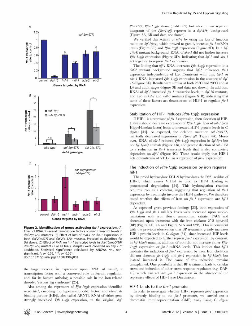
We then verified the effect of RNAi on levels of mRNA from the
endogenous ftn-1 gene. This identified four genes where RNAi
robustly reduced ftn-1 mRNA levels: hsf-1, mdl-1, ada-2 and elt-2
(Figure 2A, Table S1).
The heat-shock factor hsf-1 was previously shown to mediate
effects of IIS on gene expression [29]. The screen also confirmed
that the GATA transcription factor elt-2 plays a role in ftn-1
regulation. This is consistent with the role of elt-2 as an activator of
intestinal gene expression [30]; moreover, elt-2 is the only
previously described transcriptional activator of ftn-1 expression
[20]. Thus, identification of hsf-1 and elt-2 in this unbiased screen
is evidence of the efficacy of the screen. ada-2 encodes a homolog
of the Ada2 subunit of various histone acetyl transferase (HAT)
complexes that activate gene expression by modifying chromatin
Author Summary
Iron plays a role in many biological processes, includingenergy generation and DNA replication. But to maintainhealth, levels of cellular iron must be just right: too muchor too little iron can cause illnesses, such as anemia andhemochromatosis, respectively. Animals therefore carefullycontrol their iron levels by regulating of iron uptake,transport, and storage within protein capsules calledferritins. But how do they coordinate this? Using themodel organism C. elegans, we have discovered a networkof genes and pathways that control iron homeostasis. Wefind that ferritin is regulated by insulin/IGF-1 signaling,which also controls growth and resistance to oxidativestress in response to harsh environmental conditions.Ferritin is also regulated by the hypoxia signaling pathway,which responds to oxygen and iron levels as well as tometabolic cues. We find that the hypoxia pathway acts asan iron sensor, a role it may also play in humans. Our workdefines a network of signaling pathways that can adjustiron availability in response to a range of environmentalcues. Understanding this network in C. elegans can help usto understand the causes of iron dyshomeostasis inhumans, which can profoundly affect health.
Ferritin Regulated by IIS and Hypoxia Signaling
PLoS Genetics | www.plosgenetics.org 2 March 2012 | Volume 8 | Issue 3 | e1002498

via histone acetylation [31]. It is possible that, ada-2 influences ftn-
1 expression via effects on chromatin status.
More notable is the identification of the MAD-like transcription
factor mdl-1 as an activator of ftn-1 expression. mdl-1 plays a role in
the protective effects of reduced IIS against a tumorous germline
phenotype [32] and is upregulated in IIS mutants [10,26,32]. We
confirmed that the null mutation mdl-1(tm311) reduces ftn-1
mRNA levels in daf-2 mutants (Figure 2B).
To explore whether these four factors might be acting
downstream of DAF-16, we tested whether RNAi reduces ftn-1
expression in a daf-16; daf-2 double mutant. The results imply that
only MDL-1 does not require DAF-16 to activate ftn-1 expression.
This suggests that mdl-1 acts downstream of daf-16, or possibly in
parallel to IIS, to regulate ftn-1 expression (Figure 2C). Given that
mdl-1 is a direct transcriptional target of DAF-16 [33], the former
seems more likely.
Unexpectedly, RNAi of hsf-1 markedly increased ftn-1 expres-
sion in a daf-16; daf-2 background (Figure 2C). The effects of hsf-1
RNAi (Figure 2A) imply that HSF-1 and DAF-16 act together to
activate ftn-1 expression, as previously shown for other genes [29].
That loss of hsf-1 in daf-16; daf-2 mutants increases expression of
ftn-1 could imply a repressive role of HSF-1 in the absence of
DAF-16. Alternatively, this increase might merely reflect a stressed
state in the worms, caused by loss of both hsf-1 and daf-16 at 25uC(see Discussion).
Since ftn-1 is known to be responsive to iron levels, we also tested
whether DAF-16, HSF-1 or MDL-1 are required for iron-
dependent regulation of ftn-1. daf-16, hsf-1 and mdl-1 mutants were
treated with iron (25 mM ferric ammonium citrate, FAC) and ftn-1
transcript levels measured by qRT-PCR. Iron-induced up-regula-
tion of ftn-1 was unchanged in each case (Figure S1A), i.e. these
three factors do not mediate effects of iron on ftn-1 expression.
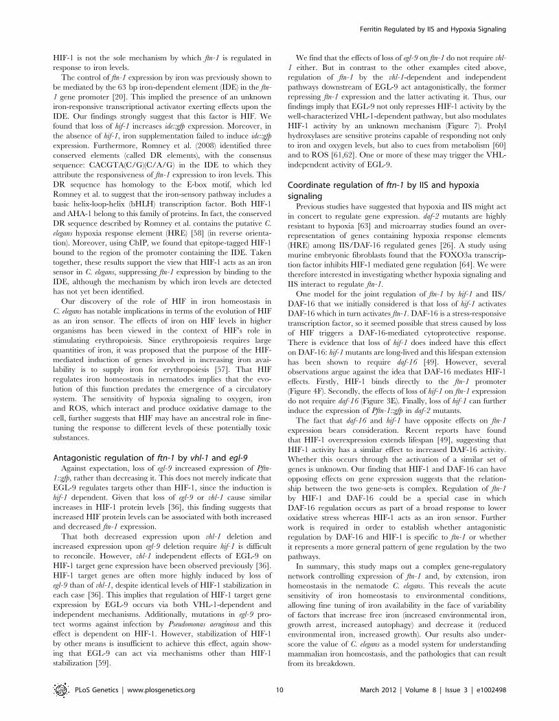
hif-1 and daf-2 act additively to repress ftn-1 expressionRNAi of 28 genes further increased expression of the Pftn-1:gfp
reporter (Table S2), already induced by daf-2(m577). Of note was
Figure 1. Regulation of the ferritin gene ftn-1 by insulin/IGF-1 signaling. (A) Effect of loss of daf-2 and daf-16 function on ftn-1 mRNA levels.Animals were grown at 15uC until the L4 stage of development, then kept at 25uC for two days prior to RNA extraction. (B) Construction of Pftn-1::gfptransgenic C. elegans. Approximately 3.8 kb of upstream sequence was fused to the GFP coding sequence. Epifluorescence images of nematodesbearing wuIs177 [Pftn-1::gfp] in daf-2(+) or daf-2(m577) backgrounds. Animals were grown at 15uC until the L4 stage and then kept at 25uC for twodays before microscopy. The same exposure time was used for both images. (C) Effect of loss of daf-2 and daf-16 function on Pftn-1::gfp expression(c.f. Figure 1A). Animals were grown at 15uC until the L4 stage of development and then transferred to RNAi plates. They were then kept at 25uC fortwo days before GFP fluorescence measurements. (D) Diagrammatic depiction of RNAi screening protocol. Eggs were isolated by alkalinehypochlorite treatment and synchronized populations were left to develop at 15uC until the L4 stage of development. L4 animals were transferred toRNAi plates and left at 25uC for two days. Quantification of GFP expression was carried out by picking 40 animals into microtitre plates and measuringfluorescence using a platereader with a GFP filter set. Statistical significance was calculated by ANOVA in all cases. ***: p,0.001.doi:10.1371/journal.pgen.1002498.g001
Ferritin Regulated by IIS and Hypoxia Signaling
PLoS Genetics | www.plosgenetics.org 3 March 2012 | Volume 8 | Issue 3 | e1002498
the large increase in expression upon RNAi of unc-62, a
transcription factor with a conserved role in ferritin regulation
and, for its human ortholog, a possible role in the iron-related
disorder ‘restless leg syndrome’ [25].
Also among the repressors of Pftn-1::gfp expression identified
were hif-1, encoding the hypoxia-inducible factor, and aha-1, its
binding partner (HIFb, also called ARNT). RNAi of either gene
strongly increased Pftn-1::gfp expression, in the original daf-
2(m577); Pftn-1::gfp strain (Table S2) but also in two separate
integrants of the Pftn-1::gfp reporter in a daf-2(+) background
(Figure 3A, 3B and data not shown).
We verified this activity of hif-1 by using the loss of function
mutation hif-1(ia4), which proved to greatly increase ftn-1 mRNA
levels (Figure 3C) and Pftn-1::gfp expression (Figure 3D). In a hif-
1(ia4) mutant background, RNAi of aha-1 did not further increase
Pftn-1::gfp expression (Figure 3D), indicating that hif-1 and aha-1
act together to repress ftn-1 expression.
The finding that hif-1 RNAi increases Pftn-1::gfp expression in a
daf-2 mutant background suggests that hif-1 influences ftn-1
expression independently of IIS. Consistent with this, hif-1 or
aha-1 RNAi increased Pftn-1::gfp expression in the absence of daf-
16 (Figure 3E). Results were similar at both 25uC and 20uC and at
L4 and adult stages (Figure 3E and data not shown). In addition,
RNAi of hif-1 increased ftn-1 transcript levels in daf-16 mutants,
and also in hsf-1 and mdl-1 mutants (Figure S1B), indicating that
none of these factors act downstream of HIF-1 to regulate ftn-1
expression.
Stabilization of HIF-1 reduces Pftn-1::gfp expressionIf HIF-1 is a repressor of ftn-1 expression, then elevation of HIF-
1 levels should decrease expression of Pftn-1::gfp. Loss of vhl-1 (von
Hippel-Lindau factor) leads to increased HIF-1 protein levels in C.
elegans [34]. As expected, the deletion mutation vhl-1(ok161)
markedly decreased expression of Pftn-1::gfp (Figure 4A). More-
over, RNAi of vhl-1 reduced Pftn-1::gfp expression in hif-1(+) but
not hif-1(ia4) animals (Figure 4B), and genetic deletion of vhl-1 led
to a reduction in ftn-1 transcript levels that is also completely
dependent on hif-1 (Figure 4C). These results imply that HIF-1
acts downstream of VHL-1 as a repressor of ftn-1 expression.
The induction of Pftn-1::gfp expression by iron requireshif-1
The prolyl hydroxylase EGL-9 hydroxylates the P621 residue of
HIF-1, which causes VHL-1 to bind to HIF-1, leading to
proteasomal degradation [34]. This hydroxylation reaction
requires iron as a cofactor, suggesting that regulation of ftn-1
expression by iron might involve the HIF-1 pathway. We therefore
tested whether the effects of iron on ftn-1 expression are hif-1
dependent.
As expected given previous findings [23], both expression of
Pftn-1::gfp and ftn-1 mRNA levels were increased upon supple-
mentation with iron (ferric ammonium citrate, FAC) and
decreased upon treatment with the iron chelator 29-2 bipyridil
(BP) (Figure 4D, 4E and Figure S2A and S2B). This is consistent
with the previous observation that BP treatment greatly increases
HIF-1 protein levels in C. elegans [34], since increased HIF levels
would be expected to further repress ftn-1 expression. By contrast,
in hif-1(ia4) mutants, addition of iron did not increase either Pftn-
1::gfp expression or ftn-1 mRNA levels. This implies that hif-1
mediates the induction of ftn-1 expression by iron. Iron chelation
did not decrease ftn-1::gfp and ftn-1 expression in hif-1(ia4), but
instead increased it. The cause of this induction remains
unexplained. One possibility is that BP treatment leads to cellular
stress and induction of other stress response regulators (e.g. DAF-
16), which can activate ftn-1 expression in the absence of the
repressive effects of HIF-1 (see Discussion).
HIF-1 binds to the ftn-1 promoterIn order to investigate whether HIF-1 represses ftn-1 expression
by directly binding to the ftn-1 promoter, we carried out a
chromatin immunoprecipitation (ChIP) assay using C. elegans
Figure 2. Identification of genes activating ftn-1 expression. (A)Effect of RNAi of several transcription factors on ftn-1 transcript levels indaf-2(m577) mutants. (B) Effect of loss of mdl-1 on ftn-1 expression inboth daf-2(m577) and daf-2(e1370) mutants. Protocol as described for(A) above. (C) Effect of RNAi on ftn-1 transcript levels in daf-16(mgDf50);daf-2(m577) mutants. For all trials, samples were collected on day 2 ofadulthood. Statistical significance calculated by ANOVA. n.s:. non-significant, *: p,0.05, ***: p,0.001.doi:10.1371/journal.pgen.1002498.g002
Ferritin Regulated by IIS and Hypoxia Signaling
PLoS Genetics | www.plosgenetics.org 4 March 2012 | Volume 8 | Issue 3 | e1002498

expressing Myc-tagged HIF-1 [35] and an anti-Myc antibody.
We used three lines: wild type (N2), ZG429 [hif-1::myc] and
GA654 [hif-1::myc vhl-1(ok161)]. Given that vhl-1 mutants have
elevated HIF-1 levels and reduced ftn-1 mRNA levels (Figure 4C),
greater levels of HIF-1::Myc binding to the ftn-1 promoter
should be detectable in vhl-1 mutants, if the interaction is in
fact direct.
We first checked that our ChIP protocol allowed us to measure
binding by HIF-1::Myc by testing binding to the promoter of a
known HIF-1 target gene, nhr-57. We designed one set of primers
to amplify the region of the promoter containing two putative
hypoxia response elements (HREs) and another set of primers
targeting an area within the 39 UTR of this gene. Quantity of
qRT-PCR amplified promoter DNA was then compared to the 39
UTR quantity as a test of enrichment of the promoter in our ChIP
DNA pools. This amplification from the 39 UTR (to which HIF-1
is not expected to bind) allowed us to control for input quantity.
We saw a large (8.3-fold) enrichment of the nhr-57 promoter
sequence in the HIF-1::Myc lines and an even greater (14.9-fold)
enrichment when HIF-1::Myc was stabilized by deletion of
vhl-1 (Figure 4F). Relative amounts of HIF-1::Myc were monitored
by Western blotting of the same ChIP samples using the same
aliquot of anti-Myc antibody used for ChIP, and we were able to
confirm that vhl-1(ok161) increases HIF-1::Myc protein levels
(Figure 4F).
We then measured binding to the ftn-1 promoter through qRT-
PCR against the promoter sequence of ftn-1. For this, we used a
primer pair specific to the IDE sequence. These results were again
normalized against the same nhr-57 39UTR in order to correct for
differences in input quantity. While weaker than binding to Pnhr-
57, enrichment of the Pftn-1 sequence in HIF-1::Myc and
stabilized HIF-1::Myc lines was statistically significantly different
to that seen in wild-type controls (Figure 4F). This is evidence that
HIF-1 represses ftn-1 expression through direct binding to its
promoter.
Iron-dependent regulation of ftn-1 is partiallyvhl-1–dependent
The repression of ftn-1 expression by HIF-1 and the
requirement for iron in the degradation of HIF-1 by the
proteasome suggests a possible mechanism for the iron-dependent
regulation of ftn-1 in which changes in iron levels alter the level of
HIF-1 protein, which in turn alter ftn-1 expression. Since the iron-
dependent degradation of HIF-1 occurs via the action of VHL-1,
HIF-1 protein levels in C. elegans are not sensitive to iron levels
when VHL-1 is absent [36].
We found that loss of vhl-1 largely abrogated the induction of
Pftn-1::gfp expression by iron supplementation, though there was
still a significant induction of lesser magnitude (Figure 5A).
Figure 3. HIF signaling regulates ftn-1 expression. (A) Effect of RNAi of hif-1 and aha-1 on the expression of Pftn-1::gfp. This result was obtainedfrom animals carrying wuIs177 [Pftn-1::gfp]. Animals were grown at 20uC, transferred to RNAi at the L4 stage and GFP fluorescence quantified twodays later. (B) Epifluorescence image of Pftn-1::gfp under control conditions (L4440) and hif-1 RNAi. Animals were grown at 20uC and photographedon day 2 of adulthood. (C) Effect of hif-1(ia4) on ftn-1 transcript levels. Animals were grown at 20uC and samples were collected on day 1 ofadulthood. (D) Effect of hif-1 and aha-1 RNAi on ftn-1 expression in wild-type and hif-1(ia4) animals. (E) Effect of hif-1, aha-1 and daf-16 RNAi on Pftn-1::gfp expression in wild type and daf-16 mutants. These measurements were carried out on L4 animals kept at 25uC. In order to quantify GFPexpression in L4 animals, 60 rather than 40 animals were transferred to each well of the microtitre plates. Statistical significance calculated by ANOVA.n.s:. non-significant, ***: p,0.001.doi:10.1371/journal.pgen.1002498.g003
Ferritin Regulated by IIS and Hypoxia Signaling
PLoS Genetics | www.plosgenetics.org 5 March 2012 | Volume 8 | Issue 3 | e1002498

Reduction of Pftn-1::gfp expression by iron chelation was not
affected by loss of vhl-1 (Figure 5B). Taken together, this suggests
that regulation of ftn-1 by iron may occur partially, but not
exclusively, through changes in HIF-1 protein levels regulated by
iron-dependent degradation.
Evidence that hif-1 mediates iron-dependent regulationvia the iron-dependent element
The induction of ftn-1 levels by iron requires a 63 bp element
(the iron-dependent element, or IDE) in the gene’s promoter [20].
Figure 4. Evidence that the HIF pathway acts as an iron sensor. (A) Effect vhl-1(ok161) on Pftn-1::gfp expression. Experiment was carried out at20uC. (B) Effect of RNAi of vhl-1 on Pftn-1::gfp expression in wild-type and hif-1(ia4) animals. Nematodes were maintained on RNAi plates for twogenerations at 25uC and GFP fluorescence quantified at the L4 stage. (C) ftn-1 transcript levels in wild type, vhl-1(ok161), hif-1(ia4) and hif-1(ia4); vhl-1(ok161) double mutants. Cultures were grown at 20uC and samples were collected at day 1 of adulthood. (D) Effect of addition of iron (25 mM FAC)on expression of Pftn-1::gfp in wild type and hif-1(ia4) mutants. (E) Effect of addition of 0.1 mM bipyridyl (BP) on expression of Pftn-1::gfp in wild typeand hif-1(ia4) mutants. (F) Chromatin immunoprecipitation (ChIP) was carried out using N2 (wild type), ZG429 (hif-1::Myc) and GA654 (hif-1::Myc vhl-1(ok161). Binding was assessed by qRT-PCR of ChIP samples using primers against the promoters of ftn-1 and the known HIF-1 target gene nhr-57.Values obtained were normalized to using qRT-PCR with primers against the 39UTR of nhr-57, to which HIF-1::Myc is not thought to bind. HIF-1::Mycprotein levels were quantified by Western blot using the same antibody aliquot as used for the ChIP experiment. Statistical significance calculated byANOVA. *: p,0.05, ***: p,0.001.doi:10.1371/journal.pgen.1002498.g004
Ferritin Regulated by IIS and Hypoxia Signaling
PLoS Genetics | www.plosgenetics.org 6 March 2012 | Volume 8 | Issue 3 | e1002498

We wondered whether the hif-1 pathway might mediate the effects
of iron on IDE-mediated gene expression. A reporter strain
carrying a ftn-1 promoter lacking the IDE is insensitive to changes
in iron levels [20]. Using these same reporters we found that
absence of the IDE abolished hif-1 RNAi-induced induction of
expression (Figure 5C).
Figure 5. HIF-1 represses expression via the iron-dependent element (IDE). (A) Expression of Pftn-1::gfp in wild-type and vhl-1(ok161)animals with or without addition of iron (25 mM FAC). GFP fluorescence was quantified using a plate reader after 18 h of iron treatment. At least tenbiological replicates were quantified. Asterisks denote statistically significant difference when compared to non-iron treated controls. (B) Expressionof Pftn-1::gfp in wild-type and vhl-1(ok161) animals with or without iron chelation (0.1 mM bipyridyl, BP). GFP fluorescence was quantified using aplate reader after 18 h of iron chelation. BP-treated animals were compared to ethanol control treated ones. (C) Effect of hif-1 RNAi on the Pftn-1::gfptransgene with or without the IDE regulatory element. Fluorescence was measured in L4 animals grown at 25uC through pixel density quantificationof epifluorescence microscopy images. hif-1 RNAi was administered for one generation. Strains used: XA6900 and XA6902. (D) ide::gfp expression inhif-1(+) and hif-1(ia4) animals with or without the addition of iron. Fluorescence measured as in (C). FAC treatment was administered from egg to L4stage of development. (E) ide::gfp expression in hif-1(+) and hif-1(ia4) animals with or without the addition of iron chelator. Due to toxic effects of BPtreatment during development, BP treatment was administered for 18 h during adulthood. Quantification was carried out on the second day ofadulthood using pixel density quantification. n.s:. non-significant, *: p,0.05, ***: p,0.001.doi:10.1371/journal.pgen.1002498.g005
Ferritin Regulated by IIS and Hypoxia Signaling
PLoS Genetics | www.plosgenetics.org 7 March 2012 | Volume 8 | Issue 3 | e1002498

Another reporter construct with just the IDE sequence fused to
a minimal promoter and driving GFP expression was previously
shown to be responsive to iron [20]. We found that loss of hif-1
increased ide::gfp expression, demonstrating that hif-1 does promote
gene expression from the IDE (Figure 5D). Moreover, addition of
iron did not induce ide::gfp expression in hif-1 mutants (Figure 5D).
However, in hif-1 mutants treatment with the iron chelator BP still
reduced ide::gfp expression (Figure 5E). This possibly reflects an
effect of BP on ftn-1 that is independent of its effects on iron levels,
or the existence of a second iron-dependent factor. These results
show that the IDE is subject to regulation by HIF-1 and suggest
that HIF-1 mediates the effects of iron on IDE-mediated gene
expression.
Loss of egl-9 increases ftn-1 expressionAs previously described, loss of vhl-1 decreases expression of
Pftn-1::gfp (Figure 4A). This is expected given that HIF-1 represses
ftn-1 expression and that loss of vhl-1 increases HIF-1 levels [34].
The prolyl hydroxylase EGL-9 targets HIF-1 for proteasomal
degradation, and loss of egl-9 causes a similarly large increase in
HIF-1 protein levels as loss of vhl-1 [36]. We therefore expected
that loss of egl-9, like that of vhl-1, would reduce Pftn-1::gfp
expression. In fact, deletion of egl-9 caused an 11-fold increase in
Pftn-1::gfp expression (Figure 6A) and a ,950-fold increase in ftn-1
mRNA levels (Figure 6B). Animals with a different allele, egl-
9(n586), also showed increased ftn-1 mRNA levels (Figure S3A).
Visible Pftn-1::gfp expression remained restricted to the intestine in
wild type, vhl-1 mutants and egl-9 mutants.
vhl-1-independent effects of EGL-9 on HIF-1 target gene
expression have been observed previously [36]. Our findings
suggest that in the case of ftn-1 regulation, egl-9 can act
independently of, and antagonistically to, vhl-1. As expected, loss
of egl-9 induced ftn-1 expression even in the absence of vhl-1
(Figure S3B and S3C). However, egl-9 RNAi did not increase ftn-1
transcript or Pftn1::gfp expression in the absence of hif-1 (Figure 6B
and Figure S3D). This implies that the inhibition of ftn-1
expression by EGL-9 also requires hif-1.
Thus, egl-9 and vhl-1 inhibit and activate expression of ftn-1,
respectively, and both activities require hif-1. One possibility is that
EGL-9 inhibits ftn-1 expression by stimulating HIF-1 activity via
an as yet unidentified pathway.
Discussion
In this study, we have investigated the regulation of the
inducible C. elegans ferritin gene ftn-1, a key determinant of iron
homeostasis. We reveal that expression of this gene is coordinately
regulated by insulin/IGF-1 and HIF signaling, pathways previ-
ously known to interact in the regulation of stress resistance and
lifespan. Our findings imply that the HIF pathway is required for
gene regulation in response to iron levels in C. elegans, and that IIS
controls iron homeostasis, potentially increasing free iron avail-
ability to support growth.
Insulin/IGF-1 signaling (IIS) regulates growth and ironhomeostasis
IIS and DAF-16 play a pivotal role in the organismal decision
between growth and diapause. Under growth-promoting condi-
tions, DAF-16 is inactivated through cytoplasmic retention, which
facilitates reproductive growth [8,37]. In the absence of sufficient
food or given exposure to certain forms of stress, DAF-16 enters
the nucleus and transcriptionally specifies a survival program. This
entails delayed reproduction, enhanced stress resistance and
increased lifespan. Modulation of DAF-16 activity is therefore
crucial for ensuring an optimal response to the worm’s
environment; with growth and reproduction under conditions
that are propitious to growth, and developmental arrest, stress
protection and increased longevity under conditions that are not.
Regulation of ftn-1 by DAF-16 suggests the existence of a trade-
off between growth and stress resistance involving iron homeosta-
sis. A role for ferritin in regulating growth via its effects on iron
homeostasis has been described previously in mammalian cells
[38]. This study found that Myc, a bHLH transcription factor with
a major role in promoting cellular proliferation, can repress H-
ferritin expression. Overexpression of ferritin in cells carrying
activated Myc led to a decrease in in vitro clonogenicity, and this
effect could be rescued by addition of iron, suggesting that Myc–
mediated repression of ferritin expression favors growth by
increasing iron availability. The study identified DNA synthesis
as a possible mechanism for iron-dependent control of cellular
proliferation by c-Myc, as DNA synthesis is increased by c-Myc in
a manner dependent on ferritin repression and the associated
increases in iron availability. This finding is consistent with the
requirement for iron in the activity of ribonucleotide reductase, the
rate-limiting enzyme in DNA synthesis. Similar mechanisms may
be at play in the regulation of ferritin expression by IIS. When
Figure 6. Regulation of ftn-1 expression by EGL-9. (A) Effect ofegl-9(sa307) deletion mutaion on Pftn-1::gfp expression. Epifluorescencemicroscopy and plate reader quantification of GFP fluorescence wascarried out on day 2 of adulthood. (B) Effect of egl-9 deletion on ftn-1transcript levels in hif-1(+) and hif-1(ia4) animals. Samples werecollected at day 1 of adulthood. Statistical significance calculated byANOVA. n.s:. non-significant, ***: p,0.001.doi:10.1371/journal.pgen.1002498.g006
Ferritin Regulated by IIS and Hypoxia Signaling
PLoS Genetics | www.plosgenetics.org 8 March 2012 | Volume 8 | Issue 3 | e1002498

conditions favor growth, and IIS is increased, reduced ftn-1
expression is expected to increase iron availability, thus fulfilling a
key requirement for growth.
While free iron is required for growth, it can also cause harm by
catalyzing the Fenton reaction, which increases levels of ROS and
molecular damage. When conditions are not suitable for growth,
IIS is reduced, and increased ftn-1 expression is expected to lower
levels of free iron and of ROS, thereby protecting against stress.
Consistent with this, induced over-expression of ftn-1 causes
resistance to oxidative stress (S. Valentini and D. Gems,
unpublished results). Thus, upregulation of ftn-1 likely contributes
to the broader increase in cytoprotection seen when IIS is reduced.
Reduced IIS also increases levels of autophagy in C. elegans
[39,40] and autophagy releases iron from ferruginous materials,
such as mitochondrial metalloproteins [41]. This predicts that
reduced IIS will increase free iron levels, and concomitant
elevation of ftn-1 expression could ensure that iron released by
autophagy does not cause molecular damage.
Transcriptional activators of ftn-1 expressionUsing an RNAi screen we identified new regulators of ftn-1,
including hsf-1 and mdl-1. It was previously shown that in daf-2
mutants the heat shock factor HSF-1 acts in concert with DAF-16
to promote expression of small heat shock proteins and other
molecular chaperones, which contribute to longevity [29]. We find
that hsf-1 is also involved in the induction of ftn-1 in daf-2 mutants,
since loss of hsf-1 reduced ftn-1 expression in daf-2 but not daf-16;
daf-2 mutants.
The MAD-like transcription factor mdl-1 is also regulated by
IIS. Microarray and qRT-PCR studies showed it to be up-
regulated in daf-2 mutants [10,26,32]. mdl-1 also contributes to the
resistance of daf-2 mutants to germline tumor formation in the gld-
1 tumor model, and to daf-2 mutant longevity [32]. That MDL-1
activates ftn-1 expression is consistent with the role of mammalian
MAD as an inhibitor of Myc, which represses ferritin expression
(see above); however, C. elegans does not possess any clear ortholog
of Myc [42,43].
A study of DAF-16 binding sites did not provide evidence that
ftn-1 is a direct regulatory target of DAF-16 [33], but suggested
that mdl-1 might be. Given that ftn-1 may be a direct target of
MDL-1 [44,45], one possibility is that activation of mdl-1
expression by DAF-16 leads to increased ftn-1 expression. This
hypothesis predicts that abrogation of mdl-1 expression should
decrease ftn-1 expression more in daf-2 than in daf-16; daf-2
animals, but this is not the case (Figure 2A, 2C). This could imply
that mdl-1 regulates ftn-1 independently of daf-16, at least in part.
ftn-1 is negatively regulated by hif-1 and aha-1We discovered that loss of hif-1 or its binding partner aha-1
greatly increased ftn-1 expression in daf-2 mutants. This implicated
hypoxia signaling in the regulation of ftn-1.
The HIF transcription factor is composed of an a and a bsubunit, encoded by the genes hif-1 and aha-1 in C. elegans. HIF
regulates the transcriptional response to hypoxia in both worms
and vertebrates and, as expected, worms lacking hif-1 are
hypersensitive to hypoxia [46]. Levels of HIFb protein are
relatively stable, whereas HIFa is constantly being degraded by
the proteasome under normal, non-hypoxic conditions. In both
worms and higher organisms, this occurs because the HIFa/HIF-1
protein is hydroxylated at conserved proline residues by prolyl
hydroxylase (PHD), encoded by the egl-9 gene in worms. After
hydroxylation by PHD/EGL-9, the von Hippel-Lindau protein
VHL-1 binds to HIFa, which targets it for degradation [34,47].
PHDs require oxygen, iron and 2-oxoglutarate for the
hydroxylation reaction. When cells are kept under hypoxic
conditions or when an iron chelator is added, the proline residue
in HIFa is not hydroxylated and the HIFa protein accumulates
[48]. That loss of hif-1 has such dramatic effects on gene
expression under normoxic conditions demonstrates that HIF-1
affects gene regulation even at the very low levels of HIF-1 found
when it is being hydroxylated and degraded. Similarly, it was
previously observed that loss of hif-1 can increase C. elegans lifespan
under normoxic conditions [49]. Consistent with this, we find
statistically significant levels of binding of the non-stabilized HIF-
1::Myc protein to both ftn-1 and nhr-57 promoters (Figure 4F).
Since iron is a required cofactor for hydroxylation of HIF by
PHD, levels of iron affect those of HIF. For example, in C. elegans,
depletion of iron using the iron chelator 2-29 bipyridyl stabilizes
HIF-1 [34], and feeding mice a low-iron diet leads to increased
HIFa levels [50]. This increase in HIF-1 levels is not without
consequence: chelation of iron has also been shown to increase
expression of the C. elegans HIF-1 target gene nhr-57, indicating
that the stabilization of HIF upon loss of iron leads to HIF-1-
dependent changes in gene expression [51]. In vertebrates, HIF
activates expression of genes involved in regulating iron homeo-
stasis, including heme oxygenase [52], the transferrin receptor
[53,54], ceruloplasmin [55], DMT1 [56] and possibly ferroportin
[57]. Loss of HIF-2a in mice causes decreased iron levels in the
plasma and livers of mice [56]. It has therefore been suggested that
HIF can act as an iron sensor: low iron levels lead to HIF
stabilization, which leads to changes in gene expression that
increase iron levels [57]. The results presented here support this
hypothesis.
Iron-dependent regulation of ftn-1 via hif-1The repression of ferritin expression by hif-1/aha-1 is consistent
with a role of HIF in increasing iron availability. By this view,
lower ferritin expression upon HIF activation would reduce iron
storage capacity, thereby increasing iron availability. We therefore
investigated whether HIF mediates iron-dependent regulation of
ftn-1, and this proved to be the case: ftn-1 regulation by iron is
blocked in hif-1 mutants. In wild-type animals iron supplementa-
tion increases ftn-1 expression while iron depletion decreases it. By
contrast, in hif-1 mutants iron supplementation does not increase
ftn-1 expression.
Treatment of hif-1 mutants with the iron chelator 2-29 bipyridyl
(BP) caused a large increase, rather than decrease, of ftn-1
expression. This was unexpected, but we noticed that BP treated
worms were somewhat sickly in appearance. One possibility is that
toxicity of BP in hif-1 mutants triggers other stress response
mediators (e.g. DAF-16) that activate ftn-1 expression. This is
consistent with our observation that stressful conditions tend to
induce expression of this reporter. Similar to treatment with BP,
RNAi of hsf-1 in daf-16(mgDf50); daf-2(m577) animals raised at
25uC also caused the worms to have a sickly appearance and
induced Pftn-1::gfp expression (Figure 2C). Moreover, we observed
that starved animals also show elevated Pftn-1::gfp expression (data
not shown).
The requirement for hif-1 in the iron-dependent regulation of
ftn-1 suggests that this regulation may occur via iron-dependent
degradation of HIF-1. However, our data implies that this is not
the whole story. Mutants of vhl-1 have constitutively stabilized
HIF-1 and its levels cannot therefore respond to changes in iron
(or oxygen) levels [34,36]. While the increase in Pftn-1::gfp
expression upon treatment with iron was greatly reduced in vhl-1
mutants, Pftn-1::gfp expression was still elevated compared to the
control treatment. This implies that iron-dependent degradation of
Ferritin Regulated by IIS and Hypoxia Signaling
PLoS Genetics | www.plosgenetics.org 9 March 2012 | Volume 8 | Issue 3 | e1002498
HIF-1 is not the sole mechanism by which ftn-1 is regulated in
response to iron levels.
The control of ftn-1 expression by iron was previously shown to
be mediated by the 63 bp iron-dependent element (IDE) in the ftn-
1 gene promoter [20]. This implied the presence of an unknown
iron-responsive transcriptional activator exerting effects upon the
IDE. Our findings strongly suggest that this factor is HIF. We
found that loss of hif-1 increases ide::gfp expression. Moreover, in
the absence of hif-1, iron supplementation failed to induce ide::gfp
expression. Furthermore, Romney et al. (2008) identified three
conserved elements (called DR elements), with the consensus
sequence: CACGTA(C/G)(C/A/G) in the IDE to which they
attribute the responsiveness of ftn-1 expression to iron levels. This
DR sequence has homology to the E-box motif, which led
Romney et al. to suggest that the iron-sensory pathway includes a
basic helix-loop-helix (bHLH) transcription factor. Both HIF-1
and AHA-1 belong to this family of proteins. In fact, the conserved
DR sequence described by Romney et al. contains the putative C.
elegans hypoxia response element (HRE) [58] (in reverse orienta-
tion). Moreover, using ChIP, we found that epitope-tagged HIF-1
bound to the region of the promoter containing the IDE. Taken
together, these results support the view that HIF-1 acts as an iron
sensor in C. elegans, suppressing ftn-1 expression by binding to the
IDE, although the mechanism by which iron levels are detected
has not yet been identified.
Our discovery of the role of HIF in iron homeostasis in
C. elegans has notable implications in terms of the evolution of HIF
as an iron sensor. The effects of iron on HIF levels in higher
organisms has been viewed in the context of HIF’s role in
stimulating erythropoiesis. Since erythropoiesis requires large
quantities of iron, it was proposed that the purpose of the HIF-
mediated induction of genes involved in increasing iron avai-
lability is to supply iron for erythropoiesis [57]. That HIF
regulates iron homeostasis in nematodes implies that the evo-
lution of this function predates the emergence of a circulatory
system. The sensitivity of hypoxia signaling to oxygen, iron
and ROS, which interact and produce oxidative damage to the
cell, further suggests that HIF may have an ancestral role in fine-
tuning the response to different levels of these potentially toxic
substances.
Antagonistic regulation of ftn-1 by vhl-1 and egl-9Against expectation, loss of egl-9 increased expression of Pftn-
1::gfp, rather than decreasing it. This does not merely indicate that
EGL-9 regulates targets other than HIF-1, since the induction is
hif-1 dependent. Given that loss of egl-9 or vhl-1 cause similar
increases in HIF-1 protein levels [36], this finding suggests that
increased HIF protein levels can be associated with both increased
and decreased ftn-1 expression.
That both decreased expression upon vhl-1 deletion and
increased expression upon egl-9 deletion require hif-1 is difficult
to reconcile. However, vhl-1 independent effects of EGL-9 on
HIF-1 target gene expression have been observed previously [36].
HIF-1 target genes are often more highly induced by loss of
egl-9 than of vhl-1, despite identical levels of HIF-1 stabilization in
each case [36]. This implies that regulation of HIF-1 target gene
expression by EGL-9 occurs via both VHL-1-dependent and
independent mechanisms. Additionally, mutations in egl-9 pro-
tect worms against infection by Pseudomonas aeruginosa and this
effect is dependent on HIF-1. However, stabilization of HIF-1
by other means is insufficient to achieve this effect, again show-
ing that EGL-9 can act via mechanisms other than HIF-1
stabilization [59].
We find that the effects of loss of egl-9 on ftn-1 do not require vhl-
1 either. But in contrast to the other examples cited above,
regulation of ftn-1 by the vhl-1-dependent and independent
pathways downstream of EGL-9 act antagonistically, the former
repressing ftn-1 expression and the latter activating it. Thus, our
findings imply that EGL-9 not only represses HIF-1 activity by the
well-characterized VHL-1-dependent pathway, but also modulates
HIF-1 activity by an unknown mechanism (Figure 7). Prolyl
hydroxylases are sensitive proteins capable of responding not only
to iron and oxygen levels, but also to cues from metabolism [60]
and to ROS [61,62]. One or more of these may trigger the VHL-
independent activity of EGL-9.
Coordinate regulation of ftn-1 by IIS and hypoxiasignaling
Previous studies have suggested that hypoxia and IIS might act
in concert to regulate gene expression. daf-2 mutants are highly
resistant to hypoxia [63] and microarray studies found an over-
representation of genes containing hypoxia response elements
(HRE) among IIS/DAF-16 regulated genes [26]. A study using
murine embryonic fibroblasts found that the FOXO3a transcrip-
tion factor inhibits HIF-1 mediated gene regulation [64]. We were
therefore interested in investigating whether hypoxia signaling and
IIS interact to regulate ftn-1.
One model for the joint regulation of ftn-1 by hif-1 and IIS/
DAF-16 that we initially considered is that loss of hif-1 activates
DAF-16 which in turn activates ftn-1. DAF-16 is a stress-responsive
transcription factor, so it seemed possible that stress caused by loss
of HIF triggers a DAF-16-mediated cytoprotective response.
There is evidence that loss of hif-1 does indeed have this effect
on DAF-16: hif-1 mutants are long-lived and this lifespan extension
has been shown to require daf-16 [49]. However, several
observations argue against the idea that DAF-16 mediates HIF-1
effects. Firstly, HIF-1 binds directly to the ftn-1 promoter
(Figure 4F). Secondly, the effects of loss of hif-1 on ftn-1 expression
do not require daf-16 (Figure 3E). Finally, loss of hif-1 can further
induce the expression of Pftn-1::gfp in daf-2 mutants.
The fact that daf-16 and hif-1 have opposite effects on ftn-1
expression bears consideration. Recent reports have found
that HIF-1 overexpression extends lifespan [49], suggesting that
HIF-1 activity has a similar effect to increased DAF-16 activity.
Whether this occurs through the activation of a similar set of
genes is unknown. Our finding that HIF-1 and DAF-16 can have
opposing effects on gene expression suggests that the relation-
ship between the two gene-sets is complex. Regulation of ftn-1
by HIF-1 and DAF-16 could be a special case in which
DAF-16 regulation occurs as part of a broad response to lower
oxidative stress whereas HIF-1 acts as an iron sensor. Further
work is required in order to establish whether antagonistic
regulation by DAF-16 and HIF-1 is specific to ftn-1 or whether
it represents a more general pattern of gene regulation by the two
pathways.
In summary, this study maps out a complex gene-regulatory
network controlling expression of ftn-1 and, by extension, iron
homeostasis in the nematode C. elegans. This reveals the acute
sensitivity of iron homeostasis to environmental conditions,
allowing fine tuning of iron availability in the face of variability
of factors that increase free iron (increased environmental iron,
growth arrest, increased autophagy) and decrease it (reduced
environmental iron, increased growth). Our results also under-
score the value of C. elegans as a model system for understanding
mammalian iron homeostasis, and the pathologies that can result
from its breakdown.
Ferritin Regulated by IIS and Hypoxia Signaling
PLoS Genetics | www.plosgenetics.org 10 March 2012 | Volume 8 | Issue 3 | e1002498

Materials and Methods
Nematode culture and strainsMaintenance and culture of C. elegans was carried out as
published [65,66,67]. The following strains were used: CB5602
vhl-1(ok161), DR1563 daf-2(e1370), DR1567 daf-2(m577), GA300
daf-16(mgDf50); daf-2(m577), GA633 daf-2(m577); wuIs177 [Pftn-
1::gfp lin-15(+)], GA636 rrf-3(pk1426); daf-2(m577); wuIs177 [Pftn-
1::gfp lin-15(+)], GA639 daf-16(mgDf50); wuIs177 [Pftn-1::gfp
lin-15(+)], GA640 wuIs176 [Pftn-1::gfp lin-15(+)], GA641 wuIs177
[Pftn-1::gfp lin-15(+)], GA642 hif-1(ia4); wuIs177 [Pftn-1::gfp lin-
15(+)], GA643 daf-16(mgDf50); daf-2(m577); wuIs177 [Pftn-1::gfp
lin-15(+)], GA654 unc-119(ed3) vhl-1(ok161) iaIs128[Phif-1::hif-
1a::myc unc-119(+)], GA675 xtEx79 [Dpes-10(+63)::GFP-his, pha-
1(+)], GA676 hif-1(ia4) xtEx79 [Dpes-10(+63)::GFP-his, pha-1(+)],
GA688 pha-1(e2123ts) xtEx79 [Dpes-10(+63)::GFP-his, pha-1(+)],
GA688 pha-1(e2123ts); hif-1(ia4) xtEx79 [Dpes-10(+63)::GFP-his,
pha-1(+)], GA694 wuIs176 [Pftn-1::gfp lin-15(+)] egl-9(sa307)],
GA1200 mdl-1(tm311), GA1203 daf-2(e1370); mdl-1(tm311),
GA1204 daf-2(m577); mdl-1(tm311), GR1307 daf-16(mgDf50),
JT307 egl-9(sa307), N2, PS3551 hsf-1(sy441), UZ96 pha-
1(e2123ts) xtEx79 [Dpes-10(+63)::GFP-his, pha-1(+)], XA6900 pha-
1(e2123ts) qaEx6902 [Pftn-1(D63)::[Dpes-10::GFP-his, pha-1(+)],
XA6902 pha-1(e2123ts) qaEx6902 [Pftn-1::[Dpes-10::GFP-his, pha-
1(+)] and ZG31 hif-1(ia4). ZG429 unc-119(ed3) iaIs128[Phif-1::hif-
1a::myc unc-119(+)] Worms were maintained at 20uC unless
otherwise indicated.
Strain constructionsMultiple mutants were created using standard methodologies
and the presence of genomic deletions was tested via PCR.
Genotyping was carried out by lysis of parent animals using
proteinase K (Sigma) and subsequent PCR using the following
primers. For daf-16(mgDf50): daf-16F1, gccactttattggaatttgagc; and
daf-16R1, atcctcccatagaaggaccatt. For hif-1(ia4): hif-1_ex_fwd1,
gctcctcctactccacctttg, hif-1_ex_rev1, gtgacgagttgtgaatgcacc, hif-
1_int_rev1.2, tcggcgatggtgtcttcagtc. For rrf-3(pk1426): rrf-
3_ex_fwd1, gagttcgcatcaagtttcac, rrf-3_ex_rev1, tgccttcgtacattt-
caacc and rrf-3_int_rev2, ggtatttattgcttcctgccac. For vhl-1(ok161):
DA75, gctgtcaatcggagcactgtc, DA76, ttgctgaggtctctggggtc, and
DA77, gttagctctgccacgaatacgatg. For egl-9(sa307): DA117, acaaa-
gacaggtgttgcgaatgag, DA118, ttgtagtgatccgagcccag, and DA119,
gatgcttctgatgttcttggagg.
The promoter::gfp transgene of ftn-1 was created using methods as
previously described [68] and the transgenic strain was created by
microinjection. The primers used for creation of the construct
were: ftn-1.5’ex, tgcttactggttctgccgag, ftn-1.5’in, tgtagggttt-
gattgtggtttg, ftn-1.3’fus, agtcgacctgcaggcatgcaagctttgacgagctagaga-
catgac. Extrachromosomal arrays were integrated by X-ray
irradiation.
Fluorescence measurementsThe method used to quantify GFP expression was adapted from
one used in an earlier study [69]. Using a worm pick, samples of
forty adult worms were transferred into the wells (V-shaped) of
microtitre plates (Greiner). Fluorescence was then measured in a
GeniosPlus plate reader (Tecan) at wavelengths appropriate for
GFP (excitation: 495 nm; emission: 535 nm) using a fixed gain of
75. Quantification of GFP expression from transgenes with low
level expression was carried out using a Leica DMRXA2
microscope using a GFP filter cube (excitation: 470/40 nm;
emission: 525/50 nm), an Orca C10600 digital camera (Hama-
matsu) and Volocity image analysis software (Improvision).
RNAi libraryThe transcription factor RNAi library used for this project was
generously provided by Dr. Weiqing Li (University of Washing-
ton). Similar libraries are now available commercially (geneservi-
ce.co.uk). Where RNAi robustly affected ftn-1 expression levels,
RNAi plasmid inserts were sequenced to confirm their identity
using the primers JJM130 (gggaagggcgatcggtgcgggcc) and JJM131
(gcgcagcgagtcagtgagcgagg).
Figure 7. ftn-1 expression is regulated by both the insulin/IGF-1and hypoxia signaling pathways. This figure provides a diagram-matic representation of the gene regulatory networking controlling ftn-1 expression. It includes previously established regulatory elements(black lines), newly established regulatory elements (blue lines) andnew, hypothetical regulatory elements (dashed blue lines). Weidentified a positive regulatory role for the genes mdl-1, hsf-1, ada-2and daf-16. In the case of mdl-1, previous work suggests that thistranscription factor acts downstream of DAF-16, but it is unclearwhether this is true for ftn-1 regulation (hence second line, dotted blue,from DAF-16 to MDL-1), or whether MDL-1 acts independently of DAF-16 in this case. Loss of ada-2 or elt-2 reduces ftn-1 expression but wewere unable to detect an effect of ada-2 or elt-2 RNAi in the absence ofDAF-16. While this may be caused by a lack of sensitivity in our assay, itcould also indicate that these factors may act together with DAF-16 orupstream of DAF-16 to regulate ftn-1 expression. We found that hif-1and aha-1 repress ftn-1 expression and that hif-1 is required for iron-dependent regulation of ftn-1, implying that HIF acts as an iron sensorin C. elegans. However, HIF-1 activity on ftn-1 expression can beregulated through both vhl-1-dependent and independent pathwaysand our data shows that these pathways act antagonistically on ftn-1expression. The VHL-1-independent inhibition of ftn-1 expression byEGL-9 could either involve activation of transcriptional repression byHIF-1 or (more parsimoniously) inhibition of transcriptional activationby HIF-1. The latter interpretation would suggest the presence of a co-regulator that turns HIF-1 into a transcriptional activator of ftn-1.doi:10.1371/journal.pgen.1002498.g007
Ferritin Regulated by IIS and Hypoxia Signaling
PLoS Genetics | www.plosgenetics.org 11 March 2012 | Volume 8 | Issue 3 | e1002498

qRT–PCRRNA was isolated from 2-day old adults after three washes,
which removed E. coli and L1 progeny from the sample. After
RNA isolation cDNA was synthesized using SuperScript II reverse
transcriptase (Invitrogen) using oligo dT (Invitrogen). qRT-PCR
was carried out using Fast SYBR Green Master Mix (Applied
Biosystems) and the 7900 HT Fast PCR system (Applied
Biosystems). Normalization of transcript quantity was carried out
using the geometric mean of three stably expressed reference genes
Y45F10D.4, pmp-3, and cdc-42 in order to control for cDNA input,
as previously described [70]. The following primers were used for
this assay. Y45F10D.4: DA90, gtcgcttcaaatcagttcagc, and DA91,
gttcttgtcaagtgatccgaca. pmp-3: DA88, gttcccgtgttcatcactcat, and
DA89, acaccgtcgagaagctgtaga. cdc-42: DA86, ctgctggacaggaagat-
tacg, and DA87: ctcggacattctcgaatgaag. ftn-1: ftn-1_fwd_RT2,
cggccgtcaataaacagattaacg, and ftn-1_rev_RT2 cacgctcctcatcc-
gattgc.
qRT-PCR of ChIP DNA pools was carried out for the nhr-57
promoter using DA130: cctcccgcgtctccacattcaatc and DA131:
cagcgaggtctgggttttccg, the nhr-57 39UTR using DA135: tggcacaa-
gatatgacgaaagctg and DA136: ggcgagaaatttgttgtaggttgcc, and the
ftn-1 promoter using DA139: aacagctcacgtagccaatgataag and
DA140: gcatcacatgagctgcccta.
Statistical analysisAll results shown are the mean of at least three independent
biological replicates and error bars represent the s.e.m. Statistical
significance was calculated by two-way or one-way ANOVA of
either raw values or log-transformed quantities, depending on
circumstances.
Chromatin immunoprecipitationThe protocol for chromatin immunoprecipitation was adapted
from Mukhopdhyay et al. [71]. C. elegans cultures were grown for
two generations in S-media with suspended OP50 at 20uC with
constant shaking at 200 rpm. The worms were collected and
washed four times in PBS buffer and then re-suspended in PBS
containing 1% formaldehyde. Samples were then partially lysed
using 8 strokes with a 1/3 turn in a 7 cm Dunce homogenizer and
then incubated for 17 minutes with gentle mixing at room
temperature. Crosslinking was stopped by addition of 200 ml
2.5 mol/L Glycine solution and 20 minutes further incubation at
room temperature. After four washes in PBS containing protease
inhibitor tablets (Complete, Roche), samples were flash frozen and
stored at 280uC. After thawing, 2 mL of HLB buffer [50 mM
HEPES-KOH, pH 7.5, 150 mM NaCl, 1 mM EDTA, 0.1%
(wt/vol) sodium deoxycholate, 1% (vol/vol) Triton X-100, 0.1%
(wt/vol) SDS and 16Complete protease inhibitor] was added and
sonication was carried out at 70% intensity for 7 bursts of
30 seconds in the Vibracell sonicator (Sonics). Protein quantity
was estimated by Bradford assay (Biorad) and 2 mg were diluted
into to 500 ml of in HLB buffer. Three 50 ml aliquots were
removed at this point. DNA isolated from these samples was
subsequently used as input controls. Samples were precleared for
1 h in prewashed salmon sperm DNA/protein-A agarose beads
(Millipore) and then incubated overnight with 10 ml of anti-Myc
Ab (9b11; Cell signalling). Samples were then incubated with
prewashed salmon sperm DNA/protein-A agarose beads for 2 h.
The beads were then washed twice in WB1 [50 mM HEPES-
KOH, pH 7.5, 150 mM NaCl, 1 mM EDTA, 1% (wt/vol)
sodium deoxycholate, 1% (vol/vol) Triton X-100, 0.1% (wt/vol)
SDS and 16Complete protease inhibitor], twice in WB2 [50 mM
HEPES-KOH, pH 7.5, 1 M NaCl, 1 mM EDTA, 1% (wt/vol)
sodium deoxycholate, 1% (vol/vol) Triton X-100, 0.1% (wt/vol)
SDS and 16 Complete protease inhibitor] and once in WB3
[50 mM Tris-HCl, pH 8, 0.25 mM LiCl, 1 mM EDTA, 0.5%
(vol/vol) NP-40 and 0.5% (wt/vol) sodium deoxycholate]. Cross-
linking was reversed by addition of proteinase K solution [50 mM
Tris-HCl, pH 8, 25 mM EDTA, 1.25% (wt/vol) SDS, 160 mg/ml
proteinase K (Qiagen)] and incubation for 2 h at 45uC and
overnight at 65uC. DNA was isolated by applying solution to
Qiagen PCR purification columns (Qiagen).
Supporting Information
Figure S1 (A) Worms carrying mutations in the genes daf-16,
mdl-1 and hsf-1 as well as wild type worms were grown at 20uCuntil early adulthood. At this point, they were washed off the plates
and transferred to plates containing 25 mM ferric ammonium
citrate (FAC). Samples were collected for qRT-PCR after 18 h. (B)
Synchronized L1 animals of these same strains were grown on
either control RNAi cultures or treated with RNAi against hif-1.
Samples were collected on the first day of adulthood for
qRT-PCR. Statistical significance calculated by ANOVA.
***: p,0.001.
(EPS)
Figure S2 (A) Effect of addition of iron (25 mM FAC) on ftn-1
transcript levels in wild type and hif-1(ia4) mutants. (B) Effect of
addition of 0.1 mM bipyridyl (BP on ftn-1 transcript levels in wild
type and hif-1(ia4) mutants. Statistical significance calculated by
ANOVA. ***: p,0.001.
(EPS)
Figure S3 (A) Effect of the n586 allele of egl-9 on expression of
ftn-1. After two rounds of outcrossing, expression of ftn-1 was
quantified in egl-9(n586) as well as N2 control animals. (B) Effect of
egl-9 deletion on Pftn-1::gfp expression in vhl-1(+) and vhl-1(ok161)
animals. (C) Effect of egl-9 deletion on ftn-1 transcript levels in vhl-
1(+) and vhl-1(ok161) animals. Samples were collected at day 1 of
adulthood. (D) Effect of egl-9 deletion on ftn-1 transcript levels in
hif-1(+) and hif-1(ia4) animals. Statistical significance calculated by
ANOVA. **: p,0.01, ***: p,0.001.
(EPS)
Table S1 RNAi of a large number of genes altered expression of
Pftn-1::gfp in the primary screen. Table S1 contains a list of RNAi
treatments that reduced expression of the transgene by at least
20% and shows which of these effect were confirmed first using an
alternative strain and then using qRT-PCR of the ftn-1 transcript.
(DOCX)
Table S2 Table S2 contains a list of RNAi treatments found to
increase expression of Pftn-1::gfp by at least 20% in the primary
screen.
(DOCX)
Acknowledgments
We thank members of the Gems lab for useful discussion and David
Weinkove, Jennifer Tullet and Laura Ackerman for critical reading of the
manuscript. We thank Elizabeth Leibold and Jo Anne Powell-Coffman for
providing us with C. elegans strains and Weiqing Li, Dhaval Patel, and
Stephen Nurrish for RNAi clones.
Author Contributions
Conceived and designed the experiments: DA DG. Performed the
experiments: DA. Analyzed the data: DA. Contributed reagents/
materials/analysis tools: DA. Wrote the paper: DA DG.
Ferritin Regulated by IIS and Hypoxia Signaling
PLoS Genetics | www.plosgenetics.org 12 March 2012 | Volume 8 | Issue 3 | e1002498
References
1. Riddle DL, Albert PS (1997) Genetic and Environmental Regulation of Dauer
Larva Development.
2. Hu PJ (2007) Dauer WormBook. pp 1–19.
3. Cassada RC, Russell RL (1975) The dauerlarva, a post-embryonic develop-
mental variant of the nematode Caenorhabditis elegans. Developmental Biology
46: 326–342.
4. Lithgow GJ, Walker GA (2002) Stress resistance as a determinate of C. elegans
lifespan. Mechanisms of Ageing and Development 123: 765–771.
5. Henis-Korenblit S, Zhang P, Hansen M, McCormick M, Lee SJ, et al. (2010)
Insulin/IGF-1 signaling mutants reprogram ER stress response regulators to
promote longevity. Proc Natl Acad Sci U S A 107: 9730–9735.
6. Murakami S, Johnson TE (1996) A genetic pathway conferring life extension
and resistance to UV stress in Caenorhabditis elegans. Genetics 143: 1207–1218.
7. Henderson ST, Johnson TE (2001) daf-16 integrates developmental and
environmental inputs to mediate aging in the nematode Caenorhabditis elegans.
Current Biology 11: 1975–1980.
8. Lin K, Hsin H, Libina N, Kenyon C (2001) Regulation of the Caenorhabditis
elegans longevity protein DAF-16 by insulin/IGF-1 and germline signaling.
Nature Genetics 28: 139–145.
9. McElwee J, Bubb K, Thomas JH (2003) Transcriptional outputs of the
Caenorhabditis elegans forkhead protein DAF-16 (vol 2, pg 111, 2003). Aging Cell
2: 341–341.
10. Murphy CT, McCarroll SA, Bargmann CI, Fraser A, Kamath RS, et al. (2003)
Genes that act downstream of DAF-16 to influence the lifespan of Caenorhabditis
elegans. Nature 424: 277–284.
11. Kenyon C, Chang J, Gensch E, Rudner A, Tabtiang R (1993) A C. elegans
mutant that lives twice as long as wild-type. Nature 366: 461–464.
12. Gems D, Doonan R (2009) Antioxidant defense and aging in C. elegans: is the
oxidative damage theory of aging wrong? Cell Cycle 8: 1681–1687.
13. Le NT, Richardson DR (2002) The role of iron in cell cycle progression and the
proliferation of neoplastic cells. Biochim Biophys Acta 1603: 31–46.
14. Hentze MW, Muckenthaler MU, Andrews NC (2004) Balancing acts: Molecular
control of mammalian iron metabolism. Cell 117: 285–297.
15. Andrews NC (2000) Iron metabolism: iron deficiency and iron overload. Annu
Rev Genomics Hum Genet 1: 75–98.
16. Mackenzie EL, Iwasaki K, Tsuji Y (2008) Intracellular iron transport and
storage: From molecular mechanisms to health implications. Antioxidants &
Redox Signaling 10: 997–1030.
17. Kaplan J, Ward DM, De Domenico I (2011) The molecular basis of iron
overload disorders and iron-linked anemias. Int J Hematol 93: 14–20.
18. Lawson DM, Treffry A, Artymiuk PJ, Harrison PM, Yewdall SJ, et al. (1989)
Identification of the ferroxidase center in ferritin. FEBS Letters 254: 207–210.
19. Kim YI, Cho JH, Yoo OJ, Ahnn J (2004) Transcriptional regulation and life-
span modulation of cytosolic aconitase and ferritin genes in C. elegans. Journal of
Molecular Biology 342: 421–433.
20. Romney SJ, Thacker C, Leibold EA (2008) An iron enhancer element in the
FTN-1 gene directs iron-dependent expression in Caenorhabditis elegans intestine.
Journal of Biological Chemistry 283: 716–725.
21. White K, Munro HN (1988) Induction of ferritin subunit synthesis by iron is
regulated at both the transcriptional and translational Levels. Journal of
Biological Chemistry 263: 8938–8942.
22. Torti FM, Torti SV (2002) Regulation of ferritin genes and protein. Blood 99:
3505–3516.
23. Gourley BL, Parker SB, Jones BJ, Zumbrennen KB, Leibold EA (2003)
Cytosolic aconitase and ferritin are regulated by iron in Caenorhabditis elegans.
Journal of Biological Chemistry 278: 3227–3234.
24. Winkelmann J, Schormair B, Lichtner P, Ripke S, Xiong L, et al. (2007)
Genome-wide association study of restless legs syndrome identifies common
variants in three genomic regions. Nature Genetics 39: 1000–1006.
25. Catoire H, Dion PA, Xiong L, Amari M, Gaudet R, et al. (2011) Restless legs
syndrome-associated MEIS1 risk variant influences iron homeostasis. Ann
Neurol.
26. McElwee JJ, Schuster E, Blanc E, Thomas JH, Gems D (2004) Shared
transcriptional signature in Caenorhabditis elegans dauer larvae and long-lived daf-2
mutants implicates detoxification system in longevity assurance. Journal of
Biological Chemistry 279: 44533–44543.
27. McElwee JJ, Schuster E, Blanc E, Piper MD, Thomas JH, et al. (2007)
Evolutionary conservation of regulated longevity assurance mechanisms.
Genome Biology 8(7): R132.
28. Vermeirssen V, Deplancke B, Barrasa MI, Reece-Hoyes JS, Arda HE, et al.
(2007) Matrix and Steiner-triple-system smart pooling assays for high-
performance transcription regulatory network mapping. Nature Methods 4:
659–664.
29. Hsu AL, Murphy CT, Kenyon C (2003) Regulation of aging and age-related
disease by DAF-16 and heat-shock factor. Science 300: 1142–1145.
30. McGhee JD, Fukushige T, Krause MW, Minnema SE, Goszczynski B, et al.
(2009) ELT-2 is the predominant transcription factor controlling differentiation
and function of the C. elegans intestine, from embryo to adult. Developmental
Biology 327: 551–565.
31. Poulin G, Dong Y, Fraser AG, Hopper NA, Ahringer J (2005) Chromatinregulation and sumoylation in the inhibition of Ras-induced vulval development
in Caenorhabditis elegans. Embo Journal 24: 2613–2623.
32. Pinkston-Gosse J, Kenyon C (2007) DAF-16/FOXO targets genes that regulatetumor growth in Caenorhabditis elegans. Nature Genetics 39: 1403–1409.
33. Schuster E, McElwee JJ, Tullet JM, Doonan R, Matthijssens F, et al. (2010)
DamID in C. elegans reveals longevity-associated targets of DAF-16/FoxO. MolSyst Biol 6: 399.
34. Epstein ACR, Gleadle JM, McNeill LA, Hewitson KS, O’Rourke J, et al. (2001)
C. elegans EGL-9 and mammalian homologs define a family of dioxygenases thatregulate HIF by prolyl hydroxylation. Cell 107: 43–54.
35. Zhang Y, Shao Z, Zhai Z, Shen C, Powell-Coffman JA (2009) The HIF-1
hypoxia-inducible factor modulates lifespan in C. elegans. PLoS ONE 4: e6348.doi:10.1371/journal.pone.0006348.
36. Shao ZY, Zhang Y, Powell-Coffman JA (2009) Two Distinct Roles for EGL-9 in
the Regulation of HIF-1-Mediated Gene Expression in Caenorhabditis elegans.Genetics 183: 821–829.
37. Lee RY, Hench J, Ruvkun G (2001) Regulation of C. elegans DAF-16 and its
human ortholog FKHRL1 by the daf-2 insulin-like signaling pathway. CurrentBiology 11: 1950–1957.
38. Wu KJ, Polack A, Dalla-Favera R (1999) Coordinated regulation of iron-
controlling genes, H-ferritin and IRP2, by c-MYC. Science 283: 676–679.
39. Melendez A, Talloczy Z, Seaman M, Eskelinen EL, Hall DH, et al. (2003)
Autophagy genes are essential for dauer development and life-span extension in
C. elegans. Science 301: 1387–1391.
40. Hansen M, Chandra A, Mitic LL, Onken B, Driscoll M, et al. (2008) A role for
autophagy in the extension of lifespan by dietary restriction in C. elegans. PLoS
Genet 4: e24. doi:10.1371/journal.pgen.0040024.
41. Kurz T, Terman A, Brunk UT (2007) Autophagy, ageing and apoptosis: the role
of oxidative stress and lysosomal iron. Arch Biochem Biophys 462: 220–230.
42. Yuan J, Tirabassi RS, Bush AB, Cole MD (1998) The C-elegans MDL-1 andMXL-1 proteins can functionally substitute for vertebrate MAD and MAX.
Oncogene 17: 1109–1118.
43. Pickett CL, Breen KT, Ayer DE (2007) A C. elegans Myc-like networkcooperates with semaphorin and Wnt signaling pathways to control cell
migration. Developmental Biology 310: 226–239.
44. Gerstein MB, Lu ZJ, Van Nostrand EL, Cheng C, Arshinoff BI, et al. (2010)Integrative analysis of the Caenorhabditis elegans genome by the modENCODE
project. Science 330: 1775–1787.
45. modencode.org (accessed May 2011).
46. Jiang HQ, Guo R, Powell-Coffman JA (2001) The Caenorhabditis elegans hif-1 geneencodes a bHLH-PAS protein that is required for adaptation to hypoxia.
Proceedings of the National Academy of Sciences of the United States ofAmerica 98: 7916–7921.
47. Kaelin WG (2005) Proline hydroxylation and gene expression. Annu Rev
Biochem 74: 115–128.
48. Mole DR (2010) Iron Homeostasis and its interaction with prolyl hydroxylases.Antioxidants & Redox Signaling 12: 445–458.
49. Leiser SF, Begun A, Kaeberlein M (2011) HIF-1 modulates longevity andhealthspan in a temperature-dependent manner. Aging Cell 10: 318–326.
50. Peyssonnaux C, Zinkernagel AS, Schuepbach RA, Rankin E, Vaulont S, et al.
(2007) Regulation of iron homeostasis by the hypoxia-inducible transcriptionfactors (HIFs). Journal of Clinical Investigation 117: 1926–1932.
51. Bishop T, Lau KW, Epstein AC, Kim SK, Jiang M, et al. (2004) Genetic analysis
of pathways regulated by the von Hippel-Lindau tumor suppressor inCaenorhabditis elegans. PLoS Biol 2: e289. doi:10.1371/journal.pbio.0020289.
52. Lee PJ, Jiang BH, Chin BY, Iyer NV, Alam J, et al. (1997) Hypoxia-inducible
factor-1 mediates transcriptional activation of the heme oxygenase-1 gene inresponse to hypoxia. Journal of Biological Chemistry 272: 5375–5381.
53. Lok CN, Ponka P (1999) Identification of a hypoxia response element in the
transferrin receptor gene. Journal of Biological Chemistry 274: 24147–24152.
54. Tacchini L, Bianchi L, Bernelli-Zazzera A, Cairo G (1999) Transferrin receptorinduction by hypoxia - HIF-1-mediated transcriptional activation and cell-
specific post-transcriptional regulation. Journal of Biological Chemistry 274:24142–24146.
55. Mukhopadhyay CK, Mazumder B, Fox PL (2000) Role of hypoxia-inducible
factor-1 in transcriptional activation of ceruloplasmin by iron deficiency. Journalof Biological Chemistry 275: 21048–21054.
56. Mastrogiannaki M, Matak P, Keith B, Simon MC, Vaulont S, et al. (2009) HIF-
2alpha, but not HIF-1alpha, promotes iron absorption in mice. Journal ofClinical Investigation 119: 1159–1166.
57. Peyssonnaux C, Nizet V, Johnson RS (2008) Role of the hypoxia inducible
factors in iron metabolism. Cell Cycle 7: 28–32.
58. Shen C, Nettleton D, Jiang M, Kim SK, Powell-Coffman JA (2005) Roles of theHIF-1 hypoxia-inducible factor during hypoxia response in Caenorhabditis
elegans. J Biol Chem 280: 20580–20588.
59. Shao Z, Zhang Y, Ye Q, Saldanha JN, Powell-Coffman JA (2010) C. elegans
SWAN-1 Binds to EGL-9 and regulates HIF-1-mediated resistance to the
bacterial pathogen Pseudomonas aeruginosa PAO1. PLoS Pathog 6: e1001075.doi:10.1371/journal.ppat.1001075.
Ferritin Regulated by IIS and Hypoxia Signaling
PLoS Genetics | www.plosgenetics.org 13 March 2012 | Volume 8 | Issue 3 | e1002498

60. Koivunen P, Hirsila M, Remes AM, Hassinen IE, Kivirikko KI, et al. (2007)
Inhibition of hypoxia-inducible factor (HIF) hydroxylases by citric acid cycleintermediates - Possible links between cell metabolism and stabilization of HIF.
Journal of Biological Chemistry 282: 4524–4532.
61. Gerald D, Berra E, Frapart YM, Chan DA, Giaccia AJ, et al. (2004) JunDreduces tumor angiogenesis by protecting cells from oxidative stress. Cell 118:
781–794.62. Guzy RD, Schumacker PT (2006) Oxygen sensing by mitochondria at complex
III: the paradox of increased reactive oxygen species during hypoxia.
Experimental Physiology 91: 807–819.63. Scott BA, Avidan MS, Crowder CM (2002) Regulation of hypoxic death in C.
elegans by the insulin/IGF receptor homolog DAF-2. Science 296: 2388–2391.64. Emerling BM, Weinberg F, Liu JL, Mak TW, Chandel NS (2008) PTEN
regulates p300-dependent hypoxia-inducible factor 1 transcriptional activitythrough Forkhead transcription factor 3a (FOXO3a). Proc Natl Acad Sci U S A
105: 2622–2627.
65. Brenner S (1974) The genetics of Caenorhabditis elegans. Genetics 77: 71–94.
66. Sulston JE, Hodjkin J (1988) The nematode Caenorhabditis elegans Cold Spring
Harbor Laboratory Press. pp 587–606.
67. Stiernagle T (2006) Maintenance of C. elegans WormBook. pp 1–11.
68. Hobert O (2002) PCR fusion-based approach to create reporter gene constructs
for expression analysis in transgenic C. elegans. Biotechniques 32: 728–730.
69. Wolff S, Ma H, Burch D, Maciel GA, Hunter T, et al. (2006) SMK-1, an
essential regulator of DAF-16-mediated longevity. Cell 124: 1039–1053.
70. Hoogewijs D, Houthoofd K, Matthijssens F, Vandesompele J, Vanfleteren JR
(2008) Selection and validation of a set of reliable reference genes for
quantitative sod gene expression analysis in C. elegans. Bmc Molecular Biology 9.
71. Mukhopadhyay A, Deplancke B, Walhout AJ, Tissenbaum HA (2008)
Chromatin immunoprecipitation (ChIP) coupled to detection by quantitative
real-time PCR to study transcription factor binding to DNA in Caenorhabditis
elegans. Nat Protoc 3: 698–709.
Ferritin Regulated by IIS and Hypoxia Signaling
PLoS Genetics | www.plosgenetics.org 14 March 2012 | Volume 8 | Issue 3 | e1002498