instar larvae of aedes aegypti - umexpert · instar larvae of aedes aegypti issn 1751-8741 received...
TRANSCRIPT

IET Nanobiotechnology
Research Article
Eco-friendly synthesis of silver nanoparticlesand its larvicidal property against fourthinstar larvae of Aedes aegypti
ISSN 1751-8741Received on 28th December 2015Revised 29th March 2016Accepted on 20th April 2016E-First on 7th June 2016doi: 10.1049/iet-nbt.2015.0123www.ietdl.org
Zainal Abidin Ali1 , Muhammad Aidil Roslan2, Rosiyah Yahya3, Wan Yusoff Wan Sulaiman2, RustamPuteh1
1Department of Physics, Faculty of Science, University of Malaya, Kuala Lumpur 50603, Malaysia2Department of Parasitology, Faculty of Medicine, University of Malaya, Kuala Lumpur 50603, Malaysia3Department of Chemistry, Faculty of Science, University of Malaya, Kuala Lumpur 50603, Malaysia
E-mail: [email protected]
Abstract: In this study, larvicidal activity of silver nanoparticles (AgNPs) synthesised using apple extract against fourth instarlarvae of Aedes aegypti was determined. As a result, the AgNPs showed moderate larvicidal effects against Ae. aegypti larvae(LC50 = 15.76 ppm and LC90 = 27.7 ppm). In addition, comparison of larvicidal activity performance of AgNPs at highconcentration prepared using two different methods showed that Ae. aegypti larvae was fully eliminated within the duration of2.5 h. From X-ray diffraction, the AgNP crystallites were found to exhibit face centred cubic structure. The average size of theseAgNPs as estimated by particle size distribution was in the range of 50–120 nm. The absorption maxima of the synthesised Agshowed characteristic Ag surface plasmon resonance peak. This green synthesis provides an economic, eco-friendly and cleansynthesis route to Ag.
1 IntroductionAedes aegypti is a vector responsible in spreading several arbovirusdiseases including dengue, chikungunya and dengue haemorrhagicfever [1]. Cases involving this vector are globally increasing. Thestatistical numbers of dengue cases have exceeded 1.2 million in2008 while >2.2 million were reported in 2010 with an estimated500,000 individuals infected with dengue (mainly severe dengue)being hospitalised annually. Of these, almost 2.5% of the affectedpopulation succumbed to death [2]. In many countries, such as inMalaysia, dengue has become endemic [3]. Since the first reportedcase in Malaysia has been documented in 1902 [4], several dengueoutbreaks have been reported in 1974, 1978, 1982 and 1990, withthe total number of dengue cases has increased [5]. There iscurrently no effective specific treatment for dengue. However, withan effective and practical control strategy such as frequentmosquito growth eradication, the transmission and spread of thedengue virus carrier can be reduced and inhibited.
Green synthesis of silver nanoparticles (AgNPs) has beenreported to possess anti-larvae application [6, 7]. Soni and Prakash[8] had reported the potential of AgNPs synthesised by a fungus F.oxysporum and found LC50 and LC90 values of 4 and 11.48 againstAe. aegypti, respectively. Owing to its simplicity and cheapprocedures, the green approach in the anti-larvae application hasbeen gaining interest worldwide. However, as cases have shown toincrease and current solution might not be sufficiently effective andpreventive, the usage of AgNPs at higher concentration should beconsidered. Previous studies [6, 7] have shown that the rate atwhich the larva is eliminated is directly dependent on theconcentration of the Ag. Therefore, using high concentration willreduce the risk of any surviving larvae to mature and becomemosquito. Cheap supply of AgNPs from green route could ensurethat this step is economic and viable.
Recently, there have been several reports in synthesising AgNPsusing apple extract [9, 10]. In this study, we attempt to use itagainst the primary dengue vector, Ae. aegypti. Apple was chosenbecause it is easily available and very cost effective. In this study,our focus will be on the application of high concentration of Ag(500 ppm) against the Ae. aegypti larvae. The motivation and idea
behind this study is to use an easy-to-prepared solution to eliminatelarvae and monitor the pattern of the elimination.
2 Methodology2.1 Synthesis of Ag nanoparticles
Red apples were bought from a local grocery stores, and silvernitrate (AgNO3) was purchased from Sigma Aldrich. The appleextract was prepared by cutting the apples into small pieces, whichwere then thoroughly washed with running tap water. About 100 gof the small cut apples were put in 200 ml of deionised water,which was heated for 1 h at 80°C. The extract was filtered usingfilter paper, and the filtrate was later used as the reducing agent forAgNPs preparation. The synthesis of AgNPs was carried out byusing 50 ml of the apple extract in 50 ml of 0.1 M aqueous AgNO3solution. Two types of AgNPs were prepared: AgNPs prepared byheating (thereafter be referred as AgNPs-T) and non-heating(thereafter be referred as AgNPs-RT) methods. To prepare theAgNPs-RT, the mixture was left for 24 h. The colourless mixturewill turn into dark-brownish solution. For the preparation ofAgNPs-T, the mixture of aqueous AgNO3 and apple extract washeated at 80°C for 60 min. Black-brownish precipitation will beobserved at the bottom of the flask. The precipitates were latercleaned with deionised water for few times, dried using oven at100°C and processed for characterisation prior for use in the anti-larvicidal test.
2.2 Characterisation
UV–visible spectroscopy was used to monitor the colour changesof the mixture. The UV–visible spectra in the wavelength region of200–700 nm were recorded on a UV-2450 Shimadzu UVspectrophotometer. To observe the morphology of the synthesisedAgNPs, images were obtained by a Field Emission ScanningElectron Microscope (FESEM) (HITACHISU-6600 model)instrument. The green-synthesised AgNPs were centrifuged toobtain the residue and subsequently washed with deionised water.This process was repeated several times before the powder wasdried in a hot air oven at 100°C for 24 h. The powdered AgNPswere analysed by a Bruker model D8 advanced powder X-ray
IET Nanobiotechnol., 2017, Vol. 11 Iss. 2, pp. 152-156© The Institution of Engineering and Technology 2016
152

diffractometer to identify their crystalline structures. Zeta potentialmeasurements were made by microelectrophoresis using a MalvernZetasizer Nanoseries Nano ZS (Malvern Instruments, Herrenberg,Germany). Particle size and its distribution (dispersity) (PSD tests)were assessed with a laser dynamic light scattering instrument(Zetasizer-nano series, Malvern Instruments Ltd, Malvern,Worcestershire, UK). The Fourier transform infrared spectroscopy(FTIR) measurements were carried out using the Perkin-Elmerinstrument at wavelength ranges from 4000 to 500 cm−1. Fortransmission electron microscopy (TEM), the AgNPs wereanalysed using Philips CM200 operated at 200 kV.
2.3 Larvicidal bioassay
The larvicidal activity was conducted using the establishedprotocols provided by WHO [11] and as per the method of [12]with some modifications introduced. Screening test to determinethe LC50 and LC90 was carried out. For each bioassay test, 20fourth instar larvae of Ae. aegypti were placed in three batchescontaining a mixture of 100 ml of distilled water and 1.0 ml of theAg solution. The control containing only 100 ml of distilled waterand 20 fourth instar larvae of Ae. aegypti was also set up in thisstudy. To assess the toxicity of the AgNPs at high concentration,500 ppm of the AgNPs were used. The numbers of dead larvaewere monitored and counted each 10 min.
3 Results3.1 Characterisation of AgNPs
The X-ray diffraction (XRD) pattern of AgNPs, as shown in Fig. 1,proved that apple extracts could reduce Ag ions and produceAgNPs. Intense peaks were observed at 2θ values of 38.24°,44.42°, 64.55° and 77.50° for AgNPs-RT and 38.21°, 44.35°,64.55° and 77.50° for AgNPs-T. These peaks correspond to (111),(200), (220) and (311) Bragg's reflections based on the face centredcubic structure of AgNPs, respectively. The average particle size ofAgNPs can be calculated using Debye–Scherrer equation
n = Kλβcos θ
where K is the Scherrer constant, where λ is the X-ray wavelength(1.5418 Å), β is the width of the XRD peak at half height and θ isthe Bragg angle. From the Scherrer equation, the average crystallitesize of the AgNPs is found to be around 13.32 and 29.20 nm forAgNPs-RT and AgNPs-T, respectively. The AgNPs were alsoevaluated through spectrophotometer in a range of wavelengthfrom 300 to 700 nm (Fig. 2). The maximum peak at 420 nmindicates the production of AgNPs. This is similar to the surfaceplasmon vibrations with characteristic peaks of AgNPs prepared bychemical reduction [6, 7].
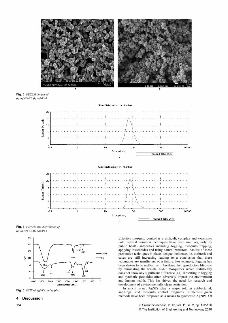
Zeta potential is an essential characteristic parameter fornanosuspensions[13]. It gives information on the stability anddispersibility of a material in a certain solution. High dispersibilityof the AgNPs in the solution allows better interaction between theAgNPs and the larvae, therefore increasing the effectiveness of thesolution. The zeta potential of AgNPs in deionised water wasobserved to be 40.1 ± 5.44 and 10.7 ± 12.5 mV for AgNPs-RT andAgNPs-T, respectively. High zeta potential value indicateselectrically stabilised particles while particles with low zeta valuetend to coagulate or aggregate. The better stability anddispersibility of the AgNPs-RT has probably contributed to itsbetter performance in eliminating the larvae at high concentration.As for the morphology of green-synthesised AgNPs, which wasviewed by FESEM (Fig. 3), the particles were identified by theirirregular, spherical and bean shapes. Agglomeration seemed to besignificant. The sizes of both samples were approximately in therange of 25–150 nm in size, in at least one dimension. Smallersizes of the AgNPs-RT may also play some roles in the anti-larvaeapplication. PSD tests measured the average size of AgNPs-RT andAgNPs-T to be 79.34 ± 26.43 and 93.18 ± 32.03 nm, respectively.The distribution of the particles is given in Fig. 4.
The FTIR spectra of AgNPs and apple extracts are shown inFig. 5. The AgNPs demonstrated peaks at 3366 cm−1 for O–Hstretching of alcohols, 1658 cm−1 for C–O stretching of amide,1142 and 1052 cm−1 for C–F stretching of alkyl halides or C–Ostretching of esters. Apple extracts revealed peaks at 3412 cm−1
attributable to O–H stretching of alcohols, 2981 cm−1 to C–Hstretching of alkanes, 1727 cm−1 to C=O stretching of carbonyl oraldehyde, 1663 cm−1 to C=O stretching of amide, 1037 cm−1 to C–O stretching of esters, 836 cm−1 to =C-H bending of alkene. Thedeviation of alcohol, amide and alkyl halides or ester observed atthe AgNPs with respect to the apple extracts indicates that thesefunctional groups could probably be involved in the synthesis ofthe AgNPs. TEM reveals the non-uniform circular shape of AgNPswith size ranging from 5 to 80 nm (Fig. 6).
3.2 Toxicity of AgNPs against fourth instar larvae of Ae.aegypti
The larvicidal activity of AgNPs, as presented in Table 1, showedmoderate toxic effect on the larvae after 24 h of exposure. TheLC50 = 15.76 ppm, LC90 = 27.7 ppm for AgNPs-RT and LC50 = 29.81 ppm, LC90 = 42..3 ppm for AgNPs-T were recorded. Forboth cases, the mortality increased as the concentration of theAgNPs increased (Table 1). Control samples were observed toshow 0% of mortality. Based on this preliminary screening results,we carried out the test of high concentration on the larvae. Thepattern of elimination is shown in Fig. 7. Comparison ofperformance between AgNPs-T and AgNPs-RT was alsoconducted. AgNPs-RT showed a slightly better performance. TheAgNPs-RT successfully eliminated all the larvae after 100 min,whereas the AgNPs-T after 160 min. The difference in the toxicitycould be attributable to smaller size and better dispersibility of theAgNPs-RT. These properties are characterised and discussed in thenext section.
Fig. 1 XRD spectrum of the synthesised AgNPs
Fig. 2 UV–visible spectra of the AgNPs
IET Nanobiotechnol., 2017, Vol. 11 Iss. 2, pp. 152-156© The Institution of Engineering and Technology 2016
153
4 Discussion
Effective mosquito control is a difficult, complex and expensivetask. Several common techniques have been used regularly bypublic health authorities including fogging, mosquito trapping,applying insecticides and using natural predators. Amidst of thesepreventive techniques in place, dengue incidence, i.e. outbreak andcases are still increasing leading to a conclusion that thesetechniques are insufficient or a failure. For example, fogging hasbeen shown to be ineffective in breaking the reproductive lifecycleby eliminating the female Aedes mosquitoes which statisticallydoes not show any significant difference [14]. Resorting to foggingand synthetic pesticides often adversely impact the environmentand human health. This has driven the need for research anddevelopment of environmentally clean pesticides.
In recent years, AgNPs play a major role in antibacterial,antifungal and mosquito control programs. Numerous greenmethods have been proposed as a means to synthesise AgNPs. Of
Fig. 3 FESEM images of(a) AgNPs-RT, (b) AgNPs-T
Fig. 4 Particle size distribution of(a) AgNPs-RT, (b) AgNPs-T
Fig. 5 FTIR of AgNPs and apple
154 IET Nanobiotechnol., 2017, Vol. 11 Iss. 2, pp. 152-156© The Institution of Engineering and Technology 2016

the AgNPs synthesised by fungus [15, 16] and bacteria [17],AgNPs synthesised using plant extract gain more interest in themosquito control [18, 19] owing to its availability, simplicity andlow cost. Synthesis of AgNPs by filamentous fungus Cochlioboluslunatus has been reported to have its larvicidal activity against thethird instar larvae of Ae. aegypti (LC50 = 1.48, LC90 = 3.33) [20].The value of LC50 = 0.79 ppm and LC90 = 1.09 ppm have beenobserved for Ae. aegypti treated with B. bassiana AgNPs [7]. Banuand Balasubramanian [21] had reported the activity of synthesisedAgNPs using Bacillus thuringiensis extract against the third larvaeof Ae. aegypti and found that LC50 = 0.10 ppm and LC90 = 0.39 ppm. Synthesised AgNPs using fungus C. tropicum extract testedagainst the third-instar larvae of Ae. aegypti resulted in the value of
LC50 and LC90 to be 4 and 8.91 ppm [22]. LC50 and LC90 againstAe. aegypti using AgNPs synthesised from mangroove plant extractwere found to be 0.585 and 2.615 mg/l.[12]. AgNPs-RTsynthesised by apple extract using non-heating method showedLC50 and LC90 values of 15.76 and 27.7 ppm against Ae. aegypti.The comparison of high dose (500 ppm) performance usingAgNPs-RT and AgNPs-T shows that there is slight difference inthe rate of mortality. This could be attributed to the smaller size ofthe AgNPs-RT. In addition, the AgNPs-RT were also observed toshow higher stability and better dispersibility in water which couldbe important factors that contribute to its improved toxicity.Therefore, these would be important factors in designing apromising and practical larvicidal dose.
In the synthesis of AgNPs, apple which acts as a reducing agentis widely available at low cost, therefore the use of highconcentration of AgNPs would not be a problem. Lokina et al. [10]reported that the –OH groups present in the apple extract couldinvolve the reduction of Ag+ to Ag0 through oxidation of alcohol toaldehyde group. In addition to that, biological molecules could alsoact as stabilising agent for the AgNPs [23].
Although the mechanism of larvicidal activity remains unclear,it is believed that the AgNPs has to be in contact with the larvae inorder to eliminate it. Sundaravadivelan [24] believed the idea putforth by Yamanaka et al. [25] on the mechanism of antibacterialstudy could also be applicable in the case of larvae. Yamanakaproposed that interaction of the biologically synthesised Ag withcytoplasm in the interior of the cell could denature the ribosome,resulting in the suppression of enzymes and proteins essential forATP production and eventually leads to cell disruption. Literaturesof a comparative study of larvicidal activity AgNPs towardsdifferent larvae are shown in Table 2 for future references.
5 ConclusionThe present investigation highlights the potential of the appleextract in the synthesis of AgNPs (AgNPs-RT and AgNPs-T) as apotential biolarvicidal agent for the dengue vector Ae. aegypti. TheAgNPs at high concentration have rapid impact on vector mosquitopopulation and thus conclude that AgNPs synthesised by appleextract could perhaps be a better, environmentally safer andgreener approach for vector control strategy. Further research couldbe carried out to find its precise formulation beforecommercialisation and integrating it into the society asbiolarvicides.
6 AcknowledgmentThe authors thank University of Malaya for providing facilities,equipments, grant (UMRG- RP019-14AFR), IPPP UM
Fig. 6 TEM of(a) AgNPs-RT, (b) AgNPs-T
Fig. 7 Pattern of the elimination of Ae. aegypti
Table 1 Larvicidal activity of AgNPs-RT and AgNPs-Tagainst fourth instar larvae of Ae. aegyptiSample Concentration,
ppm% Mortality,mean ± SD
LC50 LC90
AgNPs-RT 500 100.00 ± 0.00 15.76 ppm
27.7 ppm250 100.00 ± 0.0050 100.00 ± 0.0025 93.33 ± 7.64
12.5 16.67 ± 2.895 3.33 ± 2.86
AgNPs-T 500 100.00 ± 0.00 29.81 ppm
42..3 ppm250 100.00 ± 0.00
50 100.00 ± 0.0025 30 ± 21.74
12.5 13.33 ± 2.895 10.00 ± 5.00
IET Nanobiotechnol., 2017, Vol. 11 Iss. 2, pp. 152-156© The Institution of Engineering and Technology 2016
155

(PG009-2014A) and Ministry of Higher Education of Malaysia forMyBrain PhD Programme Scholarship (KPM(B) 870713095185).
7 References[1] TianCi, Y., Liang, L., GuiMing, F., et al.: ‘Epidemiology and vector efficiency
during a dengue fever outbreak in Cixi, Zhejiang Province, China’, J. VectorEcol., 2009, 34, (1), pp. 148–154
[2] Organization, W.H.: ‘Dengue and severe dengue’, WHO Fact sheet No117(updated September 2014). Available at http://www.who.int/mediacentre/factsheets/fs117/en, 2012
[3] Halstead, S.B.: ‘Dengue virus-mosquito interactions’, Annu. Rev. Entomol.,2008, 53, pp. 273–291
[4] Skae, F.: ‘Dengue fever in Penang’, BMJ, 1902, 2, (2185), p. 1581[5] Lam, S.: ‘Two decades of dengue in Malaysia’, J. Trop. Med., 1994, 35, (4),
pp. 195–200[6] Santhoshkumar, T., Rahuman, A.A., Rajakumar, G., et al.: ‘Synthesis of silver
nanoparticles using Nelumbo nucifera leaf extract and its larvicidal activityagainst malaria and filariasis vectors’, Parasitol. Res., 2011, 108, (3), pp.693–702
[7] Banu, A.N., Balasubramanian, C.: ‘Myco-synthesis of silver nanoparticlesusing Beauveria bassiana against dengue vector, Aedes aegypti (Diptera:Culicidae)’, Parasitol. Res., 2014, 113, (8), pp. 2869–2877
[8] Soni, N., Prakash, S.: ‘Possible mosquito control by silver nanoparticlessynthesized by soil fungus (Aspergillus niger 2587)’, 2013
[9] Umoren, S., Obot, I., Gasem, Z.: ‘Green synthesis and characterization ofsilver nanoparticles using red apple (Malus domestica) fruit extract at roomtemperature’, J. Mater. Environ. Sci., 2014, 5, pp. 907–914
[10] Lokina, S., Stephen, A., Kaviyarasan, V., et al.: ‘Cytotoxicity andantimicrobial activities of green synthesized silver nanoparticles’, Eur. J.Med. Chem., 2014, 76, pp. 256–263
[11] Larvicides, M.: ‘Guidelines for laboratory and field testing of mosquitolarvicides’, 2005
[12] Gnanadesigan, M., Anand, M., Ravikumar, S., et al.: ‘Biosynthesis of silvernanoparticles by using mangrove plant extract and their potential mosquitolarvicidal property’, Asian Pac. J. Trop. Med., 2011, 4, (10), pp. 799–803
[13] Müller, R., Jacobs, C., Kayser, O.: ‘Nanosuspensions as particulate drugformulations in therapy: rationale for development and what we can expectfor the future’, Adv. Drug Deliv. Rev., 2001, 47, (1), pp. 3–19
[14] Chua, K., Chua, I.L., Chua, I.E., et al.: ‘Effect of chemical fogging onimmature Aedes mosquitoes in natural field conditions’, Singapore Med. J.,2005, 46, (11), p. 639
[15] Soni, N., Prakash, S.: ‘Synthesis of gold nanoparticles by the fungusAspergillus niger and its efficacy against mosquito larvae’, Rep. Parasitol.,2012, 2, pp. 1–7
[16] Dhanasekaran, D., Thangaraj, R.: ‘Evaluation of larvicidal activity ofbiogenic nanoparticles against filariasis causing Culex mosquito vector’,Asian Pac. J. Trop. Dis., 2013, 3, (3), pp. 174–179
[17] Karthik, L., Kumar, G., Kirthi, A.V., et al.: ‘Streptomyces sp. LK3 mediatedsynthesis of silver nanoparticles and its biomedical application’, BioprocessBiosyst. Eng., 2014, 37, (2), pp. 261–267
[18] Jayaseelan, C., Rahuman, A.A.: ‘Acaricidal efficacy of synthesized silvernanoparticles using aqueous leaf extract of Ocimum canum againstHyalomma anatolicum anatolicum and Hyalomma marginatum isaaci (Acari:Ixodidae)’, Parasitol. Res., 2012, 111, (3), pp. 1369–1378
[19] Pasupuleti, V.R.: ‘Biogenic silver nanoparticles using Rhinacanthus nasutusleaf extract: synthesis, spectral analysis, and antimicrobial studies’, Int. J.nanomed., 2013, 8, p. 3355
[20] Suganya, G., Karthi, S., Shivakumar, M.S.: ‘Larvicidal potential of silvernanoparticles synthesized from Leucas aspera leaf extracts against denguevector Aedes aegypti’, Parasitol. Res., 2014, 113, (3), pp. 875–880
[21] Banu, A.N., Balasubramanian, C., Moorthi, P.V.: ‘Biosynthesis of silvernanoparticles using Bacillus thuringiensis against dengue vector, Aedesaegypti (Diptera: Culicidae)’, Parasitol. Res., 2014, 113, (1), pp. 311–316
[22] Soni, N., Prakash, S.: ‘Efficacy of fungus mediated silver and goldnanoparticles against Aedes aegypti larvae’, Parasitol. Res., 2012, 110, (1),pp. 175–184
[23] Roy, N., Mondal, S., Laskar, R.A., et al.: ‘Biogenic synthesis of Au and Agnanoparticles by Indian propolis and its constituents’, Colloids Surf. B,Biointerfaces, 2010, 76, (1), pp. 317–325
[24] Sundaravadivelan, C., Padmanabhan, M.N.: ‘Effect of mycosynthesized silvernanoparticles from filtrate of Trichoderma harzianum against larvae and pupaof dengue vector Aedes aegypti L’, Environ. Sci. Pollut. Res., 2014, 21, (6),pp. 4624–4633
[25] Yamanaka, M., Hara, K., Kudo, J.: ‘Bactericidal actions of a silver ionsolution on Escherichia coli, studied by energy-filtering transmission electronmicroscopy and proteomic analysis’, Appl. Environ. Microbiol., 2005, 71,(11), pp. 7589–7593
[26] Roni, M., Murugan, K., Panneerselvam, C., et al.: ‘Evaluation of leaf aqueousextract and synthesized silver nanoparticles using Nerium oleander againstAnopheles stephensi (Diptera: Culicidae)’, Parasitol. Res., 2013, 112, (3), pp.981–990
[27] Muthukumaran, U., Govindarajan, M., Rajeswary, M., et al.: ‘Synthesis andcharacterization of silver nanoparticles using Gmelina asiatica leaf extractagainst filariasis, dengue, and malaria vector mosquitoes’, Parasitol. Res.,2015, 114, (5), pp. 1–11
[28] Muthukumaran, U., Govindarajan, M., Rajeswary, M.: ‘Mosquito larvicidalpotential of silver nanoparticles synthesized using Chomelia asiatica(Rubiaceae) against Anopheles stephensi, Aedes aegypti, and Culexquinquefasciatus (Diptera: Culicidae)’, Parasitol. Res., 2014, pp. 1–11
[29] Shanmugasundaram, T., Balagurunathan, R.: ‘Mosquito larvicidal activity ofsilver nanoparticles synthesised using actinobacterium, Streptomyces sp. M25against Anopheles subpictus, Culex quinquefasciatus and Aedes aegypti’, J.Parasitic Dis., 2015, 39, (4), pp. 1–8
Table 2 Larvicidal activity AgNPsReducing agent Larvae species LC50/LC90,
ppmReferences
plantextracts
NelumbonuciferaGaertn.
An. subpictus 0.69/2.15 [6]Cx.
quinquefasciatus1.10/3.59
Neriumoleander
An. stephensi [26]
Gmelinaasiatica
An. stephensi 22.44/40.65 μg/ml
[27]
Cx.quinquefasciatus
27.83/48.92 μg/ml
Chomeliaasiatica
(Rubiaceae)
An. stephensi 17.95/33.03 μg/ml
[28]
Cx.quinquefasciatus
20.92/37.41 μg/ml
fungus F. oxysporum Cx.quinquefasciatus
8/12.3 [8]
Chrysosporiumtropicum
Ae. aegypti 3.47, 4, 2/12.30, 8.91,4 (for L1, L3.L4 after 1 h)
[22]
bacteria Streptomyces Rhipicephalusmicroplus
16.10/(N/A)mg/l
[17]
Haemaphysalisbispinosa
16.45/(N/A)mg/l
Streptomyces An. subpictus 51.34/(N/A)mg/L
[29]
Cx.quinquefasciatus
48.98/(N/A)mg/l
N/A, data not provided by the author.
156 IET Nanobiotechnol., 2017, Vol. 11 Iss. 2, pp. 152-156© The Institution of Engineering and Technology 2016