insilico analysis of mosaic virus proteins in different ... volume 3, issue 6/ijpab... · insilico...
TRANSCRIPT

Jain, P.A., et al Int. J. Pure App. Biosci. 3 (6): 257-274 (2015) ISSN: 2320 – 7051
Copyright © December, 2015; IJPAB 257
Insilico Analysis of Mosaic Virus Proteins in Different Plants of Cucurbit Family
Prashant Ankur Jain1*, Ankush Vinod Lal 1, Preetam Verma2 and Ved Kumar Mishra3
1Dept. of Computational Biology & Bioinformatics, Jacob School of Biotechnology & Bioengineering, Sam Higginbottom Institute of Agriculture, Technology & Sciences (Deemed to be University) Allahabad (U.P.)
2Dept. of MCE, Jacob School of Biotechnology & Bioengineering, Sam Higginbottom Institute of Agriculture, Technology & Sciences (Deemed to be University) Allahabad (U.P.)
3Ph.D. Scholar, Dept. of Computational Biology & Bioinformatics, Jacob School of Biotechnology & Bioengineering, Sam Higginbottom Institute of Agriculture, Technology &
Sciences (Deemed to be University) Allahabad (U.P.) *Corresponding Author E-mail: [email protected]
INTRODUCTION Cucumber mosaic virus (CMV) causing viral diseases of many important plants worldwide have been isolated from pumpkin (Cucurbitapepo L.) plant leaves. Diseased plants had light green mottled foliage. Leaves were smaller than normal, yellow mottled and crinkled. Cucumber mosaic, caused by the cucumber mosaic virus, is one of the most widespread and destructive diseases on cucumber and muskmelon. The disease has been known since the early 1900's, and is now found worldwide. The virus can infect cucumber, squash, muskmelon, and numerous other hosts in 34 plant families, including tomato, lima bean, beet, sweet corn, and sweet potato. Most often, actively growing and mature plants are affected. It rarely infects plants in the seedling stage, but will kill them quickly when it does. It causes a decrease in the number and the quality of the fruit.
Available online at www.ijpab.com
ISSN: 2320 – 7051 Int. J. Pure App. Biosci. 3 (6): 257-274 (2015)
Research Article
ABSTRACT Mosaic virus is caused by a variety of viruses which attack all members of the cucurbit family, but especially thrive on summer squash, cucumber and muskmelon plants. It is spread by a variety of methods and so is a serious disease for plants of the curcurbit family, including cucumbers, gourds, muskmelons, winter squash, summer squash, watermelons and pumpkins. The virus can infect cucumber, squash, muskmelon, and numerous other hosts in 34 plant families, including tomato, lima bean, beet, sweet corn, and sweet potato. Most often, actively growing and mature plants are affected. It rarely infects plants in the seedling stage, but will kill them quickly when it does. It causes a decrease in the number and the quality of the fruit. In this study with the help of various softwares we have elucidated the primary structure and secondary structure information and its components. The predicted and analyzed information can be further used in determining the three dimensional analysis and related studies for these proteins.
Key words:Mosaic Virus, Poly Protein, p3 Protein, sequence analysis.
DOI: http://dx.doi.org/10.18782/2320-7051.2103
Cite this article: Jain, P.A., Lal, A.V., Verma, P. and Mishra, V.K., Insilico Analysis of Mosaic Virus Proteins in Different Plants of Cucurbit Family, Int. J. Pure App. Biosci. 3 (6): 257-274 (2015). doi: http://dx.doi.org/10.18782/2320-7051.2103

Jain, P.A., et al Int. J. Pure App. Biosci. 3 (6): 257-274 (2015) ISSN: 2320 – 7051
Copyright © December, 2015; IJPAB 258
Cucumber mosaic virus (CMV) has one of the broadest host ranges. CMV as a type species of the genus Cucumovirus in the family Bromoviridae is reported to infect 1287 plant species in 518 genera belonging to 100 families3. It is geographically widespread and has been reported in Europe, Asia, Australia, North America and India. In Lithuania, this virus is spread on black currant13, leguminous14, ornamental12 and vegetable16plants, however not detected on pumpkin. The most common symptom induced by CMV is mosaic; however, severity of disease may range from no obvious symptoms in some crops to death of the host species. The virus causes fern leaf, stunting of vegetable crops and malformation of their fruits. It is transmitted by numerous species of aphid, through the sap, the seeds and dodder2,5,7. Morphologically CMV has rather characteristic about 30 nm polyhedral particles with hollow centre9. The genome consists of three plus sense single-stranded RNAs, packaged in separate particles. CMV particles contain about 18% RNA. The RNAconsists of 4 RNAs. Only largest RNA3 are required for infectivity11. The virions are not stable to freezing. Long-term storage of CMV is most reliable in the form of viral RNA, which is highly infectious, and very stable at −20ºC10. Great number of different CMV strains, serogroups, subgroups and biological variations has been described1,4,6,8,15. Unfortunately, there is no chemical control for mosaic virus, and plants need to be removed and destroyed promptly if they are infected with this viral disease. To control the spread of the disease by cucumber beetles and aphids, we need to control these insect populations with a diazinon containing insecticide repeating the application as much as necessary in seven day intervals. Although there are mosaic virus resistant cucumber varieties, so far no resistant varieties of muskmelon and summer squash are available to plant.Secondary structure in biochemistry and structural biology describes the general three-dimensional form of local segments of biopolymers such as proteins and nucleic acids (DNA/RNA). It does not, however, describe specific atomic positions in three-dimensional space, which are considered to be tertiary structure.
MATERIALS AND METHODS
The target selected was tobacco mosaic virus protein. The primary sequence from retrieved from NCBI database. In the present work an integrated approach was used for sequence analysis. The query protein sequence of interest is subjected to an exhaustive sequence similarity search conducted over all accessible sequence databases by standard sequence analysis tools and it yields in 1D topology of the sequence. In the next step of this procedure “2D topology” is obtained which provides a platform to rationalize highly conserved sequence positions in terms of structural and functional relevance and further allows gaining qualitative insights into possible helix-helix interactions. The 2D topology is a prerequisite receptor sequence representation, defining the interrelation between the family-wide sequence characteristics. Following Software and databases were used in this study. 1. Primary Sequence retrieval: NCBI (www.ncbi.nlm.nih.gov) 2. Sequence Alignment tools: BLASTP (www.ncbi.nlm.nih.gov/blast) 3. Primary Structure Analysis Protoparam: (http://an.expasy.org/tools/peotoparam.html) 4. Tm Region Prediction: TMHMM (www.cbs.dtn.dk/services/tmhmm-2.0) 5. Signal P3.0 Server Prediction: (www.cbs.dtn.dk/services/signalP) 6. Sumo PlotTM Prediction: (www.abgent.com/doc/sumoplot) 7. Secondary Structure Prediction: SOPMA:(www.cbs.dtn.dk/services/sopma) 8. Minimum System Configuration: Pentium 4 CPU, 3.8 GHz, 512 MB of RAM 9. Operating System: Windows
The sequence details were retrieved by searching GENE BANK using Entrees Browser. In predicting the 1D topology Proto Param was used for computing molecular weight, theoretical isoelectric point (p1), instability index and aliphatic index. Self Optimized prediction method (SOPM) was used to give a four state description of secondary structure (alpha helix, beta sheet, turn and coils). The output width was set at 70. Prediction of protein membrances in the target was done using Tmpred & TMHMM. Prediction of cleavage sites as well as signal peptide/non-signal peptide prediction on combination of several rtificial neural networks and hidden markov models was done using SinalP server. Small Ubiquitin – like modifier (SUMO) protein can be conjugated to substrates by enzymes that operate in ubiquitylation, which mark proteins for rapid intercellular degradation. SUMOplotTM was used to
Jain, P.A., et al Int. J. Pure App. Biosci. 3 (6): 257-274 (2015) ISSN: 2320 – 7051
Copyright © December, 2015; IJPAB 259
predict the probability for the SUMO consensus sequences (SUMO-CS) to be engaged in SUMO attachment of query sequence. Neutral network prediction for the attachment of O-linked N-acetylglucose amine (O-Glc NAC) in protein was done using Ying O Yang Server. By following all the above procedures, the protein sequence is subjected to an exhaustive sequence similarity search conducted over all accessible sequence databases by standard sequence analysis tools and yields its primary and secondary structure topology.
RESULTS& DISCUSSIONS • Sequence Retrieval: Every step in sequence / structural analysis of the target proteins is found to be crucial to end -up with an accurate model . The accuracy and the reliability of a theoretical protein model often depend on the template structures and the alignment between the target and template sequence. The target sequences were taken from NCBI. To begin with, the FASTA formats of the proteins were retrieved.
>gi|51949951|ref|YP_077272.1| P3 protein [Watermelon mosaic virus]
GEAQQRMKCETALIKSIFKPKRMIQILEDDPYILLMGLISPSILIHMYRMK HFEKGIELWISK EHSVAKIFIIMEQLTRKIAANDLLLEQLDIIAGTSQKLMDVLEDCPQSAHS YRTAKDLLAIY IERRASNNQLIENGFVDINDQLYVTHEKIYVDRLKQEWHALSWLEKSSITWQLKRFTPHTE QCLTKKVVEESSAYSRNFVSACFMNAQSHLKNVRNTFFRKCDQAWTASVRVLVRFIIATL HKCYSDIVYLVNICLIFSLLVQMVSVLQGIVSTAKRDKAFVHMHKRIEDEQ AVVHLYEMC EKMENKHPSVEEFLSHVKKVRPELLPVAKSMTGQSEDVSAQ
>gi|365777336|gb|AEW91906.1| polyprotein, partial [Papaya ringspot virus W]
AAMIESWGYGELTHQIRRFYQWVLEQAPFNELARQGRAPYVSEVGLRRLYTSERGSMDE LEAYIDKYFERERGDSPELLVYHESRIADDYQLVCSNNTHVFHQSKNEAVDAGLNEKLKE KEKQKEKEKEKQKEKEKDDASDGNDVSTSTKTGERDRDVNVGTSGTFTVPRIKSFTDKMI LPRIKGKTVLNLNHLLQYNPQQIDISNTRATQSQFEKWYEGVRNDYGLNDNEMQVMLNG LMVWCIENGTSPDISGVWVMMDGETQVDYPIKPLIEHATPSFRQIMAHFSNAAEAYIAKR NATERYMPRYGIKRNLTDISLARYAFDFYEVNSKTPDRAREAHMQMKAAAL RNTSRRMF GMDGSVSNKEENTERHTVEDVNRDMHSLLGMRN >gi|343790821|dbj|BAK61797.1| 1a protein [Cucumber mosaic virus]
MATSSFNINELVASHGDKGLLATALVDKTAHEQLEEQLQHQRRGRKVYIRNVLGVKDSE VIRNRYGGKYDLHLTQQEFAPHGLAGALRLCETLDCLDSFPSSGLRQDLVLDFGGSWVTH YLRGHNVHCCSPCLGIRDKMRHAERLMNMRKIILNDPQQFDGRQPDFCTQPAADCKVQA HFAISIHGGYDMGFRGLCEAMNAHGTTILKGTMMFDGAMMFDDQGVIPELNCQWRKIRS AFSETEDVTPLSGKLNSTVFSRVRKFKTMVAFDFINESTMSYVHDWENIKSFLTDQTYSYR GMTYGIERCVIHAGIMTYKIIGVPGMCPPELIRHCIWFPSIKDYVGLKIPASQDLVEWKTVR ILMSTLRETEEIAMRCYNDKKAWMEQFKVILGVLSAKSSTIVINGMSMQSGERIDINDYHY IGFAILLHTKMKYEQLGKMYDMWNASSISKWFAALTRPLRVFLSGVVHALF PTLRPREEK EFLIKLSTFVTFNEECSFDGGEEWDVISSAAYVATQAVTDGKILAAQKAEKLAEKLAQPVI EVSDSPEAPSQTPDDTAEVCGKEREVSELDSLSAQTRSPITRVAERATAMLEYAAYEKQLH DTTVSNLKRIWNMAGGDDKRNSLEGNLKFVFDTYFTVDPMVNIHFSTGRWMRPVPEGV VYSVGYNERGLGPKSDGELYIVNSECVICNSESLSTVTRSLQAPTGTISQVDGVAGCGKTT AIKSIFEPSTDMIVTANKKSAQDVRMALFKSSDSKEACTFVRTADSVLLNECPTVSRVLVD EVVLLHFGQLCAVMSKLKAVRAICFGDSEQIAFSSRDASFDMRFSKIIPDETSDADTTFRSP QDVVPLVRLMATKALPKGTRSKYTKWVSQSKVKRSVTSRAIVSVTLVDLDPSRFYITMTQ ADKASLISRAKEMNLPKTFWNERIKTVHESQGISEDHVTLVRLKSTKCDLFKQFSYCLVAL TRHKVTFRYEYCGVLNGDLIAECVARA

Jain, P.A., et al Int. J. Pure App. Biosci. 3 (6): 257-274 (2015) ISSN: 2320 – 7051
Copyright © December, 2015; IJPAB 260
>gi|37927249|gb|AAO45984.1| P3 protein [Zucchini yellow mosaic virus]
PYILLLGMISPTILVHMYRMRHFERGIEVWIKRDHEIGKIFVILEQLTRKV ALAEVLVDQLN LISEASPHLLEIMKGCQDNQRAYVPALDLLTIQVEREFSNKELKTNGYPDLQQTLFDMWE KMYAKQLHNSWQELSLLEKSCVTVRLKQFSIFTERNLIQRAEEGKRASSLQ
Next, a set of related sequences were submitted to the multiple sequence alignment server CLUSTALW. The templates assigned were obtained through the BLASTp search using SWISS-PROT and PDB databases. The CLUSTAL’s output format is compatible with GDE, PHYLIP or GCG packages. CLUSTALW involves a progressive strategy for aligning pairs of sequences .The CLUSTAL server was selected for sequence analysis as it exploits the fact that similar sequences are likely to be evolutionarily related and it expressed the degree of similarity in a relatively concised format . As part of its operation , the program produced information required to produce a phylogenetic tree.
Fig.1:Cladogram of protein sequences
By the analysis of the phylogram and Cladogram it can be predicted that sequences gi/3657773 and gi/34379008 are forming a clade and more closely related to each other then other sequences. � Primary Structure Analysis (ProtParam): P3 protein (Watermelon mosaic virus)
Number of amino acids:347 ;Molecular weight: 40288.0 ; Theoretical pI: 8.51
Total number of negatively charged residues (Asp + Glu): 41
Total number of positively charged residues (Arg + Lys): 45
Atomic composition:
Carbon C 1805
Hydrogen H 2887
Nitrogen N 489
Oxygen O 511
Sulfur S 21
Formula:C1805H2887N489O511S21;Total number of atoms: 5713
Instability index: The instability index (II) is computed to be 45.08. This classifies the protein as
unstable.
Aliphatic index:100.00 ;Grand average of hydropathicity (GRAVY): -0.144
Fig.2:Phylogram of protein sequences

Jain, P.A., et al Int. J. Pure App. Biosci. 3 (6): 257-274 (2015) ISSN: 2320 – 7051
Copyright © December, 2015; IJPAB 261
� Polyprotein, partial (Papaya ring spot virus W)
Number of amino acids:390 ;Molecular weight: 45144.5 ; Theoretical pI: 6.01
Total number of negatively charged residues (Asp + Glu): 61
Total number of positively charged residues (Arg + Lys): 55
Atomic composition:
Carbon C 1961
Hydrogen H 3074
Nitrogen N 572
Oxygen O 619
Sulfur S 18
Formula:C1961H3074N572O619S18 ;Total number of atoms: 6244
Instability index: The instability index (II) is computed to be 35.49. This classifies the protein as stable.
Aliphatic index:64.77 ;Grand average of hydropathicity (GRAVY): -0.864
� 1a protein (Cucumber mosaic virus)
Number of amino acids:993 ;Molecular weight: 111421.8 ; Theoretical pI: 7.49
Total number of negatively charged residues (Asp + Glu): 118
Total number of positively charged residues (Arg + Lys): 119
Atomic composition:
Carbon C 4923 Hydrogen H 7790 Nitrogen N 1354 Oxygen O 1472 Sulfur S 60
Formula:C4923H7790N1354O1472S60 ;Total number of atoms: 15599
Instability index: The instability index (II) is computed to be 42.32. This classifies the protein as
unstable.
Aliphatic index:82.27 ;Grand average of hydropathicity (GRAVY): -0.206
� P3 protein (Zucchini yellow mosaic virus)
Number of amino acids:173 ;Molecular weight: 20327.7 ; Theoretical pI: 7.42
Total number of negatively charged residues (Asp + Glu): 22
Total number of positively charged residues (Arg + Lys): 22
Atomic composition:
Carbon C 915
Hydrogen H 1468
Nitrogen N 248
Oxygen O 258
Sulfur S 8
Formula:C915H1468N248O258S8 ;Total number of atoms: 2897
Jain, P.A., et al Int. J. Pure App. Biosci. 3 (6): 257-274 (2015) ISSN: 2320 – 7051
Copyright © December, 2015; IJPAB 262
Instability index: The instability index (II) is computed to be 46.05. This classifies the protein as
unstable.
Aliphatic index:107.05 ;Grand average of hydropathicity (GRAVY): -0.218
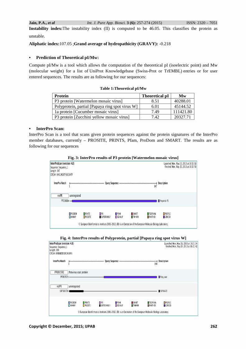
• Prediction of Theoretical pI/Mw:
Compute pI/Mw is a tool which allows the computation of the theoretical pI (isoelectric point) and Mw (molecular weight) for a list of UniProt Knowledgebase (Swiss-Prot or TrEMBL) entries or for user entered sequences. The results are as following for our sequences:
Table 1:Theoretical pI/Mw
Protein Theoretical pI Mw P3 protein [Watermelon mosaic virus] 8.51 40288.01 Polyprotein, partial [Papaya ring spot virus W] 6.01 45144.52 1a protein [Cucumber mosaic virus] 7.49 111421.80 P3 protein [Zucchini yellow mosaic virus] 7.42 20327.71
• InterPro Scan: InterPro Scan is a tool that scans given protein sequences against the protein signatures of the InterPro member databases, currently – PROSITE, PRINTS, Pfam, ProDom and SMART. The results are as following for our sequences
Fig. 3: InterPro results of P3 protein [Watermelon mosaic virus]
Fig. 4: InterPro results of Polyprotein, partial [Papaya ring spot virus W]

Jain, P.A., et al Int. J. Pure App. Biosci. 3 (6): 257-274 (2015) ISSN: 2320 – 7051
Copyright © December, 2015; IJPAB 263
Fig. 5: InterPro results of 1a protein [Cucumber mosaic virus]
Fig. 6: InterPro results of P3 protein [Zucchini yellow mosaic virus]
SignalP was used to predict the presence of signal peptide sequence in the N –terminal region. There is no signal peptide predicted and the signal peptide probability is found to be zero. Thus, it could be safely predicted that the target sequences are non-secretory proteins .
Fig.7: SignalP results of P3 protein [Watermelon mosaic virus]

Jain, P.A., et al Int. J. Pure App. Biosci. 3 (6): 257-274 (2015) ISSN: 2320 – 7051
Copyright © December, 2015; IJPAB 264
Fig.8: SignalP results of Polyprotein, partial [Papaya ring spot virus W]
Fig.9: SignalP results of 1a protein [Cucumber mosaic virus]
Fig. 10: SignalP results of P3 protein [Zucchini yellow mosaic virus]

Jain, P.A., et al Int. J. Pure App. Biosci. 3 (6): 257-274 (2015) ISSN: 2320 – 7051
Copyright © December, 2015; IJPAB 265
• The SUMOplot™ Analysis: Program predicts and scores sumoylation sites in protein. The SUMOplot™ Analysis Program predicts the probability for the SUMO consensus sequence (SUMO-CS) to be engaged in SUMO attachment. The SUMOplot™ score system is based on two criteria: direct amino acid match to SUMO-CS. substitution of the consensus amino acid residues with amino acid residues exhibiting similar hydrophobicity in the results the red colored amino acids represents Motifs with high probability, blue represents Motifs with low probability and green represents overlapping Motifs.
� P3 protein [Watermelon mosaic virus]
Table 2. Motifs position in P3 protein [Watermelon mosaic virus]
No. Pos. Group Score
1 K160 IYVDR LKQE WHALS 0.91
2 K8 EAQQR MKCE TALIK 0.80
3 K281 GIVST AKRD KAFVH 0.79
4 K179 SITWQ LKRF TPHTE 0.56
5 K308 LYEMC EKME NKHPS 0.50
6 K51 IHMYR MKHF EKGIE 0.45
7 K226 RNTFF RKCD QAWTA 0.44
8 K69 KEHSV AKIF IIMEQ 0.44
9 K284 STAKR DKAF VHMHK 0.15
� Polyprotein, partial [Papaya ring spot virus W]
Table 3. Motifs position in Polyprotein, partial [Papaya ring spot virus W]
No. Pos. Group Score
1 K172 FTVPR IKSF TDKMI 0.59
2 K136 KQKEK EKDD ASDGN 0.50
3 K66 LEAYI DKYF ERERG 0.15

Jain, P.A., et al Int. J. Pure App. Biosci. 3 (6): 257-274 (2015) ISSN: 2320 – 7051
Copyright © December, 2015; IJPAB 266
� 1a protein [Cucumber mosaic virus]
Table 4. motifs position in 1a protein [Cucumber mosaic virus]
No. Pos. Group Score
1 K433 LLHTK MKYE QLGKM 0.80
2 K346 KDYVG LKIP ASQDL 0.80
3 K68 RNRYG GKYD LHLTQ 0.67
4 K675 ERGLG PKSD GELYI 0.61
5 K287 HDWEN IKSF LTDQT 0.59
6 K529 KILAA QKAE KLAEK 0.50
7 K957 TKCDL FKQF SYCLV 0.50
8 K922 KEMNL PKTF WNERI 0.26
9 K481 LRPRE EKEF LIKLS 0.15
� P3 protein [Zucchini yellow mosaic virus]
Table 5. motifs position in P3 protein [Zucchini yellow mosaic virus]
No. Pos. Group Score
1 K32 GIEVW IKRD HEIGK 0.94
2 K149 CVTVR LKQF SIFTE 0.56
3 K39 RDHEI GKIF VILEQ 0.32
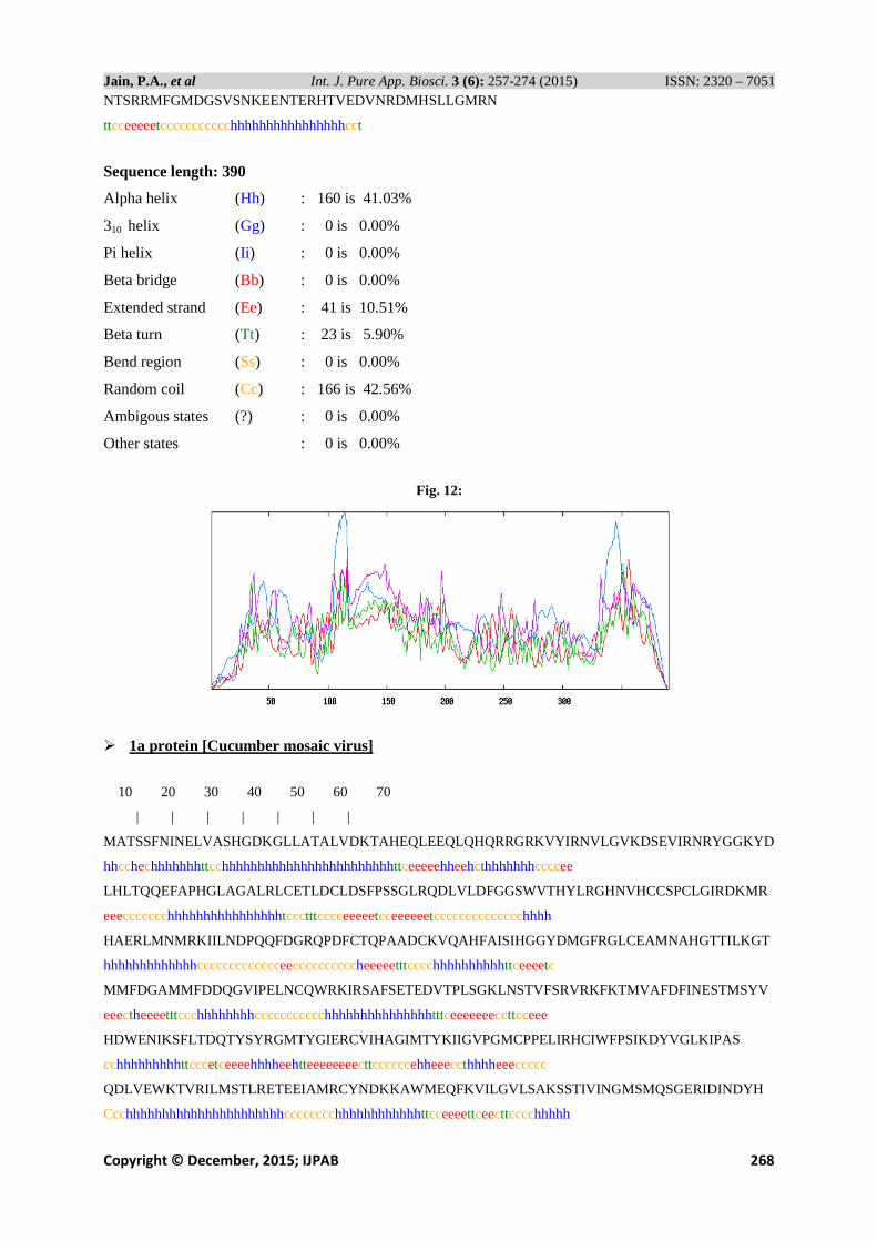
• SOPMA Secondary Structure Prediction: Self-Optimized Prediction Method (SOPM) has been described to improve the success rate in the prediction of the secondary structure of proteins. This method (SOPMA) correctly predicts 69.5% of amino acids for a three-state description of the secondary structure (alpha-helix, beta-sheet and coil) in a whole database containing 126 chains of non-homologous (less than 25% identity) proteins. Joint prediction with SOPMA and a neural networks method (PHD) correctly predicts 82.2% of residues for 74% of co-predicted amino acids.In our case we have selected the Window width-17, Similarity threshold -08 and Number of states- 04.
� P3 protein [Watermelon mosaic virus]
10 20 30 40 50 60 70
| | | | | | |
GEAQQRMKCETALIKSIFKPKRMIQILEDDPYILLMGLISPSILIHMYRMK HFEKGIELWISKEHSVAKI
chhhhhhhhhhhhhhhcccthhhhhhhttttceeeeehccttheeeehcttchhhhhhhhhcttcchhhh
FIIMEQLTRKIAANDLLLEQLDIIAGTSQKLMDVLEDCPQSAHSYRTAKDL LAIYIERRASNNQLIENGF
hhhhhhhhhhhhhhhhhhhhhhhhhhhhhhhhhhhhhcccccchhhhhhhhhhhhhhhhhcchhhhhttc
VDINDQLYVTHEKIYVDRLKQEWHALSWLEKSSITWQLKRFTPHTEQCLTKKVVEESSAYSRNFVSACFM
Eehhhhhhhhhhhhhhhhhhhhhhhhhhhhhhhhhhhhhccccccccccchhhhhccccchhhhhhhhhh

Jain, P.A., et al Int. J. Pure App. Biosci. 3 (6): 257-274 (2015) ISSN: 2320 – 7051
Copyright © December, 2015; IJPAB 267
NAQSHLKNVRNTFFRKCDQAWTASVRVLVRFIIATLHKCYSDIVYLVNICL IFSLLVQMVSVLQGIVSTA
hhhhhhhhhhhhhhhhhhhhhhhhhhhhhhhhhhhhhhhcchhhhhhhhhhhhhhhhhhhhhhhhhhhhh
KRDKAFVHMHKRIEDEQAVVHLYEMCEKMENKHPSVEEFLSHVKKVRPELLPVAKSMTGQSEDVSAQ
Hhhhhhhhhhhhhhhhhhhhhhhhhhhhhttcccchhhhhhhhhhcchhhhhhhhhhhhtcchhhhc
Sequence length: 347
Alpha helix (Hh) : 272 is 78.39%
310 helix (Gg) : 0 is 0.00%
Pi helix (Ii ) : 0 is 0.00%
Beta bridge (Bb) : 0 is 0.00%
Extended strand (Ee) : 11 is 3.17%
Beta turn (Tt) : 16 is 4.61%
Bend region (Ss) : 0 is 0.00%
Random coil (Cc) : 48 is 13.83%
Ambigous states (?) : 0 is 0.00%
Other states : 0 is 0.00%
Fig. 11:
� Polyprotein, partial [Papaya ring spot virus W]
10 20 30 40 50 60 70
| | | | | | |
AAMIESWGYGELTHQIRRFYQWVLEQAPFNELARQGRAPYVSEVGLRRLYTSERGSMDELEAYIDKYFER
hhhhhhttchhhhhhhhhhhhhhhtccchhhhhhttccchhhhhhhhhhhhccccchhhhhhhhhhhhhh
ERGDSPELLVYHESRIADDYQLVCSNNTHVFHQSKNEAVDAGLNEKLKEKEKQKEKEKEKQKEKEKDDAS
ccccccceeeecccccctteeeeettccceeetccchhhhhhhhhhhccccccccccccccccccccccc
DGNDVSTSTKTGERDRDVNVGTSGTFTVPRIKSFTDKMILPRIKGKTVLNLNHLLQYNPQQIDISNTRAT
ccccccccccccccccccccccccceeccccchhcccccccccttcceeehhheeeccccccchhccccc
QSQFEKWYEGVRNDYGLNDNEMQVMLNGLMVWCIENGTSPDISGVWVMMDGETQVDYPIKPLIEHATPSF
hhhhhhhhhhhhhhhccchhhhhhhhttheeeeecttccccccceeeeecccccccccccchhhtccchh
RQIMAHFSNAAEAYIAKRNATERYMPRYGIKRNLTDISLARYAFDFYEVNSKTPDRAREAHMQMKAAALR
hhhhhhhhhhhhhhhhhhtccccccchhccchtcchhhhhhhheeeeeecccccchhhhhhhhhhhhhhh
Jain, P.A., et al Int. J. Pure App. Biosci. 3 (6): 257-274 (2015) ISSN: 2320 – 7051
Copyright © December, 2015; IJPAB 268
NTSRRMFGMDGSVSNKEENTERHTVEDVNRDMHSLLGMRN
ttcceeeeetccccccccccchhhhhhhhhhhhhhhhcct
Sequence length: 390
Alpha helix (Hh) : 160 is 41.03%
310 helix (Gg) : 0 is 0.00%
Pi helix (Ii ) : 0 is 0.00%
Beta bridge (Bb) : 0 is 0.00%
Extended strand (Ee) : 41 is 10.51%
Beta turn (Tt) : 23 is 5.90%
Bend region (Ss) : 0 is 0.00%
Random coil (Cc) : 166 is 42.56%
Ambigous states (?) : 0 is 0.00%
Other states : 0 is 0.00%
Fig. 12:
� 1a protein [Cucumber mosaic virus]
10 20 30 40 50 60 70
| | | | | | |
MATSSFNINELVASHGDKGLLATALVDKTAHEQLEEQLQHQRRGRKVYIRNVLGVKDSEVIRNRYGGKYD
hhcchechhhhhhhttcchhhhhhhhhhhhhhhhhhhhhhhhttceeeeehheehcthhhhhhhccccee
LHLTQQEFAPHGLAGALRLCETLDCLDSFPSSGLRQDLVLDFGGSWVTHYLRGHNVHCCSPCLGIRDKMR
eeeccccccchhhhhhhhhhhhhhhhtccctttcccceeeeetcceeeeeetcccccccccccccchhhh
HAERLMNMRKIILNDPQQFDGRQPDFCTQPAADCKVQAHFAISIHGGYDMGFRGLCEAMNAHGTTILKGT
hhhhhhhhhhhhhccccccccccccceeccccccccccheeeeetttcccchhhhhhhhhhttceeeetc
MMFDGAMMFDDQGVIPELNCQWRKIRSAFSETEDVTPLSGKLNSTVFSRVRKFKTMVAFDFINESTMSYV
eeectheeeetttccchhhhhhhhccccccccccchhhhhhhhhhhhhhhtttceeeeeeeccttcceee
HDWENIKSFLTDQTYSYRGMTYGIERCVIHAGIMTYKIIGVPGMCPPELIRHCIWFPSIKDYVGLKIPAS
cchhhhhhhhhttcccetceeeehhhheehtteeeeeeeecttccccccehheeeccthhhheeeccccc
QDLVEWKTVRILMSTLRETEEIAMRCYNDKKAWMEQFKVILGVLSAKSSTI VINGMSMQSGERIDINDYH
Ccchhhhhhhhhhhhhhhhhhhhhhcccccccchhhhhhhhhhhhttcceeeettceecttcccchhhhh

Jain, P.A., et al Int. J. Pure App. Biosci. 3 (6): 257-274 (2015) ISSN: 2320 – 7051
Copyright © December, 2015; IJPAB 269
YIGFAILLHTKMKYEQLGKMYDMWNASSISKWFAALTRPLRVFLSGVVHAL FPTLRPREEKEFLIKLSTF
hhhhhhhhhhhhhhhhhhhhhhhhhhtccchhhhhhccchhhhhhhhhhhcchhhhhhhhhhhhhhhcch
VTFNEECSFDGGEEWDVISSAAYVATQAVTDGKILAAQKAEKLAEKLAQPVIEVSDSPEAPSQTPDDTAE
eeehhhhhhcccccchhhhhhhhhhhhhhhhhhhhhhhhhhhhhhhhhhhhheeccccccccccccchhc
VCGKEREVSELDSLSAQTRSPITRVAERATAMLEYAAYEKQLHDTTVSNLKRIWNMAGGDDKRNSLEGNL
ccccccccchhccccccccceecchhhhhhhhhhhhhhhhhhhhhhhhhhhhhheettcccccccchhhh
KFVFDTYFTVDPMVNIHFSTGRWMRPVPEGVVYSVGYNERGLGPKSDGELYIVNSECVICNSESLSTVTR
hhhhhhhhhhhhhheecccttceeccccteeeeeeeccttcccccccceeeeeccccehhhhhhhhhhcc
SLQAPTGTISQVDGVAGCGKTTAIKSIFEPSTDMIVTANKKSAQDVRMALFKSSDSKEACTFVRTADSVL
cccccccceeeeetcccccccceehhhccttceeeeeetccchhhhhhhhhhccccthhhheehhhhhhe
LNECPTVSRVLVDEVVLLHFGQLCAVMSKLKAVRAICFGDSEQIAFSSRDASFDMRFSKIIPDETSDADT
etcccthhheehhhheecchhhhhhhhhhhhhheeeeeccccceeeeccccceeeeeeeecccccccccc
TFRSPQDVVPLVRLMATKALPKGTRSKYTKWVSQSKVKRSVTSRAIVSVTLVDLDPSRFYITMTQADKAS
cccccchhhhhhhhhhhtcccttcccceeeeecchhhhhhccccceeeeeecccccteeeeeecccchhh
LISRAKEMNLPKTFWNERIKTVHESQGISEDHVTLVRLKSTKCDLFKQFSYCLVALTRHKVTFRYEYCGV
hhhhhhhhtccchhhhhhhhhhhhtttccccheeeeeeccccchhhhhhhhheeeehtttcceeeeeccc
LNGDLIAECVARA cccceehhhhhhh
Sequence length:993
Alpha helix (Hh) : 437 is 44.01%
310 helix (Gg) : 0 is 0.00%
Pi helix (Ii ) : 0 is 0.00%
Beta bridge (Bb) : 0 is 0.00%
Extended strand (Ee) : 178 is 17.93%
Beta turn (Tt) : 66 is 6.65%
Bend region (Ss) : 0 is 0.00%
Random coil (Cc) : 312 is 31.42%
Ambigous states (?) : 0 is 0.00%
Other states : 0 is 0.00%
Fig. 13:

Jain, P.A., et al Int. J. Pure App. Biosci. 3 (6): 257-274 (2015) ISSN: 2320 – 7051
Copyright © December, 2015; IJPAB 270
� P3 protein [Zucchini yellow mosaic virus]
10 20 30 40 50 60 70
| | | | | | |
PYILLLGMISPTILVHMYRMRHFERGIEVWIKRDHEIGKIFVILEQLTRKV ALAEVLVDQLNLISEASPH
cheeeehhccthheeehhcttchhhhhhheeccccchhhhhhhhhhhhhhhhhhhhhhhhhhhhhhhhhh
LLEIMKGCQDNQRAYVPALDLLTIQVEREFSNKELKTNGYPDLQQTLFDMWEKMYAKQLHNSWQELSLLE
hhhhhhhccccccchhhhhhhhhhhhhhcccchhhhhttceehhhhhhhhhhhhhhhhhhhhhhhhhhhh
KSCVTVRLKQFSIFTERNLIQRAEEGKRASSLQ
hhhheehhcccchhhhhhhhhhhhttcccceee
Sequence length: 173
Alpha helix (Hh) : 120 is 69.36%
310 helix (Gg) : 0 is 0.00%
Pi helix (Ii ) : 0 is 0.00%
Beta bridge (Bb) : 0 is 0.00%
Extended strand (Ee) : 16 is 9.25%
Beta turn (Tt) : 7 is 4.05%
Bend region (Ss) : 0 is 0.00%
Random coil (Cc) : 30 is 17.34%
Ambigous states (?) : 0 is 0.00%
Other states : 0 is 0.00%
Fig. 14:
• Predict transmembrane: From the analysis of TMHMM result it could be inferred that there are no transmembrane helices present in this sequence so, it is not likely to be a transmembrane protein .It also indicates that any of the predicted transmembrane helix in the N-term is not a signal peptide.
� P3 protein [Watermelon mosaic virus]
Sequence Length : 347
# Sequence Number of predicted TMHs : 1
# Sequence Exp number of AAs in TMHs : 30.61454
# Sequence Exp number, first 60 AAs : 10.07098
# Sequence Total prob of N-in : 0.74772

Jain, P.A., et al Int. J. Pure App. Biosci. 3 (6): 257-274 (2015) ISSN: 2320 – 7051
Copyright © December, 2015; IJPAB 271
Sequence POSSIBLE N-term signal sequence
Sequence TMHMM2.0 inside 1 253
Sequence TMHMM2.0 TMhelix 254 276
Sequence TMHMM2.0 outside 277 347
Fig. 15:
� Polyprotein, partial [Papaya ring spot virus W]
Sequence Length : 390
# Sequence Number of predicted TMHs : 0
# Sequence Exp number of AAs in TMHs : 0.00127
# Sequence Exp number, first 60 AAs : 0
# Sequence Total prob of N-in : 0.00659
Sequence TMHMM2.0 outside 1 390
Fig. 16:

Jain, P.A., et al Int. J. Pure App. Biosci. 3 (6): 257-274 (2015) ISSN: 2320 – 7051
Copyright © December, 2015; IJPAB 272
� 1a protein [Cucumber mosaic virus]
Sequence Length: 993
# Sequence Number of predicted TMHs : 0
# Sequence Exp number of AAs in TMHs : 0.03494
# Sequence Exp number, first 60 AAs : 0
# Sequence Total prob of N-in : 0.00152
Sequence TMHMM2.0 outside 1
Fig. 17:
993
� P3 protein [Zucchini yellow mosaic virus]
Sequence Length : 173
# Sequence Number of predicted TMHs : 0
# Sequence Exp number of AAs in TMHs : 0.4565
# Sequence Exp number, first 60 AAs : 0.4565
# Sequence Total prob of N-in : 0.03243
Sequence TMHMM2.0 outside 1 173
Fig. 18:

Jain, P.A., et al Int. J. Pure App. Biosci. 3 (6): 257-274 (2015) ISSN: 2320 – 7051
Copyright © December, 2015; IJPAB 273
CONCLUSION Several viruses that affect cucumbers, melons, pumpkins, squash, and other members of the cucurbit family e.g. Cucumber mosaic virus (CMV), zucchini yellow mosaic virus (ZYMV), watermelon mosaic virus (WMV) and papaya ring spot-W (PRSV-W). All are transmitted from diseased plants to healthy plants by aphids from plant to plant in a non-persistent manner. This means they acquire the virus from an infected plant almost immediately but are able to infect healthy plants for only a short time, usually a few days to a week. Only a small number of aphids are needed to spread the virus throughout the field. CMV is also spread by spotted and striped cucumber beetles.Cucumber mosaic, caused by the cucumber mosaic virus, is one of the most widespread and destructive diseases on cucumber and muskmelon. The disease has been known since the early 1900's, and is now found worldwide. The virus can infect cucumber, squash, muskmelon, and numerous other hosts in 34 plant families, including tomato, lima bean, beet, sweet corn, and sweet potato. Most often, actively growing and mature plants are affected. It rarely infects plants in the seedling stage, but will kill them quickly when it does. It causes a decrease in the number and the quality of the fruit. Here we have done the project about the sequence analysis and prediction of secondary structures of cucurbits. The Fasta format of the target sequences were retrieved from NCBI. The next step for sequence analysis was performed using the Multiple Sequence Alignment Server CLUSTAL W which involves a progressive strategy for aligning pairs of sequence. The CLUSTAL W server was selected for sequence analysis as it exploits the fact that similar sequences are likely to be evolutionary related and it expressed the degree of similarity in the relatively concise format.A lot more information about linear amino acid sequence was known but full understanding of the biological role of these can only be possible if we clearly analyse the secondary structure of protein. As part of secondary structure prediction process, various online servers were used. The full biological roles were understood by analysing the entire possible aspect sand feature with the help of various softwares. The study gives the insight into engineering the molecules for better study of the enzyme and obtaining the structure molecule for present and future development of the process.
Acknowledgement Author would like to thank to Department of Computational Biology and Bioinformatics, JSBB, SHIATS Allahabad, U.P.-India for supporting this work by providing a good research environment and related facilities.
REFERENCES 1. Aramburu, J., Galipienso, L., Lopez, C., Reappearance of Cucumber mosaic virus isolates belonging
to subgroup IB in tomato plants in North-eastern Spain. Journal of Phytopathology.,155:513–518 (2007).
2. Dijkstra, J. and Khan, J. A., Description of positive-sense, singlestranded RNA viruses. Handbook of Plant Virology / Khan J. A., Dijkstra J. (eds). – New York, USA, 253–388 (2006).
3. Edwardson, J.R. and Christie, R.G., Cucumoviruses. Viruses infecting forage legumes.Gainesville.,1:143–1214 (1987).
4. Finetti-Sialer, M.M., Cillo, F., Barbarossa, L. and Gallitelli, D., Differentiation of cucumber mosaic virus subgroups by RTPCR-RFLP. Journal of Plant Pathology.,81:145–149 (1999).
5. Francki, R.I.B., Mossop, D.W. and Hatta, T., Cucumber mosaic virus. CMI/AAB Descriptions of plant viruses. 213:1–6 (1979).
6. Hord, M.J., Garcia, A. and Villalobos, H. et al., Field survey of Cucumber mosaic virus subgroups I and II in crop plants in Costa Rica. Plant Disease.,85(9):952–954 (2001).
7. Kaper, J.M and Waterworth, H.E., Cucumoviruses. Handbook of plants virus infections and comparative diagnosis / Kurstak E. (ed.). – Amsterdam, Netherlands, 257–332 (1981).
8. Lin, H.X., Rubio, L. andSmythe, A. et al., Genetic diversity and biological variation among California isolates of Cucumber mosaic virus. Journal of General Virology.,84: 249–258 (2003).

Jain, P.A., et al Int. J. Pure App. Biosci. 3 (6): 257-274 (2015) ISSN: 2320 – 7051
Copyright © December, 2015; IJPAB 274
9. Palukaitis, P., Roossinck, M.J., Diecgen, R.G., Francki, R.I.B., Cucumber mosaic virus. Advances in Virus Research.,41:280–348 (1992).
10. Roossinck, M.J. and White, P.S.,Cucumovirus isolation and RNA extraction. Plant Virology Protocols from virus isolation to transgenic resistance. Methods in molecular biology / Forster G. D., Taylor S. C. (eds). – Totowa, USA, 189–196 (1998).
11. Roossinck, M.J., Zhang, L. and Hellwald, K.H., Rearrangements in the 5ʹ nontranslated region and phylogenetic analyses of Cucumber mosaic virus RNA3 indicate radial evolution of three subgroups. Journal of General Virology., 73(8):6752–6758 (1999).
12. Samuitienė, M. and Navalinskienė, M., Occurrence of Cucumber mosaic cucumovirus on ornamental plants in Lithuania. Žemdirbystė=Agriculture.,95(3):135–143 (2008).
13. Samuitienė, M., Savičienė, A., Virus ogurečnojmozajkinačernojsmorodine v Litve. Zaščitasel’skohozjajstvennyhrastenij v uslovijahprimenenijaintensivnyhtehnologij. –, č. 2:126 (in Russian) (1987).
14. Staniulis,J., Ankštiniųaugalųvirusiniųirgeltostipoligųsukėlėjai Lietuvoje: gamtosmokslųhabilitacinisdarbas. – Vilnius,. – 75 p. (in Lithuanian) (1994).
15. Yordanova, A. andHristova, D.,Serogroup differentiation of Bulgarian isolates of cucumber mosaic virus. Comptesrendus de l’Academiebulgare des Sciences.,55(2):75–80 (2002).
16. Zitikaitė, I., Diversity of vegetable viruses identified in Lithuania.Problemysochranenijabiologičeskogoraznoobrazija i ispol’zovanijabiologičeskihresursov. – Minsk, 1: 117–119 (2009).