insect cell culture - oetltd.com · oet insect cell culture user guide 2017-18 4 | p a g e 1....
TRANSCRIPT

Insect Cell Culture
User Guide
2017-18
OET Insect Cell Culture User Guide 2017-18
2 | P a g e
Insect Cell Culture Manual
2017-18
Contents
Insect cell culture reagents from OET Ltd 3
1.0 Introduction 4
1.1 Insect cell culture 4
1.2 Choice of cell line 4
1.3 Culture medium 6
1.4 General requirements for insect cell culture 7
1.5 Maintaining cell cultures 7
2.0 General Cell Culture Techniques 9
2.1 Sterile technique 9
2.2 Passaging cells 9
2.3 Adherent culture 10
2.4 Suspension culture 14
3.0 Freezing and Thawing Cells 19
3.1 Freezing cells 19
3.2 Thawing cells 21
4.0 Counting Cells and Determining Cell Viability 23
4.1 Counting cells 23
4.2 Cell viability 26
5.0 Establishing a New Culture From Living Cells 27
6.0 Establishing a New Culture of Cells From a Frozen
Ampoule 29
7.0 Adapting Cells to New Media 31
7.1 Adapting cells to baculoGROW II or ESF9 21 31
8.0 References 33
OET Insect Cell Culture User Guide 2017-18
3 | P a g e
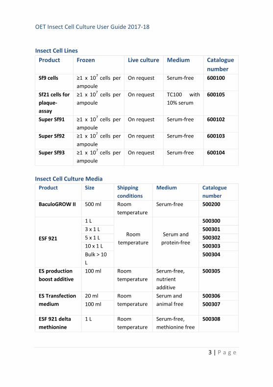
Insect Cell Lines
Product Frozen Live culture Medium Catalogue
number
Sf9 cells ≥1 x 10⁷
cells per
ampoule
On request Serum-free 600100
Sf21 cells for
plaque-
assay
≥1 x 10⁷ cells per
ampoule
On request TC100 with
10% serum
600105
Super Sf91 ≥1 x 10⁷
cells per
ampoule
On request Serum-free 600102
Super Sf92 ≥1 x 10⁷ cells per
ampoule
On request Serum-free 600103
Super Sf93 ≥1 x 10⁷
cells per
ampoule
On request Serum-free 600104
Insect Cell Culture Media
Product Size Shipping
conditions
Medium Catalogue
number
BaculoGROW II 500 ml Room
temperature
Serum-free 500200
ESF 921
1 L
Room
temperature
Serum and
protein-free
500300
3 x 1 L 500301
5 x 1 L 500302
10 x 1 L 500303
Bulk > 10
L
500304
ES production
boost additive
100 ml Room
temperature
Serum-free,
nutrient
additive
500305
ES Transfection
medium
20 ml Room
temperature
Serum and
animal free
500306
100 ml 500307
ESF 921 delta
methionine
1 L Room
temperature
Serum-free,
methionine free
500308
OET Insect Cell Culture User Guide 2017-18
4 | P a g e
1. Introduction
1.1 Insect cell culture
This manual provides a guide to the growth and maintenance of insect cell
cultures. It is extremely important that the insect cells used for the
production and analysis of recombinant baculoviruses are of the highest
quality. Insect cells can also be transfected with suitable plasmids to
produce stable cell lines expressing a gene of choice. This manual provides
tried and trusted protocols used within the labs at OET Ltd and by scientists
with over thirty-years’ experience of working with insect cell cultures and
baculoviruses. We hope it is useful to both beginners and more experienced
researchers.
1.2 Choice of cell line
The insect cells most commonly used for the baculovirus expression system
are Sf21 cells, originally derived from the pupal ovarian cells of Spodoptera
frugiperda (fall army worm)1; Sf9 cells, which are a clonal isolate of Sf21
2; or
T. ni (TnHi5™) cells3, originally derived from the ovarian cells of Trichoplusia
ni (cabbage looper)4. Generally, Sf21 or Sf9 cells are used for co-
transfections, virus amplification and plaque assays. Whilst many labs use
Sf9 for all protocols, Sf21 cells are superior for plaque-assays and monitoring
virus cytopathic effects and are more tolerant to sub-optimal conditions;
and so ideal for those new to the system. Sf9 cells are usually better for
amplification of large stocks of virus and protein production and grow very
well in large-scale fermenters or shake flasks. TnHi5™ cells are often used to
achieve maximal protein production but should not be used to produce or
amplify virus because of the increased possibility of generating virus
mutants4. For reasons that remain unclear, some genes are expressed much
better in Sf cells than T. ni cells or vice versa, so testing expression in both
cell lines at an early stage is recommended.
OET Insect Cell Culture User Guide 2017-18
5 | P a g e
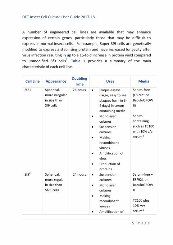
A number of engineered cell lines are available that may enhance
expression of certain genes, particularly those that may be difficult to
express in normal insect cells. For example, Super Sf9 cells are genetically
modified to express a stabilising protein and have increased longevity after
virus infection resulting in up to a 15-fold increase in protein yield compared
to unmodified Sf9 cells5. Table 1 provides a summary of the main
characteristic of each cell line.
Cell Line Appearance Doubling
Time Uses Media
Sf211 Spherical,
more irregular
in size than
Sf9 cells
24 hours Plaque-assays
(large, easy to see
plaques form in 3-
4 days) in serum
containing media
Monolayer
cultures
Suspension
cultures
Making
recombinant
viruses
Amplification of
virus
Production of
proteins
Serum-free
(ESF921 or
BaculoGROW
II)
Serum-
containing
such as TC100
with 10% v/v
serum*
Sf92 Spherical,
more regular
in size than
Sf21 cells
24 hours Suspension
cultures
Monolayer
cultures
Making
recombinant
viruses
Amplification of
Serum-free –
ESF921 or
BaculoGROW
II
TC100 plus
10% v/v
serum*
OET Insect Cell Culture User Guide 2017-18
6 | P a g e
virus
Production of
proteins
Plaque-assays
(small plaques
form in 4 days)
Super Sf9
1-35
Spherical,
more regular
in size than
Sf21 cells
24 hours + Protein
production for
secreted or
difficult to
express proteins
Serum-free -
adapted to
SF900II
(Invitrogen)
or;
TC100 plus
10% v/v
serum*
TniHi5™3 Spherical,
larger and
more irregular
in size than
Sf9 cells
18 hours Suspension
cultures
Production of
proteins
Monolayer
cultures (loose
attachment)
Serum-free
medium, e.g.,
ESF921
Table 1: Insect cell lines and characteristics
NOTE
Superscripts 1-5 in Table refer to references. *Foetal calf serum – batches
vary; always test before using a new batch, some lots may require heat-
inactivation at 60°C for 30mins.
1.3 Culture medium
Most insect cell culture medium utilizes a phosphate buffering system,
rather than the carbonate-based buffers commonly used for mammalian
cells. This means that CO2 incubators are not required. Serum is required for
the maintenance of certain cell lines, but many have now been adapted to
OET Insect Cell Culture User Guide 2017-18
7 | P a g e
serum-free conditions. There is a large variety of insect cell culture media
available and it is beyond the scope of this manual to list them all; Table 1
lists the media currently in use in our labs. Sf9, Sf21 and TnHi5™ cells can all
be grown in medium with serum or serum-free media (Table 1). Always use
a different bottle of cell culture medium for each cell line. The addition of
antibiotics is optional (penicillin and streptomycin prepared with 5 units/ml-1
penicillin G sodium and 5 μl/ml-1
streptomycin sulphate in 0.85% saline can
be used) but generally it is not recommended for virus amplification or
protein production. Certainly it is best to maintain stock cultures without
antibiotics; otherwise you may be maintaining a low-level contaminant that
may later cause inefficient virus replication or protein production. Addition
of antibiotics to plaque-assay medium is recommended.
1.4 General requirements for insect cell culture
Insect cells have a relatively high dissolved oxygen content (DOC)
requirement, particularly when infected with virus. Maintaining the
appropriate DOC is important for cell growth and virus replication, and this
can be achieved in shake, spinner and tissue culture flasks by using vented
caps and not over-tightening lids. Most insect cells can be cultivated over a
temperature range from 25-30°C. The optimal temperature for cell growth
and infection for insect cells is considered to be 27-28°C. Insect cells can
also be cultured at room temperature (about 20-22°C) when a slower
growth rate is required. Virus infection is usually carried out at 27-28°C.
We recommend carrying out any cell culture work each day prior to
handling virus and only using one cell line at a time.
1.5 Maintaining cell cultures
Insect cell lines can be maintained as either suspension cultures, in shake
flasks or in stirred vessels (Figure 1), or in monolayer adherent cultures in T
flasks or dishes. Generally, insect cells adapted to serum-free medium are
cultivated in suspension shake cultures whilst cells adapted to serum-
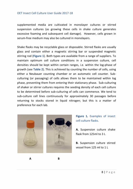
OET Insect Cell Culture User Guide 2017-18
8 | P a g e
supplemented media are cultivated in monolayer cultures or stirred
suspension cultures (as growing these cells in shake culture generates
excessive foaming and subsequent cell damage). However, cells grown in
serum-free medium may also be cultured in monolayers.
Shake flasks may be recyclable glass or disposable. Stirred flasks are usually
glass and contain either a magnetic stirring bar or suspended magnetic
stirring rod (Figure 1). Both types are available from a range of suppliers. To
maintain optimum cell culture conditions in a suspension culture, cell
densities should be kept within certain ranges, i.e. within the log-phase of
growth (see Table 2). This is achieved by counting the number of cells, using
either a Neubauer counting chamber or an automatic cell counter. Sub-
culturing (or passaging) of cells allows them to be maintained within log
phase, preventing them from entering their stationary phase. Sub-culturing
of shaker or stirrer cultures requires the seeding density of each cell culture
to be determined before sub-culturing of cells can commence. We tend to
sub-culture cell lines continuously for approximately 30 passages before
returning to stocks stored in liquid nitrogen; but this is a matter of
preference for each lab.
A B
Figure 1. Examples of insect
cell culture flasks.
A. Suspension culture shake
flask from 125ml to 3 L
B. Suspension culture stirred
vessel from 125 ml to 1 L
OET Insect Cell Culture User Guide 2017-18
9 | P a g e
2. General Cell Culture Techniques
2.1 Sterile technique
All techniques must be carried out under sterile conditions either in a Class II
or Laminar Flow safety cabinet.
2.2 Passaging cells
This is also referred to as sub-culturing cells and allows a stock of cells to be
kept within log phase and optimal viability for experimental use. For
example, if cells are not in log phase, they will not have available all the
enzymes and molecules needed for effective and efficient virus replication,
leading to poor virus titres or low level of expression. It is important that
cell cultures are passaged before the culture conditions reach stationary
phase – at this point cells are starting to die and any passaged cells may take
longer to recover. Cultures that are continually left to reach stationary
phase before passaging may suffer permanent problems and will not
support virus replication. In this case, a new culture must be established
from a frozen stock.
At each passage, record the passage number on the culture flask. Generally,
insect cell cultures can be passaged for about 30 times before returning to
frozen stocks of cells to initiate a new culture.
When initiating a new culture from a frozen stock, we strongly recommend
starting the culture as an adherent culture and once the cells are growing
well (1-2 passages), transfer them into a suspension culture. It is also easier
to monitor cells visually under the microscope when growing in monolayer
culture. It is possible to go straight from a frozen vial to a suspension
culture but this requires a high density and high viability of the frozen cells.
See notes later on in this manual.
OET Insect Cell Culture User Guide 2017-18
10 | P a g e
2.3 Adherent culture
Cells can be maintained in T25 or T75 flasks and grown at 27-28°C until the
cells just reach confluency. Cells can be maintained in serum-free media or
in media containing 10% serum (Table 1) Confluency means the cells have
just reached the point where they are touching each other, covering the
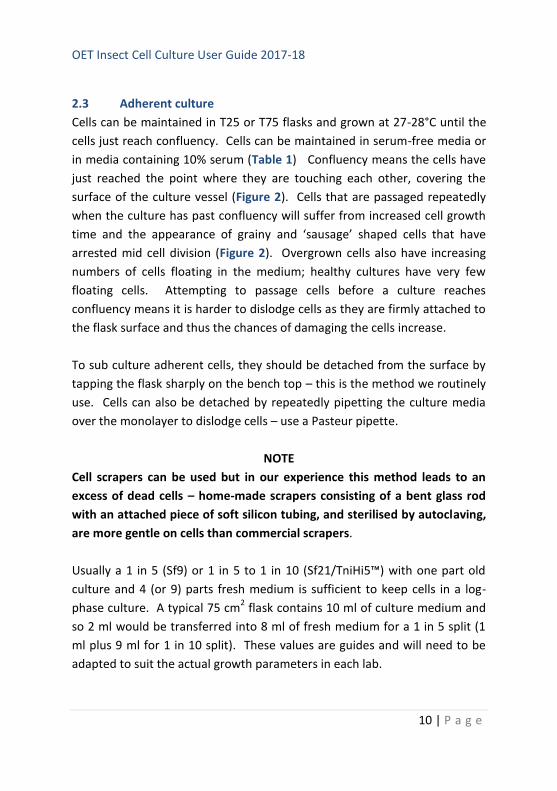
surface of the culture vessel (Figure 2). Cells that are passaged repeatedly
when the culture has past confluency will suffer from increased cell growth
time and the appearance of grainy and ‘sausage’ shaped cells that have
arrested mid cell division (Figure 2). Overgrown cells also have increasing
numbers of cells floating in the medium; healthy cultures have very few
floating cells. Attempting to passage cells before a culture reaches
confluency means it is harder to dislodge cells as they are firmly attached to
the flask surface and thus the chances of damaging the cells increase.
To sub culture adherent cells, they should be detached from the surface by
tapping the flask sharply on the bench top – this is the method we routinely
use. Cells can also be detached by repeatedly pipetting the culture media
over the monolayer to dislodge cells – use a Pasteur pipette.
NOTE
Cell scrapers can be used but in our experience this method leads to an
excess of dead cells – home-made scrapers consisting of a bent glass rod
with an attached piece of soft silicon tubing, and sterilised by autoclaving,
are more gentle on cells than commercial scrapers.
Usually a 1 in 5 (Sf9) or 1 in 5 to 1 in 10 (Sf21/TniHi5™) with one part old
culture and 4 (or 9) parts fresh medium is sufficient to keep cells in a log-
phase culture. A typical 75 cm2 flask contains 10 ml of culture medium and
so 2 ml would be transferred into 8 ml of fresh medium for a 1 in 5 split (1
ml plus 9 ml for 1 in 10 split). These values are guides and will need to be
adapted to suit the actual growth parameters in each lab.
OET Insect Cell Culture User Guide 2017-18
11 | P a g e
A B
C D
E F
OET Insect Cell Culture User Guide 2017-18
12 | P a g e
It is also important not to make the density of cells too low when passing, as
cells need to be in reasonable proximity with other cells to promote growth.
Some cells are more tolerant of this than others: Sf21 cells are the most
tolerant, Sf9 cells are the least. Figure 2 shows an example of cell density
immediately after passaging and at confluent levels for Sf21, Sf9 and
TniHi5™ cells. If the cells in the newly seeded culture are too sparse, they
may not divide and the culture will not reach become confluent.
It is essential to monitor cells under an inverted microscope prior to and
after passing to check for confluency, detachment after tapping, or any signs
of poor health or over growth. Signs of poor health include: grainy cells,
sausage-shaped cells, floaters, longer doubling time, failure to reach
confluence. Grainy cells with refractive cuboidal-like structures in the
nucleus are a sign of wild-type baculovirus contamination.
Maintain a log book of passaging and record the passage number, date and
split ratio on the culture flask. After about 30 passages of being maintained
in log phase, cultures start to lose viability and virus replication can be
impaired. The old culture should be discarded and a new one established
from a frozen stock. The log book can also be used to record any
Figure 2 (page 66). Images to illustrate
insect cells at various stages of culture.
A. Sf21 cells - sub confluent
B. Sf21 cells - confluent
C. Sf9 cells – sub confluent
D. Sf9 cells – confluent
E. Tni cells – sub confluent
F. Tni cells - confluent
OET Insect Cell Culture User Guide 2017-18
13 | P a g e
observations about the culture and this is sometimes very helpful when
trouble shooting.
Whilst it is not essential, some labs prefer to count cells at each passage and
seed a certain number of cells per flask each time. Table 2 provides an
indication of seeding density for passaging cells grown in monolayer culture.
Cell line
Cells to
seed a T25
flask
Cells to
seed a T75
flask
Cells to seed a
T150 flask
Split ratio guide
(culture : fresh
medium)
Sf21
1 x 10
6 3 x 10
6 5-6 x 10
6 1:5 to 1:10
Sf9/Super
Sf9 1.5 x 10
6 5 x 10
6 1 x 10
7 1:5
TniHi5™
0.9 x 10
6 2-3 x 10
6 4-6 x 10
6 1:5 to 1:10
Culture
volume 5-7 ml 10-15 ml 30-40 ml
Table 2: Seeding density for passaging monolayer cultures
When harvesting cells for use in experimental work, always count the
number of cells and determine their viability so that the correct seeding
density can be achieved for transfections or virus amplification.
Key points:
Check cells each day under the microscope until a confluent
monolayer has formed
Passage cells when confluent or shortly after
Do not allow cells to become overgrown
Do not split at too high a ratio
Keep a record of passage number, date and split ratio
OET Insect Cell Culture User Guide 2017-18
14 | P a g e
Start a new culture from frozen stocks after about 30 passages
Do not use antibiotics in routine cultures
2.4 Suspension culture
Maintaining insect cells in suspension culture is very easy and provides a
ready source of cells for amplifying recombinant viruses and infecting cells
for protein production. There are two main methods for small-scale
suspension cultures – shake flasks or stirred flasks (Figure 1).
Shake flasks require the use of serum-free medium as otherwise serum
creates excess froth that results in cells bursting as their membranes fuse
with the bubbles. Most serum-free medium contain surfactants to reduce
frothing. A surfactant such as Pluronic®F-68 can be added to media to
reduce frothing but in our experience even adding surfactant to serum-
containing medium does not prevent damage to cells.
There are commercial disposable shake flasks in a range of sizes that permit
cultures from 10 ml to 1.5 L. As the culture volume increases attention must
be given to aeration since insect cells, particularly those infected with virus,
have a high oxygen requirement for metabolism. This can be achieved by
selectinging an appropriate rpm, not over filling the flask to maximise the
surface area for gas exchange and ensuring lids are vented.
For those on a restricted budget, it is cheaper to use reusable glass flasks
with cotton wool and loosely covered foil caps that have been sterilised by
autoclaving and then dried. However, insect cells are very susceptible to
contaminants in flasks and so any washing-up regime must be very
stringent.
OET Insect Cell Culture User Guide 2017-18
15 | P a g e
At OET, our washing up regime is as follows:
Disinfect flask with VirkonTM
Soak in hot water with mild detergent (washing up liquid) and scrub
internal surface with nylon bottle brush to remove adherent cell
debris.
Rinse five times with hot water.
Soak in hot water for 2 hours.
Rinse in deionised water twice.
Soak in deionised water overnight.
Rinse in deionised water, dry and sterilise in an autoclave.
Dry in warm cabinet prior to use.
As insect cells do not require CO2, a shaking platform can be placed inside a
standard incubator maintained at 27-28°C or even a clean cupboard/room
maintained at this temperature.
Cells can also be grown in stirred cultures using commercial systems that
have vessels with either a vertical impeller or hanging stirring bar that sit on
a bespoke stirring device (Figure 1). There are often side ports to take
samples or add media/cells. Again, the caps should be vented or left loose
and the vessels must not be overfilled to ensure good aeration. Cost
effective home-made stirred flasks can be made using a flat-bottomed
round flask with a stirring bar sat on a conventional magnetic stirrer. Again
attention must be given to ensure the flasks are properly cleaned and
sterilised before use.
Cells detached from a healthy adherent culture should be counted and used
to set up a suspension culture according to the guidance in Table 3. It is
important that the cells have a high viability – at least 95%. When
establishing a suspension culture for the first time, set up a relatively
OET Insect Cell Culture User Guide 2017-18
16 | P a g e
modest scale flask (50-100 ml culture). Once the suspension culture is
established, a larger culture can be established if required.
Cells should be monitored daily by taking a small sample for counting and
determining cell viability. In this way a growth curve can be plotted and
monitored. Cells should be passaged before they reach stationary phase.
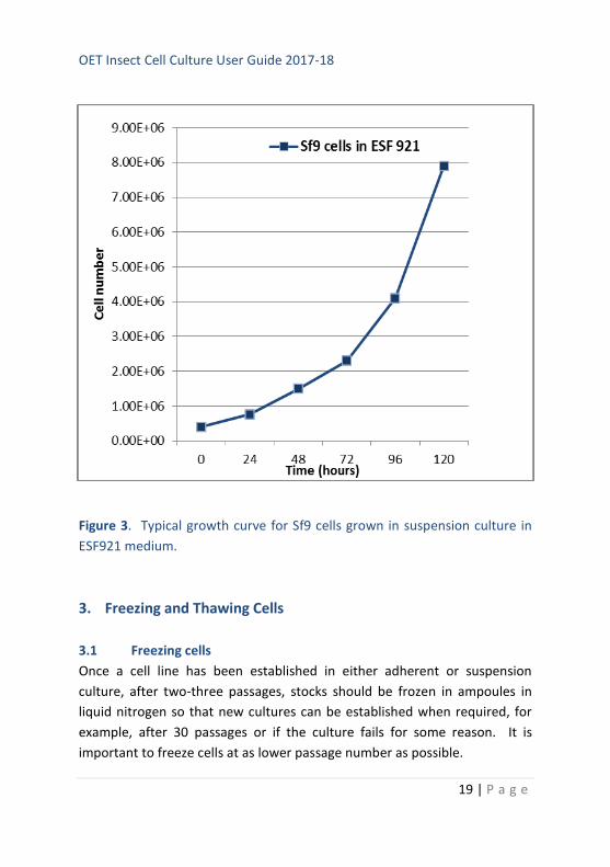
An example of a typical growth curve is shown in Figure 3. Whilst we
provide guidance on this matter, each lab should establish the optimal
conditions for their cells and media combination.
Passaging cells in suspension is very easy, after counting and determining
the viability, remove all the excess cells and, to the cells remaining in the
flask, add fresh medium to establish a new culture at the correct cell density
(Table 3). This can continue for up to about 30 passages (see notes under
adherent cultures). After this time, the culture needs to be set up from a
fresh adherent culture at low passage number.
The excess cells removed can be used to set up further suspension cultures
for virus infection – either to amplify stocks of recombinant virus or infect
cells for protein production. Cells can also be used to seed monolayer
cultures for experimental use e.g., 30mm dishes or multiwall plates for
plaque assay, co-transfections to make recombinant viruses or to test
expression levels.
Key points when setting up a new suspension culture:
Use healthy log phase cells from an adherent culture that is at least
95% viable
Count the cells and seed a culture no larger than 100 ml using the
guidelines in Table 4 using serum-free medium
Ensure good aeration by not over filling flasks, maintaining optimal
surface area to volume ratio, using an appropriate rpm and vented
lids
OET Insect Cell Culture User Guide 2017-18
17 | P a g e
Monitor cells daily and set up a growth curve
Passage cells by removing excess cells and adding fresh media to
achieve correct cell density (Table 4) before cells reach stationary
phase
Use excess cells to start cultures for virus amplification, protein
production or experimental use in monolayer cultures
Progress to larger volumes once cells established in culture
Always note passage number, date and cell count/viability data on
flask and in log book
After 30 passages (from retrieval of frozen stock – include passage
number data from adherent cultures), start a new adherent culture
from a frozen stock
Only use log phase cells for virus infections
Do not use TniHi5™ cells for making recombinant viruses or
amplifying viruses
OET Insect Cell Culture User Guide 2017-18
18 | P a g e
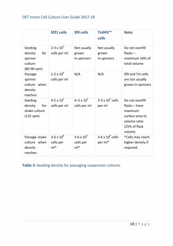
Sf21 cells Sf9 cells TniHi5™
cells
Note
Seeding
density for
spinner
culture
(80-90 rpm)
2-3 x 10⁵
cells per ml
Not usually
grown
In spinners
Not usually
grown
In spinners
Do not overfill
flasks –
maximum 50% of
total volume
Passage
spinner
culture when
density
reaches:
1-2 x 10⁶
cells per ml
N/A N/A Sf9 and Tni cells
are not usually
grown in spinners
Seeding
density for
shake culture
(135 rpm)
4-5 x 10⁵
cells per ml
4–5 x 10⁵
cells per ml
2-3 x 10⁵ cells
per ml
Do not overfill
flasks – have
maximum
surface area to
volume ratio
(25% of flask
volume
Passage shake
culture when
density
reaches:
3-6 x 10⁶
cells per
ml*
3-6 x 10⁶
cells per
ml*
3-6 x 10⁶ cells
per ml*
*Cells may reach
higher density if
required.
Table 3: Seeding density for passaging suspension cultures
OET Insect Cell Culture User Guide 2017-18
19 | P a g e
Figure 3. Typical growth curve for Sf9 cells grown in suspension culture in
ESF921 medium.
3. Freezing and Thawing Cells
3.1 Freezing cells
Once a cell line has been established in either adherent or suspension
culture, after two-three passages, stocks should be frozen in ampoules in
liquid nitrogen so that new cultures can be established when required, for
example, after 30 passages or if the culture fails for some reason. It is
important to freeze cells at as lower passage number as possible.
OET Insect Cell Culture User Guide 2017-18
20 | P a g e
There are many variations in the protocols published for the freezing down
and thawing of insect cells. The following is one that we find works well.
Protocol:
1. Set up a culture (suspension or adherent but suspension is better)
that will provide sufficient cells for freezing down (1 x 10⁷ cells per
vial). Freeze down several vials in one batch – at least 20.
2. Harvest cells from a log phase culture (mid log phase for
suspension or just prior to confluency for adherent cells). Count
cells and determine their viability. Cells need to be at least 95%
viable.
3. Place and label the required number of cryovials on ice.
4. Pellet required number of cells very gently at 500 rpm for 5 mins.
Remove and use the conditioned growth medium to prepare the
freezing mixture as follows:
Serum-free medium* Serum-containing medium
45% conditioned growth medium 40% conditioned medium
45% fresh medium 10% FBS (serum)
10% DMSO 10% DMSO
40% fresh medium
*use same freezing mix as serum-containing medium if preferred
5. Resuspend the required number of cells in freezing mixture, very
gently by pipetting up and down, to achieve a density of 1 x 10⁷
cells per ml. Place 1 ml aliquots into cryovials.
OET Insect Cell Culture User Guide 2017-18
21 | P a g e
6. Place the vials in a freezing chamber (e.g., we use Mr Frosty by
Nalgene) containing isopropanol and immediately place the
chamber at -80°C overnight before transferring to liquid nitrogen.
Or, place the vials at -20°C for one hour and then at -80°C overnight
before placing in liquid nitrogen.
7. After a few days, retrieve one vial to ensure that the freezing
process has been successful.
8. Keep a log book/e-record of where and when each cell line is
frozen, and when vials are recovered.
Key points when freezing cells:
Use healthy log phase cells with 95% or greater viability
Once DMSO has been added, cells must be cooled immediately to
avoid damage
Freeze slowly to avoid damage to cells
Check process has been successful after a few days of storage
Check liquid nitrogen levels regularly to ensure cells don’t start to
thaw during storage
Take all the normal H&S precautions when handling liquid nitrogen.
3.2 Thawing cells
It is important that cells are rapidly thawed and transferred into fresh
growth medium as soon as possible. DMSO is cytotoxic when cells are
thawed. We recommend recovering cells into an adherent culture for ease
of replacing the freezing mixture with fresh medium and monitoring the
cells under the microscope.
OET Insect Cell Culture User Guide 2017-18
22 | P a g e
Protocol:
1. Add 5 ml pre-warmed fresh growth medium to each of two T25
flasks.
2. Rapidly defrost the cells e.g., in a water bath at 37°C until just
thawed.
3. Sterilise the outside of the cryovial by misting with 70% ethanol
spray and transfer 0.5 ml of thawed cells to each flask. Mix to
generate an even distribution of cells and allow the live cells to
attach to the flask for about 1 hour at 28°C.
4. Monitor the cells under the microscope. Live cells should have
attached firmly within the hour incubation period; dead cells will
float. Remove the freezing medium and any floating cells (and
DMSO). Replace with fresh medium.
5. Continue to incubate the cells until a confluent monolayer is
formed and then passage/cub-culture as previously described,
setting up a suspension culture after 2-3 passages if required.
6. Alternatively, the whole 1 ml contents of the cryovial can be placed
in a 125 ml shake/stirred flask containing 25 ml pre-warmed fresh
medium and incubated as for shake/stirred cultures. Remove 5 ml
as a back-up into a T25 monolayer culture flask (and treat as above
steps 4-5). Continue the culture until the cell density is 2 x 10⁶
cells/ml and passage as normal.
It is recommended that suspension cultures set up in this way are
passaged two-three times before being used for experimental or
virus work.
OET Insect Cell Culture User Guide 2017-18
23 | P a g e
Key points when thawing cells:
Thaw quickly in a clean water bath
Sterilise the outside of the vial before opening
Set up a monolayer culture first so it is easy to remove dead cells
When cells are growing well, establish suspension cultures
4. Counting Cells and Determining Cell Viability
4.1 Counting cells
Before passaging cells or using cells for virus infection or transfections, cells
should be counted to establish an accurate count per ml. This can be
achieved using a commercial cell counter or by using a standard
haemocytometer. When establishing a new culture, new cell line or using a
new medium, it is well worth setting up a growth curve and monitoring cell
density every 24 hours to establish the growth pattern.
The following provides a protocol for counting cells using a Neubauer
haemocytometer.
Protocol:
1. Take a sample of cells from a shake or spinner culture or cells
harvested from a monolayer culture and using a Pasteur pipette,
load the prepared Neubauer chamber using capillary action (attach
cover slip firmly to form the counting chamber).
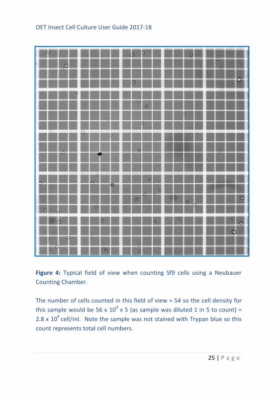
2. Count all the cells within the central 5 x 5 square grid (Figure 4) on
the counting chamber using a phase-contrast microscope (x10
objective). Count cells touching the etched triple line on the top
and left of squares. Do not count cells touching the triple lines on
the bottom or right of the squares.
OET Insect Cell Culture User Guide 2017-18
24 | P a g e
3. Count cells on both 5 x 5 grids and average the results. If the cells
are too dense to count accurately, dilute the sample an appropriate
amount to get a countable number of cells. If the cells are
clumped, they should be dispersed by gently pipetting up and
down to get single cells; otherwise the cell count will be inaccurate.
Ideally you need at least 30 cells and no more than 100.
4. The 5 x 5 square gives the number of cells present in 0.1 µl of
culture. To calculate the number of cells per ml, multiply by 10,000
(104). If the cells were diluted before counting, remember to
multiply the answer by the dilution factor.
Key points when counting cells:
Ensure cells are in a single cell suspension and not clumped
Count at least in duplicate to get an average
Don’t forget to multiply answer by any dilution factor
OET Insect Cell Culture User Guide 2017-18
25 | P a g e
Figure 4: Typical field of view when counting Sf9 cells using a Neubauer
Counting Chamber.
The number of cells counted in this field of view = 54 so the cell density for
this sample would be 56 x 104 x 5 (as sample was diluted 1 in 5 to count) =
2.8 x 10⁶
cell/ml. Note the sample was not stained with Trypan blue so this
count represents total cell numbers.
OET Insect Cell Culture User Guide 2017-18
26 | P a g e
4.2 Cell viability
Cell viability should be tested from time to time and every time cells are
used for virus work (transfections or virus infection) as poor viability is a
common reason for failure of virus to amplify to high titres or ensure high
yield of protein production.
The easiest method is Trypan blue exclusion and this can conveniently be
performed when counting cells. Trypan blue is a vital stain that is actively
excluded from live cells; therefore, dead cells take up and retain the stain
appearing blue under the microscope. Healthy cells appear refractile, bright
and clear. The percentage of dead cells can be calculated and used to
determine the overall viability of the culture. A good culture should be at
least 90% and preferably 95% viable.
Protocol:
1. Prepare a 2% (w/v) preparation of Trypan blue (e.g. Sigma) in PBS.
2. Dilute a sample of cells 1:1 with the stain (final concentration 1%)
and view cells under a phase contrast microscope immediately. It
is convenient to count the cells at this stage (see above protocol).
3. % viability = 100 - % dead cells
% dead cells = total blue cells counted/total cells counted x 100
Key points when determining viability:
Ensure cells are in a single cell suspension and not clumped
Count at least in duplicate to get an average
Don’t forget to multiply answer by two to take account of adding
stain
Don’t leave cells in stain for more than few minutes before
determining viability
OET Insect Cell Culture User Guide 2017-18
27 | P a g e
5. Establishing a New Culture from Living Cells
If cells are received from another lab or from a supplier (e.g., the
BaculoComplete kit from OET Ltd), you will need to establish a new culture
as soon as the cells arrive. You will need to ensure that you have a supply of
medium (see page 2), access to a Laminar Flow hood or Class II safety
cabinet, a supply of sterile flasks and pipettes, access to a cell counter or a
Neubauer haemocytometer to count cells, an incubator set at 27-28°C (no
CO2 required), access to a phase contrast inverted microscope and Trypan
blue stain (e.g. from Sigma)
Protocol:
1. View cells under an inverted light microscope and note if majority
of cells are floating or attached as a monolayer.
2. Decontaminate the outside of the flask by spraying with 70%
alcohol.
3. Place flask containing cells in a laminar flow hood or class 2 safety
cabinet and use aseptic technique through-out all procedures.
4. If many cells are still attached as a monolayer, decant the medium
and any floating cells into a discard container and replace with 5-6
ml fresh medium (e.g., baculoGROW II, ESF921).
If majority of cells are floating, decant medium and cells into a
sterile centrifuge tube and gently pellet cells at 1000 rpm for 5-10
min in a bench top centrifuge.
Also add 5-6 ml fresh medium to the attached cells in the original
flask and incubate at 27-28°C until just about confluent (~80%).
OET Insect Cell Culture User Guide 2017-18
28 | P a g e
5. Decant the old medium from the pelleted cells (into discard) and
gently resuspend the cells in 5-6 ml of fresh medium to obtain a
single cell suspension. Place the cells into a fresh T-flask, ensure
they are evenly dispersed over the surface, and incubate over night
as described above. Any live cells will attach overnight. The
following day, remove floating dead cells and replace with 5-6 ml
fresh medium and incubate as above until cells are just about
confluent.
6. We recommend an initial 1 in 4 to 1 in 5 split of cells when just
confluent. For example, harvest the cells by tapping the flask
sharply on the bench to detach cells, and gently pipette up and
down to get a single cell suspension. For example, place 1.25 ml
cells with 4.75 ml fresh medium into a new T-flask.
Passage the cells through at least one further round of monolayer
culture before setting up shake cultures.
7. The cells can be maintained like this in monolayer culture,
expanding into larger T-flasks as required, or the cells can be grown
in suspension/shake culture if cultured in serum-free medium.
8. To establish a shake culture, harvest cells from 2 x T25 monolayer
cultures and transfer 8 ml into a 125ml shaker flask containing 12
ml fresh medium.
Ensure the lid is not on tightly to allow for aeration and shake at
100-110 rpm at 27-28°C for 3-4 days and then count the cells.
9. Set up a fresh shake culture at 1 x 10⁶ cells/ml with the appropriate
amount of cell culture and fresh medium. Once the suspension
culture is established, seed the newly passaged cells at 0.8 x 10⁶
OET Insect Cell Culture User Guide 2017-18
29 | P a g e
cells/ml or as required. Cells will grow to a density of 5-8 x 10⁶ cells
per ml.
10. Do not allow cells to overgrow in either monolayer or shake
culture. For optimal virus infections, cells should be used in log
growth phase. Undertaking a growth curve is useful to understand
the growth characteristics of the culture.
11. We recommend that as soon as possible a batch of expanded cells
are frozen down in liquid nitrogen to act as a source of low passage
number cells for long term use. We normally passage cells for 30
passages before retrieving fresh stocks from liquid nitrogen.
12. For general information about insect cell culture read pages 3 to 8.
6. Establishing a New Culture of Cells From a Frozen Ampoule
On receipt, it is essential that the ampoule of frozen cells is either
transferred to liquid nitrogen for storage or thawed to initiate a live cell
culture. Do not freeze insect cells at -80°C.
You must use aseptic technique through-out and work in a Class II Safety
Hood or Tissue Culture Laminar Flow Hood. Rinse or mist the vial of cells
with 70% alcohol before opening.
Read through the general information about insect cell culture on pages 2
to 7 before starting and have the following materials available: suitable
growth medium (e.g., baculoGROW II, ESF921 from OET Ltd, Table 2),
T25/T75 monolayer flasks, 1ml and 10 ml sterile pipettes, incubator at 27-
28°C, water bath at ~37°C (best to use a ‘temporary bath’ such as a clean
beaker with warm clean water rather than a dirty water bath).

OET Insect Cell Culture User Guide 2017-18
30 | P a g e
Protocol:
1. On receipt, using aseptic technique, defrost the cells rapidly in a
clean water bath at 37°C until just thawed.
2. Rinse or mist the outside of the vial with 70% alcohol and then
transfer the contents of the ampoule between 2 T25/T75 flasks
containing 10-15 ml fresh culture medium (e.g., baculoGROW II,
ESF 921). Incubate the cells overnight at 27-28°C.
3. Check the cells under the microscope. Dead cells will be floating.
Live cells will have attached.
4. Decant medium containing floating cells into discard. Replace with
an appropriate amount of fresh media and continue to incubate
cells until they form a just confluent monolayer. Check daily.
5. When ready, passage cells and set up further monolayer or
suspension cultures as described above (pages 2 - 7).
6. Cells can be used to prepare recombinant viruses as soon as they
have recovered from shipping and are doubling approximately
every 24 hours with a high viability (90% or more). This may take 2-
3 passages of cells.
It is important that cells are not used to make recombinant viruses
until they are growing well in a log phase culture.
7. As soon as possible prepare a stock of cells for freezing down for
long term storage in liquid nitrogen (see page 10).
OET Insect Cell Culture User Guide 2017-18
31 | P a g e
7. Adapting Cells to New Media
On occasion it is necessary to transfer cells into a new culture medium.
Sometimes this can be achieved by simply using the new medium in place of
the old. However, more often a period of adaption is required.
The accepted standard protocol is to culture the cells in 75% old medium: 25
% new for one to two passages, then 50% old: 50% new for one to two
passages followed by 75% new: 25% old before finally culturing the cells in
the new medium. This takes time but ensure the cells adapt well to a new
medium.
A quicker version of this method can often be successful. It simply involves
culturing the cells in 50:50, old: new medium for one passage (monolayer or
suspension culture) and then continuing to grow the cells in the new
medium for at least one passage before using them for experimental work
(e.g. amplifying virus or making recombinant viruses).
For this quicker protocol to be successful, it is important that the cells are in
log phase of growth (80% confluent for monolayer or 3-5 x 10⁶ cells/ml for
suspension cultures) and that you monitor the cells each day to ensure they
are still growing well.
The cells should be passaged as soon as they are confluent (monolayer
cultures) or when they reach a density of 4-5 x 10⁶ cells/ml (suspension
culture). If the cells take a very long time to reach confluency or a density of
4-5 x 10⁶, or cell viability drops very low, the longer adaption process
described above will need to be followed.
7.1 Adapting cells to baculoGROW II or ESF921
We have successfully adapted Sf9 cells to baculoGROW II and ESF 921 (from
a variety of media) using the quick adaption protocol described below.
OET Insect Cell Culture User Guide 2017-18
32 | P a g e
Protocol:
1. Take a log phase culture of the cells to be adapted (80% confluent
monolayer culture) or cells at 3-5 x 10⁶ cells/ml for suspension
cultures. Ensure the culture is at least 90% viable.
2. For suspension cultures, dilute the culture with an equal amount of
the new medium and continue growing the cells until they reach 4-
5 x 10⁶ cells per ml.
For monolayer cultures, passage the cells using 50% old medium
saved from the previous culture and 50% new medium. Grow until
the cells are just confluent.
3. Passage the cells as normal using the new medium.
4. Use cells for experimental work after 1-2 passages in the new
medium.
OET Insect Cell Culture User Guide 2017-18
33 | P a g e
8. References
1. Vaughn, J. L., Goodwin, R. H., Tompkins, G. J. & McCawley, P.
(1977). In Vitro 13, 213-217.
2. Sf9 was cloned by G. E. Smith and C. L. Cherry from the parent line,
IPLB-SF 21 AE, by Vaughn et al., in 19771.
3. Wickham T.J., Davis T, Granados R.R., Shuler M.L. & Wood H.A.
(1992). Biotechnol Prog. 8, 391–396.
4. Hink, W. F. & Vail, P. V. (1973). J. Invertebr. Pathol. 22, 168-174.
Acknowledgements
This guide to insect cell culture was prepared by the OETeam of Robert
Possee, Adam Chambers, Olga Lissina, Aleksandra Kuczera, Linda King and
Laura Swan. We hope it is helpful.