innovative methods and applications in mucoadhesion research
TRANSCRIPT

This is a repository copy of Innovative Methods and Applications in Mucoadhesion Research..
White Rose Research Online URL for this paper:http://eprints.whiterose.ac.uk/115054/
Version: Accepted Version
Article:
Mackie, AR orcid.org/0000-0002-5681-0593, Goycoolea, FM, Menchicchi, B et al. (10 more authors) (2017) Innovative Methods and Applications in Mucoadhesion Research. Macromolecular Bioscience, 17 (8). 1600534. ISSN 1616-5187
https://doi.org/10.1002/mabi.201600534
© 2017 WILEY-VCH Verlag GmbH & Co. KGaA, Weinheim. This is the peer reviewed version of the following article: [. R. Mackie, F. M. Goycoolea, B. Menchicchi, C. M. Caramella, F. Saporito, S. Lee, K. Stephansen, I. S. Chronakis, M. Hiorth, M. Adamczak, M. Waldner, H. Mørck Nielsen, L. Marcelloni, Macromol. Biosci. 2017, 1600534.], which has been published in final form at http://doi.org/10.1002/mabi.201600534. This article may be used for non-commercial purposes in accordance with Wiley Terms and Conditions for Self-Archiving.
[email protected]://eprints.whiterose.ac.uk/
Reuse
Unless indicated otherwise, fulltext items are protected by copyright with all rights reserved. The copyright exception in section 29 of the Copyright, Designs and Patents Act 1988 allows the making of a single copy solely for the purpose of non-commercial research or private study within the limits of fair dealing. The publisher or other rights-holder may allow further reproduction and re-use of this version - refer to the White Rose Research Online record for this item. Where records identify the publisher as the copyright holder, users can verify any specific terms of use on the publisher’s website.
Takedown
If you consider content in White Rose Research Online to be in breach of UK law, please notify us by emailing [email protected] including the URL of the record and the reason for the withdrawal request.

Innovative methods and applications in mucoadhesion research 1
2
Alan R. Mackie1,2, Francisco M. Goycoolea2,3, Bianca Menchicchi4, Carla Caramella5, 3
Francesca Saporito5, Seunghwan Lee6, Karen Stephansen7, Ioannis S Chronakis7, Marianne 4
Hiorth8, Malgorzata Adamczak8, Max Waldner9, Hanne Mørck Nielsen10, Luciano 5
Marcelloni11 6
7
1. Institute of Food Research, Norwich Research Park, Norwich, UK 8
2. School of Food Science and Nutrition, University of Leeds, LS2 9JT, Leeds, UK 9
3. Institut für Biologie und Biotechnologie der Pflanzen, Westfälische Wilhelms-10
Universität Münster, Schlossgarten 3, 48149 Münster, Germany 11
4. Nanotechnology Group, Department of Plant Biology and Biotechnology, University 12
of Münster, Schlossgarten 3, 48149 Münster, Germany 13
5. Department of Drug Sciences, University of Pavia, Via Taramelli, 12, 27100 Pavia, 14
Italy 15
6. Department of Mechanical Engineering, Technical University of Denmark, 16
Produktionstorvet, 2800 Kgs. Lyngby Copenhagen, Denmark 17
7. National Food Institute, Technical University of Denmark, Søltofts Plads, 2800 Kgs. 18
Lyngby, Copenhagen, Denmark 19
8. School of Pharmacy, University of Oslo, Postboks 1068 Blindern, 0316 OSLO, 20
Norway. 21
9. Medizinische Klinik 1, Ulmenweg 18, 91054 Erlangen, Germany 22
10. Department of Pharmacy, University of Copenhagen, Universitetsparken 2, 2100 23
Copenhagen, Denmark 24
Revised Manuscript
1 2 3 4 5 6 7 8 9 10 11 12 13 14 15 16 17 18 19 20 21 22 23 24 25 26 27 28 29 30 31 32 33 34 35 36 37 38 39 40 41 42 43 44 45 46 47 48 49 50 51 52 53 54 55 56 57 58 59 60 61 62 63 64 65

11. S.I.I.T. S.r.l Pharmaceutical & Health Food Supplements, Via Canova 5/7 - 20090 1
Trezzano S/N , Milan, (ITALY) 2
3
Abstract 4
The present review is aimed at elucidating relatively new aspects of mucoadhesion/mucus 5
interaction and related phenomena that emerged from a Mucoadhesion workshop held in 6
Munster on 2-3 September 2015 as a satellite event of the ICCC 13th –EUCHIS 12th. After a 7
brief outline of the new issues, the focus is on mucus description, purification and mucus/mucin 8
characterization, all steps that are pivotal to the understanding of mucus related phenomena 9
and the choice of the correct mucosal model for in vitro and ex-vivo experiments, alternative 10
bio/mucomimetic materials are also presented. Then a selection of preparative techniques and 11
testing methods are described (at molecular as well as micro- and macroscale) that may support 12
the pharmaceutical development of mucus-interactive-systems and assist formulators in the 13
scale-up and industrialization steps. Recent applications of mucoadhesive systems (including 14
medical devices) intended for different routes of administration (oral, gastro-intestinal, vaginal, 15
nasal, ocular and intravesical) and for the treatment of difficult to treat pathologies or the 16
alleviation of symptoms are described. 17
18
1 2 3 4 5 6 7 8 9 10 11 12 13 14 15 16 17 18 19 20 21 22 23 24 25 26 27 28 29 30 31 32 33 34 35 36 37 38 39 40 41 42 43 44 45 46 47 48 49 50 51 52 53 54 55 56 57 58 59 60 61 62 63 64 65

1
Contents 2
1. Introduction ................................................................................................................................... 4 3
2. Mucoadhesion ................................................................................................................................ 4 4
3. Mucus composition and properties as a function of location .................................................... 6 5
4. Preparation of mucin or mucus ................................................................................................... 8 6
a. Purification of secreted mucins ................................................................................................ 8 7
b. Biomimetic approaches........................................................................................................... 10 8
5. Preparation of electrospun mucoadhesive formulations ............................................................. 12 9
6. Methods for molecular scale testing of mucoadhesion ................................................................ 15 10
a. Spectroscopic studies .............................................................................................................. 15 11
b. Atomic Force Microscopy ...................................................................................................... 19 12
c. Scattering techniques (SAXS, SANS, SLS and DLS) ........................................................... 21 13
7. Methods for macroscale testing of mucoadhesion .................................................................... 26 14
a. Rheology including polymer interaction in dilute solution ................................................. 28 15
c. Inclined plane .......................................................................................................................... 33 16
d. Tensile testing .......................................................................................................................... 38 17
8. Cellular methods ............................................................................................................................. 38 18
9. Methods for characterising mucus permeability ......................................................................... 40 19
10. Application specific requirement ................................................................................................. 42 20
a. Gastrointestinal drug delivery ................................................................................................. 42 21
b. Advances in the therapy of Helicobacter pylori ................................................................... 43 22
c. In the oral cavity ..................................................................................................................... 48 23
d. Colorectal drug delivery ......................................................................................................... 49 24
e. Vaginal drug delivery ............................................................................................................. 53 25
f. Nasal delivery .......................................................................................................................... 54 26
g. Ocular delivery ........................................................................................................................ 60 27
h. Intravesical drug delivery ...................................................................................................... 64 28
12. Conclusion ............................................................................................................................... 67 29
13. References ................................................................................................................................ 68 30
31
32
1 2 3 4 5 6 7 8 9 10 11 12 13 14 15 16 17 18 19 20 21 22 23 24 25 26 27 28 29 30 31 32 33 34 35 36 37 38 39 40 41 42 43 44 45 46 47 48 49 50 51 52 53 54 55 56 57 58 59 60 61 62 63 64 65

1. Introduction 1
The present review stems from a Mucoadhesion workshop held in Munster on 2-3 September 2
2015 as a satellite event of the ICCC 13th –EUCHIS 12th, held on 30 August- 2 September. 3
We are perfectly aware that there are a significant number of reviews and papers already 4
available in the current literature on mucoadhesion and on the relevant pharmaceutical 5
applications. Thus, in order to avoid duplications, the present review represents an update of 6
the topic but with special focus on some new aspects of mucoadhesion/mucus interactions, 7
taking inspiration and advantage of the multidisciplinary nature of the above conference which 8
gathered biomedical as well as food technology and physical-chemistry experts. 9
10
2. Mucoadhesion 11
Definition of Mucoadhesion: It is common knowledge that mucoadhesion is a special case of 12
bioadhesion, which is the ability of a material to adhere to a biological substrate. Thus in 13
mucoadhesion the biological substrate is represented by mucosal tissue. 14
Opportunities and applications: The advantages are at least theoretically well understood. 15
Mucoadhesive formulations are used to temporarily immobilize a delivery device on a specific 16
site for targeted release and optimal drug delivery due to intimacy and duration of contact. 17
Indeed it is no news that the short residence times of formulations (due to the various removal 18
and dilution effects depending on the route of administration) at their intended site of 19
action/absorption may result in reduced availability to the target tissue. Over the last 30 years, 20
mucoadhesive polymers and formulations thereof have been developed for buccal, nasal, 21
ocular, vaginal and oral applications. So far, a considerable number of papers focusing on the 22
mucoadhesive properties of a wide range of polymeric materials have been published [1-3]. Such 23
1 2 3 4 5 6 7 8 9 10 11 12 13 14 15 16 17 18 19 20 21 22 23 24 25 26 27 28 29 30 31 32 33 34 35 36 37 38 39 40 41 42 43 44 45 46 47 48 49 50 51 52 53 54 55 56 57 58 59 60 61 62 63 64 65

a huge effort has not been paralleled by an increase of clinical applications which are still 1
limited to a two digit number [4]. 2
Mechanisms: Concerning the mechanisms, it is recognised and experimentally proven that the 3
interaction between the mucus and mucoadhesive polymers is a result of physical entanglement 4
and secondary bonding, mainly H-bonding and van der Waals attraction, which according the 5
many authors, are mainly related to the following polymer properties: capability to create 6
strong H-bonding, high molecular weight, sufficient chain flexibility, and surface energy 7
properties favouring spreading onto mucus [5]. 8
Testing methods: It has also to be recognized that a variety of in vitro tests have been developed 9
by different research groups with the aim of understanding the phenomenon at different length 10
scales, from observational (tensile testing, flow retention experiments) to molecular, using 11
sophisticated techniques from fluorescence and confocal microscopy to a variety of 12
spectroscopic techniques. 13
The new approaches-innovative aspects: In recent years, other concepts have emerged in the 14
literature in relation to mucoadhesion. The first observation is that in many physiological 15
situations the mucus layer is the main actor, and the focus should therefore be on its nature and 16
complexity/variability depending on the anatomical site and on its sensitivity to various physio-17
pathological stimuli. It must also be recognized that mucins and mucus are quite different 18
substrates and their interactions with mucoadhesives are different and should be taken into 19
account when dealing with testing methods. 20
Mucus and food interactions: The comprehension of mucus interactions is also relevant in food 21
technology for food progression and nutrient digestion and absorption. There is a need to link 22
the knowledge acquired in this field to the problem of drug delivery. 23
1 2 3 4 5 6 7 8 9 10 11 12 13 14 15 16 17 18 19 20 21 22 23 24 25 26 27 28 29 30 31 32 33 34 35 36 37 38 39 40 41 42 43 44 45 46 47 48 49 50 51 52 53 54 55 56 57 58 59 60 61 62 63 64 65

Mucus penetration and mucoadhesion: Recently the focus has shifted to the mucus penetrating 1
systems [6, 7] and the study thereof not as an alternative but as a complementary opportunity to 2
mucoadhesion. They may work together to assure the best results. 3
Mucomimetic approaches: The recent trend aimed at the development of mucomimetics 4
substrates to formulate in vitro mucus or mucosae model for both testing and innovative 5
products should be recognised. 6
Summary: In line with the ongoing research in the field, the review will illustrate the latest 7
preparative and testing techniques that may support the pharmaceutical development of 8
optimized systems, intended for the different routes of administration. This knowledge is the 9
driving force for the pharmaceutical and related companies in the field. In addition, the review 10
aims to elucidate the above relatively new aspects of mucus characterization, mucus 11
penetration and mucomimetic phenomena that represent the basis for a science-based 12
development of any technological, in vitro, ex–vivo test and for any sustainable formulation 13
development. 14
15
3. Mucus composition and properties as a function of location 16
Mucus is a highly complex viscoelastic medium that provides a defensive barrier for many 17
different epithelial surfaces including the respiratory, reproductive and gastrointestinal (GI) 18
tracts. It performs a range of functions including lubrication, maintenance of a hydrated layer 19
and it acts as a barrier to pathogens and toxic substances while facilitating the exchange of 20
gases and nutrients with the underlying epithelium.[8] The mucus layer comprises two different 21
groups of mucins, secreted and membrane bound.[9] Membrane bound mucins form the 22
glycocalyx that provides an important link between the cell surface and the secreted gel layer. 23
On the luminal side of the membrane, these membrane bound mucins have either SEA (self-24
1 2 3 4 5 6 7 8 9 10 11 12 13 14 15 16 17 18 19 20 21 22 23 24 25 26 27 28 29 30 31 32 33 34 35 36 37 38 39 40 41 42 43 44 45 46 47 48 49 50 51 52 53 54 55 56 57 58 59 60 61 62 63 64 65

cleaving) -domains (MUC1, MUC3, MUC12, MUC13 and MUC17) or von Willebrand 1
domains (MUC4). The membrane bound mucins play a role in both cellular protection and 2
signalling [10, 11] through mechanisms such as the regulation of chemokine secretion. 3
The secreted mucins are produced by submucosal glands and goblet cells and are characterized 4
by their high molecular weight and high proportion of O-linked carbohydrate.[12] Mucus is 5
continuously secreted with nearly 10L secreted into the adult GI tract alone.[8] The composition 6
of mucus varies in different parts of the body. The mucins secreted into saliva are primarily 7
MUC5B and MUC7 and comprise about 16% of the total protein in saliva [13], whereas the 8
primary secreted mucin in the stomach is MUC5AC but with lower concentrations of MUC5B 9
and MUC6. It is possible that small amounts of MUC5B found in the stomach are pulmonary 10
in origin as pulmonary mucins are expelled via the GI tract. Intestinal secreted mucin is 11
predominantly MUC2 but again there are low concentrations of MUC6 and MUC11 in the 12
small intestine and MUC5B, MUC11 and MUC12 in the large intestine. Pulmonary secreted 13
mucins are primarily MUC5AC and MUC5B [14, 15], both of which are considered to be gel 14
forming. The secreted mucins of the female reproductive tract are primarily MUC5B but with 15
lower concentrations of MUC5AC and MUC6.[16] In addition to the mucins the mucus layer 16
contains lipids, salts, proteins, macromolecules and cellular debris.[17] In particular partially 17
degraded cellular DNA provides a significant contribution to the viscosity of the mucus layer 18
[18]. Both secretory and transmembrane mucins have been detected in the eye, namely MUC2, 19
MUC5AC, and MUC7, and MUC1, MUC4, MUC13, MUC15, MUC16, and MUC17, 20
respectively [19, 20]. In turn, transmembrane MUC1 and MUC4 are the predominant mucins 21
expressed in the normal human bladder [21]. 22
The properties of the various secreted mucins can vary significantly depending on the location 23
but they are still largely controlled by the basic properties of the mucins. Thus, they are 24
generally of high molecular weight (in excess of 1MDa) and are primarily hydrophilic. The 25
1 2 3 4 5 6 7 8 9 10 11 12 13 14 15 16 17 18 19 20 21 22 23 24 25 26 27 28 29 30 31 32 33 34 35 36 37 38 39 40 41 42 43 44 45 46 47 48 49 50 51 52 53 54 55 56 57 58 59 60 61 62 63 64 65

extensive glycosylation means that mucins are stiff, extended polymers with a persistence 1
length of 36 nm [22] and having a negative charge, often associated with sialic acid groups or 2
sulphate. The properties of mucins in solution very much depend on concentration and what 3
other components are present in the local environment. The secreted mucins are normally 4
considered to be gel forming. 5
In the GI tract the mucus layer varies widely in thickness. It is thickest in the colon and thinnest 6
in the duodenum [23]. In the intestine the mucus barrier comprises two different regions, known 7
as tightly adherent and loosely adherent.[24, 25] In the large intestine, these regions are clearly 8
delineated and under healthy conditions the tightly adherent layer provides a physical barrier 9
to bacteria. However, in the small intestine this layer is much thinner and the loosely adherent 10
layer dominates.[26] Measurements of particulate diffusion through human cervical mucus has 11
shown a network pore size of ~100 nm [27] and AFM images of intestinal mucin have shown a 12
similar pore size. [28] Despite this data on intestinal mucus, as has already been stated, the small 13
intestine is dominated by the loosely adherent layer, which is much more heterogeneous. This 14
layer has been shown to allow the passage of even 2 µm particles provided that they carry a 15
significant net negative charge. [18, 29] This will be discussed in more detail in Section 9. 16
17
4. Preparation of mucin or mucus 18
a. Purification of secreted mucins 19
There are very good books that describe the preparation of mucins, especially one edited by 20
McGuckin and Thornton [30]. As a starting point, we recommend that secreted mucus is 21
removed by gently scraping the epithelial surface with a plastic scraper and then purified [28]. 22
Because of the large size and complex structure of secreted mucins it is important to use an 23
extraction buffer containing a strong chaotrope capable of disrupting hydrogen bonding 24
1 2 3 4 5 6 7 8 9 10 11 12 13 14 15 16 17 18 19 20 21 22 23 24 25 26 27 28 29 30 31 32 33 34 35 36 37 38 39 40 41 42 43 44 45 46 47 48 49 50 51 52 53 54 55 56 57 58 59 60 61 62 63 64 65

network. For example, 4M guanadinium hydrochloride has been widely used [31]. The resulting 1
solution can then be purified using a two-step isopycnic density-gradient centrifugation, in 2
which the first step removes proteins and the second step nucleic acid. Proceed by adjusting 3
the sample to a density of 1.4 g/mL with CsCl and centrifuge (55K rpm at 10 °C for 62 h). The 4
high degree of glycation leaves the mucin strongly Alcian blue positive and this can be used to 5
identify the mucin containing fractions. Aliquots of fractions can be sampled, absorption at 280 6
nm measured and 2 たL of each fraction can be spotted and stained with Alcian blue. UV and 7
Alcian blue-positive aliquots should then be pooled and diluted in extraction buffer lacking 8
guanidinium hydrochloride (final guanidinium concentration 0.5 M), adjusted in density to 1.4 9
g/mL with CsCl, and centrifuged again (50K rpm at 10 °C for 96 h). Again aliquots can be 10
sampled, measured at 280 nm and stained with Alcian blue. The fraction at 1.4−1.55 g/mL and 11
strongly Alcian blue-positive but with weak absorption at 280 nm is identified as the mucin 12
fraction. More detailed methods for the purification of specific mucins can be gathered from 13
the literature. For example, MUC5B [32] and MUC7 [33] from saliva, MUC5B from respiratory- 14
and cervical-tract secretions[34] and MUC2 from intestinal mucus [28]. Confirmation of the 15
presence of mucin resulting from the purification should be undertaken using 16
immunoreactions. There is now wide range of antibodies available against mucins from a range 17
of animal sources indeed for many mucins it is possible to target specific regions of the 18
molecule. 19
20
Although the extraction and purification of mucins are well established methods, some 21
disadvantages related to the short conservation time, lower yield of production and batch-to-22
batch variability lead frequently to the alternative use of commercial mucin. Commercial mucin 23
the type from Sigma (Germany) or Orthana (Denmark) are purchased in lyophilized powder 24
which then can be hydrated in ultrapure water or buffers for 3h at room temperature under 25
1 2 3 4 5 6 7 8 9 10 11 12 13 14 15 16 17 18 19 20 21 22 23 24 25 26 27 28 29 30 31 32 33 34 35 36 37 38 39 40 41 42 43 44 45 46 47 48 49 50 51 52 53 54 55 56 57 58 59 60 61 62 63 64 65

gentle stirring. An extensive dialysis allows removals of small ions or low-molecular-weight 1
additive. Several treatments have been reported in the literature. Rossi et al. increased the 2
solubility of mucin from Sigma by adding 2% (w/w) SDS to 12% w/w mucins dispersion [35]. 3
SDS was then removed by 2 days dialysis against 10 volumes of 1M urea-1M NaCl, followed 4
by other 2 days dialysis against 40 volumes water and finally against 0.1 M acetate buffer pH 5
4.5. Alternatively, the mucin dispersion can be centrifuged for 1 h at 25,000xg, the supernatant 6
fraction collected, lyophilized and stored at 4°C until usage. The glycoprotein concentration 7
can be measured by colorimetric method or absorbance reading at 280 nm and calculated on 8
the basis of the difference before and after the treatment. Samples from Orthana have been 9
characterized in terms of monosaccharides composition revealing a predominant presence of 10
neutral O-linked oligosaccharides which confer high hydrophilicity and high solubility up to 11
200 mg/mL. Solution of this commercial mucin can be prepared by dispersion in water, 12
extensive dialysis and finally lyophilized. Orthana as well Sigma mucins do not show the 13
gelling properties upon lowering pH however rheological studies demonstrated the existence 14
of a concentration-dependent variation of the viscosity from dilute to semi diluted to entangled 15
state [36]. 16
17
In addition to preparative methods such as those mentioned above, analytical methods such as 18
agarose gel electrophoresis can also be used and separation monitored by lectin, 19
immunochemical or histochemical staining. This method can be used to analyse minimally 20
treated samples as long as they are protected from degradation. As the separation is based on 21
the inherent charge of the mucin, it can be used to separate different mucins [37] or different 22
glycated forms of the same mucin. [38] 23
24
b. Biomimetic approaches 25
1 2 3 4 5 6 7 8 9 10 11 12 13 14 15 16 17 18 19 20 21 22 23 24 25 26 27 28 29 30 31 32 33 34 35 36 37 38 39 40 41 42 43 44 45 46 47 48 49 50 51 52 53 54 55 56 57 58 59 60 61 62 63 64 65

In parallel with biological mucin and mucus, efforts to develop artificial mucus or mucus 1
models have been put forth for long. Easily accessible mucus models or mimics are beneficial 2
for all research disciplines requiring mucin, mucus or mucosa for a number of obvious reasons; 3
biological mucus could be difficult to be accessed by some research groups, generally 4
cumbersome to prepare, and presents ethical issue.[39] Technically, biological mucus samples 5
may reveal inconsistent structure and properties across studies due to differences between 6
individual animal sources and/or preparation details. While some mucus models have been 7
devised clearly in the context of mucoadhesion and drug delivery, some others have been 8
developed for other purposes and thus may be considered for future mucoadhesion studies. 9
Growing interests in mucus models are reflected in a few excellent review papers on this 10
subject published in recent a couple of years, including by Groo and Lagrace[40] and 11
Authimoolam and Dziubla[41] with a focus on artificial mucus, and by Cook and 12
Khutoryanskiy[39] with a focus on artificial mucosa, respectively. Briefly, mucus models can 13
be classified into glycan micro-arrays,[42-45] mucin layers,[46-50] complexes of mucins with 14
synthetic polymers,[51-55] and synthetic polymers,[56-59] roughly according to the scale. Glycan 15
micro-arrays have gained popularity for its specificity in probing glycan-binding receptors, 16
antibodies, and enzymes,[42, 43] and can be applicable to mucoadhesion too. Despite that micro-17
arrays with mucin-specific glycan arrays are also readily available,[44, 45] application in the 18
context of mucoadhesion is rare probably due to the lack of three dimensional, mechanical 19
barrier character in those systems. In fact, this is a common problem for all other types of two 20
dimensional mucus models, such as various monolayers of mucins on substrates.[46-50] Mucin-21
synthetic polymers complexes were motivated from that self-aggregated mucins, especially 22
commercially available ones, in aqueous solvent even at physiological concentration or higher 23
do not reproduce viscoelasticity comparable to that of native mucus.[60] Thus, synthetic 24
polymers, especially mucoadhesive polymers, are employed as crosslinker to enhance the 25
1 2 3 4 5 6 7 8 9 10 11 12 13 14 15 16 17 18 19 20 21 22 23 24 25 26 27 28 29 30 31 32 33 34 35 36 37 38 39 40 41 42 43 44 45 46 47 48 49 50 51 52 53 54 55 56 57 58 59 60 61 62 63 64 65

network forming capabilities of mucin aggregates. Representative polymers include guar 1
gum/borate,[53] alginate,[51] poly(acrylic acid),[52] and glutaraldehyde.[55] 2
Some hydrophilic and network-forming synthetic polymers, such as locust bean 3
gum/tetraborate[56] poly(acrylic acid)/(hydroxypropyl)methyl cellulose,[59, 61, 62] poly(styrene) 4
sulfonate,[58] and poly(ethylene glycol)-block-poly(lactic acid),[57] N-acryloyl-D-glucosamine 5
(AGA)/2-hydroxyethylmethacrylate (HEMA),[63] poly(ethylene glycol diacrylate) (PEGDA) 6
[64] have been employed even without involving mucin molecules. The assessment of synthetic 7
polymeric systems or complexes of mucin and synthetic polymers as mucus models has 8
typically been conducted via characterization of rheological properties[51-56] and adhesive 9
properties (detachment forces) against mucoadhesive drug tablets,[59] often in comparison with 10
biological mucus. These two properties represent mechanical integrity of mucus model and 11
their interfacial chemical properties against mucoadhesive polymers, respectively, in the 12
context of drug delivery researches. Nevertheless, no mucus model or mimic that can 13
universally replace biological mucus has emerged yet, presumably because of diverse and 14
complex properties required for mucoadhesion researches. 15
16
5. Preparation of electrospun mucoadhesive formulations 17
During the last two decades, electrospinning has gained increasing interest as a promising 18
technique for biomedical applications.[65-67] In drug delivery, nanofibers are appealing due to 19
their high encapsulation efficiency and flexible encapsulation capacity.[68] Moreover, 20
electrospun fibers allow for numerous delivery and encapsulation options; blend, core-shell, 21
particles combined with fibers, etc[69, 70]. Electrospun fibers have a large surface area that allows 22
for extensive interactions with the surrounding environment, which, depending on the 23
application, can be mucus or other biological components. Surprisingly, mucoadhesion of 24
nanofibers has not yet been extensively addressed. From the limited studies (examples from 25
1 2 3 4 5 6 7 8 9 10 11 12 13 14 15 16 17 18 19 20 21 22 23 24 25 26 27 28 29 30 31 32 33 34 35 36 37 38 39 40 41 42 43 44 45 46 47 48 49 50 51 52 53 54 55 56 57 58 59 60 61 62 63 64 65

the literature can be found in Table 5.1), it is evident that the mucoadhesive properties of 1
nanofibers can be manipulated by changing nanofiber properties, such as the extent of cross-2
linking.[71-73] Moreover, the inherent mucoadhesive properties of some biopolymers can be 3
exploited when developing mucoadhesive nanofibers. Thus, biopolymers with known adhesive 4
properties (such as alginate and chitosan) have been electrospun with increased bioadhesion of 5
the nanofibers compared to those made from synthetic polymers.[74, 75] However, the physico-6
chemical properties of nanofibers does not necessarily correlate with those of the unprocessed 7
material,[76] for which reason mucoadhesion of biopolymeric nanofibers in general must be 8
studied. Also, the effect of fiber morphology on the mucoadhesive properties, such as fiber 9
diameter, is yet to be explored. 10
11
The oral mucosa is permeable and vascularized, and therefore an appealing delivery.[77] The 12
group of Yang developed a delivery system for the oral mucosa, based on a semi-13
interpenetrating network (sIPN) made from gelatin.[78, 79] By cross-linking the fibers using 14
polyethylene glycol diacrylate (PEG-DA) the authors obtained stable, mucoadhesive fibers. 15
The mucoadhesion was affected by several factors: stability, porosity, swelling, and PEG 16
composition of the scaffold.[78] The sIPNs were used as a delivery system for insulin, and the 17
authors found that the transbuccal permeability of the released insulin was larger than that of 18
free insulin.[78] Another delivery system targeting the oral cavity was developed by 19
Tonglairoum et al., who developed polyvinylpyrrolidone/cyclodextrin/clotrimazole sandwich 20
patches coated with chitosan (CS) or thiolated chitosan (CS-SH) for oral candidiasis.[80] The 21
authors studied the fiber’s mucoadhesion, and thus the ability to adhere to the oral mucus. It 22
was shown that fibers coated with CS-SH exhibited a higher mucoadhesive strength compared 23
to CS coated, which is in line with thiolated chitosan providing stronger interaction with the 24
mucus.[80] The mucoadhesive properties of nanofibers can also be controlled by adding 25
1 2 3 4 5 6 7 8 9 10 11 12 13 14 15 16 17 18 19 20 21 22 23 24 25 26 27 28 29 30 31 32 33 34 35 36 37 38 39 40 41 42 43 44 45 46 47 48 49 50 51 52 53 54 55 56 57 58 59 60 61 62 63 64 65

mucoadhesive small molecules. For instance, Wongsasulak et al. obtained increased 1
mucoadhesion of zein–chitosan composite electrospun fibers by addition of alpha-tocopherol 2
(a-TOC).[72, 81] Electrospun nanofibers have also been studied for vaginal drug delivery.[82, 83] 3
In a study by Zong et al., polyethylene oxide (PEO)/polylactide composite electrospun 4
nanofibers was developed and loaded with cisplatin for local chemotherapy. The mucoadhesive 5
properties of the nanofibers caused the fibers to stay in the vagina and release the drug, whereas 6
the gel leaked out. Accordingly, the nanofibers facilitated increased bioavailability of the drug 7
as compared to a gel.[82] Electrospun fibers have shown promising results for mucosal drug 8
delivery, however the full potential is still to be revealed. 9
10
Table 5.1 Examples of electrospun formulations for drug delivery. 11
Mucosal target Fiber material Drug Ref
Buccal mucosa chitosan or thiolated chitosan/polyvinyl
alcohol
Garcinia mangostana
extract
[84]
Buccal mucosa polyvinyl alcohol Di- phenhydramine [73]
Buccal mucosa polyvinyl alcohol Docetaxel [85]
Buccal mucosa Gelatin and photo-reactive polyethylene
glycol diacrylate Nystatin, insulin [78, 79]
Buccal mucosa chitosan/polyvinyl alcohol Clotrimazole [86]
Buccal mucosa polyvinylpyrrolidone/cyclodextrin/clotri
mazole and chitosan/polyvinyl alcohol Clotrimazole [80]
Sublingual
mucosa
polyvinyl alcohol and sodium
alginate/polyvinyl alcohol
Insulin
[71]
GI mucosa polycaprolactone Diclofenec sodium [87]
1 2 3 4 5 6 7 8 9 10 11 12 13 14 15 16 17 18 19 20 21 22 23 24 25 26 27 28 29 30 31 32 33 34 35 36 37 38 39 40 41 42 43 44 45 46 47 48 49 50 51 52 53 54 55 56 57 58 59 60 61 62 63 64 65

Gastric mucosa Zein, chitosan and poly(ethylene oxide) g-tocopherol [72, 81]
Vaginal mucosa polystyrene coated with poly(allylamine
hydrochloride) or dextran sulfate sodium HIV entrapment [88]
Vaginal mucosa cellulose acetate phthalate TMC 125/Viread [83]
Vaginal mucosa poly(ethylene oxide)/polylactide Cisplatin [82]
Ocular mucosa Polyvinyl alcohol/polycaprolactone
Timolol maleate and
dorzolamide
hydrochloride
[89]
1
2
6. Methods for molecular scale testing of mucoadhesion 3
a. Spectroscopic studies 4
Over the last 20 years a range of spectroscopic methods have been used for the in vitro analysis 5
of the mucoadhesive behaviour of polymeric materials, and the determination of their affinity 6
toward mucin at the molecular level.[90-92] In particular, the interactions between 7
glycoproteins\mucins with mucoadhesive polymers have been investigated by 1H and/or 13C 8
Nuclear Magnetic Resonance (NMR) spectroscopy or by NMR diffusion measurements. 9
Analysis using NMR is advantageous, as no sample derivatization or pre-treatment is needed 10
and due to the advantage of non-alteration of the normal bio-functionality of the biomolecules. 11
Uccello-Barretta and co-workers have used proton selective relaxation rate NMR 12
measurements for the determination of mucoadhesive properties of different 13
polysaccharides.[93] Mucoadhesivity can be determined by exploiting the possibility to detect 14
changes of affinity to mucin of small probe molecules due to the mucin–polysaccharide 15
interactions. They have demonstrated the affinity of ketotifen fumarate (KT) to mucin, and they 16
1 2 3 4 5 6 7 8 9 10 11 12 13 14 15 16 17 18 19 20 21 22 23 24 25 26 27 28 29 30 31 32 33 34 35 36 37 38 39 40 41 42 43 44 45 46 47 48 49 50 51 52 53 54 55 56 57 58 59 60 61 62 63 64 65

have used KT as an interaction probe to compare the bovine submaxillary mucin affinities of 1
tamarind-seed polysaccharide and larch arabinogalactan.[94] Diclofenac sodium salt also has 2
high affinity for mucin (and low affinity for the polysaccharides), and was also employed as a 3
mucoadhesivity probe for polysaccharide mixtures containing tamarind seed polysaccharide 4
and hyaluronic acid.[95] It has been shown that the selective relaxation rate of the ligand is a 5
more sensitive indicator of binding than the non-selective relaxation rate. Earlier studies using 6
1H and 13C Nuclear Magnetic Resonance, recognised that the hydrogen bonds formed between 7
the carboxylic acid of poly(acrylic acid) and the glycoprotein component of mucus, play a 8
significant role in the process of mucoadhesion.[96, 97] Moreover, Griffiths et al., used pulsed-9
gradient spin-echo (PGSE-NMR) diffusion measurements to study the interactions of various 10
model polymer therapeutics with mucin and to quantify their diffusion within mucin 11
solutions.[98] A strong interaction with mucin was observed for a series of polyamidoamine 12
dendrimers and hyperbranched poly(ethylene imine), which displayed a characteristic pH-13
dependent profile and led to significant reductions in their rates of diffusion. 14
15
The use of attenuated total reflectance-Fourier transform infra-red spectral analysis (ATR-16
FTIR) is another spectroscopy method to study the interfacial interaction/absorption, and the 17
diffuse phase across the interface of mucoadhesive polymers and mucin segments.[99] 18
Sriamornsak et al., studied the mechanisms of gastrointestinal mucoadhesion of different 19
pectin films in contact with mucin in different media.[100] The diffusion of water was used as 20
an indirect measurement of any change resulting from the interpenetration of polymer–mucin 21
chains at the aqueous solution-polymer film interface.[101] The ATR-FTIR data confirmed the 22
formation of hydrogen bonds and the changes resulting from the interpenetration of pectin–23
mucin chains at the film interface. Furthermore, by using ATR–FTIR spectroscopy Xiang and 24
Li suggested that intra-polymer interactions, and inter-surface interactions played opposite 25
1 2 3 4 5 6 7 8 9 10 11 12 13 14 15 16 17 18 19 20 21 22 23 24 25 26 27 28 29 30 31 32 33 34 35 36 37 38 39 40 41 42 43 44 45 46 47 48 49 50 51 52 53 54 55 56 57 58 59 60 61 62 63 64 65
roles in the mucoadhesion performance of cationic polymers at the negatively charged buccal 1
mucosa surface.[102] The intra-polymer interactions can increase the crosslinking within the 2
polymer and lead to the decrease of mucoadhesion, while the inter-surface interactions can 3
promote mucoadhesion of the polymer. Optimal mucoadhesion can be achieved by balancing 4
these two interactions. In a recent study, ATR-FTIR was used to investigate the molecular 5
interactions between a chitosan hydrogel (consisting of non-ionic surfactant vesicles, 6
niosomes, with chlorotoxin) and various cell lines for cancer therapy. The specific 7
accumulation of mucoadhesive chitosan on the surface of ovarian epithelial carcinoma cells 8
was confirmed, demonstrating chitosan跨s specificity in targeting of mucin antigen 9
overexpressing tumor cells.[103] 10
11
Several of the mucoadhesive studies focus on bulk polymers, however, interest in the 12
mucoadhesion at the nanoscale has been growing.[104, 105] In fact, the mucoadhesion ability of 13
nanoparticulate systems is affected by their surface properties (hydrophobic, hydrophilic), 14
surface charges and their size. To detect the mucoadhesive phenomena in the intestinal tract 15
after oral administration of nanoparticulate systems, confocal laser scanning microscopy 16
(CLSM) has been used.[106, 107] Chen et al., investigated the adhesion of chitosan-modified 17
liposomes, (average diameter of ~200 nm) using CLSM and fluorophotometry with coumarin 18
6 as the fluorescent probe.[108] Their studies indicated that the positively charged surface charge 19
of the liposome particles played an important role in their interaction with the negatively 20
charged mucin fibres. In another study, the in vivo mucoadhesion of pH-responsive thiolated 21
chitosan nanoparticles for oral low-molecular weight heparin delivery was assessed using 22
CLSM.[109] Fluorescein-5-isothiocyanate (FITC)-labelled nanoparticles were prepared and the 23
intensity of green fluorescence in the small intestine epithelium of rats after oral administration 24
were evaluated. It is to note, that the CLSM method is sensitive to detect the organic dye-25
1 2 3 4 5 6 7 8 9 10 11 12 13 14 15 16 17 18 19 20 21 22 23 24 25 26 27 28 29 30 31 32 33 34 35 36 37 38 39 40 41 42 43 44 45 46 47 48 49 50 51 52 53 54 55 56 57 58 59 60 61 62 63 64 65

labelled association of nanoparticles to the mucosal layer of the animal intestine, and does not 1
modify the properties of the developed formulations of the nanoparticles. Instead of organic 2
fluorescence materials, orally administered quantum dots (QDs, semiconductor nanocrystals 3
with diameters of 1–10 nm), could be used as fluorescence markers. Tahara and co-workers 4
have developed QD-loaded liposomes which had high biocompatibility and low toxicity in 5
Caco-2 cells.[110] By using CLSM, the fluorescent signal of QDs in the liposomes could be 6
detected in the intestinal mucosa after oral administration. Thus, QDs can be used for tracing 7
and detecting bioadhesion and uptake of liposomes in in vivo applications. The relaxation NMR 8
approach, using dexamethasone 21-phosphate as a mucoadhesivity probe, confirmed the in 9
vitro mucoadhesivity of nanoparticles obtained from quaternary ammonium chitosan 10
conjugates.[111] The high surface area of nanoparticulate aggregates significantly enhanced the 11
interactions with bovine submaxillary mucin. In addition to nanoparticles and liposomes, block 12
polymeric micelles were also tested for the development of mucoadhesive drug loaded 13
nanovehicles. The mucoadhesivity of solutions of micelles having acrylated end groups was 14
characterized by using 1H NMR.[112] To quantify the extent of reaction, the decreased area 15
under the curve in the vinyl proton regime of the NMR spectra, (indicating interactions between 16
the acrylates and thiols present in cysteine residues of the mucin), was evaluated. 17
18
Overall, spectroscopic studies are very useful to investigate the interactions between polymers 19
or nanoparticulate systems with mucus. The choice of the mucoadhesion spectroscopy method 20
affects the characterization of their bioadhesive\diffusion properties and the determination of 21
the mucoadhesive strength. 22
23
1 2 3 4 5 6 7 8 9 10 11 12 13 14 15 16 17 18 19 20 21 22 23 24 25 26 27 28 29 30 31 32 33 34 35 36 37 38 39 40 41 42 43 44 45 46 47 48 49 50 51 52 53 54 55 56 57 58 59 60 61 62 63 64 65

b. Atomic Force Microscopy 1
Atomic force microscopy (AFM) is another method that has been used in mucoadhesion 2
measurements. The imaging mode can provide essential information about the amount and 3
conformation of material adhering to the sample, while force spectroscopy enables sensitive 4
adhesion measurements. In order to increase the surface contact area between the tip and the 5
sample in force measurements, it is advantageous to prepare a so-called ‘colloidal probe’. As 6
shown in Figure 6.1, a colloidal-sized particle is attached to the AFM cantilever using two 7
component epoxy glue. The colloidal probe and the sample surface can be further 8
functionalized with molecules of interest (mucin, APTES, -COOH, -NH3, -OH groups, 9
antibodies and others). Later on, the cantilever is moved towards the surface in the vertical 10
direction. The deflection of the cantilever is measured during the approach and retracts of the 11
probe; as a result, a force-distance profile is obtained. The maximum force of adhesion (Fadh) 12
and the work of adhesion (Wadh) can be determined from the retract curve. 13
1 2 3 4 5 6 7 8 9 10 11 12 13 14 15 16 17 18 19 20 21 22 23 24 25 26 27 28 29 30 31 32 33 34 35 36 37 38 39 40 41 42 43 44 45 46 47 48 49 50 51 52 53 54 55 56 57 58 59 60 61 62 63 64 65

1
Figure 6.1 Scheme of experimental setup and force-distance profile for mucoadhesion measurements. 2
3
The colloidal probe approach has been used by Cleary et al. in order to measure the adhesion 4
between a Pluronic-PAA modified glass bead and the mucous substrate [46]. The mucoadhesion 5
was studied in conditions of varying pH and ionic strength. It was also found that the time of 6
contact between the probe and the sample affects the adhesive forces. Prolonged contact favors 7
interdiffusion and interpenetration of polymer chains and mucin network, resulting in increased 8
adhesive force. Pettersson and Dedinaite investigated the interactions between mica surface 9
and silica particles coated with mucin and mucin-chitosan layers [113]. In order to mimic the 10
daily oral care procedure and its influence on mucous layers, the films were exposed to the 11
anionic surfactant SDS. Another interesting approach to the colloidal probe method was 12
1 2 3 4 5 6 7 8 9 10 11 12 13 14 15 16 17 18 19 20 21 22 23 24 25 26 27 28 29 30 31 32 33 34 35 36 37 38 39 40 41 42 43 44 45 46 47 48 49 50 51 52 53 54 55 56 57 58 59 60 61 62 63 64 65

presented by Iijima et al., who have measured the interactions between mucin layers and 1
stimuli-responsive drug delivery vehicles [48]. Instead of using the colloidal sized, glass or silica 2
particle attached to the AFM cantilever, the nanogel particles were freeze-dried and the 3
resulting granules were directly adhered to the tip by means of micromanipulation system. 4
Joergensen et al. used the image analysis of AFM scans in order to evaluate the mucoadhesive 5
properties of different pectins [114]. Mucin coated mica was scanned in AFM liquid cell before 6
and after incubation with polymer solution, followed by comparison of the roughness 7
parameters extracted from the images. Sriamornsak et al. investigated the structures of mucin, 8
pectin and their mixtures in acidic medium and deionized water, observing formation of large 9
aggregates in neutral pH conditions [50]. Similar study by Deacon et al. assessed the interactions 10
between pig gastric mucin and chitosan [115]. 11
AFM in mucoadhesion measurements presents both advantages and limitations. It allows 12
sensitive force measurements as a function of pH, ionic strength or time of contact, but it is 13
also time-consuming and can be affected by a choice of place in the case of heterogeneous 14
samples. 15
16
17
c. Scattering techniques (SAXS, SANS, SLS and DLS) 18
The detailed macromolecular structure of mucin has been addressed at molecular level using 19
high-resolution scattering techniques, namely, synchrotron SAXS [116-118], SANS [117, 119] and 20
static and dynamic light scattering [36]. This has allowed accounting for the properties of mucin 21
samples of different biological origin and methods of preparation. Thus, the cylindrical model, 22
and more recently, the double-globular (or “dumbbell”) comb model, has been used to describe 23
1 2 3 4 5 6 7 8 9 10 11 12 13 14 15 16 17 18 19 20 21 22 23 24 25 26 27 28 29 30 31 32 33 34 35 36 37 38 39 40 41 42 43 44 45 46 47 48 49 50 51 52 53 54 55 56 57 58 59 60 61 62 63 64 65

the complex mucin structure [36, 116, 119]. The schematic structure of mucin at different length 1
scales and its mechanical response at varying pH are represented in Figure 6.2. 2
3
Figure 6.2. Schematic representation of the biochemical structure of gel-forming mucin at 4
different magnifications: A) entangled mucin network; B) mucin monomers cross-linked 5
through disulfide bonds ; C) mucin monomer with globular naked-protein regions and D) low 6
scale representation of the bottle-brush highly glycosylated region of mucin (Sources: Modified 7
from [8, 120]; mechanical spectra of pig’s gastric mucin (PGM) as a function of pH taken from 8
Celli et al. [121]; double-globular comb structural parameters taken from Di Cola et al. [116] and 9
corresponding to pharmaceutical mucin sample “Orthana” in aqueous medium in absence of 10
salt). With permission of American Chemical Society and Elsevier. 11
12
1 2 3 4 5 6 7 8 9 10 11 12 13 14 15 16 17 18 19 20 21 22 23 24 25 26 27 28 29 30 31 32 33 34 35 36 37 38 39 40 41 42 43 44 45 46 47 48 49 50 51 52 53 54 55 56 57 58 59 60 61 62 63 64 65

Table S1 summarizes the results of biophysical studies, based on scattering techniques, namely, 1
synchrotron SAXS, SANS, and SLS and DLS, that have addressed the structural properties of 2
purified mucins of different biological origin. Fundamental parameters probed include the 3
radius of gyration (Rg), the coil overlap concentration (c*) and the slope of the intensity 4
scattering curves (also known as the fractal dimension (df)). These parameters have been 5
determined at varying conditions of pH, solvent, concentration and temperature. The wealth of 6
documented studies has contributed to the elucidation of the mechanisms and molecular events 7
that govern the properties of mucin that underlie its biological functions such as the formation 8
of gel networks. Indeed, mucin participates in the formation of the gel which prevents the 9
digestion of stomach epithelia caused by the acidic gastric juice. This is a function of pH, but 10
also mucin concentration and ionic strength. At physiological conditions, the high 11
concentration of mucin (> 20 mg/mL) and the high-molecular-weight of the molecules, favor 12
the formation and stabilization of an entangled network which behaves as a weak reversible 13
gel [122]. On the other hand, mucin undergoes to sol-gel transition [121, 123] a low pH (pH < 4) 14
due to a conformational change in which hydrophobic domains of the non-glycosylated 15
cysteine-rich regions become exposed and the negative charges of the sugars residues 16
responsible of maintaining the expanded structure get protonated. As observed in vitro for 17
native mucin, this phenomenon is accompanied by increase of the size at pH ~2 [124, 125] due to 18
aggregation of mucin by a combination of hydrophobic and electrostatic interaction and 19
entanglement of the sugar chains resulting in an increase of the viscosity of the solutions [121, 20
126]. In support of the model proposed for mucin gelation, AFM images have shown that mucin 21
is in an extended fiber-like shape at pH 6.0, whereas it forms well-defined clusters at pH 2.0 22
[124]. Consequently, the different conformation of mucin throughout the mucus layer allows 23
selective diffusion of HCl. At low concentration [123], in presence of high ionic strength [126], 24
commercial mucin [60], does not gel. However, pH-dependent interactions, as shown by DLS 25
1 2 3 4 5 6 7 8 9 10 11 12 13 14 15 16 17 18 19 20 21 22 23 24 25 26 27 28 29 30 31 32 33 34 35 36 37 38 39 40 41 42 43 44 45 46 47 48 49 50 51 52 53 54 55 56 57 58 59 60 61 62 63 64 65

and CD-spectroscopy, are attributed to a conformational transition of mucin at pH < 4.0 [127, 1
128] that imparts some fluidic viscoelasticity to the bulk sample [121]. 2
Recent studies using synchrotron SAXS have aimed to gain insight into the interaction between 3
soluble commercial pig gastric mucin and alginates of high-molecular-weight (~ 400 kDa) and 4
low-molecular-weight (~4 kDa) [129]. Firstly, the structure of mucin alone (at 3 mg/mL), at three 5
different values of pH, namely at 1.2, 2.5 and 4.0, was investigated. The scattering curves were 6
characterized by a single fractal dimension, df = -1.6 at pH 4.0, which at low-q range, increased 7
to df = -2.6 when lower pH were assessed. This observation is consistent with a pH-driven 8
conformational transition in the mucin, in agreement with observations in other mucin samples 9
differing in origin and preparation methods, as revealed from other techniques. The structure 10
of mucin in three different concentration (namely, at 0.3, 1.5 and 3.0 mg/mL) was characterized 11
by different scattering profile, being the one at lower concentration ideal to calculate the radius 12
of gyration (Rg) that afforded a value of ~18 nm. Interestingly, when the more diluted mucin 13
was mixed with two different types of alginates, different effects in the high-q range of the 14
intensity scattering plot were observed. Indeed, the addition of the low-molecular-weight 15
alginate produced a scattering profile in which the high-q range resembled the one of mucin 16
solutions at high concentration (3 mg/mL). By contrast, this effect was less pronounced when 17
adding the high-molecular-weight alginate, where the high-q region resembled more closely 18
the behavior of mucin at low concentration. Based on this evidence, along with that from 19
fluorescence quenching spectroscopy, viscosimetry and DLS studies, a general model was 20
proposed to explain the interaction of soluble mucin with polyanions. This model accounts for 21
the influence of molecular weight, charge and degree of chain contraction (Figure 6.3). 22
Although the overall net charge of mucin is negative, positively charged patches are expected 23
to occur in the non-glycosylated protein globular regions of mucin due to the presence of 24
1 2 3 4 5 6 7 8 9 10 11 12 13 14 15 16 17 18 19 20 21 22 23 24 25 26 27 28 29 30 31 32 33 34 35 36 37 38 39 40 41 42 43 44 45 46 47 48 49 50 51 52 53 54 55 56 57 58 59 60 61 62 63 64 65

histidine, arginine and lysine. These positive patches represent sites for the interaction with 1
negatively charged polysaccharides. 2
3
Figure 6.3 Model of interaction between the mucin in its double-globular comb mucin 4
structure and alginate as a function of alginate´s Mw (Alg 4 = 4 kDa; and Alg400 = 400 kDa) 5
and chain flexibility [129]. With permission of American Chemical Society. 6
7
Low-molecular-weight and stiff polyanions will interact mainly with the sites available on the 8
globular regions without influencing the preferred conformation of mucin. Thus, minimal 9
variation of the bulk properties such as size and viscosity are expected to occur. However, due 10
to the small size, low-molecular-weight polyanions are able to penetrate in the globular 11
structure inducing eventually rearrangement of the protein. On the other hand, high-molecular-12
weight and more flexible polyanions, due to the large size, might act as bridges between distant 13
available sites thus influencing the initial conformation of mucin and favoring a reduction of 14
the overall hydrodynamic volume. 15
16
1 2 3 4 5 6 7 8 9 10 11 12 13 14 15 16 17 18 19 20 21 22 23 24 25 26 27 28 29 30 31 32 33 34 35 36 37 38 39 40 41 42 43 44 45 46 47 48 49 50 51 52 53 54 55 56 57 58 59 60 61 62 63 64 65

7. Methods for macroscale testing of mucoadhesion 1
The methods to study mucoadhesion can be classified depending on the underlying physical 2
phenomena involved and also depending on the type of formulation that can be tested. Table 3
7.1 and Table S2 summarize the investigative techniques available. In this Section, we focus 4
on those that probe macroscale phenomena. 5
Table 7.1. In vitro methods used to study mucoadhesion as classified on the basis of the 6
physical phenomena involved. 7
Test method Formulation Mucosal surface/mucosa
mimetic material/mucin
Methods based on the mechanical force determination [130]
Texture Analyzer
Compressed polymers tablets
[59]; Polymers solutions [131];
Cast polymer films [132, 133];
Polymer gels [134-136];
Compacted polymer
microparticles into tablet [137]
Animal mucosal tissue [133,
135, 137]; Mucosa-mimetic
hydrogels [59]; Mucin-coated
(Sigma) filter papers [131,
132]; Mucin (Sigma) disc
[134]; PGM (Sigma) gels [136]
Modified balance/modified
surface tensiometer
Polymer coated glass [138];
Compressed polymer [139, 140];
Polymer cups [141]
Animal mucosal tissue [139-
142];
Tensile tester Polymer paste [143]; Hydrogels
[144]
Plexiglas® disk [143]; gelled
BSM [144]
Tensile stress tester Composite hydrocolloids [145] Filter paper [145]
1 2 3 4 5 6 7 8 9 10 11 12 13 14 15 16 17 18 19 20 21 22 23 24 25 26 27 28 29 30 31 32 33 34 35 36 37 38 39 40 41 42 43 44 45 46 47 48 49 50 51 52 53 54 55 56 57 58 59 60 61 62 63 64 65

Rotational cylinder Compressed polymer tablets [1,
140]
Animal mucosal tissue [1,
140]
Atomic Force Microscopy
(AFM)
Polymer coated glass
microsphere [46]; Polymer
solution [146]; Mucin-polymer
complexes [50, 115]
Human buccal cells [146];
Freshly purified PGM [115];
PGM (Sigma) [50]
Methods based on mucoadhesive interaction
Surface Plasmon
Resonance (BIACORE®)
Covalently-bound polymer
on CM5 chip [147]
Submicron-sized commercial
PGM suspension [147]
Dynamic light scattering
(DLS)
Mucin-polymer complexes
[128, 148]
Turbidity Mucin-polymer complexes
[128, 149]
IR-NMR
Freeze-dried mucin-polymer
mixed solutions [150]
Crude homogenized porcine
gastric mucus [150]; PGM
solution [150]:
Analytical ultracentrifuge Polymer-mucin mixed
solutions [151-153]
PGM from different gastric
regions [151]; HGM [152]
Impedance crystal quartz
microbalance (QCM)
Polymer solutions; Polymer-
micelles [154]
BSM (Sigma) solution [154]
Method based on flow forces
1 2 3 4 5 6 7 8 9 10 11 12 13 14 15 16 17 18 19 20 21 22 23 24 25 26 27 28 29 30 31 32 33 34 35 36 37 38 39 40 41 42 43 44 45 46 47 48 49 50 51 52 53 54 55 56 57 58 59 60 61 62 63 64 65

Flow through systems
Fluorescent labeled
nanoparticles [155, 156];
Polymer microparticles [157]
Ocular tissue [155]; PGM
(Sigma) solution [156]; Isolated
small rat intestine [157];
Method based on fluorescent probes
Fluorescence
determination
Fluorescent labeled-
nanoparticles [158];
Fluorescent labeled-polymer
solutions [159, 160]; Polymer
solutions [161]
Animal mucosal tissue [158-160]
Pyrene-labeled human
conjunctival epithelial cells
[161]
Multiple Particle Tracking Fluorescent particles [162] Purified PGM hydrogels [162]
Method based on rheological solution properties
Viscometer Polymer-mucin mixed
solution [129, 149, 163, 164] Mucin (Sigma) solutions [129,
131, 149, 164-166]; Homogenised
porcine gastric mucus [167, 168] Rheometer Polymer-mucin mixed
solution [131, 148, 149, 165, 166]
1
2
3
4
a. Rheology including polymer interaction in dilute solution 5
The interaction occurring between mucus and mucoadhesive polymers in mixed systems 6
produces variation in the flow properties of the mixtures with respect to those of the single 7
1 2 3 4 5 6 7 8 9 10 11 12 13 14 15 16 17 18 19 20 21 22 23 24 25 26 27 28 29 30 31 32 33 34 35 36 37 38 39 40 41 42 43 44 45 46 47 48 49 50 51 52 53 54 55 56 57 58 59 60 61 62 63 64 65

components. Thus, the study of the rheological properties of mixtures of mucus or mucin in 1
solution with mucoadhesive polymers has been widely exploited. Steady-shear measurements 2
of viscosity, (defined as the resistance of a fluid to the imposed shearing force), and 3
oscillatory shear determinations of the mechanical viscoelastic moduli (namely, storage and 4
loss moduli, G’ and G一, respectively), have been used to study liquid and gel-like systems, 5
respectively [169]. In general, when two different macromolecular species (e.g., polysaccharide 6
and protein) are mixed in solution, either attractive or repulsive interactions can take place [170] 7
(Figure 7.1). Attractive interactions can result in the formation of a complex that either remains 8
as a soluble colloidal complex or precipitates as a coacervate. Repulsive interactions in turn, 9
depending on the concentration of the macromolecular species, can lead to phase separation or 10
co-solubility [170]. In the case of associative interactions, the bulk viscosity of dilute mixed 11
solutions is expected to decrease due to overall reduction of the hydrodynamic volume of the 12
macromolecules when they are combined. However, in some other cases, cooperative intra and 13
inter-polymer interaction can induce increase in viscosity which is higher than the expected 14
sum of the individual contribution, up to physical gelation. This “synergistic” interaction was 15
previously observed in xanthan and galactomannan or in plasma proteins and egg albumin 16
mixed system [169, 171]. In repulsive interactions, the viscosity of mixed solutions is expected to 17
remain similar to those of the individual stocks. However, if the conformation of one of the 18
molecules changes due to the exclusion into a segregated phase, then the viscosity of the 19
mixture can also deviate from the expected additive line. Viscosity synergism cannot 20
distinguish between binding interaction and exclusion effects [172], unless experimental criteria 21
are applied. In the experimental conditions in which polysaccharides and mucin solutions are 22
in the dilute regime (rel ~2; sp ~ 1), polymer exclusion effects are assumed to be negligible 23
[169] being the polymers well below the overlap coil concentration. 24
1 2 3 4 5 6 7 8 9 10 11 12 13 14 15 16 17 18 19 20 21 22 23 24 25 26 27 28 29 30 31 32 33 34 35 36 37 38 39 40 41 42 43 44 45 46 47 48 49 50 51 52 53 54 55 56 57 58 59 60 61 62 63 64 65

1
Figure 7.1 Schematic representation of the type of interaction that can occur in protein-2
polysaccharides blends in dilute solution mixtures (modified from [170]). 3
4
Mucus is a weak viscoelastic gel biological material which possesses both flow (viscosity) and 5
deformation (elasticity) properties [120]. Such properties are regulated for example during 6
peristaltic movement or copulation [8]. At higher concentration mucus is characterized by a 7
shear thinning behavior (i.e. decrease in viscosity upon increase of the shear rate) typical of an 8
entangled network. However, the soluble fraction of PGM (Sigma) at concentration of ~ 8 9
mg/mL (in 0.1 M TRIS pH 7.4) was found to behave as Newtonian fluid since any shear-10
dependence of the viscosity was observed [164]. The addition of human albumin produces an 11
increase in viscosity due to association of albumin and mucin [164]. In the context of mucin and 12
1 2 3 4 5 6 7 8 9 10 11 12 13 14 15 16 17 18 19 20 21 22 23 24 25 26 27 28 29 30 31 32 33 34 35 36 37 38 39 40 41 42 43 44 45 46 47 48 49 50 51 52 53 54 55 56 57 58 59 60 61 62 63 64 65

polymer interactions, a rheological approach to screen the mucoadhesive properties of polymer 1
was described by Hassan and Gallo [163]. The mucoadhesion strength of several polysaccharides 2
was evaluated by studying the viscosity enhancement occurring upon mixing solution of 3
polymers with commercial mucin sample using a viscosimeter Brookfield Model RTV 4
(Brookfield Engineering Laboratories, Stoughton, MD). The increase in viscosity (positive 5
synergism) respect to the sum of the individual viscosities of the two components measured in 6
the same conditions as the mixture (in terms of concentration, temperature, time and rate of 7
shear) but with an Ostwald capillary viscosimeter (Fisher Scientific Co., Pittsburgh, PA) was 8
attributed to physical entanglement between the two species and defined as component of 9
bioadhesion (b). For each polymer-mucin system, b was calculated with the following 10
equation: 11
b t = - m - p Eq. 1 12
where is the measured viscosity of the mixture and p and m the individual viscosity of the 13
polymer and mucin, respectively. 14
The b values were found to be inverse proportional to the rate of shear per second (), thus, 15
the force of bioadhesion F, defined as intermolecular friction force per area unit was calculated 16
using the equation: 17
F = b × ߛሶ Eq. 2 18
Based on this pioneering protocol, several subsequent studies aimed to test the mucoadhesion 19
of polymers. This procedure could distinguish between positive synergism (interaction), lack 20
of synergism (no-interaction) or negative synergism of the viscosity or of the mechanical 21
properties. Mortazavi et al., [173] reported the gel strengthening effect of poly-acrylic acid on 22
homogenized mucus and observed that was characterized by increased values of G’ (which 23
reflects the ability of a viscoelastic material to store the elastic energy and recover its initial 24
1 2 3 4 5 6 7 8 9 10 11 12 13 14 15 16 17 18 19 20 21 22 23 24 25 26 27 28 29 30 31 32 33 34 35 36 37 38 39 40 41 42 43 44 45 46 47 48 49 50 51 52 53 54 55 56 57 58 59 60 61 62 63 64 65

shape) and decrease in G一 (which reflects the loss of energy as liquid-like flow). Madsen et al., 1
[167] described the effect of mucoadhesive type and concentration on the profiles of the 2
mechanical spectra of the mixtures in order to determine the type of gel formed. Some of the 3
most relevant works based on rheological methods that have contributed to a systematic 4
description of the mucoadhesive properties of polysaccharides are summarized in Tables S2 5
and S3. Sometimes, different outcomes have been observed for the same polymer-mucin 6
mixture, such as in the case of chitosan-mucin [149, 163] or cellulose derivative-mucin [166, 167] 7
depending on different experimental conditions, particularly the polymer concentration [165] or 8
mucin source, making direct comparisons and interpretations challenging [174]. 9
Recent evidence [175], has shown that mixing two stock solutions of chitosan and mucin of 10
matched rel ~ 2.0, at increasing f ratio (mass proportion of mucin respect the total mass in the 11
mixture) a reduction in rel to a minimum value (fさmin) occurs beyond which, upon a subsequent 12
increase in f, the rel increases again to approach that of mucin stock solution. Such behavior 13
describes a skewed U-shaped curve both in water and 0.1M NaCl (pH 4.5) as shown in Figure 14
7.2a and b, respectively, for a representative CS-mucin systems. This approach enables to 15
determine, in a quantitative manner, the degree of interaction, given by the value of the area 16
under the curve of the relative deviation from the theoretical additive line (or line of “no 17
interaction”). Also, the method enables to determine the maximum stoichiometry of the 18
interaction given by the f ratio of minimum rel (fさmin). 19
Mechanical force studies or rheological synergism are diagnostic of mucus (or mucin)-polymer 20
interactions, however, no detailed information regarding the underlying molecular mechanisms 21
of interaction can be deduced from these techniques. Table S3 offers a summary of the major 22
rheological methods that have been used to study the interactions of polymers and proteins 23
with mucin solutions and mucus gels. 24
1 2 3 4 5 6 7 8 9 10 11 12 13 14 15 16 17 18 19 20 21 22 23 24 25 26 27 28 29 30 31 32 33 34 35 36 37 38 39 40 41 42 43 44 45 46 47 48 49 50 51 52 53 54 55 56 57 58 59 60 61 62 63 64 65

Figure 7.2 Relative viscosity (さrel) of chitosan–mucin mixtures of varying compositions 1
expressed as the mass fraction of mucin (f) respect the total mass in a)water and b)0.1M NaCl 2
(37°C, pH 4.5, inclination angle 50°). The red dotted line in a) and b) represents the calculated 3
values of さrel of the mixtures assuming there is no interaction (additive line). The さrel values at 4
f = 0 and 1 are the relative viscosities of the chitosan and mucin stock solutions, respectively. 5
The lower panels show the normalized data expressed as percentage deviation from the additive 6
line in c) water and d) 0.1 M NaCl, both at pH 4.5 (mean values ± minimum and maximum, 7
n=2). The blue shaded areas in plots c) and d) represent the integrated area under the curve 8
calculated using a trapezoid approximation available in Origin 8.5 (Origin Lab Corp., 9
Northampton, MA) [175]. 10
11
c. Inclined plane 12
1 2 3 4 5 6 7 8 9 10 11 12 13 14 15 16 17 18 19 20 21 22 23 24 25 26 27 28 29 30 31 32 33 34 35 36 37 38 39 40 41 42 43 44 45 46 47 48 49 50 51 52 53 54 55 56 57 58 59 60 61 62 63 64 65

As pointed out in the introduction, a variety of methods can be used to study mucoadhesion 1
and in Table 7.1 a classification of methods is given based on the physical phenomena involved. 2
From a practical point of view it is useful to distinguish between mechanistic methods and 3
functionality (or performance) test methods; the first ones (the most common are rheological 4
and spectroscopic methods) give information on the events that occur at the mucoadhesive joint 5
in order to prove the interaction mechanisms, whereas the second ones are aimed at evaluating 6
the actual mucoadhesive properties/performance of formulations. In turn, they can be divided 7
into mechanical tests (the most common are tensile testing and rotational cylinder) intended to 8
measure the force needed to detach the formulation from the substrate and dynamic tests 9
(among which flow through or flow retention methods) intended to mimic the physiological 10
clearance mechanisms and to follow the fate of the formulation/loaded drug (retention on or 11
removal from the mucosal substrate). Mechanical and dynamic methods are believed to 12
provide information on the overall performance of the formulation as a delivery system. 13
The inclined plane method [176, 177] can be classified as a special dynamic test that measures 14
mucoadhesiveness as a function of the retention of the mucoadhesive material in contact with 15
a mucosal substrate (mucin film or mucosal tissue). It has been devised to test liquid or 16
semisolid formulations endowed with intrinsic flowing properties at test temperature. It is not 17
applicable to solid formulations or very thick gels. 18
19
Description of the apparatus 20
The inclined plane apparatus basically consists of a plexiglas support whose angle of 21
inclination with respect to the horizontal can be varied between 30° and 60°, thermostated at 22
37°C and placed above an electronic microbalance interfaced with a personal computer. An 23
illustrated picture of the apparatus, including details of the plexiglass support (which is 24
composed of a thermostated plate and an adapted substrate holder) is given in Figure 7.3. 25
1 2 3 4 5 6 7 8 9 10 11 12 13 14 15 16 17 18 19 20 21 22 23 24 25 26 27 28 29 30 31 32 33 34 35 36 37 38 39 40 41 42 43 44 45 46 47 48 49 50 51 52 53 54 55 56 57 58 59 60 61 62 63 64 65

1
2
Figure 7.3 Illustration of the inclined plane apparatus 3
4
The substrate holder (hosting two parallel channels) may be coated with a thin mucin film 5
(prepared by casting) or covered with mucosal tissue. The surface area coated is normally 28 6
cm2. The whole apparatus is placed in a transparent box allowing constant temperature to be 7
maintained and avoiding disturbances during the measurements. An overall picture of the 8
assembled apparatus is given in Figure 7.4. 9
10
1 2 3 4 5 6 7 8 9 10 11 12 13 14 15 16 17 18 19 20 21 22 23 24 25 26 27 28 29 30 31 32 33 34 35 36 37 38 39 40 41 42 43 44 45 46 47 48 49 50 51 52 53 54 55 56 57 58 59 60 61 62 63 64 65

Figure 7.4 Overall picture of the assembled apparatus 1
2
Description of the operational procedure for measuring mucoadhesive properties 3
The substrate holder is coated with the mucin film and equilibrated. Porcine gastric mucin is 4
normally used as biological substrate. Mucin films are prepared directly on the Plexiglas holder 5
in the horizontal position, by pouring a measured volume of 8% w/w mucin dispersion in water 6
then drying at 45°C for 45 min. A weighed amount of the formulation is placed on top of the 7
substrate holder, still held horizontal and until equilibrated. The support plate is then inclined 8
(at a given angle) and the amount of formulation dropped on the microbalance is recorded as a 9
function of time. Blank measurements are performed in the absence of the mucin film on a 10
weighed amount of sample using the same experimental conditions employed in the presence 11
of mucin. The amount of formulation dropped down the inclined plate is recorded by means of 12
suitable software as a function of time until a plateau is reached. The amount adhering to the 13
inclined plate is calculated as the difference between the amount of formulation loaded and the 14
amount dropped down from the balance (non-adherent) and expressed as a percentage (% 15
adhered). An example is given in Figure 7.5. 16
17
18
0
10
20
30
40
50
60
70
80
0 20 40 60 80 100 120 140
% n
on
ad
he
red
time(s)
Measurement with mucin
% adhered=49.74±1.10
Blank measurement
% adhered=31.89±2.62
1 2 3 4 5 6 7 8 9 10 11 12 13 14 15 16 17 18 19 20 21 22 23 24 25 26 27 28 29 30 31 32 33 34 35 36 37 38 39 40 41 42 43 44 45 46 47 48 49 50 51 52 53 54 55 56 57 58 59 60 61 62 63 64 65

Figure 7.5 Plots of the amount of sample dropped (non- adherent) on the balance as a function 1
of time. 2
3
A normalized mucoadhesion parameter is calculated as follows: (% adhered mucin-% adhered 4
blank)/% adhered blank and is equal to 56%.This parameter allows the mucoadhesive 5
properties of a given formulation to be measured independently of the consistency of the 6
sample, since the blank measurement allows for normalization [170].. 7
8
Validation of the method 9
The inclination angle, quantity of mucin, length and width of the channels engraved on the 10
sample holder, sample weight influence test results and their reliability and must be optimized 11
to manage sample and testing variabilities. Recently these parameters have been the object of 12
a validation exercise aimed at 1) evaluating the capability of the method to discriminate 13
between different prototypes of a formulation intended for marketing and 2) assessing the 14
precision and reproducibility of the method as well as the robustness with respect to operational 15
parameters. This exercise could lead to the proposal of the method as a routine control method 16
for the quality of the product. 17
18
Applications 19
The method has been profitably used to test the mucoadhesive properties of polymeric 20
solutions, liquid or gel formulations (mouthwashes, vaginal washings, eye drops, buccal 21
sprays, nasal washings, nasal sprays) [177] and even melted suppositories. The method has also 22
been employed to test mucoadhesive systems characterized by in situ gelling properties, like 23
swallowable gels intended for esophageal lining, or in situ gelling solutions used in diagnostic 24
colonoscopy, since it enables evaluation of the contribution of gelation time to mucoadhesive 25
performance [171]. 26
1 2 3 4 5 6 7 8 9 10 11 12 13 14 15 16 17 18 19 20 21 22 23 24 25 26 27 28 29 30 31 32 33 34 35 36 37 38 39 40 41 42 43 44 45 46 47 48 49 50 51 52 53 54 55 56 57 58 59 60 61 62 63 64 65

1
d. Tensile testing 2
A texture analyzer can be used for the quantification of the tensile strength i.e. the force 3
required to remove the formulation from a mucosal surface, which can be used as a measure of 4
the mucoadhesive strength. In a generalized setup, the formulation is fixed on a probe which is 5
subsequently lowered into a mucus sample. After incubation that ensures full contact between 6
the formulation and the mucus, the mucoadhesive strength is measured as the force of 7
detachment. Formulations such as tablets [178], films, [179, 180] hydrogels, [181] and fibers [80, 89] 8
can be studied using this technique. 9
8. Cellular methods 10
Use of mucosal tissue of animal origin in mucoadhesion studies presents certain drawbacks, 11
such as limited availability, time-consuming tissue preparation, small surface area, or 12
significant variability of the obtained results [182]. In vitro study of mucoadhesion on cell 13
cultures is an example of alternative method of measurements, based on the interactions 14
between mucin and material of interest. Mucus-secreting HT29-MTX cell line is derived from 15
human colon adenocarcinoma and often used as a model for human intestinal adsorption or 16
buccal tissue in the oral cavity. Mucus secretion depends on the culture period and usually 17
reaches maximum thickness after ca. 3 weeks of cell growth [183, 184]. Figure 8 presents the 18
HT29-MTX cells 1 and 14 days after passaging, respectively. 19
1 2 3 4 5 6 7 8 9 10 11 12 13 14 15 16 17 18 19 20 21 22 23 24 25 26 27 28 29 30 31 32 33 34 35 36 37 38 39 40 41 42 43 44 45 46 47 48 49 50 51 52 53 54 55 56 57 58 59 60 61 62 63 64 65

1
Figure 8.1 Morphology of the HT29-MTX cells 1 (a) and 14 days (b) after passaging. 2
3
There are several publications reporting use of cellular methods in mucoadhesion 4
measurements. Jintapattanakit et al. assessed the mucoadhesion of trimethyl chitosan and 5
PEGylated chitosan using HT29-MTX-E12 monolayers [185]. Fluorescently labeled polymers 6
were incubated with cells for 2 hours. Later on the cells were lysed and the uptake of polymers 7
was determined as the amount of fluorescence per unit weight of cellular protein. The obtained 8
results were similar when compared to mucin particle method. Chen et al. evaluated the 9
mucoadhesion of probiotic alginate microcapsules by counting Lactobacillus reuteri strain 10
released from the capsules adhering to the HT29-MTX after 1 hour of incubation. [186] In 11
another study, mucoadhesion of fluorescently labeled, non-coated and polymer-coated 12
liposomes was measured after 2 hour of incubation with HT29-MTX cell monolayer [187]. The 13
amount of liposomes adhering to the mucus was determined by measuring the fluorescence 14
intensity both directly on the cell monolayer and indirectly from the supernatant solution after 15
incubation. 16
17
1 2 3 4 5 6 7 8 9 10 11 12 13 14 15 16 17 18 19 20 21 22 23 24 25 26 27 28 29 30 31 32 33 34 35 36 37 38 39 40 41 42 43 44 45 46 47 48 49 50 51 52 53 54 55 56 57 58 59 60 61 62 63 64 65

9. Methods for characterising mucus permeability 1
One of the primary functional roles of mucins is to provide a barrier to bacteria while at the 2
same time allowing the passage of smaller components such as nutrients, bioactive agents or 3
gasses depending on location. As a result the permeability of mucus and factors that can affect 4
it are of great interest. The permeability of mucus depends on the pore size of the network, the 5
size of the objects trying to pass through it and the interactions between the two. Ideally, it 6
should be possible to mathematically model the diffusion through mucus in order to provide 7
some degree of prediction and understanding of the phenomena involved [188]. A range of 8
approaches are suitable to measure the diffusion of objects of different sizes ranging from 9
single molecules to nano- and micro-particles. At the molecular level, the diffusion chamber 10
has been used to determine the diffusion of molecules through mucus[189] but this has rather 11
been superseded by other more relevant methods that can be applied directly on live animals 12
in the gut [190] or nasal cavity [191]. 13
14
Another commonly used method is NMR[192-194] and most recently the use of F-19 entrapped 15
in different cyclodextrins was successfully used to probe the pore size of submaxillary and 16
nasal mucins [193]. This method shows great promise for the future. There has been a significant 17
amount of research on the use of particles as delivery systems through mucus and as a 18
consequence a wide range of methods have been used many of which have been described an 19
a recent review by Greissinger et al. [195]. The quartz crystal microbalance with dissipation 20
(QCM-D) has been used to assess the combined properties between mucoadhesion and 21
permeability of thiolated chitosans and their complexes with a layer of native porcine gastric 22
mucin [196]. 23
24
1 2 3 4 5 6 7 8 9 10 11 12 13 14 15 16 17 18 19 20 21 22 23 24 25 26 27 28 29 30 31 32 33 34 35 36 37 38 39 40 41 42 43 44 45 46 47 48 49 50 51 52 53 54 55 56 57 58 59 60 61 62 63 64 65

One of the most widely used approaches uses the tracking of particles of different sizes to 1
determine mucus pore size [27, 197-199]. The advantage of using particles of a well-defined size is 2
that the diffusion coefficient can be used to calculate the microviscosity of the mucus sample 3
(Stokes viscosity). For example, a study on human cervicovaginal mucus using multiple 4
particle tracking, revealed that the average pore size 340 +/- 70 nm and that the range was 5
approximately 50–1800 nm [199]. For smaller particles, fluorescence recovery after 6
photobleaching (FRAP) has been used to determine diffusion coefficients in mucus [27]. Using 7
a combination of particle tracking and FRAP it was proposed that human cervical mucus had 8
a pore size of 100nm. Particle tracking has also been used on particles as small as HIV virus 9
[200] showing that mucus has complex microrheological properties. This approach has also been 10
extended to include gastric and intestinal mucus, MUC5AC and MUC2 respectively. In this 11
case the diffusion of 500 nm latex beads was determined as a function of mucin concentration. 12
The results showed that lowering pH caused both mucins to gel and the addition of a polyphenol 13
(epigallocatechin gallate) to porcine gastric mucin caused a similar effect. A combination of 14
FRAP and particle tracking has been used to show that soluble dietary fiber can decrease the 15
permeability of porcine intestinal mucus [201]. FRAP has also been used to characterize the 16
superficial mucus layer and the periciliary fluid layer of the human bronchial system revealing 17
hyperviscous airway periciliary and mucous liquid layers in cystic fibrosis [202]. 18
The disadvantage of both FRAP and particle tracking is that they rely on the passive diffusion 19
of the probe in order to determine the physical properties of the microenvironment. Another 20
approach that has been used is the use of optical tweezers to probe the microrheology and 21
particularly, the rigidity of the mucus scaffold in a noninvasive way on the micrometer scale 22
[203]. All of these physical methods are in marked contrast to others who have tried to estimate 23
intestinal mucus permeability based on structural parameters [204]. This would indicate a pore 24
size of about 1 micron for intestinal MUC2 mucus. Such a size is significantly larger than that 25
1 2 3 4 5 6 7 8 9 10 11 12 13 14 15 16 17 18 19 20 21 22 23 24 25 26 27 28 29 30 31 32 33 34 35 36 37 38 39 40 41 42 43 44 45 46 47 48 49 50 51 52 53 54 55 56 57 58 59 60 61 62 63 64 65

measured by AFM in reconstituted MUC2 mucin that suggested a distribution of pore sizes 1
from 20 to 200 nm [28]. 2
In addition to the pore size, the charge on mucus is a key factor in determining permeability 3
[205]. The net negative charge carried by the mucins under neutral pH conditions means that any 4
positively charged particles or polymers tend to be mucoadhesive. This has led to the 5
widespread use of cationic biopolymers such as chitosan for their mucoadhesive properties [206]. 6
In the small intestine, it has been shown that 500 nm latex beads can diffuse through intestinal 7
mucus when bile salts adsorbed to the surface while they were unable to do so in the absence 8
of bile [29]. In the same article the authors were able to show that E. coli were unable to diffuse 9
through the same mucus regardless of the presence of bile. This suggested that a zeta potential 10
of at least -20 mV was required for permeability of these particles. In addition to charge, 11
hydrophobicity also plays a role in mucoadhesion and permeability [128, 207], and seems to play 12
a role aggregation and structure formation by mucins as well as interactions with mucoadhesive 13
polymers such as chitosans. 14
15
10. Application specific requirement 16
a. Gastrointestinal drug delivery 17
Oral delivery is the most commonly used and compliant form of drug administration. 18
Moreover, the gastrointestinal tract (GI) is characterized by its highly absorptive surface which 19
plays a role both for local and systemic effects. An important disadvantage of this 20
administration route is however represented by the harsh conditions of the stomach which pose 21
challenging goals for the development of carriers for the delivery of poorly stable drugs such 22
as antibiotic and proteins and which goes towards the production of always more innovative 23
and complex micro- and nanoformulations. In this context, the mucus layer lining the whole 24
1 2 3 4 5 6 7 8 9 10 11 12 13 14 15 16 17 18 19 20 21 22 23 24 25 26 27 28 29 30 31 32 33 34 35 36 37 38 39 40 41 42 43 44 45 46 47 48 49 50 51 52 53 54 55 56 57 58 59 60 61 62 63 64 65

GI tract, with its variable thickness and composition, represents the major protective barrier 1
against foreign particulate (e.g., bacteria but also micro-and nanoformulations). Nevertheless, 2
this location also represents an important anchoring site for mucoadhesive drugs formulations 3
which avoids their rapid clearance and improves their residence time in the GI. 4
Several diseases affect all of the GI tract including the accessory organs such as liver, pancreas 5
and bladder. Gastrointestinal diseases cover acute, recurrent and chronic diseases including 6
inflammatory bowel disease (IBD) and functional dyspepsia. Diseases of the upper part of the 7
gastrointestinal tract are often associated with the hostile presence of Helicobacter pylori, 8
the spiral-shaped Gram-negative bacteria which infect about half of the world population and 9
establish life-lasting bacteria-host relationship [208]. Because of the lack of symptoms in ~ 10
60% of the infected people, its presence as a commensal or pathogenic bacteria is still 11
controversial [209]. However, in a significant percentage of cases, the perpetuation of the 12
inflammation of the gastric mucosa produces tissue damage that turns into more severe 13
pathologies such as gastric and duodenal ulcer, adenocarcinoma of the distal stomach or 14
MALT-lymphoma [210]. 15
b. Advances in the therapy of Helicobacter pylori 16
The first-line therapy for the management of H.pylori infection, based on the concomitant 17
use of a proton-pump-inhibitor and a combination of two antibiotics (i.e. triple therapy), is 18
facing failure in 20-30 % of the cases. This daunting efficacy is due to the alarming increase 19
in the antibiotic resistance in association with low patient compliance and different disease’s 20
status, high bacterial load and polymorphism between strains and poor drug stability in the 21
acidic environment [211, 212]. Improving drug stability, gastroretention at pH 1.2 and release at 22
H. pylori surviving condition (pH 6-7) and site-specific targeting of H. pylori are therefore the 23
major requirements for new tailored and effective eradication therapy. Along with the 24
preparation of dosage forms whose prolonged residence time in the stomach is due to their 25
1 2 3 4 5 6 7 8 9 10 11 12 13 14 15 16 17 18 19 20 21 22 23 24 25 26 27 28 29 30 31 32 33 34 35 36 37 38 39 40 41 42 43 44 45 46 47 48 49 50 51 52 53 54 55 56 57 58 59 60 61 62 63 64 65

ability to float in the gastric fluid as the consequence of their low density[213], or to unfold and 1
expand as a result of their swelling properties [214, 215], great attempts have focused on the 2
formulations of polymeric micro-or nanoparticles with enhanced mucoadhesion ability [216]. 3
Several research groups have demonstrated proof-of-principle of the superior antimicrobial 4
activity of mucoadhesive micro- or nanoformulations both in vitro and in vivo with respect to 5
the plain drug [217-219]. 6
Besides the large portfolio of synthetic polymers such as poly-acrylic acid (PAA) and 7
poly(lactic-co-glycol acid) (PLGA) and derivatives with proven mucoadhesive properties, 8
proteins, and polysaccharides, used singularly or together like building blocks of drug delivery 9
systems, also represent potential materials. Their advantages include biodegradability, 10
biosafety, ubiquity, nutritional value but also amenability to being chemically manipulated [220]. 11
In this regard, nanoparticles comprising gelatin [221] or gliadin (gluten-derived proteins) have 12
been developed for the release of amoxicillin [219], clarithromycin-omeprazole [222] or 13
clarithromycin-amoxicillin-omeprazole [223] for the treatment of H. pylori. While alginate, 14
heparin or chitosan are the first choices for the formulation of mucoadhesive polysaccharide-15
based micro-and nanoformulations. The following section aims to review some strategies 16
adopted to improve the performance of mucoadhesive polymer-based micro-and 17
nanoformulations for gastric drug delivery. 18
19
Micro- and nanoformulation with improved gastroretention 20
Among mucoadhesive polysaccharides, chitosan, the semi-synthetic cationic 21
aminopolysaccharide derived by partial deacetylation of chitin, remains the most adopted 22
mucoadhesive biopolymer for the preparation of matrix-type or core-shell micro- and 23
nanosystems for the delivery of low-molecular-weight drugs such as antibiotics. But chitosan 24
is also a unique building block of galenic formulations due to its adjuvant properties and 25
1 2 3 4 5 6 7 8 9 10 11 12 13 14 15 16 17 18 19 20 21 22 23 24 25 26 27 28 29 30 31 32 33 34 35 36 37 38 39 40 41 42 43 44 45 46 47 48 49 50 51 52 53 54 55 56 57 58 59 60 61 62 63 64 65

antimicrobial activity toward pathogenic bacteria like H. pylori [224, 225]. Chitosan-based micro- 1
or nanoparticles can be prepared by ionotropic gelation with tripolyphosphate (TPP)[226], 2
covalent crosslinking and emulsification techniques [227]. The high solubility of chitosan at low 3
pH [228] and the high porosity of chitosan-microspheres, however, restrict its applications in 4
controlled release devices in the gastric compartment. Manipulating the crosslinking properties 5
[229] or the solubility of chitosan [230] represents, therefore, a strategy to overcome this limitation. 6
Exposing chitosan microsphere to reacetylation with acetic anhydride can modulate the release 7
of amoxicillin or metronidazole with respect the un-reacetylated formulation but can decrease 8
the encapsulation efficiency of metronidazole and also the antimicrobial activity if the exposure 9
time is too high [230]. Also, chemical crosslinking of chitosan microsphere with glutaraldehyde 10
[231] or with genipin [232], the low cytotoxic agent derived from hydrolysis of geniposide, can 11
prevent their rapid dissolution in simulated gastric fluid. Nevertheless, this procedure can 12
adversely influence the mucoadhesive properties of the microsphere if the crosslinking is 13
superior to optimal time [232]. Zhu et al. [233] solved the inconsistency between mucoadhesives 14
and controlled release by encapsulating a model drug into Eudragit® cores into chitosan/gelatin 15
microsphere only slightly crosslinked with TPP. In this study, the authors evaluated the effect 16
of type and density of crosslinking regard swelling properties, mucin adsorption on the surface 17
and in situ retention [233]. To protect chitosan-glutamic acid nanoparticles from rapid dissolution 18
at low gastric pH, Chang et al. [234] proposed an original approach. They included the 19
nanoparticles in a pH-sensitive gel comprising alginate-Ca2+ -gelatin which would adhere first 20
to the gastric mucosa, would shrink at pH 1.2 and protect the nanoparticle from rapid 21
dissolution, swell up to 50% at pH 4.5, and then collapse at pH 7 allowing 80% release 22
amoxicillin-nanoparticles. Beside chitosan, improved mucoadhesive properties can be 23
achieved using other polymer mixtures such as dextran derivatives (e.g., dextran sulfate) and 24
1 2 3 4 5 6 7 8 9 10 11 12 13 14 15 16 17 18 19 20 21 22 23 24 25 26 27 28 29 30 31 32 33 34 35 36 37 38 39 40 41 42 43 44 45 46 47 48 49 50 51 52 53 54 55 56 57 58 59 60 61 62 63 64 65

cellulose acetate [235], cholestyramine and cellulose acetate butyrate [236] or ethylcellulose and 1
carbopol-934P [237]. 2
3
pH sensitive formulations 4
Due to its almost unique property among the biopolymers of bearing positive charges along its 5
chain, chitosan is often used in combination with other mucoadhesive negatively charged 6
polymers such as alginate (Arora et al 2011) or heparin [238, 239] to form polyelectrolyte 7
complexes. Because of the formation of polyelectrolyte complexes is often enthalpically 8
driven, (i.e., they are formed mainly by electrostatic interactions or hydrogen bonding), they 9
are more promising as stimuli-responsive materials [240]. Beside the fact that heparin has shown 10
to accelerate gastric ulcer healing, chitosan-heparin nanocomplex for the delivery of berberine, 11
a natural isoquinoline used in traditional Eastern medicine to treat gastro-enteritis, showed a 12
pH-dependent drug release which was up to 19% of the initial amount at pH 1.2, ~ 10% at pH 13
6 and ~ 50% at pH 7 due to collapse of the complex [239]. Chitosan-gold nanoparticles of size 14
below 50 nm were used to stabilize the surface of negatively charged liposomes comprising L-15
g-phosphatidylcholine and 1,2-dioleoyl-sn-glycero-3-phosphate and prevent rapid liposome 16
aggregation and fusion [241]. Thus, the coated liposomes were able to release only 10% of 17
doxycycline at pH 1.2 and a release up to 90% at pH 7.4 within 24 h and to fuse with the H. 18
pylori only at pH 7.4. Moreover, only the coated doxycycline-loaded liposomes were able to 19
inhibit the bacterial growth completely respect to the plain drug or the empty coated liposomes 20
[241]. 21
22
Site-specific drug targeting 23
Formulations of higher complexity comprise a functionalization of the surface which allows 24
specific interaction directly with the surface of H. pylori. With this purpose, Umamaheshwari 25
1 2 3 4 5 6 7 8 9 10 11 12 13 14 15 16 17 18 19 20 21 22 23 24 25 26 27 28 29 30 31 32 33 34 35 36 37 38 39 40 41 42 43 44 45 46 47 48 49 50 51 52 53 54 55 56 57 58 59 60 61 62 63 64 65

et al. [242] anchored a lipid bilayer of phosphatidylethanolamine (PE) on the surface of polyvinyl 1
alcohol beads for the release of acetohydroxamic acid with the aim of plug-and-seal specific 2
receptor on H. pylori surface. Besides their ability to inhibit the bacterial growth completely 3
and to be more stable than normal liposomes, they also appeared to prevent the adhesion of H. 4
pylori to a cell monolayer and gastric tissue section [242]. The concept of plug-and-seal as an 5
approach to prevent bacterial infection is the topic of intense research of discovery of new 6
potential inhibitors (e.g., polysaccharide) and their usage as anti-adhesive preparation [243]. 7
Due to the presence of adhesines on H. pylori surface able to recognize fucose-bearing antigens 8
on the epithelial/mucosal surface, fucose has been introduced in nanoformulation as targeting 9
moiety. Ramteke et al. used a carbodiimide method to covalently conjugate fucose to chitosan 10
[244]. This conjugate was used to prepare chitosan-glutamic acid nanoparticles for the 11
concomitant delivery of amoxicillin, clarithromycin and omeprazole which were able to 12
eradicate H. pylori from Swiss albino mice respect the unconjugated chitosan-glutamate 13
nanoparticles or plain drugs [244]. 14
Lin et al. [218] combined the fucose-conjugated chitosan and genipin-crosslinking technology 15
to formulate a chitosan-heparin nanocomplex for the delivery of amoxicillin. Such a 16
formulation was obviously more effective in eradicating H. pylori from infected mice than 17
plain amoxicillin due to the most efficient interaction with bacterial receptor recognizing 18
fucose and also to reduce the H. pylori-associated gastric inflammation as concluded by 19
histological inspection [218]. 20
More recently, a site-specific chitosan/TPP nanoparticle loaded with amoxicillin was produced 21
by conjugating chitosan with the ureidododecanoid acid, introducing, therefore, a moiety 22
recognized by the urease-transporter protein present on H. pylori surface [245]. The ureido-23
modified nanoparticles were superior in inhibiting the bacterial growth respect plain 24
amoxicillin and unmodified chitosan/TPP ones. Moreover, such inhibition of the growth was 25
1 2 3 4 5 6 7 8 9 10 11 12 13 14 15 16 17 18 19 20 21 22 23 24 25 26 27 28 29 30 31 32 33 34 35 36 37 38 39 40 41 42 43 44 45 46 47 48 49 50 51 52 53 54 55 56 57 58 59 60 61 62 63 64 65

reduced by the addition of competitive substrate urea suggesting that the antibacterial activity 1
is due to a direct delivery of amoxicillin on the bacterial surface as evidenced by flow cytometry 2
analysis, CLSM imaging and OD measurements of bacterial growth [245]. 3
4
c. In the oral cavity 5
The oral cavity includes different structures, most important being the lips, the cheeks, the 6
palate, the floor of the mouth and the tongue. The inner surface of the oral cavity is protected 7
by a mucous membrane. The secretion of saliva moisturizes the mucus membrane and forms 8
the acquired enamel pellicle at the teeth and is thus very important in order to have a good oral 9
health [246]. Saliva is constantly produced from three major glands and is composed of inorganic 10
ions such as phosphate and calcium as well as organic constituents such as proteins, 11
carbohydrates and lipids. The microflora of the oral cavity is rich and more than 700 different 12
bacteria species can be found here [247]. The pH of a normal healthy mouth is around 6.5-7.5 13
[248]. 14
The oral cavity can be used for both local drug delivery for treating different infections of the 15
oral mucosa and diseases connected to the teeth such as dental caries and periodontitis in 16
addition to systemic drug delivery via the buccal route. 17
The greatest challenge when aiming for drug delivery to the oral cavity is the secretion of saliva 18
which could be as high as up to 7 ml/min [249]. Saliva will efficiently flush any foreign 19
substances, also drugs, away. Also the gingival crevicular fluid (GCV) dilutes and flush away 20
substances placed in the periodontal pockets [250]. A mucoadhesive formulation has therefore 21
been proposed in order to prolong the residence time in the oral cavity. However, the mucus 22
layer of the oral cavity has a turnover rate of 12-24 hours [248]. This implies that the residence 23
time in the oral cavity can never be longer than this period of time. In addition, eating, drinking, 24
swallowing and chewing lower this period of time even further. Many new formulations have 25
1 2 3 4 5 6 7 8 9 10 11 12 13 14 15 16 17 18 19 20 21 22 23 24 25 26 27 28 29 30 31 32 33 34 35 36 37 38 39 40 41 42 43 44 45 46 47 48 49 50 51 52 53 54 55 56 57 58 59 60 61 62 63 64 65

lately been approved for treating periodontitis [251, 252]. The success of these formulations is due 1
to the use of mucoadhesive polymers enabling the formulation to stay as a reservoir in the 2
periodontal pockets for an extended period of time. However, the periodontal pockets can 3
perhaps be seen as an easier target than the oral mucosa; when the formulation is placed in the 4
pocket the environmental challenge is more predictable. 5
Different studies have revealed positively charged materials such as chitosan and positively 6
charged lipids to exert the highest mucoadhesive/bioadhesive properties in the oral cavity [253]. 7
However, toxicity studies have shown that positively charged formulations seem to be more 8
toxic than their negative counterparts [254]. This complicates the picture since the formulations 9
with the highest degree of mucoadhesion also seem to give the highest toxicity. 10
A formulation, especially a nano- or micro formulation, placed in the oral cavity should also 11
be non-reactive towards saliva. Saliva is composed of globular proteins that can react with 12
the formulation. A study by Nguyen et al. showed that positively charged liposomes reacted 13
strongly with saliva. Also some of the negatively charged liposome reacted dependent on the 14
type of negatively charged lipid used [255]. However, when the liposomes were coated with 15
the biopolymer pectin, the interaction disappeared [256]. 16
d. Colorectal drug delivery 17
The colorectal mucosa can be regarded as an optimal site for drug delivery, following oral or 18
rectal (e.g. suppositories, enemas etc.) administration. For instance, the colonic mucosa 19
contains less digestive enzymes and therefore harbors reduced proteolytic activity than the 20
mucosa of the stomach or small intestine [257]. Thus, especially small peptides or proteins can 21
be absorbed in higher concentrations due to less degradation. Furthermore, colonic bacteria can 22
be exploited for the metabolism of prodrugs into effective metabolites [258]. 23
1 2 3 4 5 6 7 8 9 10 11 12 13 14 15 16 17 18 19 20 21 22 23 24 25 26 27 28 29 30 31 32 33 34 35 36 37 38 39 40 41 42 43 44 45 46 47 48 49 50 51 52 53 54 55 56 57 58 59 60 61 62 63 64 65

These characteristics rendered the colorectal mucosa an important target for systemic drug 1
delivery. Furthermore, local administration of various drugs poses an important basis for the 2
treatment of various colorectal diseases such as infectious colitis, inflammatory bowel diseases 3
(IBD) or colorectal cancer. In this regard, it is challenging to optimize the bioavailability of 4
drugs with a maximum concentration at the absorbing/inflamed site for a prolonged time 5
together with minimal systemic side effects. Micro-and nanoformulations can be designed 6
either to increase the stability of drugs, to optimize the ratio between the loaded amount and 7
the loading volume, and to perform a passive or active delivery in the colorectal mucosa [259]. 8
Charged biocompatible polymers (e.g polysaccharides) are able to interact with both healthy 9
and inflamed mucosa by virtue of their numerous charges, high molecular weight and chain 10
flexibility. In addition, some of them are characterized by pH-dependent properties (e.g 11
solubility), which render them optimal materials for the generation of micro-and 12
nanoformulations with mucoadhesive properties. This further enables a prolonged contact with 13
the mucosa and favors cellular uptake. Additionally, pH sensitivity protects the drug from the 14
acidic environment of the stomach and allows its release in lower GI regions [260]. 15
Thus, mucoadhesive strategies have been exploited to improve systemic or local drug 16
administration in various ways. In the subsequent sections, we will discuss challenges and 17
strategies for colorectal targeting of mucoadhesive formulations and possible medical 18
indications. 19
Micro- and nanoformulations for colorectal delivery 20
Regarding the design of micro-and nanoformulation for colorectal application, large attention 21
has been posed to the use of polysaccharides due to numerous advantages namely i) their 22
susceptibility to degradation by glycosidases produced by the intestinal microflora, which 23
avoids their accumulation and favor drug release, ii) their ability to interact with the mucosa, 24
1 2 3 4 5 6 7 8 9 10 11 12 13 14 15 16 17 18 19 20 21 22 23 24 25 26 27 28 29 30 31 32 33 34 35 36 37 38 39 40 41 42 43 44 45 46 47 48 49 50 51 52 53 54 55 56 57 58 59 60 61 62 63 64 65

which favors prolonged contact with the absorption site, and in special cases iii) to function as 1
absorption enhancer or as promoter of wound healing [261]. 2
Almost three decades ago, the ability of chitosan to promote drug absorption through the 3
intestinal epithelium and also other mucosa such as the nasal one was observed in models of 4
cell monolayers [262]. Since then, chitosan has been intensively used to generate challenging 5
formulations for the oral delivery of drugs, e.g.insulin and others, in form of TPP-crosslinked 6
nanoparticles [263] alone or entrapped into a liposome structure [264], in combination with 7
alginate [265, 266], gum arabic [267], hyaluronic acid [268], lecithin [269] among many others. 8
Chitosan-based formulations can be tailored for specific purposes addressing for instance pH-9
sensitivity by using a multi-ion crosslinking strategy based on TPP, SO42- and Mg2+ as recently 10
reported by Lin et al.,[270] or adding selective interaction with globet cells by chemically 11
modifying N-trimethyl chitosan chloride with a CSK targeting peptide [271]. Other authors use 12
chitosan and albumin to coat pH-sensitive insulin-loaded alginate/dextran sulfate nanoparticles 13
and investigated the delivery of insulin in presence or absence of inhibitors of permeability 14
[272]. Mucoadhesive formulations have not been proposed only for incorporation and delivery 15
of drugs for already established treatment (e.g insulin, steroids, IBD therapeutics [273]) as will 16
be discussed in the following section, but also for new therapeutic strategies such as delivery 17
of antisense RNA sequences (e.g siRNA technology)[274]. 18
Mucoadhesive formulations for the treatment of colorectal inflammation and cancer 19
As previously discussed, mucoadhesive formulations have been evaluated in order to optimize 20
the local treatment of colorectal diseases. Currently available studies were mainly aiming at an 21
improved treatment of infectious colitis, inflammatory bowel diseases (IBD) such as Crohn’s 22
disease (CD) and ulcerative colitis (UC) and colorectal cancer (CRC). 23
1 2 3 4 5 6 7 8 9 10 11 12 13 14 15 16 17 18 19 20 21 22 23 24 25 26 27 28 29 30 31 32 33 34 35 36 37 38 39 40 41 42 43 44 45 46 47 48 49 50 51 52 53 54 55 56 57 58 59 60 61 62 63 64 65
Regarding infectious colitis, mucoadhesive formulations have been developed for the treatment 1
of Clostridium difficile infection (CDI). Clostridium difficile is a toxin-producing, gram-2
positive bacterium that causes mild to severe colitis, frequently following previous treatment 3
with antibiotics. Symptoms range from asymptomatic carriage to severe disease with toxic 4
megacolon and standard treatment includes antibiotics such as vancomycin or metronidazole. 5
As infection reoccurs in about 10 to 40 percent of cases following initially successful therapy, 6
there is a huge demand for improved therapeutics. In order to increase colonic delivery of 7
vancomycin for the treatment of CDI, Bigucci et al. created vancomycin-containing 8
chitosan/pectin polyelectrolyte complexes [275]. These complexes show pH-dependent swelling 9
and drug release together with colonic mucoadhesion, which suggest superior drug delivery in 10
comparison to standard formulations. However, in vivo data supporting this concept are 11
missing so far. 12
In addition to infectious colitis, IBD also pose an interesting target for mucoadhesive drug 13
formulations. Both CD and UC result in a chronic relapsing inflammation of the 14
gastrointestinal tract that leads to severe complaints including diarrhea, abdominal pain, fever 15
and rectal bleeding in affected patients. Whereas UC is restricted to the large intestine, CD can 16
affect every part of the gastrointestinal tract including the large intestine. Treatment of IBD 17
includes various immunomodulatory approaches including salicylates, steroids, 18
immunosuppressives and biologicals such as anti-TNF therapeutics depending on the severity 19
of disease activity. Especially mild active disease is frequently treated with locally 20
administrated drugs such as salicylates and steroids as oral formulations, enemas or 21
suppositories. Oral drug delivery especially, harbors the challenge of selective targeting of the 22
colorectal region in patients with Crohn’s colitis or ulcerative colitis. In this regard, 23
mucoadhesive strategies have been evaluated to improve colorectal drug delivery for IBD 24
treatment. Similar to colorectal drug targeting for systemic therapy, mucoadhesive 25
1 2 3 4 5 6 7 8 9 10 11 12 13 14 15 16 17 18 19 20 21 22 23 24 25 26 27 28 29 30 31 32 33 34 35 36 37 38 39 40 41 42 43 44 45 46 47 48 49 50 51 52 53 54 55 56 57 58 59 60 61 62 63 64 65

formulations containing chitosan and/or alginate particles have been exploited to deliver 1
standard IBD therapeutics such as 5-aminosalicylic acid [276] or prednisolone to the large 2
intestine [277, 278]. Interestingly, negatively charged liposomes show an improved drug delivery 3
in comparison to the free drug solution in experimental models of colitis, possibly due to an 4
increased adhesion of negatively charged liposomes to the inflamed mucosa. Again, most of 5
these data have been generated either in vitro or in vivo with preclinical models of intestinal 6
inflammation. Thus, a proof for a transfer of these strategies for the treatment of human 7
diseases is still missing. 8
e. Vaginal drug delivery 9
The vaginal tract has a relatively large surface area of 60 cm2 and a rich blood supply. The pH 10
of the vaginal tract is controlled by the bacteria Lactobacillus converting glycogen and 11
carbohydrates to lactic acid. The pH varies between 4 and 5 depending on the menstrual cycle 12
[279]. There are no secreting glands in the vagina and the amount of fluid is therefore sparse i.e. 13
around 6g [280]. Only small amount of additional liquid can be held without starting to leak out. 14
The vaginal fluid consists of inorganic and organic salts, mucin, proteins, carbohydrates, urea 15
and fatty acids. The vaginal fluid acts as a protecting barrier and consists of different 16
antimicrobial substances [281]. The surface of the vaginal tract is covered by a mucous 17
membrane. The mucin-layer consists of two different types of mucins; cell-associated mucin 18
and secreted mucin forming the outer layer [282]. The secreted mucin has a rapid turnover and 19
can trap foreign particles which will then be efficiently cleared away. 20
The vaginal tract can in principle be used for both systemic and local delivery of drugs, where 21
local drug delivery for combating for instance fungal, bacterial or viral infections has been the 22
interest of many studies. 23
There are many challenges related to achieving local drug delivery to the vaginal tract. The 24
formulation must be able withstand changes in the pH, release the drug in the small amount of 25
1 2 3 4 5 6 7 8 9 10 11 12 13 14 15 16 17 18 19 20 21 22 23 24 25 26 27 28 29 30 31 32 33 34 35 36 37 38 39 40 41 42 43 44 45 46 47 48 49 50 51 52 53 54 55 56 57 58 59 60 61 62 63 64 65

fluid present and also be able to penetrate the mucus layer before being cleared away by the 1
self-cleaning action of vagina. In addition, the formulation must not leak out of the vagina [282]. 2
A mucoadhesive formulation will have the possibility of enhancing the time the formulation 3
stays in the vagina, however; often the formulation also needs to penetrate the mucus layer in 4
order to achieve the desired effect. This could be complicated if the formulation sticks too well 5
to the mucus layer. In addition, mucoadhesive particles may change the protective properties 6
of the mucin-layer, letting both the drug and pathogens permeate the mucus barrier [283]. The 7
size of the particle seems to be important in order to be able to penetrate the mucus-layer. 8
Nanoparticles have therefore been proposed to be a promising formulation in order to achieve 9
local vaginal drug delivery [284]. A new approach for obtaining local vaginal drug delivery is 10
muco-resistant nanoparticles such as polyethylene glycol (PEG) coated particles [285]. These 11
particles can diffuse faster through the mucus-layer than mucoadhesive particles. The size of 12
the particles should be between 200-500 nm. 13
14
f. Nasal delivery 15
Since the pioneering studies on chitosan as a nasal penetration enhancer [286], the nasal route 16
has become an attractive option for transmucosal drug delivery, especially for protein/peptide 17
drugs. The nasal mucosa consists of epithelial cells underlined with rich vascularity that 18
provides direct entry of the drug into systemic circulation via passive diffusion and eventual 19
rapid onset of the pharmacological effect. The area available for drug absorption is relatively 20
large but still limited and not easily available. Similarly to the intestinal mucosa, nasal mucosa 21
is monostratified and characterized by the presence of tight junctions and by an abundant 22
mucus secretion. Approximately 1.5–2.0 litres of mucus are secreted daily by goblet cells and 23
serous glands within the nasal cavity, known to contain ca. 1% of proteins, including several 24
proteases[287]. Moreover, mucociliary clearance is a local defence mechanism with regard to 25
1 2 3 4 5 6 7 8 9 10 11 12 13 14 15 16 17 18 19 20 21 22 23 24 25 26 27 28 29 30 31 32 33 34 35 36 37 38 39 40 41 42 43 44 45 46 47 48 49 50 51 52 53 54 55 56 57 58 59 60 61 62 63 64 65

respiratory airways that tends to remove foreign bodies (namely bacteria) including 1
formulations from the mucosal site. 2
3
Due to the above anatomical and physiological constraints, administration of drugs with the 4
aim of systemic absorption inevitably requires the use of absorption promoters, typically 5
chemical penetration enhancers, but also of enzyme inhibitors, to escape peptidase action and 6
the use of mucoadhesive formulations to increase the duration and intimacy of contact with the 7
nasal mucosa. Finally, a prerequisite for nasally applied formulations is that it does not interfere 8
with normal nasal functioning. Since the nasal mucosa is prone to damage by penetration 9
enhancers, which would impair its functions, special attention should be given to the 10
cytotoxicity of absorption promoters and the reversibility of their effect on the nasal membrane 11
should be ascertained. 12
13
Besides the vulnerability and the above limitations, the nasal route can be easily managed with 14
an appropriate mucoadhesive formulation for the administration of poorly absorbable 15
peptide/protein drugs[288] or by avoiding hepatic first-pass effect. The use of chitosan and 16
chitosan derivatives, well-known multifunctional polymers, certainly has represented a valid 17
strategy to overcome such limitations. [289, 290] The mucoadhesion properties of chitosan can be 18
exploited also in nasal administration of vaccines.[291] Due to the rapid entry to blood 19
circulation, the nasal route is also promising in the management of crisis situations and intense 20
acute pain, such as heart attack, hypoglycaemia, seizure, severe nausea and vomiting, or 21
breakthrough cancer pain. 22
23
Finally, nasal mucosa is also the target for locally applied, locally acting products intended for 24
the treatment of various upper respiratory syndromes such as flu-like symptoms (common cold) 25
1 2 3 4 5 6 7 8 9 10 11 12 13 14 15 16 17 18 19 20 21 22 23 24 25 26 27 28 29 30 31 32 33 34 35 36 37 38 39 40 41 42 43 44 45 46 47 48 49 50 51 52 53 54 55 56 57 58 59 60 61 62 63 64 65

and allergic rhinitis. These pathological conditions are characterised by an abundant mucus 1
production accompanied by headache and discomfort. Those symptoms might be caused by 2
viral/bacterial infections that are common pathologies affecting both adults and, more 3
frequently, children of school age. They might also be caused by acute and chronic reactions 4
to allergens. 5
6
Pharmacological treatment requires oral administration of anti-inflammatory drugs (aspirin, 7
paracetamol, NSAIDs) and antibiotics as well as local treatment, through sprays or aerosol, 8
with corticosteroids and antihistamines, which is likely to produce side effects such as mucosal 9
dryness and secondary fungal infections. Given that mucus overproduction is one of the most 10
fastidious symptoms, likely to be severe in children, a valuable approach could be to exploit 11
the well-known interaction between chitosan and mucins to obtain a mucolytic effect, thus 12
counteracting the mucus excess. The supposed mechanism of action is the physical interaction 13
between the negatively charged mucin macromolecules and the positively charged side groups 14
of chitosan. The approach has led to the development of a liquid nasal spray, having low 15
viscosity to ensure sprayability, intended to reduce or eliminate the excess of mucous liquid 16
production in rhino-faryngeal diseases (Figure 10.1). 17
Figure 10.1 Nasal spray 18
19
1 2 3 4 5 6 7 8 9 10 11 12 13 14 15 16 17 18 19 20 21 22 23 24 25 26 27 28 29 30 31 32 33 34 35 36 37 38 39 40 41 42 43 44 45 46 47 48 49 50 51 52 53 54 55 56 57 58 59 60 61 62 63 64 65
The commercial formulation, developed as a medical device and named Captomucil ® contains 1
a specially devised chitosan grade sourced from fungi having an average MW of 15000 Da and 2
a deacetylation degree of 70%, soluble at neutral pH (6-7) and lacking any irritancy towards 3
nasal mucosa (Patent App. N. MI2014A000825) and likely to produce a mucolytic effect even 4
at very low concentrations. To assess the functionality of the medical device, a rheological 5
approach was used, based on two different techniques [165, 292] to measure the interaction 6
between chitosan and mucin. Both techniques require the use of highly-purified mucin, sourced 7
either from the submascillary cavity of cows or from the stomach of pigs. Two sets of 8
formulations were tested, that differed for the salt contents, since it is known that tonicity has 9
an effect on mucus rheology and on nasal mucociliary clearance [293]. In the first sets of 10
experiment, performed according Rossi S. et al.[165], a hypotonic Captomucil® formulation 11
(i.e., without salt addition, with chitosan concentration ranging between 0.15-0.16 w/v), was 12
examined for its capability of reducing the viscosity of the submascillary bovine mucin solution 13
in the concentration range 0.5-6% w/w of mucin. The liquid formulation was mixed with mucin 14
solution at different concentrations (see Figure 10.2) in a volumetric ratio mimicking the 15
physiological one. Blank samples (mucin or formulation) were also prepared by diluting with 16
distilled water appropriate volumes of either mucin dispersion (mucin blank) or liquid 17
formulation (formulation blank). 18
Rheological measurements were made at 37°C on each and every set of samples (mucin blank, 19
formulation blank and mucin-formulation mixture) with a rotational rheometer. The 20
rheological interaction between mucin and chitosan was quantified by means of the rheological 21
synergism parameter 〉傾 (mPa.s), calculated with equation 3. 22
〉傾= 傾 mix - (傾 f + 傾 muc ) Eq. 3 23
Where: 傾mix=viscosity of the formulation and mucin mixture, 傾f=viscosity of formulation 24
blank and 傾muc=viscosity of mucin blank. The results (Figure 10.2) showed positive rheological 25
1 2 3 4 5 6 7 8 9 10 11 12 13 14 15 16 17 18 19 20 21 22 23 24 25 26 27 28 29 30 31 32 33 34 35 36 37 38 39 40 41 42 43 44 45 46 47 48 49 50 51 52 53 54 55 56 57 58 59 60 61 62 63 64 65
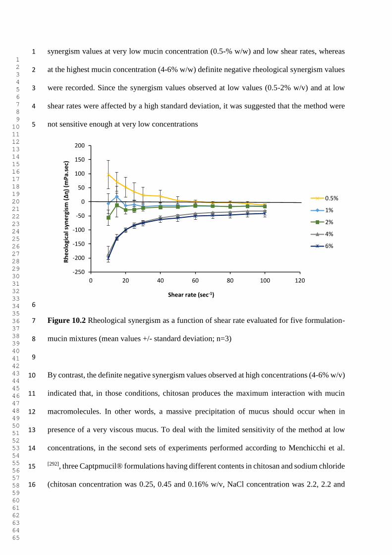
synergism values at very low mucin concentration (0.5-% w/w) and low shear rates, whereas 1
at the highest mucin concentration (4-6% w/w) definite negative rheological synergism values 2
were recorded. Since the synergism values observed at low values (0.5-2% w/v) and at low 3
shear rates were affected by a high standard deviation, it was suggested that the method were 4
not sensitive enough at very low concentrations 5
6
Figure 10.2 Rheological synergism as a function of shear rate evaluated for five formulation-7
mucin mixtures (mean values +/- standard deviation; n=3) 8
9
By contrast, the definite negative synergism values observed at high concentrations (4-6% w/v) 10
indicated that, in those conditions, chitosan produces the maximum interaction with mucin 11
macromolecules. In other words, a massive precipitation of mucus should occur when in 12
presence of a very viscous mucus. To deal with the limited sensitivity of the method at low 13
concentrations, in the second sets of experiments performed according to Menchicchi et al. 14
[292], three Captpmucil® formulations having different contents in chitosan and sodium chloride 15
(chitosan concentration was 0.25, 0.45 and 0.16% w/v, NaCl concentration was 2.2, 2.2 and 16
-250
-200
-150
-100
-50
0
50
100
150
200
0 20 40 60 80 100 120
Rh
eo
log
ica
l sy
ne
rgis
m (ȴɻ
) (m
Pa
.se
c)
Shear rate (sec-1)
0.5%
1%
2%
4%
6%
1 2 3 4 5 6 7 8 9 10 11 12 13 14 15 16 17 18 19 20 21 22 23 24 25 26 27 28 29 30 31 32 33 34 35 36 37 38 39 40 41 42 43 44 45 46 47 48 49 50 51 52 53 54 55 56 57 58 59 60 61 62 63 64 65

0.40 w/v%, respectively) were examined for dynamic viscosity using a falling sphere 1
viscometer. 2
Using this method it is possible to work at very low shear rate with precision and probe 3
differences in rheological behavior with a very high sensitivity. The deviation of the viscosity 4
of the mixture mucin/formulation from the sum of the viscosity of mucin solution plus the 5
viscosity of the formulation represents the rheological synergism that may be either positive or 6
negative depending on the sign of the deviation. The method allowed to explore 7
mucin/formulation ratio much lower than the other method ranging between 0.1 to 1.0 The 8
results obtained for the 3 formulation tested are given in Figure 10.3. 9
10
Figure 10.3 Relative deviation (in %) from the additive line (i.e., of no interaction) of the 11
relative viscosity (さrel) of chitosan-based Captomucil® formulations (as in label) in the 12
presence of pig gastric mucin as a function of mucin/formulation mass ratio f (37 °C). 13
1 2 3 4 5 6 7 8 9 10 11 12 13 14 15 16 17 18 19 20 21 22 23 24 25 26 27 28 29 30 31 32 33 34 35 36 37 38 39 40 41 42 43 44 45 46 47 48 49 50 51 52 53 54 55 56 57 58 59 60 61 62 63 64 65

1
A negative deviation is observed within the whole range of mucin/captomucil ratios tested and 2
the maximum negative deviation, meaning the maximum interaction and mucus precipitation 3
thereof, occurs in the whole range of mucin/formulation ratio for the mixture 1c (green 4
symbols) having the lower Captomucil® concentration and with the lower salt concentration 5
(slightly hypertonic formulation) whereas higher concentrations of Captomucil® and salt in 1a 6
and 1b (black and red symbols) (hypertonic formulation) caused a weaker interaction 7
specifically not in the whole range of ratios examined. 8
9
These results allowed to choose the best concentration of chitosan and salts for the final 10
formulation. Moreover, the results obtained demonstrate that the rheological approaches, 11
appropriately used according to physiological conditions; can be profitably used to measure 12
the precipitation capacity of mucolytic formulations. 13
14
g. Ocular delivery 15
To avoid physical and biochemical insults, the eye is isolated from the rest of the body by a 16
number of barriers. It is protected by the blood-retinal and blood-anterior chamber barriers. 17
Also, to meet its complex metabolic demands, the eye is supplied by an efficient 18
microvasculature that enables the supply of nutrients and oxygen, and the removal of waste 19
products at high fluxes. Due to this physiological and anatomical constraints, drug delivery to 20
the eye poses important challenges. Among these include to overcome physical and metabolic 21
barriers. Namely, structural barriers on the periphery of the retina comprised by tear secretion 22
and blinking, the conjunctival lining, connective tissue barriers of sclera and cornea, and blood 23
retinal barriers. Other factors that limit the transport of drugs to the eye include transporter 24
expression, melanin binding, and the physical state of the vitreous humour. Altogether, these 25
1 2 3 4 5 6 7 8 9 10 11 12 13 14 15 16 17 18 19 20 21 22 23 24 25 26 27 28 29 30 31 32 33 34 35 36 37 38 39 40 41 42 43 44 45 46 47 48 49 50 51 52 53 54 55 56 57 58 59 60 61 62 63 64 65

factors result in a short residence time, drug drainage, and need for frequent instillation. 1
Typically, drops instilled onto the corneal surface would be removed within 1-2 min. Also, in 2
general, only a negligible fraction of systemically administered drugs reach the ocular tissues. 3
Hence, increasing the bioavailability of topically administered drugs to the eye, has been a 4
major challenge [294]. 5
The structure of the surface of the eye is very complex. A pocket formed by the eyelids fold 6
over the cornea and conjunctiva. This pocket contains the tears (ca. 7 µL) that sit on a mucus 7
layer that coats the lining of the pocket. Two important tissues in this region are the conjunctiva 8
and the cornea. The conjunctiva has a high density of mucus-producing goblet cells, while the 9
cornea has no goblet cells. Hence, the mucin that is secreted by the conjunctival goblet cells 10
stretches across and loosely attaches to the cornea. Hence, any mucoadhesive formulation 11
needs to attach to the conjunctival mucus and presumably not to the corneal mucus, given its 12
loose attachment to the underlying tissue. Besides, binding of any material to the cornea might 13
interfere adversely with the vision process. Eye mucus is produced by goblet cells in the 14
conjunctiva. More specifically, these goblet cells are the crypts of Henle in the conjunctival 15
surface of the upper and lower tarsus and the glands of Manz on the limbal conjunctiva. Mucus-16
producing goblet cells are contained in the nonkeratinized columnar epithelium. The mucus is 17
wiped over the surface of the cornea by the windshield-wiper-like movement of the upper 18
eyelid. The human tear film is approximately 3.0 µm thick as determined by reflectance 19
spectroscopy and optical coherence tomography [295]. The mucin’s turnover rate is ca. 15-20 h. 20
The innermost mucus layer has been estimated to be 0.05 to 1.5 µm thick. The occurring 21
transmembrane and secretory mucins outlined in Section 3, play several crucial roles in 22
increasing the wettability of the ocular surface glycocalyx, mediating intracellular signal 23
transduction, as well as in modulating the viscoelastic properties and surface tension of the tear 24
film [296]. 25
1 2 3 4 5 6 7 8 9 10 11 12 13 14 15 16 17 18 19 20 21 22 23 24 25 26 27 28 29 30 31 32 33 34 35 36 37 38 39 40 41 42 43 44 45 46 47 48 49 50 51 52 53 54 55 56 57 58 59 60 61 62 63 64 65

1
Given that a number of eye pathologies are “silent” diseases with no major symptoms other 2
than the gradual loss of vision (e.g., glaucoma or cataracts), patient-friendly prolonged release 3
formulations are essential to comply with the dosing regimen. Drug delivery to the eye can be 4
either through the corneal or the non-corneal (scleral and conjunctival) route. Mucoadhesive 5
polymers have been extensively evaluated either as solutions, inserts, in situ gelling systems or 6
as colloidal particulate systems to enhance the drug penetration across the cornea epithelium 7
for ocular drug delivery. Examples include polyacrylic acid complexed to pilocarpine [297]; 8
polyacrylic acid crosslinked with divinyl alcohol for the delivery of progesterone [298]; 9
polyvinyl alcohol inserts loaded with antibiotics, sulfonamide and atropine [299]. Also, natural 10
polymers such as polysaccharides have been used as ocular delivery systems, namely pullulan, 11
hyaluronic acid and chitosan [300]. 12
13
Colloidal delivery systems such as liposomes [301], biodegradable nanoparticles [302] and 14
nanocapsules [303] offer special features as compared to other alternatives. Ex vivo studies [304] 15
demonstrated that the corneal penetration of encapsulated indomethacin in the form of 16
nanoparticles, nanocapsules and nanoemulsions was 3-fold higher than the commercial 17
formulation (Indocollyere®). No differences in drug absorption were noticed among the three 18
different systems, thus suggesting that the colloidal nature was the key factor, regardless of the 19
composition of the system. Independently, it was realized that a greater spreading coefficient 20
and lower contact angle was attained between an excised corneal tissue and a drop of a cationic 21
nanoemulsion, as compared to an anionic one [305]. This study already pointed to the importance 22
of particles with capacity to interact with the negatively charged surface of the cornea that 23
would prolong the residence time and drug absorption. In studies led by the group of Prof. 24
Maria J. Alonso [306], they determined the ocular drug disposition after instillation of 14C-25
1 2 3 4 5 6 7 8 9 10 11 12 13 14 15 16 17 18 19 20 21 22 23 24 25 26 27 28 29 30 31 32 33 34 35 36 37 38 39 40 41 42 43 44 45 46 47 48 49 50 51 52 53 54 55 56 57 58 59 60 61 62 63 64 65

indomethacin-loaded chitosan-based nanocapsules to conscious rabbits. The results showed 1
that chitosan nanocapsules increased the drug levels in cornea and aqueous humor to a 2
significantly greater extent than the commercial drug preparation or the drug loaded uncoated 3
systems. Later, it was shown that ionically crosslinked nanoparticles comprised by chitosan-4
tripolyphopspahte (TPP) were effective for the ocular delivery the hydrophobic 5
immunosuppressive peptide cyclsporin A to conscious rabbits, as a promising therapy for 6
management of external inflammatory/autoimmune ocular diseases, such as 7
keratoconjunctivitis sicca or dry eye disease. These studies led to the conclusion that chitosan 8
nanoparticles adhere to the ocular surface and some of them enter the conjunctival and the 9
corneal epithelia. It was also found that the retention of the nanoparticles was more important 10
in the conjunctiva than in the cornea. Overall, these results indicated that the affinity of chitosan 11
for the ocular surface (either cornea or conjunctiva) is greater when it is in a particulate form 12
than when in solution. Similar conclusions were reached in studies with stearylamine positively 13
charged liposomes for the ocular administration of acyclovir to rabbits [307]. These liposomes 14
would bind intimately on the cornea surface to lead to an increase of residence time and to the 15
complete coating of the corneal surface. In turn, the negatively charged liposomes were 16
expelled by the cornea surface and the drug concentration in the cornea after 2.5 h of 17
administration was half that of the positively charged liposomes. 18
19
More recently, mucoadhesive nanoparticles comprised by chitosan and hyaluronic acid have 20
been found with very low cytotoxicity and with the ability to enter the corneal epithelial cells 21
and to be taken up by CD44 receptor-mediated endocytosis [308]. These new type of particles 22
were found effective for the delivery of DNA into both the corneal epithelium (HCE) and 23
conjunctival cells (NHC) with high leves of expression of the CD44 receptor. Another study, 24
showed that polyethylenimine (PEI)/DNA polyplexes coated with low molecular weight 25
1 2 3 4 5 6 7 8 9 10 11 12 13 14 15 16 17 18 19 20 21 22 23 24 25 26 27 28 29 30 31 32 33 34 35 36 37 38 39 40 41 42 43 44 45 46 47 48 49 50 51 52 53 54 55 56 57 58 59 60 61 62 63 64 65

hyaluronic acid were effective for transfecting human corneal epithelial (HCE) cells. More 1
studies, however, are necessary to understand in greater depth at the sub-cellular level the 2
interplay between drug targeting, trafficking in the corneal epithelium. The role of 3
mucoadhesion in these systems needs to be elucidated in further detail. Particularly, aiming to 4
correlate in vitro studies with their in vivo counterparts. 5
6
h. Intravesical drug delivery 7
The bladder is a hollow organ that comprises multiple layers of tissue. From the luminal to the 8
outermost surface the layers are: urothelium, detrusor muscle and adventitia. The urothelium 9
is the innermost layer and it serves as a permeability barrier [309]. In fact, the water tight barrier 10
between blood and urine formed by urothelium is the toughest known barrier to drug delivery 11
[310]. It has exceptionally high transepithelial resistance ranging from 10 000 to >75 000 Ω/cm2 12
owing to paracellular resistance of tight junctions pooled with apical plasma membrane 13
transcellular resistance. The urothelium is composed of three different cell types (from the 14
detrusor to the apical side), namely, basal, intermediate and umbrella cells (named after their 15
characteristic shape) [311]. The umbrella cells comprise a water impermeable barrier that is 16
armored by multiple rigid-looking plaques in consort with tight junctions joining its apical 17
surface and uroplakins further enhanced by a layer of mucin and other glycosylaminoglycans 18
(GAGs) (Figure 10.4). Membrane-associated MUC1 and MUC4 are the predominant mucins 19
of the glycocalyx (Section 3), while heparan sulfate, chondroitin sulfate and dermatan sulfate, 20
have been identified among the main GAGs [312]. The primary role of the mucin/GAG layer 21
may be more in line with an antibacterial adherence function [313] rather than its role as a barrier 22
[314]. The GAG layer may also be important for the formation and attachment of particulates to 23
the urothelium and stone formation [315]. 24
25
1 2 3 4 5 6 7 8 9 10 11 12 13 14 15 16 17 18 19 20 21 22 23 24 25 26 27 28 29 30 31 32 33 34 35 36 37 38 39 40 41 42 43 44 45 46 47 48 49 50 51 52 53 54 55 56 57 58 59 60 61 62 63 64 65
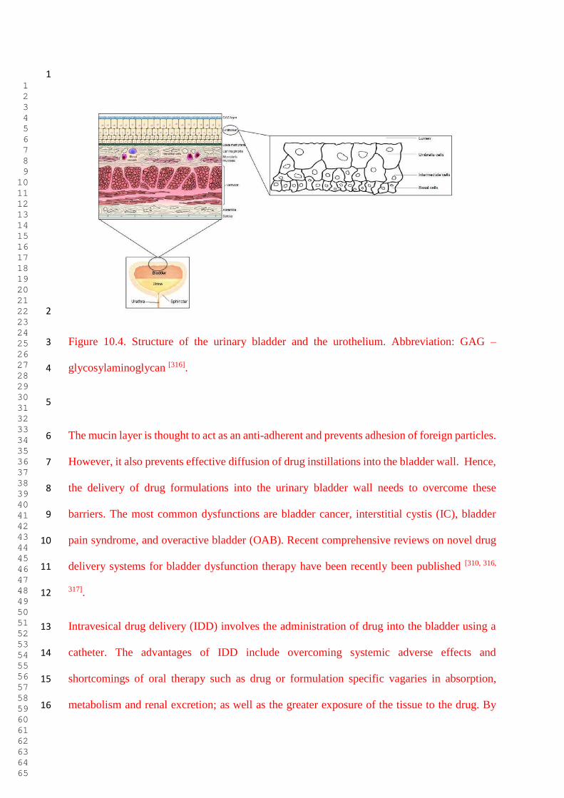
1
2
Figure 10.4. Structure of the urinary bladder and the urothelium. Abbreviation: GAG – 3
glycosylaminoglycan [316]. 4
5
The mucin layer is thought to act as an anti-adherent and prevents adhesion of foreign particles. 6
However, it also prevents effective diffusion of drug instillations into the bladder wall. Hence, 7
the delivery of drug formulations into the urinary bladder wall needs to overcome these 8
barriers. The most common dysfunctions are bladder cancer, interstitial cystis (IC), bladder 9
pain syndrome, and overactive bladder (OAB). Recent comprehensive reviews on novel drug 10
delivery systems for bladder dysfunction therapy have been recently been published [310, 316, 11
317]. 12
Intravesical drug delivery (IDD) involves the administration of drug into the bladder using a 13
catheter. The advantages of IDD include overcoming systemic adverse effects and 14
shortcomings of oral therapy such as drug or formulation specific vagaries in absorption, 15
metabolism and renal excretion; as well as the greater exposure of the tissue to the drug. By 16
1 2 3 4 5 6 7 8 9 10 11 12 13 14 15 16 17 18 19 20 21 22 23 24 25 26 27 28 29 30 31 32 33 34 35 36 37 38 39 40 41 42 43 44 45 46 47 48 49 50 51 52 53 54 55 56 57 58 59 60 61 62 63 64 65

contrast, there is substantial drug dilution during urine voiding, there is low urothelial drug 1
permeability, and the need for repeated catheterizations. To counteract the limitations 2
associated with low drug permeability, mucoadhesive formulations offer great promise. IDD 3
approach is amenable to modulating the release and absorption characteristics of instilled drugs 4
through coupling them to novel carriers, such as liposomes, microspheres, nanoparticles and in 5
situ gelling systems. 6
Dysfunction of the GAG/mucin layer is the currently prevailing hypothesis for IC. Intravesical 7
administration of hyaluronic acid, as surrogate of the GAGs of the urothelium has been reported 8
[318-320]. This approach was found to result in inhibition of leukocyte aggregation, cell migration 9
and promoted adherence of immune complexes to polymorphonuclear cells. Also, it has been 10
shown that hyaluronic acid suppresses the secretion of pro-inflammatory interleukins IL-6 and 11
IL-8, and increases the synthesis of sulphated GAGs. 12
The main challenge in IDD is to increase drug transport. This can be achieved either by physical 13
or chemical methods. We will only address here the latter. Mucoadhesive formulations for IDD 14
must fulfill three basic criteria, namely that the carrier should have rapid attachment or 15
adhesion to the bladder wall after instillation into the bladder, must not obstruct the flow of 16
urine or any of the normal functions of the bladder, and it should be able to stay attached to the 17
affected site for a number of hours even after voiding of urine [310]. A number of mucoadhesive 18
IDD formulations have been researched. These include the use of polymers of both natural and 19
synthetic type, such as chitosan, carbomers, polycarbophil (PC) gelatin, polyethylene glycol, 20
poly(methylidene malonate-2.1.2), and cellulose derivatives [309]. In a comparative study, 21
chitosan was found to exhibit greater mucoadhesion to the bladder wall than CMC and 22
polycarbophyl, thus resulting in a slower drug release and prolonged residence time [321]. 23
1 2 3 4 5 6 7 8 9 10 11 12 13 14 15 16 17 18 19 20 21 22 23 24 25 26 27 28 29 30 31 32 33 34 35 36 37 38 39 40 41 42 43 44 45 46 47 48 49 50 51 52 53 54 55 56 57 58 59 60 61 62 63 64 65

In a different study, chitosan-thioglycholic acid (TGA) nanoparticles loaded with trimethoprim 1
have been used for IDD in an in vitro study using porcine urinary bladders [322]. It was found 2
that the thiol groups and disulfide bonds introduced to chitosan-TGA conferred greater 3
stability, superior mucoadhesion and more sustained and controlled release than the 4
corresponding unmodified chitosan nanoparticles. The adhesion time of chitosan-TGA NP was 5
around 14-fold increased, while unmodified chitosan NP were washed out after three hours and 6
six micturitions. 7
Downregulation of sensory nerves by using neurotoxins like capsaicin, resiniferatoxin (RFX) 8
or botulinum toxin has proven itself a viable approach in urology, particularly for urinary 9
contingency and OAB [314]. Formulations based on liposomes and thermosensitive hydrogels 10
loaded with these drugs have also been investigated in the therapy of bladder dysfunction [323-11
325]. Capsaicin-loaded chitosan-based nanocapsules have been found to increase of paracellular 12
drug permeability in various epithelial cell lines (namely, MDCK and Caco-2) [326-328]. 13
Knowing that these nanocapsules have also proven to be mucoadhesive (see previous section 14
on ocular delivery), it would be reasonable to expect that these formulations may prove to be 15
highly effective in IDD in future studies. 16
17
12. Conclusion 18
In summary, throughout this review, we present an updated overview of the recent progress on 19
the experimental methods and applications in mucoadhesion research. As illustrated with 20
reference to specific cases, the method of choice to examine the mucoadhesive properties of a 21
given biomaterial (namely a medical device or pharmaceutical formulation) can vary widely. 22
A sounder understanding of the phenomena at play that influence the molecular level 23
interactions of mucin and mucus with a large diversity of materials is likely to contribute to the 24
development of innovative applications on a more rational basis. Novel methods currently 25
1 2 3 4 5 6 7 8 9 10 11 12 13 14 15 16 17 18 19 20 21 22 23 24 25 26 27 28 29 30 31 32 33 34 35 36 37 38 39 40 41 42 43 44 45 46 47 48 49 50 51 52 53 54 55 56 57 58 59 60 61 62 63 64 65

under development and that have not been addressed in this review include the use of in vitro 1
cell culture or biomicrofluidics approaches. 2
3
13. References 4
[1] V. Grabovac, D. Guggi, A. Bernkop-Schnurch, Advanced Drug Delivery Reviews 2005, 5
57, 1713. 6
[2] A. B. Khan, R. S. Thakur, Current Drug Delivery 2014, 11, 112. 7
[3] R. Shaikh, T. R. Raj Singh, M. J. Garland, A. D. Woolfson, R. F. Donnelly, Journal of 8
pharmacy & bioallied sciences 2011, 3, 89. 9
[4] D. S. Jones, A. D. Woolfson, A. F. Brown, W. A. Coulter, C. McClelland, C. R. Irwin, 10
Journal of Controlled Release 2000, 67, 357. 11
[5] J. D. Smart, Advanced Drug Delivery Reviews 2005, 57, 1556. 12
[6] L. M. Ensign, R. Cone, J. Hanes, Advanced Drug Delivery Reviews 2012, 64, 557. 13
[7] B. C. Tang, M. Dawson, S. K. Lai, Y. Y. Wang, J. S. Suk, M. Yang, P. Zeitlin, M. P. 14
Boyle, J. Fu, J. Hanes, Proceedings of the National Academy of Sciences of the United States 15
of America 2009, 106, 19268. 16
[8] R. A. Cone, Adv Drug Deliver Rev 2009, 61, 75. 17
[9] M. E. V. Johansson, H. Sjövall, G. C. Hansson, Nature reviews Gastroenterology 18
Hepatology 2013. 19
[10] J. L. McAuley, S. K. Linden, C. W. Png, R. M. King, H. L. Pennington, S. J. Gendler, T. 20
H. Florin, G. R. Hill, V. Korolik, M. A. McGuckin, J.Clin.Invest 2007, 117, 2313. 21
[11] Y. H. Sheng, S. Triyana, R. Wang, I. Das, K. Gerloff, T. H. Florin, P. Sutton, M. A. 22
McGuckin, Mucosal Immunology 2013, 6, 557. 23
[12] A. P. Corfield, Biochimica Et Biophysica Acta-General Subjects 2015, 1850, 236. 24
1 2 3 4 5 6 7 8 9 10 11 12 13 14 15 16 17 18 19 20 21 22 23 24 25 26 27 28 29 30 31 32 33 34 35 36 37 38 39 40 41 42 43 44 45 46 47 48 49 50 51 52 53 54 55 56 57 58 59 60 61 62 63 64 65

[13] S. A. Rayment, B. Liu, G. D. Offner, F. G. Oppenheim, R. F. Troxler, Journal of dental 1
research 2000, 79, 1765. 2
[14] K. C. Kim, Pulmonary pharmacology & therapeutics 2012, 25, 415. 3
[15] D. J. Thornton, K. Rousseau, M. A. McGuckin, Annu.Rev.Physiol 2008, 70, 459. 4
[16] I. K. Gipson, Frontiers in bioscience : a journal and virtual library 2001, 6, D1245. 5
[17] S. K. Lai, Y. Y. Wang, J. Hanes, Advanced Drug Delivery Reviews 2009, 61, 158. 6
[18] A. Macierzanka, A. R. Mackie, B. H. Bajka, N. M. Rigby, F. Nau, D. Dupont, Plos One 7
2014, 9, e95274. 8
[19] R. M. Corrales, D. J. Galarreta, J. M. Herreras, M. Calonge, F. J. Chaves, Archivos de la 9
Sociedad Espanola de Oftalmologia 2003, 78, 375. 10
[20] J. E. Jumblatt, L. T. Cunningham, Y. Li, M. M. Jumblatt, Cornea 2002, 21, 818. 11
[21] J. N'Dow, J. P. Pearson, M. K. Bennett, D. E. Neal, C. N. Robson, The Journal of 12
urology 2000, 164, 1398. 13
[22] A. N. Round, M. Berry, T. J. McMaster, A. P. Corfield, M. J. Miles, Journal of 14
Structural Biology 2004, 145, 246. 15
[23] C. Atuma, V. Strugala, A. Allen, L. Holm, American Journal of Physiology-16
Gastrointestinal and Liver Physiology 2001, 280, G922. 17
[24] M. E. Johansson, M. Phillipson, J. Petersson, A. Velcich, L. Holm, G. C. Hansson, 18
Proc.Natl.Acad.Sci.U.S.A 2008, 105, 15064. 19
[25] M. E. V. Johansson, J. M. H. Larsson, G. C. Hansson, Proceedings of the National 20
Academy of Sciences of the United States of America 2011, 108, 4659. 21
[26] B. H. Bajka, N. M. Rigby, K. Cross, A. Macierzanka, A. R. Mackie, Colloids and 22
Surfaces B: Biointerfaces 2015, 135, 73. 23
[27] S. S. Olmsted, J. L. Padgett, A. I. Yudin, K. J. Whaley, T. R. Moench, R. A. Cone, 24
Biophysical Journal 2001, 81, 1930. 25
1 2 3 4 5 6 7 8 9 10 11 12 13 14 15 16 17 18 19 20 21 22 23 24 25 26 27 28 29 30 31 32 33 34 35 36 37 38 39 40 41 42 43 44 45 46 47 48 49 50 51 52 53 54 55 56 57 58 59 60 61 62 63 64 65

[28] A. N. Round, N. M. Rigby, A. Garcia de la Torre, A. Macierzanka, E. N. C. Mills, A. R. 1
Mackie, Biomacromolecules 2012, 13, 3253. 2
[29] A. Macierzanka, N. M. Rigby, A. P. Corfield, N. Wellner, F. Böttger, E. N. C. Mills, A. 3
R. Mackie, Soft Matter 2011, 7, 8077. 4
[30] "Mucins Methods and protocols", 2012, p. 325. 5
[31] D. J. Thornton, I. Carlstedt, M. Howard, P. L. Devine, M. R. Price, J. K. Sheehan, 6
Biochemical Journal 1996, 316, 967. 7
[32] B. D. Raynal, T. E. Hardingham, D. J. Thornton, J. K. Sheehan, Biochem J 2002, 362, 8
289. 9
[33] R. Mehrotra, D. J. Thornton, J. K. Sheehan, Biochem J 1998, 334 ( Pt 2), 415. 10
[34] C. Wickstrom, J. R. Davies, G. V. Eriksen, E. C. Veerman, I. Carlstedt, Biochem J 1998, 11
334 ( Pt 3), 685. 12
[35] S. Rossi, M. C. Bonferoni, G. Lippoli, M. Bertoni, F. Ferrari, C. Caramella, U. Conte, 13
Biomaterials 1995, 16, 1073. 14
[36] G. E. Yakubov, A. Papagiannopoulos, E. Rat, R. L. Easton, T. A. Waigh, 15
Biomacromolecules 2007, 8, 3467. 16
[37] S. Kirkham, J. K. Sheehan, D. Knight, P. S. Richardson, D. J. Thornton, Biochemical 17
Journal 2002, 361, 537. 18
[38] D. J. Thornton, N. Khan, R. Mehrotra, M. Howard, E. Veerman, N. H. Packer, J. K. 19
Sheehan, Glycobiology 1999, 9, 293. 20
[39] M. T. Cook, V. V. Khutoryanskiy, Int J Pharm 2015, 495, 991. 21
[40] A. C. Groo, F. Lagarce, Drug discovery today 2014, 19, 1097. 22
[41] S. P. Authimoolam, T. D. Dziubla, Polymers 2016, 8. 23
[42] C. D. Rillahan, J. C. Paulson, Annual review of biochemistry 2011, 80, 797. 24
1 2 3 4 5 6 7 8 9 10 11 12 13 14 15 16 17 18 19 20 21 22 23 24 25 26 27 28 29 30 31 32 33 34 35 36 37 38 39 40 41 42 43 44 45 46 47 48 49 50 51 52 53 54 55 56 57 58 59 60 61 62 63 64 65

[43] S. M. Muthana, J. C. Gildersleeve, Cancer biomarkers : section A of Disease markers 1
2014, 14, 29. 2
[44] K. Godula, D. Rabuka, K. T. Nam, C. R. Bertozzi, Angewandte Chemie (International 3
ed. in English) 2009, 48, 4973. 4
[45] M. Kilcoyne, J. Q. Gerlach, R. Gough, M. E. Gallagher, M. Kane, S. D. Carrington, L. 5
Joshi, Anal Chem 2012, 84, 3330. 6
[46] J. Cleary, L. Bromberg, E. Magner, Langmuir 2004, 20, 9755. 7
[47] N. V. Efremova, Y. Huang, N. A. Peppas, D. E. Leckband, Langmuir 2002, 18, 836. 8
[48] M. Iijima, M. Yoshimura, T. Tsuchiya, M. Tsukada, H. Ichikawa, Y. Fukumori, H. 9
Kamiya, Langmuir 2008, 24, 3987. 10
[49] D. Li, H. Yamamoto, H. Takeuchi, Y. Kawashima, European Journal of Pharmaceutics 11
and Biopharmaceutics 2010, 75, 277. 12
[50] P. Sriamornsak, N. Wattanakorn, H. Takeuchi, Carbohydrate Polymers 2010, 79, 54. 13
[51] C. Taylor, J. P. Pearson, K. I. Draget, P. W. Dettmar, O. Smidsrod, Carbohydrate 14
Polymers 2005, 59, 189. 15
[52] M. Boegh, S. G. Baldursdottir, A. Mullertz, H. M. Nielsen, European Journal of 16
Pharmaceutics and Biopharmaceutics 2014, 87, 227. 17
[53] B. T. Burruano, R. L. Schnaare, D. Malamud, Contraception 2002, 66, 137. 18
[54] C. V. Duffy, L. David, T. Crouzier, Acta Biomaterialia 2015, 20, 51. 19
[55] R. Hamed, J. Fiegel, Journal of Biomedical Materials Research Part A 2014, 102, 1788. 20
[56] M. D. Anwarul Hasan, C. F. Lange, M. L. King, Journal of Non-Newtonian Fluid 21
Mechanics 2010, 165, 1431. 22
[57] S. P. Authimoolam, A. L. Vasilakes, N. M. Shah, D. A. Puleo, T. D. Dziubla, 23
Biomacromolecules 2014, 15, 3099. 24
1 2 3 4 5 6 7 8 9 10 11 12 13 14 15 16 17 18 19 20 21 22 23 24 25 26 27 28 29 30 31 32 33 34 35 36 37 38 39 40 41 42 43 44 45 46 47 48 49 50 51 52 53 54 55 56 57 58 59 60 61 62 63 64 65

[58] M. Doellinger, F. Groehn, D. A. Berry, U. Eysholdt, G. Luegmair, Journal of Speech 1
Language and Hearing Research 2014, 57, S637. 2
[59] D. J. Hall, O. V. Khutoryanskaya, V. V. Khutoryanskiy, Soft Matter 2011, 7, 9620. 3
[60] J. KocevarNared, J. Kristl, J. SmidKorbar, Biomaterials 1997, 18, 677. 4
[61] O. V. Khutoryanskaya, M. Potgieter, V. V. Khutoryanskiy, Soft Matter 2010, 6, 551. 5
[62] O. V. Khutoryanskaya, A. C. Williams, V. V. Khutoryanskiy, Macromolecules 2007, 40, 6
7707. 7
[63] M. T. Cook, S. L. Smith, V. V. Khutoryanskiy, Chemical Communications 2015, 51, 8
14447. 9
[64] T. Eshel-Green, S. Eliyahu, S. Avidan-Shlomovich, H. Bianco-Peled, International 10
Journal of Pharmaceutics 2016, 506, 25. 11
[65] T. J. Sill, H. a. von Recum, Biomaterials 2008, 29, 1989. 12
[66] Q. P. Pham, U. Sharma, A. G. Mikos, Tissue Engineering 2006, 12, 060509065116001. 13
[67] S. Ramakrishna, M. Zamani, M. P. Prabhakaran, International Journal of Nanomedicine 14
2013, 8, 2997. 15
[68] K. Stephansen, M. García-Díaz, F. Jessen, I. S. Chronakis, H. M. Nielsen, International 16
Journal of Pharmaceutics 2015, 495, 58. 17
[69] S.-F. Chou, D. Carson, K. A. Woodrow, Journal of Controlled Release 2015, 220, 584. 18
[70] A. Balaji, M. V. Vellayappan, A. A. John, A. P. Subramanian, S. K. Jaganathan, E. 19
Supriyanto, S. I. A. Razak, RSC Adv. 2015, 5, 57984. 20
[71] A. Sharma, A. Gupta, G. Rath, A. Goyal, R. B. Mathur, S. R. Dhakate, Journal of 21
Materials Chemistry B 2013, 1, 3410. 22
[72] S. Wongsasulak, S. Pathumban, T. Yoovidhya, Journal of Food Engineering 2014, 120, 23
110. 24
1 2 3 4 5 6 7 8 9 10 11 12 13 14 15 16 17 18 19 20 21 22 23 24 25 26 27 28 29 30 31 32 33 34 35 36 37 38 39 40 41 42 43 44 45 46 47 48 49 50 51 52 53 54 55 56 57 58 59 60 61 62 63 64 65
[73] C. Dott, C. Tyagi, L. K. Tomar, Y. E. Choonara, P. Kumar, L. C. du Toit, V. Pillay, 1
Journal of Nanomaterials 2013, 2013, 1. 2
[74] R. Sridhar, S. Sundarrajan, A. Vanangamudi, G. Singh, T. Matsuura, S. Ramakrishna, 3
Macromolecular Materials and Engineering 2014, 299, 283. 4
[75] F. Ding, H. Deng, Y. Du, X. Shi, Q. Wang, Nanoscale 2014, 6, 9477. 5
[76] K. Stephansen, M. García-Díaz, F. Jessen, I. S. Chronakis, H. M. Nielsen, Molecular 6
Pharmaceutics 2016, 13, 748. 7
[77] H. Seager, The Journal of pharmacy and pharmacology 1998, 50, 375. 8
[78] D. C. Aduba, J. A. Hammer, Q. Yuan, W. Andrew Yeudall, G. L. Bowlin, H. Yang, Acta 9
Biomaterialia 2013, 9, 6576. 10
[79] L. Xu, N. Sheybani, S. Ren, G. L. Bowlin, W. A. Yeudall, H. Yang, Pharmaceutical 11
Research 2015, 32, 275. 12
[80] P. Tonglairoum, T. Ngawhirunpat, T. Rojanarata, S. Panomsuk, R. Kaomongkolgit, P. 13
Opanasopit, Carbohydrate Polymers 2015, 132, 173. 14
[81] S. Wongsasulak, N. Puttipaiboon, T. Yoovidhya, Journal of Food Science 2013, 78, 15
N926. 16
[82] S. Zong, X. Wang, Y. Yang, W. Wu, H. Li, Y. Ma, W. Lin, T. Sun, Y. Huang, Z. Xie, Y. 17
Yue, S. Liu, X. Jing, European Journal of Pharmaceutics and Biopharmaceutics 2015, 93, 18
127. 19
[83] C. Huang, S. J. Soenen, E. van Gulck, G. Vanham, J. Rejman, S. Van Calenbergh, C. 20
Vervaet, T. Coenye, H. Verstraelen, M. Temmerman, J. Demeester, S. C. De Smedt, 21
Biomaterials 2012, 33, 962. 22
[84] W. Samprasit, R. Kaomongkolgit, M. Sukma, T. Rojanarata, T. Ngawhirunpat, P. 23
Opanasopit, Carbohydrate Polymers 2015, 117, 933. 24
1 2 3 4 5 6 7 8 9 10 11 12 13 14 15 16 17 18 19 20 21 22 23 24 25 26 27 28 29 30 31 32 33 34 35 36 37 38 39 40 41 42 43 44 45 46 47 48 49 50 51 52 53 54 55 56 57 58 59 60 61 62 63 64 65

[85] H. Singh, R. Sharma, M. Joshi, T. Garg, A. K. Goyal, G. Rath, Artificial Cells, 1
Nanomedicine, and Biotechnology 2015, 43, 263. 2
[86] P. Tonglairoum, T. Ngawhirunpat, T. Rojanarata, R. Kaomongkolgit, P. Opanasopit, 3
Colloids and Surfaces B: Biointerfaces 2015, 126, 18. 4
[87] H. Grewal, S. R. Dhakate, A. K. Goyal, T. S. Markandeywar, B. Malik, G. Rath, 5
Artificial Cells, Blood Substitutes, and Biotechnology 2012, 40, 146. 6
[88] C. Huang, S. J. Soenen, E. van Gulck, J. Rejman, G. Vanham, B. Lucas, B. Geers, K. 7
Braeckmans, V. Shahin, P. Spanoghe, J. Demeester, S. C. De Smedt, Polymers for Advanced 8
Technologies 2014, 25, 827. 9
[89] Gagandeep, T. Garg, B. Malik, G. Rath, A. K. Goyal, European Journal of 10
Pharmaceutical Sciences 2014, 53, 10. 11
[90] E. Zartler, J. Yan, H. Mo, A. Kline, M. Shapiro, Current Topics in Medicinal Chemistry 12
2003, 3, 25. 13
[91] S. Monti, I. Manet, G. Marconi, Physical Chemistry Chemical Physics 2011, 13, 20893. 14
[92] S. Guglieri, M. Hricovni, R. Raman, L. Polito, G. Torri, B. Casu, R. Sasisekharan, M. 15
Guerrini, Biochemistry 2008, 47, 13862. 16
[93] G. Uccello-Barretta, F. Balzano, F. Aiello, Polysaccharides: Bioactivity and 17
Biotechnology 2015, 1299. 18
[94] G. Uccello-Barretta, S. Nazzi, F. Balzano, M. Sansò, International Journal of 19
Pharmaceutics 2011, 406, 78. 20
[95] G. Uccello-Barretta, F. Balzano, L. Vanni, M. Sansò, Carbohydrate Polymers 2013, 91, 21
568. 22
[96] M. M. Patel, J. D. Smart, T. G. Nevell, R. J. Ewen, P. J. Eaton, J. Tsibouklis, 23
Biomacromolecules 2003, 4, 1184. 24
[97] S. A. Mortazavi, International Journal of Pharmaceutics 1995, 124, 173. 25
1 2 3 4 5 6 7 8 9 10 11 12 13 14 15 16 17 18 19 20 21 22 23 24 25 26 27 28 29 30 31 32 33 34 35 36 37 38 39 40 41 42 43 44 45 46 47 48 49 50 51 52 53 54 55 56 57 58 59 60 61 62 63 64 65

[98] P. C. Griffiths, P. Occhipinti, C. Morris, R. K. Heenan, S. M. King, M. Gumbleton, 1
Biomacromolecules 2010, 11, 120. 2
[99] M. Davidovich-Pinhas, H. Bianco-Peled, Expert Opinion on Drug Delivery 2010, 7, 3
259. 4
[100] P. Sriamornsak, N. Wattanakorn, J. Nunthanid, S. Puttipipatkhachorn, Carbohydrate 5
Polymers 2008, 74, 458. 6
[101] F. Saiano, G. Pitarresi, G. Cavallaro, M. Licciardi, G. Giammona, Polymer 2002, 43, 7
6281. 8
[102] J. Xiang, X. Li, Journal of Applied Polymer Science 2004, 94, 2431. 9
[103] R. Falahat, E. Williams, M. Wiranowska, R. Toomey, N. Alcantar, Cancer Research 10
2014, 74, 5410. 11
[104] N. A. Peppas, Y. Huang, Advanced Drug Delivery Reviews 2004, 56, 1675. 12
[105] J. das Neves, M. F. Bahia, M. M. Amiji, B. Sarmento, Expert Opinion on Drug 13
Delivery 2011, 8, 1085. 14
[106] H. Takeuchi, Y. Matsui, H. Sugihara, H. Yamamoto, Y. Kawashima, International 15
Journal of Pharmaceutics 2005, 303, 160. 16
[107] H. Takeuchi, J. Thongborisute, Y. Matsui, H. Sugihara, H. Yamamoto, Y. Kawashima, 17
Advanced Drug Delivery Reviews 2005, 57, 1583. 18
[108] D. Chen, D. Xia, X. Li, Q. Zhu, H. Yu, C. Zhu, Y. Gan, International Journal of 19
Pharmaceutics 2013, 449, 1. 20
[109] B. Fan, Y. Xing, Y. Zheng, C. Sun, G. Liang, Drug delivery 2016, 23, 238. 21
[110] K. Tahara, S. Fujimoto, F. Fujii, Y. Tozuka, T. Jin, H. Takeuchi, Journal of 22
Pharmaceutics 2013, 2013, 1. 23
[111] G. Uccello-Barretta, F. Balzano, F. Aiello, A. Senatore, A. Fabiano, Y. Zambito, 24
International Journal of Pharmaceutics 2014, 461, 489. 25
1 2 3 4 5 6 7 8 9 10 11 12 13 14 15 16 17 18 19 20 21 22 23 24 25 26 27 28 29 30 31 32 33 34 35 36 37 38 39 40 41 42 43 44 45 46 47 48 49 50 51 52 53 54 55 56 57 58 59 60 61 62 63 64 65

[112] T. Eshel-Green, H. Bianco-Peled, Colloids and Surfaces B: Biointerfaces 2016, 139, 1
42. 2
[113] T. Pettersson, A. Dedinaite, Journal of Colloid and Interface Science 2008, 324, 246. 3
[114] L. Joergensen, B. Klosgen, A. C. Simonsen, J. Borch, E. Hagesaether, International 4
Journal of Pharmaceutics 2011, 411, 162. 5
[115] M. P. Deacon, S. McGurk, C. J. Roberts, P. M. Williams, S. J. B. Tendler, M. C. 6
Davies, S. S. Davis, S. E. Harding, Biochemical Journal 2000, 348, 557. 7
[116] E. Di Cola, G. E. Yakubov, T. A. Waigh, Biomacromolecules 2008, 9, 3216. 8
[117] P. Georgiades, E. di Cola, R. K. Heenan, P. D. A. Pudney, D. J. Thornton, T. A. Waigh, 9
Biopolymers 2014, 101, 1154. 10
[118] Y. Watanabe, Y. Inoko, J Appl Crystallogr 2007, 40, S209. 11
[119] T. A. Waigh, A. Papagiannopoulos, A. Voice, R. Bansil, A. P. Unwin, C. D. Dewhurst, 12
B. Turner, N. Afdhal, Langmuir 2002, 18, 7188. 13
[120] S. K. Lai, Y. Y. Wang, D. Wirtz, J. Hanes, Adv Drug Deliv Rev 2009, 61, 86. 14
[121] J. P. Celli, B. S. Turner, N. H. Afdhal, R. H. Ewoldt, G. H. McKinley, R. Bansil, S. 15
Erramilli, Biomacromolecules 2007, 8, 1580. 16
[122] R. Bansil, H. E. Stanley, J. T. Lamont, Annual Review of Physiology 1995, 57, 635. 17
[123] X. Cao, R. Bansil, K. R. Bhaskar, B. S. Turner, J. T. LaMont, N. Niu, N. H. Afdhal, 18
Biophysical Journal 1999, 76, 1250. 19
[124] Z. N. Hong, B. Chasan, R. Bansil, B. S. Turner, K. R. Bhaskar, N. H. Afdhal, 20
Biomacromolecules 2005, 6, 3458. 21
[125] A. Maleki, G. Lafitte, A. L. Kjoniksen, K. Thuresson, B. Nystrom, Carbohyd Res 2008, 22
343, 328. 23
[126] K. R. Bhaskar, D. H. Gong, R. Bansil, S. Pajevic, J. A. Hamilton, B. S. Turner, J. T. 24
LaMont, Am J Physiol 1991, 261, G827. 25
1 2 3 4 5 6 7 8 9 10 11 12 13 14 15 16 17 18 19 20 21 22 23 24 25 26 27 28 29 30 31 32 33 34 35 36 37 38 39 40 41 42 43 44 45 46 47 48 49 50 51 52 53 54 55 56 57 58 59 60 61 62 63 64 65

[127] S. Lee, M. Muller, K. Rezwan, N. D. Spencer, Langmuir 2005, 21, 8344. 1
[128] I. A. Sogias, A. C. Williams, V. V. Khutoryanskiy, Biomacromolecules 2008, 9, 1837. 2
[129] B. Menchicchi, J. P. Fuenzalida, A. Hensel, M. J. Swamy, L. David, C. Rochas, F. M. 3
Goycoolea, Biomacromolecules 2015, 16, 924. 4
[130] C. Woertz, M. Preis, J. Breitkreutz, P. Kleinebudde, European Journal of 5
Pharmaceutics and Biopharmaceutics 2013, 85, 843. 6
[131] S. Rossi, M. C. Bonferoni, F. Ferrari, M. Bertoni, C. Caramella, European Journal of 7
Pharmaceutical Sciences 1996, 4, 189. 8
[132] E. Hagesaether, M. Hiorth, S. A. Sande, European Journal of Pharmaceutics and 9
Biopharmaceutics 2009, 71, 325. 10
[133] M. Bogataj, T. Vovk, M. Kerec, A. Dimnik, I. Grabnar, A. Mrhar, Biological & 11
Pharmaceutical Bulletin 2003, 26, 743. 12
[134] D. S. Jones, A. D. Woolfson, A. F. Brown, International Journal of Pharmaceutics 13
1997, 151, 223. 14
[135] H. Hägerström, K. Edsman, Journal of Pharmacy and Pharmacology 2001, 53, 1589. 15
[136] S. Tamburic, D. Q. M. Craig, European Journal of Pharmaceutics and 16
Biopharmaceutics 1997, 44, 159. 17
[137] I. A. Sogias, A. C. Williams, V. V. Khutoryanskiy, International Journal of 18
Pharmaceutics 2012, 436, 602. 19
[138] A. P. Sam, J. T. M. van den Heuij, J. J. Tukker, International Journal of Pharmaceutics 20
1992, 79, 97. 21
[139] J. D. Smart, International Journal of Pharmaceutics 1991, 73, 69. 22
[140] C. E. Kast, A. Bernkop-Schnürch, Biomaterials 2001, 22, 2345. 23
[141] K. G. H. Desai, T. M. P. Kumar, Aaps Pharmscitech 2004, 5. 24
1 2 3 4 5 6 7 8 9 10 11 12 13 14 15 16 17 18 19 20 21 22 23 24 25 26 27 28 29 30 31 32 33 34 35 36 37 38 39 40 41 42 43 44 45 46 47 48 49 50 51 52 53 54 55 56 57 58 59 60 61 62 63 64 65

[142] H. S. Chng, H. Park, P. Kelly, J. R. Robinson, Journal of Pharmaceutical Sciences 1
1985, 74, 399. 2
[143] R. Gurny, J.-M. Meyer, N. A. Peppas, Biomaterials 1984, 5, 336. 3
[144] Y. B. Huang, W. Leobandung, A. Foss, N. A. Peppas, Journal of Controlled Release 4
2000, 65, 63. 5
[145] F. Ferrari, M. Bertoni, S. Rossi, M. C. Bonferoni, C. Caramella, M. J. Waring, M. E. 6
Aulton, Drug Dev Ind Pharm 1996, 22, 1223. 7
[146] D. Patel, J. R. Smith, A. W. Smith, N. Grist, P. Barnett, J. D. Smart, International 8
Journal of Pharmaceutics 2000, 200, 271. 9
[147] H. Takeuchi, J. Thongborisute, Y. Matsui, H. Sugihara, H. Yamamoto, Y. Kawashima, 10
Adv Drug Deliv Rev 2005, 57, 1583. 11
[148] A. Fuongfuchat, A. M. Jamieson, J. Blackwell, T. A. Gerken, Carbohydrate Research 12
1996, 284, 85. 13
[149] S. Rossi, F. Ferrari, M. C. Bonferoni, C. Caramella, European Journal of 14
Pharmaceutical Sciences 2000, 10, 251. 15
[150] M. M. Patel, J. D. Smart, T. G. Nevell, R. J. Ewen, P. J. Eaton, J. Tsibouklis, 16
Biomacromolecules 2003, 4, 1184. 17
[151] M. P. Deacon, S. S. Davis, R. J. White, H. Nordman, I. Carlstedt, N. Errington, A. J. 18
Rowe, S. E. Harding, Carbohydrate Polymers 1999, 38, 235. 19
[152] I. Fiebrig, Davis, S.S., Harding, S.E., "Method used to develop drug delivery systems: 20
bioadhesion in the gastrointestinal tract", in Biopolymer mixtures, S.E. Harding, Hill,S.E., 21
Mitchell,J.R., Ed., Nottingham University Press, Nottingham, 1995, p. 737. 22
[153] S. E. Harding, Trends in Food Science & Technology 2006, 17, 255. 23
[154] S. Chayed, F. M. Winnik, European Journal of Pharmaceutics and Biopharmaceutics 24
2007, 65, 363. 25
1 2 3 4 5 6 7 8 9 10 11 12 13 14 15 16 17 18 19 20 21 22 23 24 25 26 27 28 29 30 31 32 33 34 35 36 37 38 39 40 41 42 43 44 45 46 47 48 49 50 51 52 53 54 55 56 57 58 59 60 61 62 63 64 65

[155] W. Chaiyasan, S. P. Srinivas, W. Tiyaboonchai, J Ocul Pharmacol Th 2013, 29, 200. 1
[156] Y. Bin Choy, J. H. Park, M. R. Prausnitz, J Phys Chem Solids 2008, 69, 1533. 2
[157] Y. Miyazaki, K. Ogihara, S. Yakou, T. Nagai, K. Takayama, International Journal of 3
Pharmaceutics 2003, 258, 21. 4
[158] I. Bravo-Osuna, C. Vauthier, A. Farabollini, G. F. Palmieri, G. Ponchel, Biomaterials 5
2007, 28, 2233. 6
[159] J. Schmidgall, E. Schnetz, A. Hensel, Planta Med 2000, 66, 48. 7
[160] J. Schmidgall, A. Hensel, Int J Biol Macromol 2002, 30, 217. 8
[161] K. Park, J. R. Robinson, International Journal of Pharmaceutics 1984, 19, 107. 9
[162] O. Lieleg, I. Vladescu, K. Ribbeck, Biophys J 2010, 98, 1782. 10
[163] E. E. Hassan, J. M. Gallo, Pharm Res 1990, 7, 491. 11
[164] S. J. List, B. P. Findlay, G. G. Forstner, J. F. Forstner, Biochemical Journal 1978, 175, 12
565. 13
[165] S. Rossi, F. Ferrari, M. C. Bonferoni, C. Caramella, European Journal of 14
Pharmaceutical Sciences 2001, 12, 479. 15
[166] S. Rossi, M. C. Bonferoni, G. Lippoli, M. Bertoni, F. Ferrari, C. Caramella, U. Conte, 16
Biomaterials 1995, 16, 1073. 17
[167] F. Madsen, K. Eberth, J. D. Smart, Biomaterials 1998, 19, 1083. 18
[168] C. Taylor, J. P. Pearson, K. I. Draget, P. W. Dettmar, O. Smidsrød, Carbohydrate 19
Polymers 2005, 59, 189. 20
[169] F. M. Goycoolea, E. R. Morris, M. J. Gidley, Carbohydrate Polymers 1995, 28, 351. 21
[170] D. J. McClements, Biotechnology Advances 2006, 24, 621. 22
[171] N. K. Howell, "Synergism and interactions in mixed protein systems", in Biopolymer 23
mixtures, S.E. Harding, Hill,S.E., Mitchell,J.R., Ed., Notthingam University Press, 24
Notthingam, 1995, p. 329. 25
1 2 3 4 5 6 7 8 9 10 11 12 13 14 15 16 17 18 19 20 21 22 23 24 25 26 27 28 29 30 31 32 33 34 35 36 37 38 39 40 41 42 43 44 45 46 47 48 49 50 51 52 53 54 55 56 57 58 59 60 61 62 63 64 65

[172] E. R. Morris, "Polysaccharides synergism: more question than answers?", in 1
Biopolymer Mixtures, S.E. Harding, Hill,S.E., Mitchell,J.R., Ed., Nottingham University 2
Press, Nottingham, 1995, p. 247. 3
[173] S. A. Mortazavi, B. G. Carpenter, J. D. Smart, International Journal of Pharmaceutics 4
1993, 94, 195. 5
[174] C. Woertz, M. Preis, J. Breitkreutz, P. Kleinebudde, European Journal of 6
Pharmaceutics and Biopharmaceutics 2013, 85, 843. 7
[175] B. Menchicchi, J. P. Fuenzalida, K. B. Bobbili, A. Hensel, M. J. Swamy, F. M. 8
Goycoolea, Biomacromolecules 2014, 15, 3550. 9
[176] G. Sandri, S. Rossi, M. C. Bonferoni, F. Ferrari, M. Mori, C. Caramella, Journal of 10
Drug Delivery Science and Technology 2012, 22, 275. 11
[177] G. Sandri, S. Rossi, F. Ferrari, M. C. Bonferoni, N. Zerrouk, C. Caramella, Journal of 12
Pharmacy and Pharmacology 2004, 56, 1083. 13
[178] E. SzymaMska, K. Winnicka, A. Amelian, U. Cwalina, Chemical and Pharmaceutical 14
Bulletin 2014, 62, 160. 15
[179] M. Bayarri, N. Oulahal, P. Degraeve, A. Gharsallaoui, Journal of Food Engineering 16
2014, 131, 18. 17
[180] Y. Li, W. Yokoyama, J. Wu, J. Ma, F. Zhong, RSC Adv. 2015, 5, 105844. 18
[181] M. A. Güler, M. K. Gök, A. K. Figen, S. Özgümüş, Applied Clay Science 2015, 112-19
113, 44. 20
[182] C. S. Kolli, I. Pather, "Characterization Methods for Oral Mucosal Drug Delivery", in 21
Oral Mucosal Drug Delivery and Therapy, M. Rathbone, S. Senel, and I. Pather, Eds., 22
Springer, 2015, p. 125. 23
[183] C. Pontier, J. Pachot, R. Botham, B. Lenfant, P. Arnaud, Journal of Pharmaceutical 24
Sciences 2001, 90, 1608. 25
1 2 3 4 5 6 7 8 9 10 11 12 13 14 15 16 17 18 19 20 21 22 23 24 25 26 27 28 29 30 31 32 33 34 35 36 37 38 39 40 41 42 43 44 45 46 47 48 49 50 51 52 53 54 55 56 57 58 59 60 61 62 63 64 65

[184] E. Hagesaether, E. Christiansen, M. E. Due-Hansen, T. Ulven, International Journal of 1
Pharmaceutics 2013, 452, 276. 2
[185] A. Jintapattanakit, V. B. Junyaprasert, T. Kissel, Journal of Pharmaceutical Sciences 3
2009, 98, 4818. 4
[186] S. Chen, Y. Cao, L. R. Ferguson, Q. Shu, S. Garg, J. Microencapsul. 2013, 30, 103. 5
[187] M. I. Adamczak, E. Hagesaether, G. Smistad, M. Hiorth, International Journal of 6
Pharmaceutics 2016, 498, 225. 7
[188] Y. Cu, W. M. Saltzman, Advanced Drug Delivery Reviews 2009, 61, 101. 8
[189] M. A. Desai, P. Vadgama, Analyst 1991, 116, 1113. 9
[190] L. Szentkuti, Journal of Controlled Release 1997, 46, 233. 10
[191] K. Ohtake, H. Natsume, H. Ueda, Y. Morimoto, Journal of Controlled Release 2002, 11
82, 263. 12
[192] P. C. Griffiths, P. Occhipinti, C. Morris, R. K. Heenan, S. M. King, M. Gumbleton, 13
Biomacromolecules 2010, 11, 120. 14
[193] K. T. H. Nguyen, E. V. Mathias, E. Porter, Y. Ba, Journal of Inclusion Phenomena and 15
Macrocyclic Chemistry 2016, 86, 273. 16
[194] P. Occhipinti, P. C. Griffiths, Advanced Drug Delivery Reviews 2008, 60, 1570. 17
[195] J. Griessinger, S. Dunnhaupt, B. Cattoz, P. Griffiths, S. Oh, S. B. I. Gomez, M. Wilcox, 18
J. Pearson, M. Gumbleton, M. Abdulkarim, I. P. de Sousa, A. Bernkop-Schnurch, European 19
Journal of Pharmaceutics and Biopharmaceutics 2015, 96, 464. 20
[196] S. Oh, S. Borros, Colloid Surf. B-Biointerfaces 2016, 147, 434. 21
[197] P. Georgiades, P. D. A. Pudney, D. J. Thornton, T. A. Waigh, Biopolymers 2014, 101, 22
366. 23
1 2 3 4 5 6 7 8 9 10 11 12 13 14 15 16 17 18 19 20 21 22 23 24 25 26 27 28 29 30 31 32 33 34 35 36 37 38 39 40 41 42 43 44 45 46 47 48 49 50 51 52 53 54 55 56 57 58 59 60 61 62 63 64 65

[198] S. K. Lai, D. E. O'Hanlon, S. Harrold, S. T. Man, Y. Y. Wang, R. Cone, J. Hanes, 1
Proceedings of the National Academy of Sciences of the United States of America 2007, 104, 2
1482. 3
[199] S. K. Lai, Y. Y. Wang, K. Hida, R. Cone, J. Hanes, Proc.Natl.Acad.Sci.U.S.A 4
2010/1/12, 107, 598. 5
[200] F. Boukari, S. Makrogiannis, R. Nossal, H. Boukari, Journal of Biomedical Optics 6
2016, 21. 7
[201] A. R. Mackie, A. Macierzanka, K. Aarak, N. M. Rigby, R. Parker, G. A. Channell, S. 8
E. Harding, B. H. Bajka, Food Hydrocolloids 2016, 52, 749. 9
[202] N. Derichs, B. J. Jin, Y. L. Song, W. E. Finkbeiner, A. S. Verkman, Faseb Journal 10
2011, 25, 2325. 11
[203] J. Kirch, A. Schneider, B. Abou, A. Hopf, U. F. Schaefer, M. Schneider, C. Schall, C. 12
Wagner, C.-M. Lehr, Proceedings of the National Academy of Sciences of the United States 13
of America 2012, 109, 18355. 14
[204] D. Ambort, M. E. V. Johansson, J. K. Gustafsson, H. E. Nilsson, A. Ermund, B. R. 15
Johansson, P. J. B. Koeck, H. Hebert, G. C. Hansson, Proceedings of the National Academy 16
of Sciences of the United States of America 2012, 109, 5645. 17
[205] O. Lieleg, I. Vladescu, K. Ribbeck, Biophysical Journal 2010, 98, 1782. 18
[206] S. R. Mao, W. Sun, T. Kissel, Advanced Drug Delivery Reviews 2010, 62, 12. 19
[207] R. Bansil, B. S. Turner, Current Opinion in Colloid & Interface Science 2006, 11, 164. 20
[208] T. L. Cover, M. J. Blaser, Gastroenterology 2009, 136, 1863. 21
[209] S. Mishra, European journal of clinical microbiology & infectious diseases : official 22
publication of the European Society of Clinical Microbiology 2013, 32, 301. 23
[210] J. Parsonnet, G. D. Friedman, D. P. Vandersteen, Y. Chang, J. H. Vogelman, N. 24
Orentreich, R. K. Sibley, The New England journal of medicine 1991, 325, 1127. 25
1 2 3 4 5 6 7 8 9 10 11 12 13 14 15 16 17 18 19 20 21 22 23 24 25 26 27 28 29 30 31 32 33 34 35 36 37 38 39 40 41 42 43 44 45 46 47 48 49 50 51 52 53 54 55 56 57 58 59 60 61 62 63 64 65

[211] V. De Francesco, F. Giorgio, C. Hassan, G. Manes, L. Vannella, C. Panella, E. Ierardi, 1
A. Zullo, Journal of gastrointestinal and liver diseases : JGLD 2010, 19, 409. 2
[212] P. Malfertheiner, F. Megraud, C. A. O'Morain, J. Atherton, A. T. R. Axon, F. Bazzoli, 3
G. F. Gensini, J. P. Gisbert, D. Y. Graham, T. Rokkas, E. M. El-Omar, E. J. Kuipers, T. E. H. 4
S. Group, Gut 2012, 61, 646. 5
[213] P. S. Rajinikanth, B. Mishra, Journal of controlled release : official journal of the 6
Controlled Release Society 2008, 125, 33. 7
[214] A. Streubel, J. Siepmann, R. Bodmeier, Expert Opinion on Drug Delivery 2006, 3, 217. 8
[215] E. A. Klausner, E. Lavy, M. Friedman, A. Hoffman, Journal of controlled release : 9
official journal of the Controlled Release Society 2003, 90, 143. 10
[216] D. Lopes, C. Nunes, M. C. L. Martins, B. Sarmento, S. Reis, Journal of Controlled 11
Release 2014, 189, 169. 12
[217] J. K. Patel, M. M. Patel, Curr Drug Deliv 2007, 4, 41. 13
[218] Y.-H. Lin, S.-C. Tsai, C.-H. Lai, C.-H. Lee, Z. S. He, G.-C. Tseng, Biomaterials 2013, 14
34, 4466. 15
[219] R. B. Umamaheshwari, S. Ramteke, N. K. Jain, Aaps Pharmscitech 2004, 5, 60. 16
[220] A. O. Elzoghby, W. M. Samy, N. A. Elgindy, Journal of Controlled Release 2012, 161, 17
38. 18
[221] J. Wang, Y. Tauchi, Y. Deguchi, K. Morimoto, Y. Tabata, Y. Ikada, Drug delivery 19
2000, 7, 237. 20
[222] S. Ramteke, N. K. Jain, J Drug Target 2008, 16, 65. 21
[223] S. Ramteke, N. Ganesh, S. Bhattacharya, N. K. Jain, J Drug Target 2008, 16, 694. 22
[224] D. Luo, J. Guo, F. Wang, J. Sun, G. Li, X. Cheng, M. Chang, X. Yan, Journal of 23
biomaterials science. Polymer edition 2009, 20, 1587. 24
[225] F. Nogueira, I. C. Goncalves, M. C. Martins, Acta Biomater 2013, 9, 5208. 25
1 2 3 4 5 6 7 8 9 10 11 12 13 14 15 16 17 18 19 20 21 22 23 24 25 26 27 28 29 30 31 32 33 34 35 36 37 38 39 40 41 42 43 44 45 46 47 48 49 50 51 52 53 54 55 56 57 58 59 60 61 62 63 64 65

[226] R. Fernandez-Urrusuno, P. Calvo, C. Remunan-Lopez, J. L. Vila-Jato, M. J. Alonso, 1
Pharm Res 1999, 16, 1576. 2
[227] P. Calvo, C. Remuñán-López, J. L. Vila-Jato, M. J. Alonso, Colloid and Polymer 3
Science, 275, 46. 4
[228] C. K. S. Pillai, W. Paul, C. P. Sharma, Progress in Polymer Science 2009, 34, 641. 5
[229] J. A. Raval, J. K. Patel, M. M. Patel, Acta pharmaceutica (Zagreb, Croatia) 2010, 60, 6
455. 7
[230] A. Portero, C. Remunan-Lopez, M. T. Criado, M. J. Alonso, J Microencapsul 2002, 19, 8
797. 9
[231] R. Hejazi, M. Amiji, Int J Pharm 2004, 272, 99. 10
[232] M. Fernandes, I. C. Goncalves, S. Nardecchia, I. F. Amaral, M. A. Barbosa, M. C. 11
Martins, Int J Pharm 2013, 454, 116. 12
[233] X. Zhu, D. Zhou, S. Guan, P. Zhang, Z. Zhang, Y. Huang, Journal of Materials 13
Science: Materials in Medicine 2012, 23, 983. 14
[234] C.-H. Chang, Y.-H. Lin, C.-L. Yeh, Y.-C. Chen, S.-F. Chiou, Y.-M. Hsu, Y.-S. Chen, 15
C.-C. Wang, Biomacromolecules 2010, 11, 133. 16
[235] Y. Miyazaki, K. Ogihara, S. Yakou, T. Nagai, K. Takayama, International journal of 17
pharmaceutics 2003, 258, 21. 18
[236] R. B. Umamaheshwari, S. Jain, N. K. Jain, Drug delivery 2003, 10, 151. 19
[237] Z. Liu, W. Lu, L. Qian, X. Zhang, P. Zeng, J. Pan, Journal of Controlled Release 2005, 20
102, 135. 21
[238] C.-H. Chang, W.-Y. Huang, C.-H. Lai, Y.-M. Hsu, Y.-H. Yao, T.-Y. Chen, J.-Y. Wu, 22
S.-F. Peng, Y.-H. Lin, Acta Biomaterialia 2011, 7, 593. 23
[239] Y. H. Lin, C. H. Chang, Y. S. Wu, Y. M. Hsu, S. F. Chiou, Y. J. Chen, Biomaterials 24
2009, 30, 3332. 25
1 2 3 4 5 6 7 8 9 10 11 12 13 14 15 16 17 18 19 20 21 22 23 24 25 26 27 28 29 30 31 32 33 34 35 36 37 38 39 40 41 42 43 44 45 46 47 48 49 50 51 52 53 54 55 56 57 58 59 60 61 62 63 64 65

[240] J. P. Fuenzalida, F. M. Goycoolea, Current protein & peptide science 2015, 16, 89. 1
[241] S. Thamphiwatana, V. Fu, J. Zhu, D. Lu, W. Gao, L. Zhang, Langmuir : the ACS 2
journal of surfaces and colloids 2013, 29, 12228. 3
[242] R. B. Umamaheshwari, N. K. Jain, Journal of controlled release : official journal of 4
the Controlled Release Society 2004, 99, 27. 5
[243] B. Menchicchi, A. Hensel, F. M. Goycoolea, Current Pharmaceutical Design 2015, 21, 6
4888. 7
[244] S. Ramteke, N. Ganesh, S. Bhattacharya, N. K. Jain, Journal of Drug Targeting 2009, 8
17, 225. 9
[245] Z.-W. Jing, Y.-Y. Jia, N. Wan, M. Luo, M.-L. Huan, T.-B. Kang, S.-Y. Zhou, B.-L. 10
Zhang, Biomaterials 2016, 84, 276. 11
[246] F. Dost, C. S. Farah, Australian Dental Journal 2013, 58, 11. 12
[247] J. A. Aas, B. J. Paster, L. N. Stokes, I. Olsen, F. E. Dewhirst, Journal of clinical 13
microbiology 2005, 43, 5721. 14
[248] K. Netsomboon, A. Bernkop-Schnürch, European Journal of Pharmaceutics and 15
Biopharmaceutics 2016, 98, 76. 16
[249] J. D. Smart, Crit Rev Ther Drug Carrier Syst 2004, 21, 319. 17
[250] N. Jain, G. K. Jain, S. Javed, Z. Iqbal, S. Talegaonkar, F. J. Ahmad, R. K. Khar, Drug 18
discovery today 2008, 13, 932. 19
[251] G. Mayor-Subirana, J. Yagüe-García, E. Valmaseda-Castellón, J. Arnabat-Domínguez, 20
L. Berini-Aytés, C. Gay-Escoda, Medicina oral, patologia oral y cirugia bucal 2014, 19, 21
e192. 22
[252] X. Song, T. Yaskell, V. Klepac-Ceraj, M. C. Lynch, N. S. Soukos, Journal of 23
periodontology 2014, 85, 335. 24
[253] M. I. Adamczak, E. Hagesaether, G. Smistad, M. Hiorth, Int. J. Pharm. 2016, 498, 225. 25
1 2 3 4 5 6 7 8 9 10 11 12 13 14 15 16 17 18 19 20 21 22 23 24 25 26 27 28 29 30 31 32 33 34 35 36 37 38 39 40 41 42 43 44 45 46 47 48 49 50 51 52 53 54 55 56 57 58 59 60 61 62 63 64 65

[254] G. Smistad, J. Jacobsen, S. A. Sande, International Journal of Pharmaceutics 2007, 1
330, 14. 2
[255] S. Nguyen, M. Hiorth, M. Rykke, G. Smistad, European Journal of Pharmaceutics and 3
Biopharmaceutics 2011, 77, 75. 4
[256] S. Nguyen, M. Hiorth, M. Rykke, G. Smistad, European Journal of Pharmaceutical 5
Sciences 2013, 50, 78. 6
[257] M. K. Chourasia, S. K. Jain, Journal of pharmacy & pharmaceutical sciences : a 7
publication of the Canadian Society for Pharmaceutical Sciences, Societe canadienne des 8
sciences pharmaceutiques 2003, 6, 33. 9
[258] N. Cui, D. R. Friend, R. N. Fedorak, Gut 1994, 35, 1439. 10
[259] E. Cario, Mucosal Immunol 2012, 5, 2. 11
[260] S. Hua, E. Marks, J. J. Schneider, S. Keely, Nanomedicine 2015, 11, 1117. 12
[261] M. K. Chourasia, S. K. Jain, Drug delivery 2004, 11, 129. 13
[262] P. Artursson, T. Lindmark, S. S. Davis, L. Illum, Pharm Res 1994, 11, 1358. 14
[263] Y. Pan, Y. J. Li, H. Y. Zhao, J. M. Zheng, H. Xu, G. Wei, J. S. Hao, F. D. Cui, Int J 15
Pharm 2002, 249, 139. 16
[264] E. L. Carvalho, A. Grenha, C. Remunan-Lopez, M. J. Alonso, B. Seijo, Methods in 17
enzymology 2009, 465, 289. 18
[265] B. Sarmento, A. Ribeiro, F. Veiga, P. Sampaio, R. Neufeld, D. Ferreira, Pharm Res 19
2007, 24, 2198. 20
[266] F. M. Goycoolea, G. Lollo, C. Remunan-Lopez, F. Quaglia, M. J. Alonso, 21
Biomacromolecules 2009, 10, 1736. 22
[267] M. R. Avadi, A. M. Sadeghi, N. Mohammadpour, S. Abedin, F. Atyabi, R. Dinarvand, 23
M. Rafiee-Tehrani, Nanomedicine 2010, 6, 58. 24
1 2 3 4 5 6 7 8 9 10 11 12 13 14 15 16 17 18 19 20 21 22 23 24 25 26 27 28 29 30 31 32 33 34 35 36 37 38 39 40 41 42 43 44 45 46 47 48 49 50 51 52 53 54 55 56 57 58 59 60 61 62 63 64 65

[268] S. Al-Qadi, M. Alatorre-Meda, M. Martin-Pastor, P. Taboada, C. Remuñán-López, 1
Colloids and Surfaces B: Biointerfaces 2016, 141, 223. 2
[269] L. Liu, C. Zhou, X. Xia, Y. Liu, Int J Nanomedicine 2016, 11, 761. 3
[270] Y.-H. Lin, K. Sonaje, K. M. Lin, J.-H. Juang, F.-L. Mi, H.-W. Yang, H.-W. Sung, 4
Journal of Controlled Release 2008, 132, 141. 5
[271] P. Zhang, Y. Xu, X. Zhu, Y. Huang, International Journal of Pharmaceutics 2015, 496, 6
993. 7
[272] M. Lopes, N. Shrestha, A. Correia, M.-A. Shahbazi, B. Sarmento, J. Hirvonen, F. 8
Veiga, R. Seiça, A. Ribeiro, H. A. Santos, Journal of Controlled Release. 9
[273] H. Laroui, G. Dalmasso, H. T. Nguyen, Y. Yan, S. V. Sitaraman, D. Merlin, 10
Gastroenterology 2010, 138, 843. 11
[274] B. Xiao, H. Laroui, E. Viennois, S. Ayyadurai, M. A. Charania, Y. Zhang, Z. Zhang, 12
M. T. Baker, B. Zhang, A. T. Gewirtz, D. Merlin, Gastroenterology 2014, 146, 1289. 13
[275] F. Bigucci, B. Luppi, T. Cerchiara, M. Sorrenti, G. Bettinetti, L. Rodriguez, V. Zecchi, 14
European Journal of Pharmaceutical Sciences 2008, 35, 435. 15
[276] K. Mladenovska, R. S. Raicki, E. I. Janevik, T. Ristoski, M. J. Pavlova, Z. 16
Kavrakovski, M. G. Dodov, K. Goracinova, Int J Pharm 2007, 342, 124. 17
[277] S. Wittaya-areekul, J. Kruenate, C. Prahsarn, Int J Pharm 2006, 312, 113. 18
[278] V. Araujo, A. Gamboa, N. Caro, L. Abugoch, M. Gotteland, F. Valenzuela, H. A. 19
Merchant, A. W. Basit, C. Tapia, J Pharm Sci 2013, 102, 2748. 20
[279] J. das Neves, M. F. Bahia, International Journal of Pharmaceutics 2006, 318, 1. 21
[280] D. H. Owen, D. F. Katz, Contraception 1999, 59, 91. 22
[281] D. P. Wolf, J. E. Sokoloski, M. Litt, Biochimica et biophysica acta 1980, 630, 545. 23
[282] J. das Neves, R. Nunes, A. Machado, B. Sarmento, Advanced Drug Delivery Reviews 24
2015, 92, 53. 25
1 2 3 4 5 6 7 8 9 10 11 12 13 14 15 16 17 18 19 20 21 22 23 24 25 26 27 28 29 30 31 32 33 34 35 36 37 38 39 40 41 42 43 44 45 46 47 48 49 50 51 52 53 54 55 56 57 58 59 60 61 62 63 64 65

[283] S. L. McGill, H. D. C. Smyth, Mol. Pharm. 2010, 7, 2280. 1
[284] Ž. Vanić, N. Škalko-Basnet, European Journal of Pharmaceutical Sciences 2013, 50, 2
29. 3
[285] S. K. Lai, D. E. O'Hanlon, S. Harrold, S. T. Man, Y.-Y. Wang, R. Cone, J. Hanes, 4
Proceedings of the National Academy of Sciences 2007, 104, 1482. 5
[286] L. Illum, N. F. Farraj, S. S. Davis, Pharmaceutical Research 1994, 11, 1186. 6
[287] A. M. Hillery, A. W. Lloyd, J. Swarbrick, "Drug Delivery and Targeting: For 7
Pharmacists and Pharmaceutical Scientists", Taylor & Francis, 2003. 8
[288] P. Tengamnuay, A. Sahamethapat, A. Sailasuta, A. K. Mitra, International Journal of 9
Pharmaceutics 2000, 197, 53. 10
[289] C. Caramella, F. Ferrari, M. C. Bonferoni, S. Rossi, G. Sandri, Journal of Drug 11
Delivery Science and Technology 2010, 20, 5. 12
[290] C. Prego, F. M. Goycoolea, "Nanostructures Overcoming the Nasal Barrier: Protein 13
and Peptide Delivery Strategies.", in Nanostructured Biomaterials for Overcoming Biological 14
Barriers, M.J. Alonso and N. Csaba, Eds., The Royal Society of Chemistry, 2012, p. 133. 15
[291] A. Vila, A. Sanchez, M. Tobio, P. Calvo, M. J. Alonso, Journal of Controlled Release 16
2002, 78, 15. 17
[292] B. Menchicchi, J. P. Fuenzalida, K. B. Bobbili, A. Hensel, M. J. Swamy, F. M. 18
Goycoolea, Biomacromolecules 2014, 15, 3550. 19
[293] J. J. Homer, A. C. Dowley, L. Condon, P. El-Jassar, S. Sood, Clinical Otolaryngology 20
2000, 25, 558. 21
[294] U. A., "Nanostructures Overcoming the Ocular Barrier: Drug Delivery Strategies", in 22
Nanostructured Biomaterials for Overcoming Biological Barriers. RSC Drug Discovery 23
Series No. 22. The Royal Society of Chemistry, M.J.A.a. N.S.Csaba., Ed., Cambridge, 24
2012190. 25
1 2 3 4 5 6 7 8 9 10 11 12 13 14 15 16 17 18 19 20 21 22 23 24 25 26 27 28 29 30 31 32 33 34 35 36 37 38 39 40 41 42 43 44 45 46 47 48 49 50 51 52 53 54 55 56 57 58 59 60 61 62 63 64 65

[295] J. Wang, D. Fonn, T. L. Simpson, L. Jones, Investigative ophthalmology & visual 1
science 2003, 44, 2524. 2
[296] H. Zhao, J. E. Jumblatt, T. O. Wood, M. M. Jumblatt, Cornea 2001, 20, 873. 3
[297] Y. X., R. J.R., "Bioadhesion in Mucosal Drug Delivery", in Biorelated Polymer and 4
Gels., T. Okano, Ed., Academic Press., San Diego, CA, 1998135. 5
[298] H. W. Hui, J. R. Robinson, International Journal of Pharmaceutics 1985, 26, 203. 6
[299] Y. F. Maichuk, Antibiotik 1967, 5, 435. 7
[300] J. L. Greaves, C. G. Wilson, Adv. Drug Del. Rev. 2011, 11, 349. 8
[301] G. Smolin, M. Okumoto, S. Feiler, D. Condon, American journal of ophthalmology 9
1981, 91, 220. 10
[302] C. Losa, P. Calvo, E. Castro, J. L. Vila-Jato, M. J. Alonso, The Journal of pharmacy 11
and pharmacology 1991, 43, 548. 12
[303] C. Losa, L. Marchal-Heussler, F. Orallo, J. L. Vila-Jato, M. J. Alonso, Pharm. Res. 13
1993, 10, 80. 14
[304] P. Calvo, J. L. Vila-Jato, M. J. Alonso, J Pharm Sci 1996, 85, 530. 15
[305] S. Klang, M. Abdulrazik, S. Benita, Pharmaceutical development and technology 2000, 16
5, 521. 17
[306] P. Calvo, J. L. Vila-Jato, M. J. Alonso, Int. J. Pharm. 1997, 153, 41. 18
[307] L. S.L., H. K.J., C. C.H., Journal of Controlled Release, 2000, 63, 135. 19
[308] M. de la Fuente, B. Seijo, M. J. Alonso, Investigative ophthalmology & visual science 20
2008, 49, 2016. 21
[309] C.-C. Hsu, Y.-C. Chuang, M. B. Chancellor, Int. J. Urol. 2013, 20, 552. 22
[310] P. Tyagi, P. C. Wu, M. Chancellor, Y. N., L. Huang, Mol. Pharm. 2006, 3, 369. 23
[311] S. A. Lewis, Am. J. Physiology. Renal Physiol. 2000, 278, F867. 24
1 2 3 4 5 6 7 8 9 10 11 12 13 14 15 16 17 18 19 20 21 22 23 24 25 26 27 28 29 30 31 32 33 34 35 36 37 38 39 40 41 42 43 44 45 46 47 48 49 50 51 52 53 54 55 56 57 58 59 60 61 62 63 64 65

[312] D. A. W. Janssen, X. M. R. van Wijk, K. C. F. J. Jansen, T. H. H. van Kuppevelt, 1
J.P.F.A., J. A. Schalken, J. Urology 2013, 189, 336. 2
[313] P. M. Hanno, R. W. Fritz, S. G. Mulholland, A. J. Wein, Urology 1981, 28, 273. 3
[314] M. O. Fraser, J. P. Lavelle, M. S. Sacks, M. B. Chancellor, Reviews in Urology 2002, 4, 4
1. 5
[315] F. Grases, L. G. Ferragut, A. Bosta-Bauza, Urol Res. 1996, 24, 305. 6
[316] M. M. Zacchè, S. Srikrishna, L. Cardozo, Research and Reports in Urology 2015, 7, 7
169. 8
[317] A. Giannantoni, S. M. Di Stasi, M. B. Chancellor, E. Costantini, M. Porena, European 9
Urology 2006, 50, 1183. 10
[318] L. K. Daha, C. R. Riedl, D. Lazar, G. Hohlbrugger, H. Pfluger, Eur. Urol. 2005, 47, 11
393. 12
[319] M. C. Lai, K. Y.C., H. C. Kuo, Int J Urol. 2013, 20, 203. 13
[320] P. Rooney, A. Srivastava, L. Watson, L. R. Quinlan, A. Pandit, Acta Biomater. 2015, 14
19, 66. 15
[321] M. Burjak, M. Bogataj, M. Velnar, I. Grabnar, A. Mrhar, Int. J. Pharm. 2001, 224, 123. 16
[322] J. Barthelmes, P. G., J. Hombach, S. Dunnhaupt, A. Bernkop-Schnurch, Int. J. Pharm. 17
2011, 416, 339. 18
[323] T. W. Cannon, M. B. Chancellor, Clin Obstet Gynecol 2002, 45, 205. 19
[324] M. B. Chancellor, N. Yoshimura, Urology 2004, 63, 85. 20
[325] P. Tyagi, M. B. Chancellor, Z. Li, J. Urol. 2004, 171, 483. 21
[326] M. Kaiser, F. Lankamp, F. M. Goycoolea, "Nanoencapsulation of capsaicin attenuates 22
the cytotoxic effect on Caco-2 cells", in Gums and Stabilisers for the Food Industry 18: 23
Hydrocolloid Functionality for Affordable and Sustainable Global Food Solutions., P.A. 24
Williams and G.O. Phillips, Eds., The Royal Society of Chemistry, Cambridge, 2016176. 25
1 2 3 4 5 6 7 8 9 10 11 12 13 14 15 16 17 18 19 20 21 22 23 24 25 26 27 28 29 30 31 32 33 34 35 36 37 38 39 40 41 42 43 44 45 46 47 48 49 50 51 52 53 54 55 56 57 58 59 60 61 62 63 64 65

[327] M. Kaiser, S. Pereira, L. Pohl, S. Ketelhut, B. Kemper, C. Gorzelanny, H.-J. Galla, B. 1
M. Moerschbacher, F. M. Goycoolea, Scientific reports 2015, 5, 10048. 2
[328] T. Shiobara, T. Usui, J. Han, H. Isoda, Y. Nagumo, PLoS One 2013, 8, e79954. 3
4
5
1 2 3 4 5 6 7 8 9 10 11 12 13 14 15 16 17 18 19 20 21 22 23 24 25 26 27 28 29 30 31 32 33 34 35 36 37 38 39 40 41 42 43 44 45 46 47 48 49 50 51 52 53 54 55 56 57 58 59 60 61 62 63 64 65