innervation of histaminergic tuberomammillary neurons by ... · innervation of histaminergic...
TRANSCRIPT
Innervation of Histaminergic Tuberomammillary Neurons byGABAergic and Galaninergic Neurons in the Ventrolateral PreopticNucleus of the Rat
Jonathan E. Sherin,1,2 Joel K. Elmquist,1 Fernando Torrealba,1,3 and Clifford B. Saper1
1Department of Neurology and Program in Neuroscience, Beth Israel Deaconess Medical Center, Harvard MedicalSchool, Boston, Massachusetts 02215, 2Committee on Neurobiology, University of Chicago, Chicago, Illinois 60637, and3Facultad Ciencias Biologicas, Pontificia Universidad Catolica de Chile, Santiago 22, Chile
The tuberomammillary nucleus (TMN) is the major source ofhistaminergic innervation of the mammalian brain and isthought to play a major role in regulating wake–sleep states. Werecently found that sleep-active neurons in the ventrolateralpreoptic nucleus (VLPO) provide a major input to the TMN, butthe specificity of this projection and the neurotransmitters in-volved remain unknown. In this study, we examined the rela-tionship of VLPO efferents to the TMN using both retrogradeand anterograde tracing, combined with immunocytochemistry.We found that the descending projection from the VLPO selec-tively targets the cell bodies and proximal dendrites of thehistaminergic TMN. In addition, VLPO axons could be tracedinto the brainstem, where they provided terminals in the theserotoninergic dorsal and median raphe nuclei, and the core ofthe noradrenergic locus coeruleus. Approximately 80% of the
VLPO neurons that were retrogradely labeled by tracer injec-tions including the TMN were immunoreactive either for galaninor for glutamic acid decarboxylase (GAD), the synthetic enzymefor GABA. Virtually all of the galaninergic neurons in the VLPOwere also GAD positive. Our results indicate that the VLPO mayprovide inhibitory GABAergic and galaninergic inputs to the cellbodies and proximal dendrites of the TMN and other compo-nents of the ascending monoaminergic arousal system. Be-cause these cell groups are simultaneously inhibited duringsleep, the VLPO sleep-active neurons may play a key role insilencing the ascending monoaminergic arousal system duringsleep.
Key words: sleep; arousal; basal forebrain; preoptic area;posterior hypothalamus; histamine
The tuberomammillary nucleus (TMN), located in the caudola-teral hypothalamus, is the sole source of histaminergic innerva-tion of the mammalian CNS (Snyder et al., 1974; Wilcox andSeybold, 1982; Watanabe et al., 1983, 1984; Wouterloud et al.,1986). Histaminergic output from the TMN is thought to play animportant role in mediating forebrain arousal (Lin et al., 1986,1988, 1990, 1994, 1996; Schwartz et al., 1991; Wada et al., 1991;Monti, 1993). For example, pharmacological augmentation ofhistaminergic transmission produces arousal (Monnier et al.,1970; Kalivas, 1982; Lin et al., 1990, 1994, 1996; Monti et al.,1991). Conversely, sleep is promoted by pharmacological block-ade of central histaminergic receptors (Kiyono et al., 1985; Ni-cholson et al., 1985; Monti et al., 1986; White and Rhumbold,1988; Tasaka et al., 1989), inhibition of histamine synthetic en-zymes (Kiyono et al., 1985; Lin et al., 1988; Monti et al., 1988;Itowi et al., 1991), lesions of the TMN region (Lindsley et al.,1949; Swett and Hobson, 1968; Sallanon et al., 1988), or hyper-polarization of the TMN area with GABAergic agonists (Lin etal., 1989; Sallanon et al., 1989). TMN neurons demonstrate max-imal rates of firing (and presumably transmitter release) duringarousal, whereas firing decreases during slow wave sleep andvirtually ceases during REM sleep (Vanni-Mercier et al., 1984;Steininger et al., 1996). The slowing of TMN firing during sleep
is accompanied by an increase in GABA release in the TMNregion (Nitz and Siegel, 1996).
Electron microscopic studies have demonstrated synapses ontoTMN neurons by axon terminals that are immunoreactive forGABA or galanin (Kohler et al., 1986; Ericson et al., 1991b).Because both GABA and galanin inhibit monoaminergic neurons(Sundstrom and Melander, 1988; Seutin et al., 1989; Schonrock etal., 1991; Yang and Hatton, 1994; Pieribone et al., 1995), it wouldbe important to know the sources of these inputs to the TMN cellbodies, which might play an important role in regulating wake-fulness. Unfortunately, earlier studies of afferents to the TMNwere able to identify only a few sources of sparse inputs toits cell-dense core (Wouterloud et al., 1987, 1988; Wouterloudand Gaykema, 1988; Ericson et al., 1991a; Wouterloud andTuinhof, 1992).
Recently, we identified a group of neurons in the ventrolateralpreoptic area (VLPO) that are retrogradely labeled by injectionsof retrograde tracers into the TMN (Sherin et al., 1996). VLPOneurons demonstrate Fos protein accumulation, suggesting thatthey are especially active specifically during sleep. Both GABAer-gic and galaninergic neurons are found clustered in the region ofthe VLPO (Mugnaini and Oertl, 1985; Melander et al., 1986),suggesting that VLPO neurons may inhibit the histaminergicTMN neurons during sleep. However, the terminal distributionof the descending projection from the VLPO to the TMN is notknown, nor have the neurotransmitters in the pathway beenidentified. We therefore combined anterograde and retrogradetracing methods with immunocytochemistry for several putativeneurotransmitters involved in this projection and its targets to
Received Dec. 4, 1997; revised March 2, 1998; accepted April 7, 1998.Correspondence should be addressed to Dr. Clifford B. Saper, Department of
Neurology, Beth Israel Deaconess Medical Center, 330 Brookline Avenue, Boston,MA 02215.Copyright © 1998 Society for Neuroscience 0270-6474/98/184705-17$05.00/0
The Journal of Neuroscience, June 15, 1998, 18(12):4705–4721
characterize the pathway from the VLPO to the TMN and itsneurotransmitters.
MATERIALS AND METHODSAll experiments were performed on male Sprague Dawley rats weighing250–350 gm and were performed using protocols that had been approvedby the Harvard Medical School and Beth Israel Deaconess MedicalCenter Animal Care and Use Committees.Retrograde tracer studies. Animals were anesthetized with chloral hydrate(350 mg/kg, i.p.) and placed in a stereotaxic apparatus, and a small burrhole was made above the posterolateral hypothalamus. Pressure injec-tions of retrograde tracers were placed from a glass micropipette, usingmethods described previously (Herbert and Saper, 1990; Elmquist andSaper, 1996). Injections consisting of fast blue (I lling GmBH) (7.0% insaline, 10 nl; n 5 35), diamido yellow (I lling GmBH) (7.0% in saline, 10nl; n 5 3), cholera-toxin B subunit (CTB) (List Biologic, Campbell, CA;0.1% in saline, 1–5 nl; n 5 63), or gold-conjugated CTB (List Biologic;50.0% in saline, 3–9 nl; n 5 20) were placed into the caudolateralhypothalamus at coordinates approximating the largest cluster of hista-minergic neurons, the ventral TMN (TMNv) [anteroposterior (AP),24.2; dorsoventral (DV), 29.1; left-right (LR), 6 1.35; in the flat skullposition].
In another group of animals, extracellular recording was used to guideiontophoretic deposits of Fluorogold (Fluorochrome; 1.0% in sodiumacetate buffer, pH 3.3, with 1 mA positive current, 7 sec on/7 sec off for1 min; n 5 21), which were placed through micropipettes using methodssimilar to those described previously (Aston-Jones et al., 1986; Pieriboneand Aston-Jones, 1988). In brief, pipettes with tip diameters of 1–5 mmwere positioned stereotaxically to the vicinity of the TMNv. Extracellu-lar single unit and multiunit activity were then recorded using standardmethods, and TMN neurons were identified as described previously(Reiner and McGeer, 1987). Injections of Fluorogold were placed spe-cifically at these sites.
Animals were reanesthetized 8–10 d later and perfused with 50 mlsaline (0.9%) followed by 500 ml formalin (10% in phosphate buffer, pH7.0). Subsets of the animals that received injections of gold-conjugatedCTB (n 5 20) or fluorescent tracers (n 5 23) received intracerebroven-tricular injections of colchicine (10 mg/ml in saline; 6 ml in each lateralventricle) under chloral hydrate anesthesia 36–40 hr before they werekilled to optimize immunocytochemical visualization (Moga and Saper,1994; Herbert and Saper, 1990). Brains were removed, post-fixed for 4 hrin the same fixative, and then equilibrated in 20% sucrose in PBS (0.1 M;0.9%, pH 7.0). Entire brains were sectioned (40 mm; one in four series)on a freezing microtome, and sections were stored at 4°C in tissue culturewells in PBS with sodium azide (0.02%) until they were used. Immuno-cytochemical procedures were used to detect CTB (Ericson et al., 1991a;Elmquist and Saper, 1996), whereas gold-conjugated CTB was detectedusing a commercially available silver intensification kit (Amersham,Arlington Heights, IL; Intense BL) (Llewellyn-Smith et al., 1990).
Anterograde tracing. Anterograde tracer experiments were drawn froma large series of cases (n 5 189) of injections into the preoptic area andadjacent basal forebrain; VLPO injections had coordinates at approxi-mately AP 20.45, DV 28.5, LR 61.0. Injections of the anterogradetracer biotinylated dextran were made using methods that were essen-tially the same as the placement of retrograde tracer injections. Tracerwas expelled by iontophoresis (Molecular Probes, Eugene, OR) (25% indH20 with 4 mA positive current, 7 sec on/7 sec off, for 10–20 min; n 544) or an air pressure delivery system (12.5% in saline, 0.5–3.0 nl; n 5145). Animals were reanesthetized 5–10 d later and perfused, and thebrains were sectioned, as in the retrograde transport experiments.
Biotinylated dextrans were visualized in one series of sections byincubation in peroxidase-conjugated avidin (Vector Elite ABC kit, 1:500;Vector Laboratories, Burlingame, CA) for 1 hr, followed by a 3,3 diami-nobenzidine solution (DAB; 0.05%) containing hydrogen peroxide(0.01%). After sections were mounted on gelatin-coated slides, dehy-drated in graded alcohols, and cleared in xylene, the DAB reactionproduct was intensified using a silver–gold intensification procedure, andsections were Giemsa-counterstained (de Lacalle et al., 1994). A secondseries of sections from several of these brains was prepared similarly forbiotinylated dextran visualization to examine the relationship of labeledVLPO terminals with immunolabeled cell bodies and dendrites of po-tentially recipient neurons. Sections for this series were first incubated inperoxidase-conjugated avidin, as before, but stained blue-black (DABand 0.01% cobalt chloride). Selected sections were then immunocyto-chemically stained brown (DAB alone) for various chemical markers (see
below) found in neurons providing ascending projections associated withdiffuse cortical or thalamic innervation that are thought to be associatedwith arousal or behavioral state control (Hallenger et al., 1987; Saper,1987; Steriade, 1988; Steriade et al., 1993).
Immunocytochemistry. To identify CTB retrogradely labeled neurons,we used an antiserum raised against CTB (List Biologic) (goat,1:100,000). To determine whether retrogradely labeled VLPO neuronscontained GABA or galanin, we used antisera against glutamic aciddecarboxylase (GAD) (Chemicon, Temecula, CA; rabbit, 1:10,000)(Gritti et al., 1994) and galanin (Peninsula Labs; rabbit, 1:10,000)(Elmquist et al., 1992). We used previously characterized antisera di-rected against choline acetyltransferase (gift of Dr. Lou Hersh, Univer-sity of Kentucky; rabbit, 1:10,000) to demarcate cholinergic neurons inthe diagonal band, pedunculopontine, and laterodorsal tegmental nuclei(de Lacalle, 1994); melanin-concentrating hormone (gift of Dr.TerriMaratos-Flier, Harvard Medical School; rabbit, 1:10,000) to demarcatecortically projecting tuberal lateral hypothalamic neurons (Saper et al.,1986; Bittencourt et al., 1992); adenosine deaminase (gift of RodneyKellems, Baylor College of Medicine; sheep, 1:30,000) to demarcatehistaminergic TMN neurons (Senba et al., 1985); tyrosine hydroxylase(Eugene Tech; rabbit, 1:10,000) to demarcate dopaminergic ventral teg-mental area, substantia nigra, and noradrenergic locus coeruleus neu-rons); and serotonin (Incstar; rabbit, 1:10,000) to demarcate serotonin-ergic dorsal and median raphe neurons. For all immunocytochemicalprocedures, sections were kept at room temperature under gentle agita-tion in reagents diluted in PBS (0.1 M phosphate buffer, 0.9% NaCl, pH7.0). Repeated washes between steps were performed in PBS. Sodiumazide (0.025%), normal donkey serum (3.0%), and Triton X-100 (0.25%)were added to all incubations in antisera. Sections were incubated atroom temperature in primary antiserum overnight (12–16 hr) and inbiotinylated secondary antiserum (Jackson ImmunoResearch, WestGrove, PA; donkey, 1:1000) for 1 hr. After incubation in secondaryantiserum, sections were incubated in Cy3-conjugated avidin (JacksonImmunoResearch; 1:250) for fluorescence visualization or in peroxidase-conjugated avidin (Vector Elite Kit; 1:500 in PBS) for bright-field visu-alization of immunoreactivity (ir) for 1 hr followed by DAB (0.05%) withhydrogen peroxide (0.01%). All sections were mounted onto gelatin-coated slides, dehydrated in graded ethanols, placed into xylenes, andcoverslipped with Permaslip.
In addition, preoptic sections from several colchicine-treated animalswere stained sequentially, using double-labeling immunofluorescencetechniques, for both GAD-ir and galanin-ir. For these procedures,GAD-ir was visualized using a secondary antiserum that was directlyconjugated to Cy-3 (Jackson ImmunoResearch; 1:250), and galanin-irwas visualized using a biotinylated secondary antiserum and FITC-conjugated avidin (Jackson ImmunoResearch; 1:250). Sequential stainswere conducted in both directions (GAD and then galanin in one series,galanin and then GAD in another series). All sections were mounted ongelatinized slides, air-dried, washed in distilled water, dehydrated inalcohols of ascending concentration, put into xylenes overnight, and thencoverslipped with Permount.
Immunocytochemical controls. For single-label controls, preoptic sec-tions were incubated in GAD or galanin antisera that were preabsorbedwith their respective antigens (50 mg/ml diluted serum). For double-labelcontrols, because both primary antisera were raised in rabbits, preopticsections were stained sequentially but with the second primary antiserumomitted or replaced with normal rabbit serum.
Electron microscopy. Sections from two rats with injections of biotin-ylated dextran into the preoptic area were prepared for electron micros-copy. These animals were prepared as above, except that they wereperfused with 2% paraformaldehyde–2.5% glutaraldehyde in 0.1 M PB,pH 7.4. Fifty micrometer sections were cut through the posterior hypo-thalamus using a vibrating microtome. Processing for biotinylated dex-trans was as above, except that Triton X-100 was omitted. Sections weretrimmed down to the TMN region, post-fixed in 1% osmium tetroxide,dehydrated in ethanol, and flat-embedded in Durcopan ACM (Fluka,Buchs, Switzerland) between a glass slide coated with Teflon spray(Polysciences, Warrington, PA) and a plastic coverslip. The block wastrimmed further, and silver–gold thin sections were cut on an ultramic-rotome and picked up on nickel grids for post-embedding immunocyto-chemistry to reveal GABA-ir. Thin sections were incubated in rabbitanti-GABA antibodies (Incstar) at 1:300 dilution for 24 hr at roomtemperature and then in a goat anti-rabbit secondary antiserum conju-gated to 10 nm gold colloid gold particles, at 1:15 dilution for 2 hr at 37°C.After post-fixation in 2% glutaraldehyde in cacodylate buffer for 10 min
4706 J. Neurosci., June 15, 1998, 18(12):4705–4721 Sherin et al. • Inhibitory Inputs to the Tuberomammillary Nucleus
and subsequent rinses in buffer, the sections were counterstained withuranyl acetate and lead citrate. The specificity of the primary antibodywas tested by omission or by preincubation with 10 mM GABA. In bothcases, no immunolabeling was observed.
Data analysis. Series processed for fluorescence microscopy wereviewed through UV (for fluorogold, fast blue, and diamido yellow),rhodamine (for ADA-ir, GAD-ir, galanin-ir), and FITC (for galanin-irin double-stained tissue) filter cubes (Leica, Nussloch, Germany). Seriesprocessed for light-microscopy were viewed under bright- or dark-fieldillumination. The precise location of each retrograde tracer injection sitewith respect to the TMN was determined by examining its relationship toADA-ir cell bodies that, at the ventrolateral surface of the caudolateralhypothalamus, specifically demarcate the core of the TMNv (Senba et al.,1985). Analysis of retrograde tracer data involved correlating patterns ofretrograde label throughout the brain with injection sites that were eitherconfined to the core of the TMNv, included the core of the TMNv andadjacent structures, or were adjacent to but did not include the core ofthe TMNv. Immunostained preoptic sections from colchicine-pretreatedanimals that received injections of retrograde tracer that included thecore of the TMNv were inspected for VLPO neurons containing retro-grade label and GAD-ir or galanin-ir. The percentage of retrogradelylabeled VLPO neurons that were immunolabeled for GAD or galaninwas then calculated. Double-immunostained sections containing theVLPO from colchicine-pretreated animals were inspected for bothGAD-ir and galanin-ir to determine whether these markers were presentin the same neurons.
Giemsa-counterstained sections from anterograde tracer cases wereanalyzed under both dark- and bright-field illumination to determine therelationship between cell groups included in injection sites in the VLPOregion and resultant fiber connections throughout the brain. The injec-tion sites were plotted onto drawings of Giemsa-stained sections throughthe preoptic area that were used as a reference. The course of fibersemanating from injection sites were plotted using a camera lucida appa-ratus. Double-stained sections were analyzed under high-power bright-field illumination to determine the relationships between immunostainedcell bodies and dendrites (brown) and fibers originating in the VLPO(blue-black).
Electron microscopic material was examined for both GABA-immunoreactive and biotinylated dextran-labeled axon terminals makingcontacts with TMN neurons. Dense intracellular precipitate markedanterogradely labeled terminals. GABA-immunoreactivity was deter-mined by measuring the density of colloid-gold particles over axonterminals and postsynaptic structures. There was a clear bimodal distri-bution of level of labeling. One group of structures had labeling similarto background (0–5 particles/mm 2) and was considered unlabeled. Asecond group of structures with labeling of greater than 10 particles/mm 2
was considered to be labeled.Photography. Bright-field photomicrographs were produced by captur-
ing images with a digital camera (Kodak DCS 460) mounted directly onthe microscope. Fluorescence photomicrographs were photographed oncolor slide film (Kodak, Ektachrome 400), and slides were converted todigital images (Nikon, Coolscan). Image-editing software (Adobe Pho-toshop version 3.0) was used to combine photographs and to createmontages from different focal planes of the same field. Contrast, bright-ness, and sharpness were adjusted, and figures were printed by a dyesublimation printer (Kodak 8600).
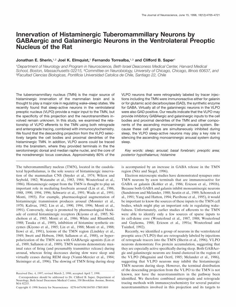
RESULTSCytoarchitectureWe adopted a nomenclature for the preoptic area that synthesizesour own data with that of a large body of literature (Simerly et al.,1984; Simerly, 1995). Although the VLPO is difficult to demar-cate on cytoarchitectonic grounds alone, it may be distinguishedin coronal sections as a small, roughly triangular-shaped cellgroup, with its base along the flat surface of the brain between theoptic chiasm and the diagonal band. Its neurons are wedged alongthe medial aspect of the horizontal limb of nucleus of the diag-onal band, and they are often separated from it by a smallpenetrating blood vessel (Fig. 1a, Table 1). The VLPO is mostprominent in the caudal half of the preoptic area and is founddorsal and lateral to the rostral pole of the supraoptic nucleus. Itextends caudally to about the level of the rostral pole of the
suprachiasmatic nucleus. The VLPO can be most easily delin-eated with markers for Fos-ir after periods of sleep (Sherin et al.,1996) (Fig. 1b) or galanin-immunoreactivity in colchicine-pretreated animals (Melander et al., 1986) (Fig. 1c).
For the TMN we use the terminology of Ericson et al. (1987)who recognized the major cluster of histaminergic neurons in theTMNv as well as a smaller cluster medially (TMNm) and adiffuse collection of cells (TMNd) in between. However, weconsidered the rostral and caudal extensions of the TMN to bepart of the TMNv rather than separate tuberal and caudal mag-nocellular nuclei (Bleier et al., 1979).
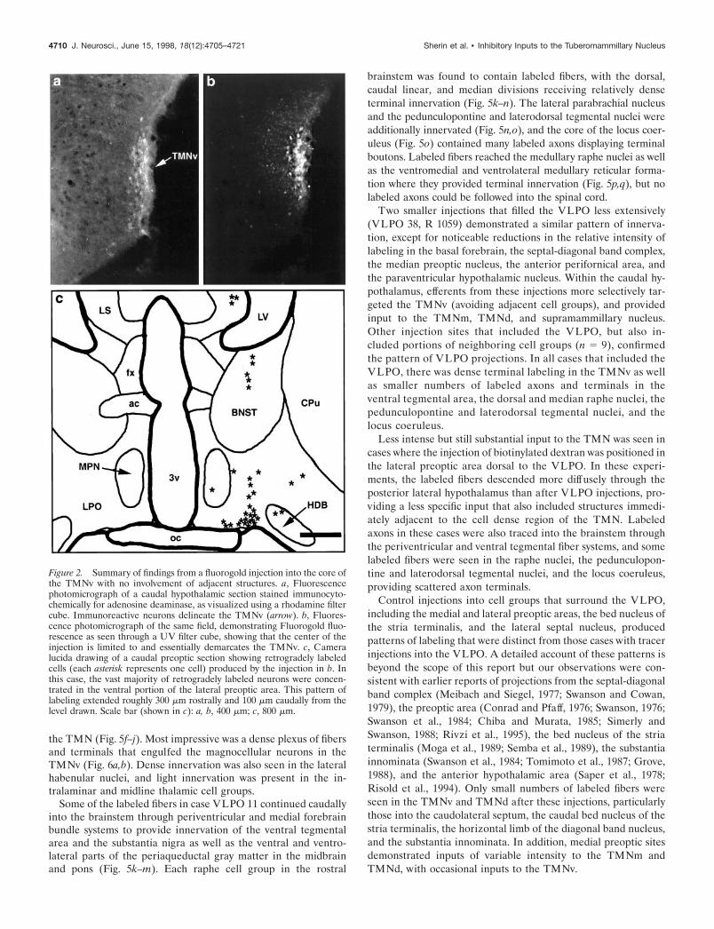
Retrograde tracing studiesThe relationship of injection sites to the TMN was most easilyappreciated in the cases that involved the TMNv. In most casesthe injection site also involved various amounts of surroundingtissue. However, in one case, J-13, a small injection of Fluorogoldplaced by electrophysiological guidance was tightly confined tothe cell-dense core of the TMNv as demonstrated in sections thatwere counterstained for adenosine deaminase-ir (Fig. 2a,b). Inthis case, the vast majority of retrogradely labeled neurons wasconcentrated in a longitudinal column, roughly 0.4 mm long,which extended rostrocaudally along the ventral-most portion ofthe lateral preoptic area (VLPO) (Fig. 2). Smaller numbers ofindividual, retrogradely labeled neurons streamed dorsally andmedially from this column into adjacent cell groups in the pre-optic area and were scattered diffusely in the lateral septum, thebed nucleus of the stria terminalis, the substantia innominata, andthe horizontal limb of the diagonal band nucleus (Fig. 2c). Noretrogradely labeled neurons were identified in other forebrain orbrainstem cell groups after this injection.
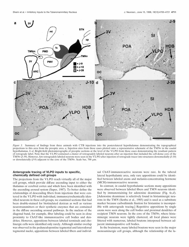
The pattern of retrograde labeling seen in case J-13 was foundin 49 additional cases in which the injection site included the coreof the TMNv (e.g., see J-39 in Fig. 3). However, because thesecases also included regions surrounding the TMN, a more wide-spread pattern of retrograde labeling was observed, as reportedpreviously by Ericson et al. (1991a). Nevertheless, the amount ofretrograde labeling in the VLPO consistently corresponded withthe degree to which the core of the TMNv was involved byretrograde tracer injection.
In 52 cases in which injection of retrograde tracer involvedstructures adjacent to but not including the core of the TMNv(see J-9 and J-10 in Fig. 3), retrogradely labeled cells wereessentially absent from the VLPO but were found concentratedin adjacent preoptic and basal forebrain cell groups. Other thanthe VLPO projection to the core of the TMN, we found noconsistent differences in patterns of retrograde labeling betweenthe cases that included TMN in the injection site and those thatdid not. Hence, although it can be concluded that the VLPO is amajor source of afferents to the TMN, these experiments cannotrule out the possibility that smaller numbers of afferents to thecore of the TMNv arise from adjacent parts of the preoptic areaand basal forebrain.
Anterograde tracer studiesTo better define the sources and terminal distributions of inputsfrom the preoptic area to the TMN, we examined cases drawnfrom a large series of 189 experiments in which the projections ofthe preoptic area and surrounding basal forebrain were system-atically explored with small injections of the anterograde tracerbiotinylated dextran. In 12 of these cases, the injection sitesinvolved the VLPO to varying degrees (for summary of injection
Sherin et al. • Inhibitory Inputs to the Tuberomammillary Nucleus J. Neurosci., June 15, 1998, 18(12):4705–4721 4707
sites, see Fig. 4). In each of three cases (VLPO 11, VLPO 38, R1059) the injection site was largely confined to the VLPO. Ex-periment VLPO 11 best demonstrated the full pattern of efferentprojections from this cell group (Fig. 5a). This relatively largeiontophoretic injection essentially filled the VLPO, with minimalspread into neighboring structures such as the supraoptic nucleus,the medial preoptic area, the dorsal lateral preoptic area, thediagonal band nucleus, or the anterior hypothalamic area (Fig.1d). However, a few scattered neurons were retrogradely labeled
in other preoptic cell groups as well as in the lateral septum andthe bed nucleus of the stria terminalis.
In case VLPO 11, labeled fibers left the injection site ipsilat-erally through medial (periventricular), dorsal (stria medullaris),and lateral (medial forebrain bundle) pathways. Minor contralat-eral projections were found to cross in the ventral supraopticcommissure and provide similarly distributed projections to thecontralateral side of the brain. Fibers that ascended into thetelencephalon ramified throughout the basal forebrain and septal-
Figure 1. A series of bright-field photomicrographs illustrating various histological markers for identifying the ventrolateral preoptic nucleus. a showsthe appearance of the VLPO in Giemsa-stained sections as a small triangular cluster of neurons along the ventral surface of the brain, with its lateraledge bordering the nucleus of the horizontal limb of the diagonal band. b illustrates the Fos-immunoreactive neuronal nuclei in the VLPO after a 1 hrperiod spent predominantly asleep. In c, the VLPO is clearly demarcated as a galanin-immunoreactive cell group in a colchicine-pretreated animal. dshows an injection site of biotinylated dextran (BD) in case VLPO 11; the section is counterstained with Giemsa. The borders of the injectionsite correspond closely with the location of the cluster of Fos-positive and galanin-positive neurons seen in b and c. Scale bar (shown in b): a, 500 mm;b–d, 300 mm.
4708 J. Neurosci., June 15, 1998, 18(12):4705–4721 Sherin et al. • Inhibitory Inputs to the Tuberomammillary Nucleus
diagonal band complex. Ascending and local fibers within thediencephalon infiltrated most preoptic structures, particularly theventromedial preoptic area and the median preoptic nucleus(Elmquist and Saper, 1996; Elmquist et al., 1996). Very littleinput was seen to the suprachiasmatic nucleus. Descending fibers(which constitute the largest contingent of fibers produced byinjection of tracer into the VLPO) traversed the hypothalamus inthe medial forebrain bundle and targeted many anterior, lateral,and posterior hypothalamic nuclei. Particularly intense hypotha-lamic innervation was seen in the perifornical area, the tuberallateral hypothalamic area, the parvocellular parts of the paraven-tricular nucleus, the supramammillary region, and all divisions of
Table continues.
ot Optic tractOVLT Organum vasculosum of the lamina terminalisPAG Periaqueductal gray matterpc Posterior commissurePF Parafascicular nucleusPHA Posterior hypothalamic areaPMD Dorsal premammillary nucleusPMV Ventral premammillary nucleusPn Pontine nucleiPr5 Principal sensory trigeminal nucleusPTA Pretectal areaPVH,r Paraventricular hypothalamic nucleus, rostral polePVT Paraventricular thalamic nucleuspy Pyramidal tractRe Reuniens thalamic nucleusRM Raphe magnus nucleusRN Red nucleusRP Raphe pontis nucleusRpa Raphe pallidus nucleusRPT Reticular tegmental nucleus of the ponsRT Reticular thalamic nucleusSCN,r Suprachiasmatic nucleus, rostral poleSFO Subfornical organSHy Septohypothalamic nucleussm Stria medullarisSNc,r Substantia nigra, pars compacta, pars reticulataSO Superior olivary complexSON,r Supraoptic nucleus, rostral poleSp 5 Spinal trigeminal nucleussp5 Spinal trigeminal tractSUM Supramammillary nucleustb Trapezoid bodyTMNm,v Tuberomammillary nucleus, medial and ventral
partsVA Ventral anterior thalamic nucleusVB Ventrobasal thalamic complexVDB Vertical limb of the diagonal band nucleusVLL Ventral nucleus of the lateral lemniscusVL Ventral lateral thalamic nucleusVLM Ventrolateral medullaVLPO Ventrolateral preoptic nucleusVMH Ventromedial hypothalamic nucleusVMPO Ventromedial preoptic nucleusVTA Ventral tegmental areaVTN Ventral tegmental nucleus (of Gudden)ZI Zona incerta
Table 1. Abbreviations used in figures
3 Oculomotor nucleus3V Third ventricle4V Fourth ventricle5 Trigeminal nucleus (motor division)6 Abducens nucleus7n Facial nerve (g, genu)12 Hypoglossal nucleusac Anterior commissureACB Nucleus accumbensAHN Anterior hypothalamic nucleusAM Anteromedial thalamic nucleusAmb Nucleus ambiguusAPFX Anterior perifornical nucleusARC Arcuate nucleusAV Anteroventral thalamic nucleusAVPV Anteroventral preoptic nucleusbc Brachium conjunctivumBD Biotinylated dextran injection siteBNST Bed nucleus of the stria terminalisCA Cerebral aqueductCb Cerebellumcc Corpus collosumCM Centromedial thalamic nucleuscp Cerebral peduncleCPu Caudate-putamencst Corticospinal tractDB Diagonal band nucleusDMH Dorsomedial hypothalamic nucleusDT Dorsal tegmental nucleusEP Entopeduncular nucleusfr Fasciculus retroflexusfx FornixGP Globus pallidusHDB Horizontal limb of the diagonal band nucleusic Internal capsuleIO Inferior oliveIP Interpeduncular nucleusLC Locus coeruleusLDT Laterodorsal tegmental nucleusLH Lateral habenular nucleusLS Lateral septal nucleusLSV Ventral lateral septal nucleusLV Lateral ventricleMCPO Magnocellular preoptic nucleusMD Mediodorsal thalamic nucleusMe5 Mesencephalic trigeminal nucleusMH Medial habenular nucleusml Medial lemniscusMLF Medial longitudinal fasciculusMM Medial mammillary nucleusMnPO Median preoptic nucleusMPB Medial parabrachial nucleusMPN Medial preoptic nucleusMR Median raphe nucleusmt Mammillothalamic tractNLOT Nucleus of the lateral olfactory tractNST Nucleus of the solitary tractNTB Nucleus of the trapezoid bodyoc Optic chiasm
Sherin et al. • Inhibitory Inputs to the Tuberomammillary Nucleus J. Neurosci., June 15, 1998, 18(12):4705–4721 4709
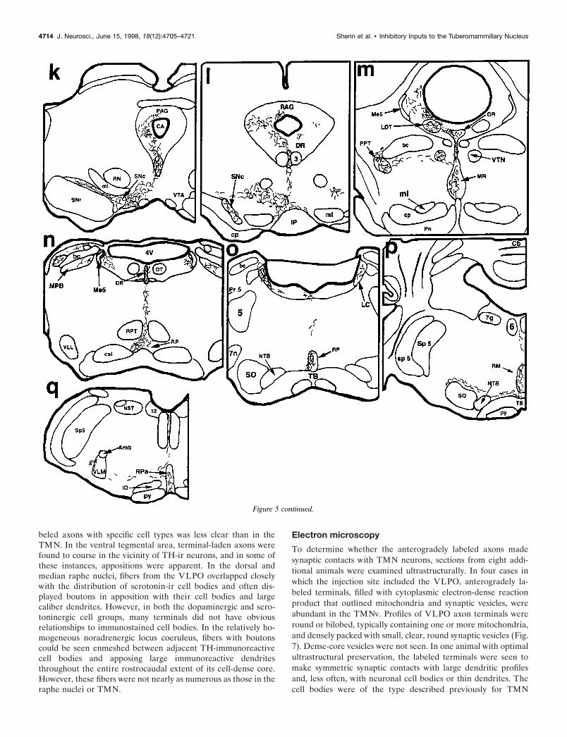
the TMN (Fig. 5f–j). Most impressive was a dense plexus of fibersand terminals that engulfed the magnocellular neurons in theTMNv (Fig. 6a,b). Dense innervation was also seen in the lateralhabenular nuclei, and light innervation was present in the in-tralaminar and midline thalamic cell groups.
Some of the labeled fibers in case VLPO 11 continued caudallyinto the brainstem through periventricular and medial forebrainbundle systems to provide innervation of the ventral tegmentalarea and the substantia nigra as well as the ventral and ventro-lateral parts of the periaqueductal gray matter in the midbrainand pons (Fig. 5k–m). Each raphe cell group in the rostral
brainstem was found to contain labeled fibers, with the dorsal,caudal linear, and median divisions receiving relatively denseterminal innervation (Fig. 5k–n). The lateral parabrachial nucleusand the pedunculopontine and laterodorsal tegmental nuclei wereadditionally innervated (Fig. 5n,o), and the core of the locus coer-uleus (Fig. 5o) contained many labeled axons displaying terminalboutons. Labeled fibers reached the medullary raphe nuclei as wellas the ventromedial and ventrolateral medullary reticular forma-tion where they provided terminal innervation (Fig. 5p,q), but nolabeled axons could be followed into the spinal cord.
Two smaller injections that filled the VLPO less extensively(VLPO 38, R 1059) demonstrated a similar pattern of innerva-tion, except for noticeable reductions in the relative intensity oflabeling in the basal forebrain, the septal-diagonal band complex,the median preoptic nucleus, the anterior perifornical area, andthe paraventricular hypothalamic nucleus. Within the caudal hy-pothalamus, efferents from these injections more selectively tar-geted the TMNv (avoiding adjacent cell groups), and providedinput to the TMNm, TMNd, and supramammillary nucleus.Other injection sites that included the VLPO, but also in-cluded portions of neighboring cell groups (n 5 9), confirmedthe pattern of VLPO projections. In all cases that included theVLPO, there was dense terminal labeling in the TMNv as wellas smaller numbers of labeled axons and terminals in theventral tegmental area, the dorsal and median raphe nuclei, thepedunculopontine and laterodorsal tegmental nuclei, and thelocus coeruleus.
Less intense but still substantial input to the TMN was seen incases where the injection of biotinylated dextran was positioned inthe lateral preoptic area dorsal to the VLPO. In these experi-ments, the labeled fibers descended more diffusely through theposterior lateral hypothalamus than after VLPO injections, pro-viding a less specific input that also included structures immedi-ately adjacent to the cell dense region of the TMN. Labeledaxons in these cases were also traced into the brainstem throughthe periventricular and ventral tegmental fiber systems, and somelabeled fibers were seen in the raphe nuclei, the pedunculopon-tine and laterodorsal tegmental nuclei, and the locus coeruleus,providing scattered axon terminals.
Control injections into cell groups that surround the VLPO,including the medial and lateral preoptic areas, the bed nucleus ofthe stria terminalis, and the lateral septal nucleus, producedpatterns of labeling that were distinct from those cases with tracerinjections into the VLPO. A detailed account of these patterns isbeyond the scope of this report but our observations were con-sistent with earlier reports of projections from the septal-diagonalband complex (Meibach and Siegel, 1977; Swanson and Cowan,1979), the preoptic area (Conrad and Pfaff, 1976; Swanson, 1976;Swanson et al., 1984; Chiba and Murata, 1985; Simerly andSwanson, 1988; Rivzi et al., 1995), the bed nucleus of the striaterminalis (Moga et al., 1989; Semba et al., 1989), the substantiainnominata (Swanson et al., 1984; Tomimoto et al., 1987; Grove,1988), and the anterior hypothalamic area (Saper et al., 1978;Risold et al., 1994). Only small numbers of labeled fibers wereseen in the TMNv and TMNd after these injections, particularlythose into the caudolateral septum, the caudal bed nucleus of thestria terminalis, the horizontal limb of the diagonal band nucleus,and the substantia innominata. In addition, medial preoptic sitesdemonstrated inputs of variable intensity to the TMNm andTMNd, with occasional inputs to the TMNv.
Figure 2. Summary of findings from a fluorogold injection into the core ofthe TMNv with no involvement of adjacent structures. a, Fluorescencephotomicrograph of a caudal hypothalamic section stained immunocyto-chemically for adenosine deaminase, as visualized using a rhodamine filtercube. Immunoreactive neurons delineate the TMNv (arrow). b, Fluores-cence photomicrograph of the same field, demonstrating Fluorogold fluo-rescence as seen through a UV filter cube, showing that the center of theinjection is limited to and essentially demarcates the TMNv. c, Cameralucida drawing of a caudal preoptic section showing retrogradely labeledcells (each asterisk represents one cell) produced by the injection in b. Inthis case, the vast majority of retrogradely labeled neurons were concen-trated in the ventral portion of the lateral preoptic area. This pattern oflabeling extended roughly 300 mm rostrally and 100 mm caudally from thelevel drawn. Scale bar (shown in c): a, b, 400 mm; c, 800 mm.
4710 J. Neurosci., June 15, 1998, 18(12):4705–4721 Sherin et al. • Inhibitory Inputs to the Tuberomammillary Nucleus
Anterograde tracing of VLPO inputs to specific,chemically defined cell groupsThe projections from the VLPO reach virtually all of the majorcell groups, which provide diffuse ascending input to either thethalamus or cerebral cortex and which have been identified withthe ascending arousal system (Saper, 1987). To better define therelationships of descending fibers from injections that were cen-tered in the VLPO with individual, immunocytochemically iden-tified neurons in these cell groups, we examined sections that hadbeen double-stained for biotinylated dextran as well as variousneurotransmitters or their synthetic enzymes that are containedin the diffuse ascending arousal pathways. In the nucleus of thediagonal band, for example, fiber labeling could be seen in closeproximity to ChAT-like immunoreactive cell bodies and den-drites. However, appositions between labeled terminals and cho-linergic cells were identified only rarely. Although similar overlapwas observed in the pedunculopontine tegmental and laterodorsaltegmental nuclei, appositions between labeled fibers and individ-
ual ChAT-immunoreactive neurons were rare. In the tuberallateral hypothalamic area, only rare appositions could be identi-fied between labeled axons and melanin-concentrating hormone(MCH)-immunoreactive neurons.
In contrast, in caudal hypothalamic sections many appositionswere observed between labeled fibers and TMN neurons identi-fied by immunostaining for adenosine deaminase (Fig. 6c,d).[Adenosine deaminase is selectively found in histaminergic neu-rons in the TMN (Senba et al., 1985) and is used as a substitutemarker because carbodiimide fixation for histamine is incompat-ible with anterograde tracing.] Repetitive appositions by singleaxons were seen along the cell bodies and proximal dendrites ofrecipient TMN neurons. In the core of the TMNv, where hista-minergic neurons were tightly clustered, all focal planes werelittered with numerous, labeled boutons interposed between ad-jacent immunopositive cell bodies.
In the brainstem, many labeled boutons were seen in the majormonoaminergic cell groups, although the relationship of the la-
Figure 3. Summary of findings from three animals with CTB injections into the posterolateral hypothalamus demonstrating the topographicalprojections to this area from the preoptic area. a, Injection sites from these cases plotted onto a representative schematic of the TMNv in the caudalhypothalamus. b–d, Bright-field photomicrographs of preoptic sections at the level of the VLPO from these cases demonstrating the resultant patternof retrograde label. Note that the VLPO contained a cluster of retrogradely labeled neurons after an injection that included the cell-dense core of theTMNv (J-39). However, few retrogradely labeled neurons were seen in the VLPO after injection of retrograde tracer into structures dorsomedially (J-10)or dorsolaterally (J-9) adjacent to the core of the TMNv. Scale bar, 700 mm.
Sherin et al. • Inhibitory Inputs to the Tuberomammillary Nucleus J. Neurosci., June 15, 1998, 18(12):4705–4721 4711

Figure 4. A summary diagram illustrating biotinylated dextran injection sites at six levels of the preoptic area. The VLPO is most prominent inschematics c, c9, d, and d9 (in light gray). Asterisks denote cases VLPO 11, VLPO 38, and R 1059 in which biotinylated dextran injections werepredominantly located within the VLPO (see Results).
4712 J. Neurosci., June 15, 1998, 18(12):4705–4721 Sherin et al. • Inhibitory Inputs to the Tuberomammillary Nucleus
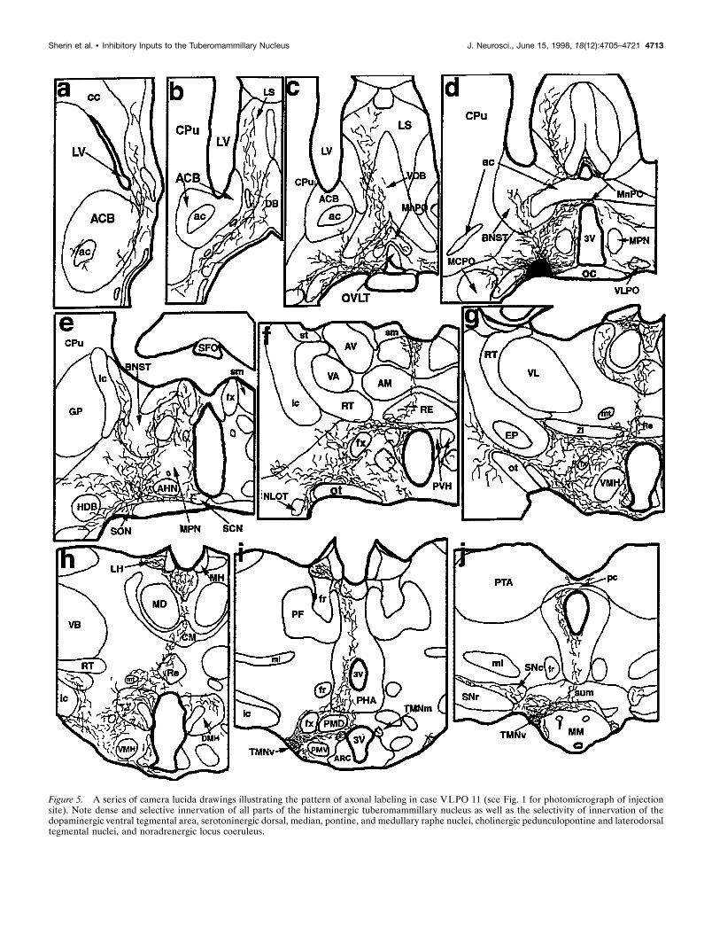
Figure 5. A series of camera lucida drawings illustrating the pattern of axonal labeling in case VLPO 11 (see Fig. 1 for photomicrograph of injectionsite). Note dense and selective innervation of all parts of the histaminergic tuberomammillary nucleus as well as the selectivity of innervation of thedopaminergic ventral tegmental area, serotoninergic dorsal, median, pontine, and medullary raphe nuclei, cholinergic pedunculopontine and laterodorsaltegmental nuclei, and noradrenergic locus coeruleus.
Sherin et al. • Inhibitory Inputs to the Tuberomammillary Nucleus J. Neurosci., June 15, 1998, 18(12):4705–4721 4713
beled axons with specific cell types was less clear than in theTMN. In the ventral tegmental area, terminal-laden axons werefound to course in the vicinity of TH-ir neurons, and in some ofthese instances, appositions were apparent. In the dorsal andmedian raphe nuclei, fibers from the VLPO overlapped closelywith the distribution of serotonin-ir cell bodies and often dis-played boutons in apposition with their cell bodies and largecaliber dendrites. However, in both the dopaminergic and sero-toninergic cell groups, many terminals did not have obviousrelationships to immunostained cell bodies. In the relatively ho-mogeneous noradrenergic locus coeruleus, fibers with boutonscould be seen enmeshed between adjacent TH-immunoreactivecell bodies and apposing large immunoreactive dendritesthroughout the entire rostrocaudal extent of its cell-dense core.However, these fibers were not nearly as numerous as those in theraphe nuclei or TMN.
Electron microscopy
To determine whether the anterogradely labeled axons madesynaptic contacts with TMN neurons, sections from eight addi-tional animals were examined ultrastructurally. In four cases inwhich the injection site included the VLPO, anterogradely la-beled terminals, filled with cytoplasmic electron-dense reactionproduct that outlined mitochondria and synaptic vesicles, wereabundant in the TMNv. Profiles of VLPO axon terminals wereround or bilobed, typically containing one or more mitochondria,and densely packed with small, clear, round synaptic vesicles (Fig.7). Dense-core vesicles were not seen. In one animal with optimalultrastructural preservation, the labeled terminals were seen tomake symmetric synaptic contacts with large dendritic profilesand, less often, with neuronal cell bodies or thin dendrites. Thecell bodies were of the type described previously for TMN
Figure 5 continued.
4714 J. Neurosci., June 15, 1998, 18(12):4705–4721 Sherin et al. • Inhibitory Inputs to the Tuberomammillary Nucleus
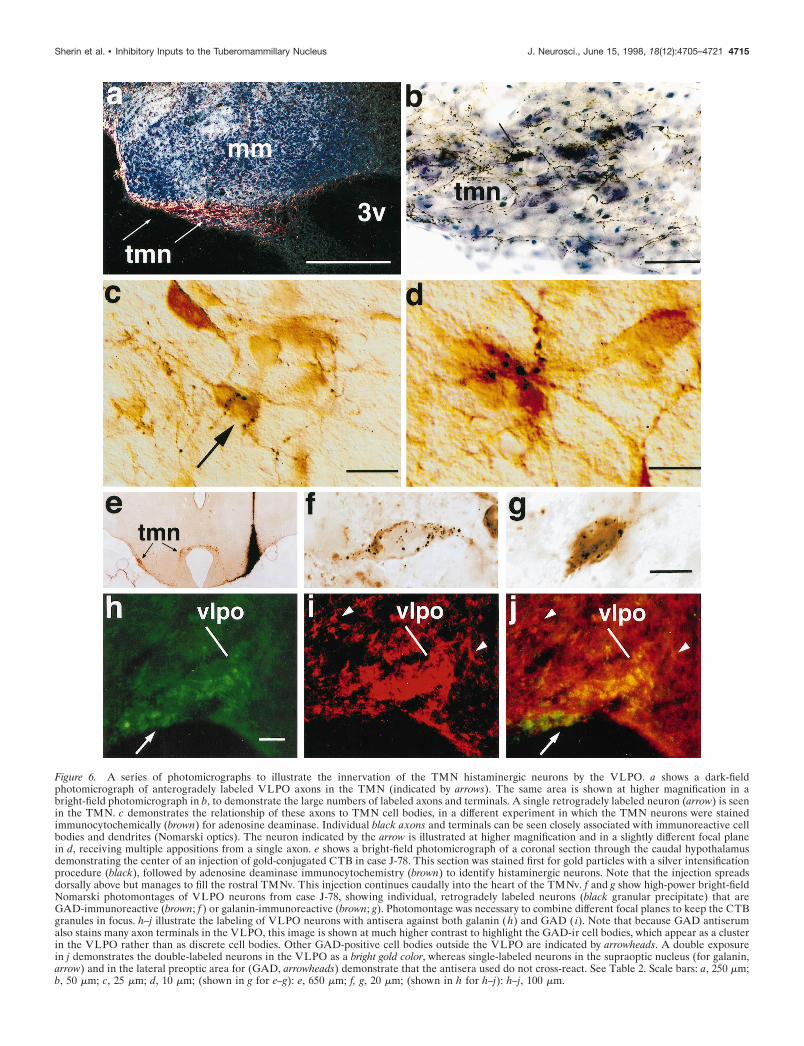
Figure 6. A series of photomicrographs to illustrate the innervation of the TMN histaminergic neurons by the VLPO. a shows a dark-fieldphotomicrograph of anterogradely labeled VLPO axons in the TMN (indicated by arrows). The same area is shown at higher magnification in abright-field photomicrograph in b, to demonstrate the large numbers of labeled axons and terminals. A single retrogradely labeled neuron (arrow) is seenin the TMN. c demonstrates the relationship of these axons to TMN cell bodies, in a different experiment in which the TMN neurons were stainedimmunocytochemically (brown) for adenosine deaminase. Individual black axons and terminals can be seen closely associated with immunoreactive cellbodies and dendrites (Nomarski optics). The neuron indicated by the arrow is illustrated at higher magnification and in a slightly different focal planein d, receiving multiple appositions from a single axon. e shows a bright-field photomicrograph of a coronal section through the caudal hypothalamusdemonstrating the center of an injection of gold-conjugated CTB in case J-78. This section was stained first for gold particles with a silver intensificationprocedure (black), followed by adenosine deaminase immunocytochemistry (brown) to identify histaminergic neurons. Note that the injection spreadsdorsally above but manages to fill the rostral TMNv. This injection continues caudally into the heart of the TMNv. f and g show high-power bright-fieldNomarski photomontages of VLPO neurons from case J-78, showing individual, retrogradely labeled neurons (black granular precipitate) that areGAD-immunoreactive (brown; f ) or galanin-immunoreactive (brown; g). Photomontage was necessary to combine different focal planes to keep the CTBgranules in focus. h–j illustrate the labeling of VLPO neurons with antisera against both galanin (h) and GAD ( i). Note that because GAD antiserumalso stains many axon terminals in the VLPO, this image is shown at much higher contrast to highlight the GAD-ir cell bodies, which appear as a clusterin the VLPO rather than as discrete cell bodies. Other GAD-positive cell bodies outside the VLPO are indicated by arrowheads. A double exposurein j demonstrates the double-labeled neurons in the VLPO as a bright gold color, whereas single-labeled neurons in the supraoptic nucleus (for galanin,arrow) and in the lateral preoptic area for (GAD, arrowheads) demonstrate that the antisera used do not cross-react. See Table 2. Scale bars: a, 250 mm;b, 50 mm; c, 25 mm; d, 10 mm; (shown in g for e–g): e, 650 mm; f, g, 20 mm; (shown in h for h–j): h–j, 100 mm.
Sherin et al. • Inhibitory Inputs to the Tuberomammillary Nucleus J. Neurosci., June 15, 1998, 18(12):4705–4721 4715
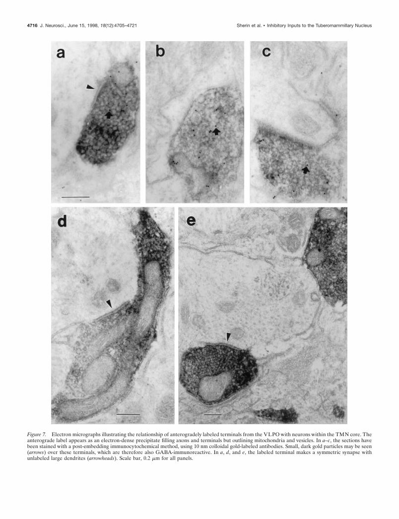
Figure 7. Electron micrographs illustrating the relationship of anterogradely labeled terminals from the VLPO with neurons within the TMN core. Theanterograde label appears as an electron-dense precipitate filling axons and terminals but outlining mitochondria and vesicles. In a–c, the sections havebeen stained with a post-embedding immunocytochemical method, using 10 nm colloidal gold-labeled antibodies. Small, dark gold particles may be seen(arrows) over these terminals, which are therefore also GABA-immunoreactive. In a, d, and e, the labeled terminal makes a symmetric synapse withunlabeled large dendrites (arrowheads). Scale bar, 0.2 mm for all panels.
4716 J. Neurosci., June 15, 1998, 18(12):4705–4721 Sherin et al. • Inhibitory Inputs to the Tuberomammillary Nucleus

histaminergic neurons (Hayaishi et al., 1984; Ericson et al., 1987;Yamamoto et al., 1990). We observed no contacts with dendriticspines or axons.
The post-embedding immunogold technique revealed a bi-modal distribution of immunolabeling of a random sample ofaxon terminals. About one-third (25 of 69) of unlabeled axonterminals were considered GABA-immunoreactive, with a meanof ;30 particles/mm2. Gold particles over labeled terminals weretypically associated with both vesicles and mitochondria, as hasbeen described previously (Merighi and Polak, 1993). The re-maining axon terminals, with 0–5 gold particles//mm2, werejudged to be GABA-negative. Virtually all of the anterogradelylabeled VLPO terminals were in the clearly GABA-immunoreactive group. For a sample of 53 anterogradely labeledVLPO terminals, there was a mean of 20.09 gold particles/mm2 61.73 (SEM). Postsynaptic elements or other cell bodies or den-drites were never immunoreactive (mean of 1.56 6 0.23 goldparticles/mm2; n 5 90). Earlier studies have reported that TMNneurons are GABAergic (Vincent et al., 1983; Ericson et al.,1991b), but the post-embedding method for electron microscopicimmunocytochemistry is considerably less sensitive than pre-embedding methods that were used previously, and our animalswere not treated with colchicine. Hence the density of goldparticles over VLPO terminals was ;13 times as great as thatover TMN dendrites ( p , 0.000001; Kolmogorov–Smirnov test).
Retrograde tracing plus immunocytochemistry forGAD or galaninBecause of the presence of clusters of both GABAergic andgalaninergic neurons in the VLPO area, and the known relation-ship of GABAergic and galaninergic terminals to TMN neurons,we examined a series of 11 additional cases in which retrogradelabeling from the TMN was combined with immunocytochemis-try for GAD or galanin. In four of these, the quality of both theretrograde transport and the immunocytochemistry was adequateto allow a semiquantitative analysis of the neurotransmitter con-tent of VLPO neurons that projected to the TMNv. In each case,the injection site included the core of the TMNv but extendedbeyond this region. Hence the specificity of the projection for theTMNv was based on the observation above that VLPO neuronsare only retrogradely labeled when injections into the posterolat-eral hypothalamus include the TMNv, and that the descendingVLPO axons have a high specificity for TMN neurons.
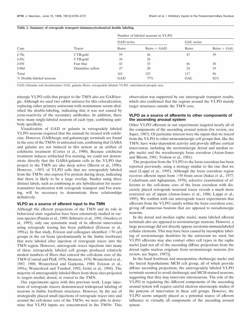
In these four cases, we found that roughly 80% of all retro-gradely labeled VLPO neurons contained GAD-ir and a similarnumber contained galanin-ir (Table 2, Fig. 6e–g). Regions outsidethe VLPO, particularly the dorsal portion of the lateral preopticarea and the ventromedial preoptic area, also contained severaldouble-labeled neurons, although they were scattered among amajority of other neurons containing only one label. No specificstaining was obtained in tissue that was incubated in preabsorbedprimary antibody for either GAD or galanin.
Preoptic sections containing the VLPO were stained immun-ofluorescently for both GAD-ir and galanin-ir in three cases.These experiments demonstrated that GAD-immunoreactiveneurons were abundant in cell groups throughout the rostralhypothalamus and adjacent basal forebrain (Mugnaini and Oer-tel, 1985), whereas the distribution of galanin-immunoreactiveneurons was more restricted. Within the VLPO, almost everyneuron found to contain galanin-ir was also found to containGAD-ir (Fig. 6h–j). Several double-labeled neurons were foundin regions outside the VLPO, including the dorsal portion of the
lateral preoptic area and the ventromedial preoptic area, althoughthey were scattered among a majority of other neurons containingonly one label. Intensely single-labeled neurons were found in thesame sections in areas other than the VLPO, such as the cortex,reticular thalamic nucleus, and lateral preoptic area (GAD-ir;Fig. 6, compare i,j) and the supraoptic nucleus (galanin-ir; Fig. 6,compare h,j), so that that the double-staining of VLPO neuronscould not have represented a cross-reactivity of the antisera. Asfurther controls, when either the GAD or galanin antiserum wasomitted or replaced with normal rabbit serum, no specific stainingwas seen. In addition, in the double immunocytochemistry exper-iment, when the second primary antiserum in each sequence wasomitted or substituted with normal rabbit serum, staining only forthe first primary antiserum was seen.
DISCUSSIONOur observations demonstrate a massive and selective input froma small group of neurons in the ventral portion of the lateralpreoptic area to the major cluster of histaminergic neurons in thebrain, the TMNv. Electron microscopic observations confirm thatthe VLPO axon terminals form symmetric synapses onto TMNvproximal dendrites and cell bodies. Our results further hint thatthis is a topographically organized projection, with additionalinput primarily to the TMNm and TMNd from the medialpreoptic area and to the TMNv from the dorsal lateral preopticarea. Approximately 70–80% of the cells that provide thisprojection contain both GABA and galanin, suggesting that theyare inhibitory in nature. Furthermore, descending axons fromthe VLPO innervate the other monoaminergic cell groups in thebrainstem that provide diffuse cortical projections, includingthe dopaminergic ventral tegmental area, the serotoninergic dor-sal and median raphe nuclei, and the noradrenergic locus coer-uleus. Hence, the VLPO is ideally situated to hyperpolarizesimultaneously the monoaminergic components of the ascendingarousal system. Because many VLPO neurons that project to theTMN are sleep active (Sherin et al., 1996), these observationssupport the hypothesis that the VLPO may play an importantrole in the regulation of wake–sleep states.
Technical considerationsWe used a combination of retrograde and anterograde tracerexperiments to define the VLPO projections more definitivelythan either approach can alone. Iontophoretic injections of ret-rograde tracers that are confined to the TMNv (case J-13) may betoo small to label all of its afferents, whereas large injectionsnecessarily include surrounding tissue, and retrogradely labeledcells may not project to the TMN. However, our anterogradetransport experiments confirm that the TMNv is filled with an-terogradely labeled fibers and boutons that appose TMN cellbodies and proximal dendrites only when the injection sites hitthe VLPO.
TMN neurons may receive other types of afferents on theirdistal dendrites, which can extend for a considerable distanceoutside the borders of the nucleus. However, the inputs to theircell bodies are confirmed by our electron microscopic findings tobe predominantly GABAergic. These VLPO terminals would bein an excellent position to hyperpolarize the TMN neurons, asconfirmed by the demonstration that electrical stimulation ofpreoptic inputs to the TMN results in inhibitory postsynapticpotentials that are abolished by GABAA antagonists (Yang andHatton, 1994).
Our double-labeling results indicate that nearly all of the gala-
Sherin et al. • Inhibitory Inputs to the Tuberomammillary Nucleus J. Neurosci., June 15, 1998, 18(12):4705–4721 4717
ninergic VLPO cells that project to the TMN also are GABAer-gic. Although we used two rabbit antisera for this colocalization,replacing either primary antiserum with nonimmune serum abol-ished the double-labeling, indicating that it was not caused bycross-reactivity of the secondary antibodies. In addition, therewere many single-labeled neurons of each type, confirming anti-body specificity.
Visualization of GAD or galanin in retrogradely labeledVLPO neurons required that the animals be treated with colchi-cine. However, GABAergic and galaninergic terminals are foundin the core of the TMNv in untreated rats, confirming that GABAand galanin are not induced in this system as an artifact ofcolchicine treatment (Cortes et al., 1990). Because colchicinetreatment induces artifactual Fos staining, we could not demon-strate directly that the GABA/galanin cells in the VLPO thatproject to the TMN are also sleep active (Sherin et al., 1996).However, .50% of VLPO cells that are retrogradely labeledfrom the TMNv also express Fos protein during sleep, indicatingthat there is likely to be a large overlap. Studies using threedistinct labels, such as combining in situ hybridization for neuro-transmitter localization with retrograde transport and Fos stain-ing, will be necessary to demonstrate these relationshipsdefinitively.
VLPO as a source of afferent input to the TMNAlthough the efferent projections of the TMN and its role inbehavioral state regulation have been extensively studied in var-ious species (Panula et al., 1989; Schwartz et al., 1991; Onodera etal., 1993), only one systematic study of its afferent innervationusing retrograde tracing has been published (Ericson et al.,1991a). In that study, Ericson and colleagues identified .70 cellgroups in the rat brain (predominantly in the limbic forebrain)that were labeled after injection of retrograde tracer into theTMN region. However, anterograde tracer injections into manyof these retrogradely labeled cell groups produced relativelymodest numbers of fibers that entered the cell-dense core of theTMN (Conrad and Pfaff, 1976; Swanson, 1976; Wouterlood et al.,1987, 1988; Wouterlood and Gaykema, 1988; Ericson et al.,1991a; Wouterlood and Tuinhof, 1992; Gritti et al., 1994). Themajority of anterogradely labeled fibers from these sites projectedto targets medial, dorsal, or rostral to the TMN.
Our experiments agree with this previous work. Large injec-tions of retrograde tracers demonstrated widespread labeling ofneurons in limbic forebrain structures. However, by the use ofstrategically placed small injections of retrograde tracer into andaround the cell-dense core of the TMNv, we were able to deter-mine that VLPO inputs are concentrated in the TMNv. This
observation was supported by our anterograde transport results,which also confirmed that the regions around the VLPO mainlytarget structures outside the TMN core.
VLPO as a source of afferents to other components ofthe ascending arousal systemOther VLPO efferents in our experiments targeted nearly all ofthe components of the ascending arousal system (for review, seeSaper, 1987). Of particular interest were the inputs that we tracedfrom the VLPO to other monoaminergic cell groups that, like theTMN, have wake-dependent activity and provide diffuse corticalinnervation, including the serotoninergic dorsal and median ra-phe nuclei and the noradrenergic locus coeruleus (Aston-Jonesand Bloom, 1981; Trulson et al., 1981).
The projection from the VLPO to the locus coeruleus has beenreported previously, using a strategy similar to the one that weused (Luppi et al., 1995). Although the locus coeruleus regionreceives afferent input from .50 brain areas (Sakai et al., 1977;Cedarbaum and Aghajanian, 1978), selective examination of af-ferents to the cell-dense core of the locus coeruleus with dis-cretely placed retrograde neuronal tracer reveals a much morerestricted set of inputs (Aston-Jones et al., 1986; Luppi et al.,1995). We confirm with our anterograde tracer experiments thatafferents from the VLPO ramify within the locus coeruleus core,giving off numerous boutons that are apposed to noradrenergicneurons.
In the dorsal and median raphe nuclei, many labeled afferentterminals also are apposed to serotoninergic neurons. However, alarge percentage did not directly appose serotonin-immunolabeledcellular elements. This may have been caused by incomplete label-ing of serotoninergic dendrites by the antiserum we used, butVLPO afferents may also contact other cell types in the raphenuclei [and not all of the ascending diffuse projections from thedorsal raphe nucleus originate from serotoninergic neurons (forreview, see Saper, 1987)].
In the basal forebrain and mesopontine cholinergic nuclei andthe lateral hypothalamic MCH cell group, all of which providediffuse ascending projections, the anterogradely labeled VLPOterminals seemed to avoid cholinergic and MCH-stained neurons,suggesting that they may innervate interneurons. The role of theVLPO in regulating the different components of the ascendingarousal system will require careful electron microscopic studies ofthe patterns of innervation in these cell groups. However, theVLPO seems uniquely placed as a potential source of afferentinfluence to virtually all components of the ascending arousalsystem.
Table 2. Summary of retrograde transport–immunocytochemical double labeling
Case Tracer
Number of labeled neurons in VLPO
GAD series GAL series
Retro Retro 1 GAD Retro Retro 1 GAL
J-78c CTB-gold 59 46 47 39J-85c CTB-gold 34 26J-85f Fast blue 43 33 46 38J-88d Diamidino yellow 27 20 24 19
Total 163 125 117 96% Double-labeled neurons GAD 77% GAL 82%
GAD, Glutamic acid decarboxylase; GAL, galanin; Retro, retrogradely labeled; VLPO, ventrolateral preoptic area.
4718 J. Neurosci., June 15, 1998, 18(12):4705–4721 Sherin et al. • Inhibitory Inputs to the Tuberomammillary Nucleus
VLPO as a potential regulator of wake–sleep statesThe region of the preoptic area containing the VLPO may play acritical role in inducing sleep. Lesions in this area cause insomnia,whereas electrical or chemical stimulation causes sleep (Stermanand Clemente, 1962; McGinty and Sterman, 1968; Szymusiak andMcGinty, 1986; Sallanon et al., 1989; John et al., 1994). Nauta(1946) proposed a simple mechanism by which sleep-promotingneurons in the preoptic area would inhibit wake-promoting neu-rons in the posterior hypothalamus to produce sleep.
Our results are remarkably consistent with this model. Wefound previously that .50% of the VLPO neurons that project tothe TMN are sleep active, as demonstrated by Fos-immunoreactivity (Sherin et al., 1996). The VLPO provides anintense and specific GABAergic and galaninergic set of inputs tothe cell bodies and proximal dendrites of the histaminergic tube-romammillary nucleus in the posterior hypothalamus. Further-more, the VLPO provides inputs to the serotoninergic dorsal andmedian raphe nuclei and to the noradrenergic locus coeruleus. Allthree of these monoaminergic populations are known to fire moreslowly during slow wave sleep and to cease firing during REMsleep. The VLPO is an attractive candidate for simultaneouslyhyperpolarizing neurons in all three populations of monoamin-ergic neurons during sleep.
Finally, the relationship of VLPO terminals to interneurons inthe cholinergic and MCH-immunoreactive systems of diffuse pro-jection neurons is intriguing. These findings suggest that theVLPO neurons may play a key role in the function of a widerange of cell groups that contribute to the wakeful state. Furtherstudy of the relationship of descending VLPO inputs to thefunction of these cell groups is likely to provide important in-sights into the mechanisms for regulation of the wake–sleep state.
REFERENCESAston-Jones G, Bloom FE (1981) Activity of norepinephrine containing
locus coeruleus neurons in behaving rats anticipates fluctuations in thesleep-waking cycle. J Neurosci 1:876–886.
Aston-Jones G, Ennis M, Pieribone VA, Nickell WT, Shipley MT (1986)The brain nucleus locus coeruleus. Afferent control of a broad efferentnetwork. Science 234:734–737.
Bittencourt JC, Presse F, Arias C, Peto C, Vaughan J, Nahon JL, Vale W,Sawchenko PE (1992) The melanin-concentrating hormone system ofthe rat brain. An immunocytochemical and hybridization histochemicalcharacterization. J Comp Neurol 319:218–245.
Bleier R, Cohn P, Siggelkow IR (1979) A cytoarchitectonic atlas of thehypothalamus and hypothalamic third ventricle of the rat. Anatomy ofthe hypothalamus. In: Handbook of the hypothalamus, Vol 1 (MorganePJ, Panksepp J, eds), pp 137–220. New York: Marcel Dekker.
Cedarbaum JM, Aghajanian GK (1978) Afferent projections to the ratlocus coeruleus as determined by a retrograde tracing technique.J Comp Neurol 178:1–16.
Chiba T, Murata Y (1985) Afferent and efferent connections of themedial preoptic area in the rat: A WGA-HRP study. Brain Res Bull14:261–272.
Conrad LCA, Pfaff DW (1976) Efferents from medial basal forebrainand hypothalamus in the rat. I. An autoradiographic study of the medialpreoptic area. J Comp Neurol 169:185–220.
Cortes R, Ceccatelli S, Schalling M, Hokfelt T (1990) Differential effectsof intercerebroventricular colchicine administration of the expressionof mRNAs for neuropeptides and neurotransmitter enzymes with spe-cial emphasis on galanin: an in situ hybridization study. Synapse6:369–391.
de Lacalle S, Lim C, Sobreviela T, Mufson EJ, Hersh LB, Saper CB(1994) Cholinergic innervation in the human hippocampal formationincluding the entorhinal cortex. J Comp Neurol 345:321–344.
Elmquist JK, Saper CB (1996) Activation of neurons projecting to theparaventricular nucleus of the hypothalamus by intravenous lipopoly-saccharide. J Comp Neurol 374:315–331.
Elmquist JK, Fox CA, Ross LR, Jacobson CD (1992) Galanin-like im-
munoreactivity in the adult and developing Brazilian opossum brain.Dev Brain Res 67:161–179.
Elmquist JK, Scammell TE, Jacobson CD, Saper CB (1996) Distributionof Fos-like immunoreactivity in the rat brain following intravenouslipopolysaccharide administration. J Comp Neurol 371:85–103.
Ericson H, Watanabe T, Kohler C (1987) Morphological analysis of thetuberomammillary nucleus in the rat brain: delineation of subgroupswith antibody against L-histidine decarboxylase as a marker. J CompNeurol 263:1–24.
Ericson H, Blomqvist A, Kohler C (1991a) Origin of neuronal inputs tothe region of the tuberomammillary nucleus of the rat brain. J CompNeurol 311:45–64.
Ericson HC, Kohler C, Blomqvist A (1991b) GABA-like immunoreac-tivity in the tuberomammillary nucleus: an electron microscopic studyin the rat. J Comp Neurol 305:462–469.
Gritti I, Mainville L, Jones BE (1994) Projections of GABAergic andcholinergic basal forebrain and GABAergic preoptic-anterior hypotha-lamic neurons to the posterior lateral hypothalamus of the rat. J CompNeurol 339:251–268.
Grove EA (1988) Efferent connections of the substantia innominata.J Comp Neurol 277:347–364.
Hallanger A, Levey AI, Lee HJ, Rye DB, Wainer BH (1987) The originsof cholinergic and other subcortical afferents to the thalamus in the rat.J Comp Neurol 262:105–124.
Hayaishi H, Takagi H, Takeda Y, Kubota M, Tohyama M, Watanabe T,Wada H (1984) Fine structure of histaminergic neurons in the caudalmagnocellular nucleus of the rat as demonstrated by immunohisto-chemistry using histidine decarboxylase as a marker. J Comp Neurol229:233–241.
Herbert H, Saper CB (1990) Cholecystokinin-, galanin-, andcorticotropin-releasing factor-like immunoreactive projections from thenucleus of the solitary tract to the parabrachial nucleus in the rat.J Comp Neurol 293:581–598.
Itowi N, Yamatodani A, Kiyono S, Hiraiwa ML, Wada H (1991) Effectof histamine depletion on the circadian amplitude of the sleep-wakefulness cycle. Physiol Behav 49:643–646.
John J, Kumar VM, Gopinath G, Ramesh V, Mallick H (1994) Changesin sleep-wakefulness after kainic acid lesion of the preoptic area in rats.Jpn J Physiol 44:231–242.
Kalivas PW (1982) Histamine-induced arousal in the conscious andpentobarbital-pretreated rat. J Pharmacol Exp Ther 222:37–42.
Kiyono S, Seo ML, Shibagaki M, Watanabe T, Maeyama K, Wada H(1985) Effects of alpha-fluoromethylhistidine on sleep-waking param-eters in rats. Physiol Behav 34:615–617.
Kohler C, Ericson H, Watanabe T, Polak J, Palay SL, Palay V, Chan-PalayV (1986) Galanin immunoreactivity in hypothalamic histamine neu-rons: further evidence for multiple chemical messengers in the tube-romammillary nucleus. J Comp Neurol 250:58–64.
Lin JS, Sakai K, Jouvet M (1986) Role of hypothalamic histaminergicsystems in the regulation of states of vigilance in the cat. C R AcadScience III 303:469–474.
Lin JS, Sakai K, Jouvet M (1988) Evidence for histaminergic arousalmechanisms in the hypothalamus of cats. Neuropharmacology27:111–122.
Lin JS, Sakai K, Vanni-Mercier G, Jouvet M (1989) A critical role forthe posterior hypothalamus in the mechanisms of wakefulness deter-mined by microinjection of muscimol in freely moving cats. Brain Res479:225–240.
Lin JS, Sakai K, Vanni-Mercier G, Arrang JM, Garbarg M, Schwartz JC,Jouvet M (1990) Involvement of histaminergic neurons in arousalmechanisms demonstrated with H3-receptor ligands in the cat. BrainRes 523:325–330.
Lin JS, Sakai K, Vanni-Mercier G, Jouvet M (1994) Hypothalamo-preoptic histaminergic projections in sleep-wake control in the cat. EurJ Neurosci 66:618–625.
Lin JS, Hou Y, Sakai K, Jouvet J (1996) Histaminergic descendinginputs to the mesopontine tegmentum and their role in the control ofcortical activation and wakefulness in the cat. J Neurosci 16:1523–1537.
Lindsley DB, JW Bowden, Magoun HW (1949) Effect upon the EEG ofacute injury to the brain stem activating system. ElectroencephalogrClin Neurophysiol 1:475.
Llewellyn-Smith IJ, Minson JB, Wright AP, Hodgson AJ (1990) Choleratoxin B-gold, a retrograde tracer that can be used in light and electronmicroscopic immunocytochemical studies. J Comp Neurol294:179–191.
Sherin et al. • Inhibitory Inputs to the Tuberomammillary Nucleus J. Neurosci., June 15, 1998, 18(12):4705–4721 4719
Luppi PH, Aston-Jones, Akaoka H, Chouvet G, Jouvet M (1995) Affer-ent projections to the rat locus coeruleus demonstrated by retrogradeand anterograde tracing with cholera-toxin B subunit and PHA-L.Neuroscience 65:119–160.
McGinty DJ, Sterman MB (1968) Sleep suppression after basal fore-brain lesions in the cat. Science 160:1253–1255.
Meibach RC, Siegel A (1977) Efferent connections of the septal area inthe rat: an analysis utilizing retrograde and anterograde transportmethods. Brain Res 119:1–20.
Melander T, Hokfelt T, Rokeaus A (1986) Distribution of galanin-likeimmunoreactivity in the rat central nervous system. J Comp Neurol248:475–517.
Merighi A, Polak JM (1993) Post-embedding immunogold staining. In:Immunohistochemistry II. IBRO Handbook Series: Methods in theneurosciences, Vol 14 (Cuello AC, ed), pp 229–264. Chichester, UK:Wiley.
Moga MM, Saper CB (1994) Neuropeptide-immunoreactive neuronsprojecting to the paraventricular hypothalamic nucleus in the rat.J Comp Neurol 346:137–150.
Moga MM, Saper CB, Gray TS (1989) The bed nucleus of the striaterminalis: cytoarchitecture, immunohistochemistry and projection tothe parabrachial nucleus in the rat. J Comp Neurol 283:315–332.
Monnier M, Sauer R, Hatt AM (1970) The activating effect of histamineon the central nervous system. Int Rev Neurobiol 12:265–305.
Monti JM (1993) Involvement of histamine in the control of the wakingstate. Life Sci 53:1331–1338.
Monti JM, Pellejero T, Jantos H (1986) Effects of H1 and H2 histaminereceptor antagonists on sleep and wakefulness in the rat. J NeuralTransm 66:1–11.
Monti JM, D’Angelo L, Jantos H, Pazos S (1988) Effects of alpha-fluoromethylhistidine on sleep and wakefulness in the rat. J NeuralTransm 72:141–145.
Monti JM, Jantos H, Boussard M, Altier H, Orellana C, Olivera S (1991)Effects of selective activation or blockade of the histamine H3 receptoron sleep and wakefulness. Eur J Pharmacol 205:283–287.
Mugnaini E, Oertel WH (1985) An atlas of the distribution of GABAer-gic neurons and terminals in the rat CNS as revealed by GAD immu-nohistochemistry. In: Handbook of chemical neuroanatomy, Vol 4,GABA and neuropeptides in the CNS: Part 1 (Bjorklund A, Hokfelt T,eds), pp 436–608. Amsterdam: Elsevier.
Nauta WJH (1946) Hypothalamic regulation of sleep in rats. An exper-imental study. J Neurophysiol 9:285–316.
Nicholson AN, Pascoe PA, Stone BM (1985) Histaminergic systems andsleep: studies in man with H1 and H2 antagonists. Neuropharmacology24:245–250.
Nitz D, Siegel JM (1996) GABA release in posterior hypothalamusacross sleep-wake cycle. Am J Physiol 271:R1707–R1712.
Onodera K, Yamatodani A, Watanabe T, Wada H (1993) Neurophar-macology of the histaminergic neuron system in the brain and itsrelationship with behavioral disorders. Prog Neurobiol 42:685–702.
Panula P, Pirvola U, Auvinen S, Airaksinen MS (1989) Histamine-immunoreactive nerve fibers in the rat brain. Neuroscience 28:585–610.
Pieribone VA, Aston-Jones G (1988) The iontophoretic application offlouro-gold for the study of afferents to deep brain nuclei. Brain Res475:259–271.
Pieribone VA, Xu ZQ, Zhang X, Grillner S, Bartfai T, Hokfelt T (1995)Galanin induces a hyperpolarization of norepinephrine-containing lo-cus coeruleus neurons in the brainstem slice. Neuroscience 64:861–874.
Reiner PB, McGeer EG (1987) Electrophysiological properties of corti-cally projecting histamine neurons of the rat hypothalamus. NeurosciLett 73:43–47.
Risold PY, Canteras NS, Swanson LE (1994) Origin of projections fromthe anterior hypothalamic nucleus: a PHA-L study in the rat. J CompNeurol 348:1–40.
Rizvi TA, Murphy AZ, Ennis M, Behbehani MM, Shipley MT (1995)Medial preoptic area afferents to periaqueductal gray medullo-outputneurons: a combined fos and tract tracing study. J Neurosci 16:333–344.
Sakai K, Touret M, Salver D, Leger L, Jouvet M (1977) Afferent pro-jections to the cat locus coeruleus as visualized by the horseradishperoxidase technique. Brain Res 119:21–41.
Sallanon M, Sakai M, Buda C, Puymartin M, Jouvet M (1988) Increaseof paradoxical sleep induced by microinjections of ibotenic acid into theventrolateral part of the posterior hypothalamus in the cat. Arch ItalBiol 126:87–97.
Sallanon M, Denoyer M, Kitahama K, Aubert C, Gay N, Jouvet M (1989)
Long-lasting insomnia induced by preoptic neuron lesions and itstransient reversal by muscimol injection into the posterior hypothala-mus in the cat. Neuroscience 32:669–683.
Saper CB (1987) Diffuse cortical projection systems: anatomical organi-zation and role in cortical function. In: Handbook of physiology. Thenervous system V. (Plum F, ed), pp 169–210. Bethesda, MD: AmericanPhysiological Society.
Saper CB, Swanson LW, Cowan WM (1978) The efferent connections ofthe anterior hypothalamic area of the rat, cat and monkey. J CompNeurol 182:575–600.
Saper CB, Akil H, Watson SJ (1986) Lateral hypothalamic innervationof the cerebral cortex: immunoreactive staining for a peptide resem-bling but immunochemically distinct from pituitary/arcuatea-melanocyte stimulating hormone. Brain Res Bull 16:107–120.
Schonrock B, Busselberg D, Haas HL (1991) Properties of tuberomam-millary histamine neurones and their response to galanin. AgentsActions 33:135–137.
Schwartz J, Arrang JM, Garbarg M, Pollard H, Ruat M (1991) Hista-minergic transmission in the mammalian brain. Physiol Rev 71:1–51.
Semba K, Reiner P, McGeer EG, Fibiger H (1989) Brainstem projectingneurons in the rat basal forebrain: neurochemical, topographical, andphysiological distinctions from cortically projecting cholinergic neu-rons. Brain Res Bull 22:501–509.
Senba E, Daddona PE, Watanabe T, Wu JY, Nagy JI (1985) Coexist-ence of adenosine deaminase, histidine decarboxylase, and glutamatedecarboxylase in hypothalamic neurons of the rat. J Neurosci5:3393–3402.
Seutin VP, Verbanck P, Massotte L, Dresse A (1989) Galanin decreasesthe activity of locus coeruleus neurons in vitro. Eur J Pharmacol164:373–376.
Sherin JE, Shiromani PJ, McCarley RW, Saper CB (1996) Activation ofventrolateral preoptic neurons during sleep. Science 271:216–219.
Simerly RB, Swanson LW (1988) Projections of the medial preopticnucleus: a Phaseolus vulgaris leucoagglutinin anterograde tract-tracingstudy in the rat. J Comp Neurol 270:209–241.
Simerly RB, Swanson LW, Gorski RA (1984) Demonstration of a sexualdimorphism in the distribution of serotonin-immunoreactive fibers inthe medial preoptic nucleus of the rat. J Comp Neurol 225:151–166.
Simerly RB (1995) Anatomical substrates of hypothalamic integration.In: The rat nervous system (Paxinos G, ed), pp 353–376. San Diego:Academic.
Snyder SH, Brown B, Kuhar MJ (1974) The subsynaptosomal localiza-tion of histamine, histidine decarboxylase and histamine methyltrans-ferase in rat hypothalamus. J Neurochem 23:37–45.
Steininger TL, Alam MN, Szymusiak R, McGinty D (1996) State-dependent discharge of neurons in the rat posterior hypothalamus. SocNeurosci Abstr 22:689.
Steriade M (1988) New vistas on the morphology, chemical transmittersand physiological actions of the ascending brainstem reticular system.Arch Ital Biol 126:225–238.
Steriade M, McCormick DA, Sejnowski TJ (1993) Thalamocortical os-cillations in the sleeping and aroused brain. Science 262:679–685.
Sterman MB, Clemente CD (1962) Forebrain inhibitory mechanisms:cortical synchronization induced by basal forebrain stimulation. ExpNeurol 6:91–102.
Sundstrom E, Melander T (1988) Effects of galanin on 5-HT neurons inthe rat CNS. Eur J Pharmacol 146:327–329.
Swanson LW (1976) An autoradiographic study of the efferent connec-tions of the preoptic region in the rat. J Comp Neurol 167:227–256.
Swanson LW, Cowan WM (1979) The connections of the septal regionin the rat. J Comp Neurol 186:621–655.
Swanson LW, Mogenson GJ, Gerfen CR, Robinson P (1984) Evidencefor a projection from the lateral preoptic area and substantia innomi-nata to the mesencephalic locomotor region in the rat. Brain Res295:161–178.
Swett CP, Hobson JA (1968) The effects of posterior hypothalamic le-sions on behavioral and electrographic manifestations of sleep andwaking in cats. Arch Ital Biol 106:283–293.
Szymusiak R, McGinty D (1986) Sleep suppression following kainicacid-induced lesions of the basal forebrain. Exp Neurol 94:598–614.
Tasaka K, Chung YH, Sawada K, Mio M (1989) Excitatory effect ofhistamine on the arousal system and its inhibition by H1 blockers. BrainRes Bull 22:271–275.
Tomimoto H, Kamo H, Kameyama M, McGeer PL, Kimura H (1987)Descending projections of the basal forebrain in the rat demonstrated
4720 J. Neurosci., June 15, 1998, 18(12):4705–4721 Sherin et al. • Inhibitory Inputs to the Tuberomammillary Nucleus
by the anterograde neural tracer Phaseolus vulgaris leucoagglutinin(PHA-L). Brain Res 425:248–255.
Trulson ME, Jacobs BL, Morrison AR (1981) Raphe unit activity dur-ing REM sleep in normal cats and in pontine lesioned cats displayingREM sleep without atonia. Brain Res 226:75–91.
Vanni-Mercier G, Sakai K, Jouvet M (1984) “Waking-state specific”neurons in the caudal hypothalamus of the cat. C R Acad Sci III298:195–200.
Vincent SR, Hokfelt T, Skirboll LR, Wu J-Y (1983) Hypothalamicgamma-aminobutyric acid neurons project to the neocortex. Science220:1309–1311.
Wada H, Inagaki N, Yamatodani A, Watanabe T (1991) Is the histamin-ergic neuron system a regulatory center for whole brain activity?Trends Neurosci 14: 415–418.
Watanabe T, Taguchi Y, Hayashi H, Tanaka J, Shiosaka S, Tohyama M,Kubota H, Terano Y, Wada H (1983) Evidence for the presence of ahistaminergic neuron system in the rat brain; an immunocytochemicalanalysis. Neurosci Lett 39:249–254.
Watanabe T, Taguchi Y, Shiosaka S, Tanaka J, Kubota H, Terano Y,Tohyama M, Wada H (1984) Distribution of the histaminergic neuronsystem in the central nervous system of the rat: a fluorescent immuno-cytochemical analysis with histidine decarboxylase as a marker. BrainRes 295:13–25.
White JM, Rumbold GR (1988) Behavioral effect of histamine and itsantagonists. A review. Psychopharmacology 1–14.
Wilcox BJ, Seybold VS (1982) Localization of neuronal histamine in ratbrain. Neurosci Lett 29:105–110.
Wouterlood FG, Gaykema RPA (1988) Innervation of histaminergic
neurons in the posterior hypothalamic region by medial preoptic neu-rons. Anterograde tracing with Phaseolus vulgaris-leucoagglutinin com-bined with immunocytochemistry of histidine decarboxylase in the rat.Brain Res 455:170–176.
Wouterlood FG, Tuinhof R (1992) Subicular efferents to histaminergicneurons in the posterior hypothalamic region of the rat studied withPHA-L tracing method combined with histidine decarboxylase immu-nocytochemistry. J Hirnforsch 33:451–465.
Wouterloud FG, Sauren YMHF, Steinbusch HWM (1986) Histaminer-gic neurons in the rat brain: correlative immunocytochemistry, Golgiimpregnation, and electron microscopy. J Comp Neurol 252:227–244.
Wouterlood FG, Steinbusch HWM, Luiten PGM, Bol JGJM (1987)Projection from the prefrontal cortex to histaminergic cell groups in theposterior hypothalamic region of the rat. Anterograde tracing withPhaseolus vulgaris-leucoagglutinin combined with immunocytochemis-try of histidine decarboxylase. Brain Res 406:330–336.
Wouterlood FG, Gaykema RPA, Steinbusch HWM, Watanabe T, WadaH (1988) The connections between the septum-diagonal band complexand histaminergic neurons in the posterior hypothalamus of the rat.Anterograde tracing with Phaseolus vulgaris-leucoagglutinin combinedwith immunocytochemistry of histidine decarboxylase. Neuroscience26:827–845.
Yamamoto T, Ochi J, Daddona PE, Nagy JI (1990) Ultrastructural im-munolocalization of adenosine deaminase in histaminergic neurons ofthe tuberomammillary nucleus of the rat. Brain Res 527:335–341.
Yang QZ, Hatton GI (1994) Excitatory and inhibitory inputs to hista-minergic tuberomammillary nucleus neurons. Soc Neurosci Abstr20:346.
Sherin et al. • Inhibitory Inputs to the Tuberomammillary Nucleus J. Neurosci., June 15, 1998, 18(12):4705–4721 4721