inhibition of cd44 gene expression in human skin models, using self-delivery short interfering rna...
TRANSCRIPT
Inhibition of CD44 Gene Expression in HumanSkin Models, Using Self-Delivery Short Interfering
RNA Administered by Dissolvable Microneedle Arrays
Maria Fernanda Lara,1,2 Emilio Gonzalez-Gonzalez,2 Tycho J. Speaker,1 Robyn P. Hickerson,1
Devin Leake,3 Leonard M. Milstone,4 Christopher H. Contag,2,5 and Roger L. Kaspar1,2
Abstract
Treatment of skin disorders with short interfering RNA (siRNA)-based therapeutics requires the development ofeffective delivery methodologies that reach target cells in affected tissues. Successful delivery of functionalsiRNA to the epidermis requires (1) crossing the stratum corneum, (2) transfer across the keratinocyte mem-brane, followed by (3) incorporation into the RNA-induced silencing complex. We have previously demon-strated that treatment with microneedle arrays loaded with self-delivery siRNA (sd-siRNA) can achieveinhibition of reporter gene expression in a transgenic mouse model. Furthermore, treatment of human culturedepidermal equivalents with sd-siRNA resulted in inhibition of target gene expression. Here, we demonstrateinhibition of CD44, a gene that is uniformly expressed throughout the epidermis, by sd-siRNA both in vitro(cultured human epidermal skin equivalents) and in vivo (full-thickness human skin equivalents xenografted onimmunocompromised mice). Treatment of human skin equivalents with CD44 sd-siRNA markedly decreasedCD44 mRNA levels, which led to a reduction of the target protein as confirmed by immunodetection in epi-dermal equivalent sections with a CD44-specific antibody. Taken together, these results demonstrate that sd-siRNA, delivered by microneedle arrays, can reduce expression of a targeted endogenous gene in a human skinxenograft model.
Introduction
The discovery of RNA interference (RNAi), coupled withthe development and synthesis of short interfering RNAs
(siRNAs) with minimal off-target and immunostimulatoryactivities, has resulted in intense efforts to develop this newclass of nucleic acid-based therapeutics. siRNAs have enteredclinical trials for a number of indications (for reviews seeVaishnaw et al., 2010; Burnett et al., 2011; Chen and Zhaori,2011), including skin (Leachman et al., 2010). Skin representsan attractive target tissue for siRNA therapeutics because ofits accessibility, the availability of rapid outcome measures,and the existence of a large number of dominant geno-dermatoses as well as skin cancers that could benefit fromsiRNA-based therapies (Pfutzner and Vogel, 2000; Khavariet al., 2002; Leachman et al., 2008; McLean and Moore, 2011; Raet al., 2011; Leslie Pedrioli et al., 2012). However, difficulties in
delivering the siRNA across the outermost stratum corneumbarrier and inefficient cellular uptake have hampered trans-lation to the clinic (Leachman et al., 2010). We have shown thata delivery method composed of dissolvable microneedle ar-rays loaded with self-delivery siRNA (sd-siRNA) cargo canlargely overcome these barriers in a transgenic mouse model(Gonzalez-Gonzalez et al., 2010b) and that sd-siRNA can in-hibit mutant gene expression in an organotypic human epi-dermal model in the absence of transfection reagents such ascationic liposomes (Hickerson et al., 2011a).
The CD44 family contains transmembrane proteins(Screaton et al., 1992) that colocalize with hyaluronanthroughout the epidermis (Wang et al., 1992) and bindhyaluronan at the cell surface through a common amino-terminal domain (Aruffo et al., 1990). More than 80% of CD44in epidermis and cultured keratinocytes is expressed asepican, a heparan/chondroitin sulfate proteoglycan (Zhou
1TransDerm, Santa Cruz, CA 95060.2Department of Pediatrics and Program in Molecular Imaging, Stanford University, Stanford, CA 94305.3Dharmacon Products/Thermo Fisher Scientific, Lafayette, CO 80026.4Department of Dermatology, Yale University, New Haven, CT 06510.5Department of Radiology and Department of Microbiology and Immunology Stanford University, Stanford, CA 94305.
HUMAN GENE THERAPY 23:1–8 (XXXXX 2012)ª Mary Ann Liebert, Inc.DOI: 10.1089/hum.2011.211
1
et al., 1999) that (1) uses an alternatively spliced CD44 coreprotein (Kugelman et al., 1992), (2) is specific for stratifiedsquamous epithelia (L.M. Milstone and J. Zhou, unpub-lished), and (3) is expressed from the basal layer through thegranular layer (Haggerty et al., 1992). The epidermis is adynamic structure composed primarily of keratinocytes atvarious stages of differentiation with variable gene expression,making analysis of siRNA functional activity in all stratachallenging for many if not most genes. The uniform distri-bution of CD44 within the live layers of the epidermis makesthis an attractive target for studying siRNA skin delivery.
In the present study, we demonstrate that a combination oftwo siRNA delivery technologies results in selective reduc-tion of CD44 gene expression in cultured human epidermalequivalents and in human skin xenografts. Dissolvable mi-croneedle arrays allow direct penetration through the stratumcorneum barrier (Gonzalez-Gonzalez et al., 2010b), whileself-delivery modifications facilitate keratinocyte uptake(Hickerson et al., 2011a). This combination has been success-fully used with engineered reporter genes (Kaspar et al., 2009)and here we demonstrate that this utility may be extended todelivery of siRNA targeting an endogenous human gene inhuman skin equivalents.
Materials and Methods
siRNAs
So-called ‘‘self delivery’’ (Accell proprietary modificationsallow cellular uptake in the absence of traditional transfectionreagents). Versions of five siRNAs targeting human CD44mRNA (Supplementary Fig. S1; supplementary data areavailable online at www.liebertonline.com/hum) were de-signed and synthesized by Dharmacon Products/ThermoFisher Scientific (Lafayette, CO); these siRNAs and this tech-nology are available commercially from this source. The siRNAsense strand (antisense sequence not shown) sequences are asfollows: CD44 sd-siRNA-2 and CD44 non-sd-siRNA-2 (referredto as CD44 sd-siRNA and CD44 non-sd-siRNA, respectively,hereafter), 5¢-GGCGCAGAUCGAUUUGAAU; CD44 sd-siR-NA-14, 5¢-CUCUGAGCAUCGGAUUUGA; CD44 sd-siRNA-15, 5¢-CCAUUCACCUUUAUGUUAU; CD44 sd-siRNA-16,5¢-CCUUUGAUCAGUAUAAUUU; CD44 sd-siRNA-17, 5¢-CUGUUUUAUCAGAGGAGUA. As negative controls, twononspecific sd-siRNAs were used: Accell nontargeting #1 sd-siRNA (sense strand, 5¢-UGGUUUACAUGUCGACUAA;Thermo Fisher Scientific) and K6a_513a.12 Accell sd-siRNA(targets a keratin 6a mutation not present in the skin modelsystems used; Hickerson et al., 2008).
Mice
Female 6- to 8-week-old severe combined immunodefi-cient (SCID) hairless mice (SHO; Charles River, Wilmington,MA) were used according to the Guide for the Care and Use ofLaboratory Animals (National Research Council) and withstrict adherence to a protocol approved by the TransDerm(Santa Cruz, CA) Institutional Animal Care and Use Com-mittee.
Preparation and treatment of epidermal equivalents
Human primary epidermal keratinocyte progenitor(HPEKp) cells (CELLnTEC, Bern, Switzerland) were cultured
and used to generate three-dimensional (3D) epidermalequivalents according to the manufacturer’s instructions andas previously described (Hickerson et al., 2011a).
Grafting of human skin equivalentonto immune-deficient mice
Full-thickness 3D human skin equivalents (StrataTest)were obtained from a commercial source (Stratatech, Madi-son, WI) (Schurr et al., 2009). Grafting of 2 · 2 cm StrataTestskin equivalents was performed as previously described(Gonzalez-Gonzalez et al., 2011) (summarized in Supple-mentary Fig. S2) under sterile conditions. Mice wereanesthetized by intraperitoneal injection of Avertin (2,2,2-tribromoethanol; Sigma-Aldrich, St. Louis, MO). A squareregion of mouse back skin (dorsal midline, approximately1.8 · 1.8 cm) was removed and the StrataTest skin equivalentwas placed over the defect, carefully aligning mouse andhuman skin equivalent edges. After waiting 5 min (to allowadherence of the graft to the mouse), the grafted area wascovered with two layers of Vaseline gauze (Kendall, Mans-field, MA) precoated with Bacitracin cream (Perrigo Phar-maceuticals, Allegan, MI), a layer of Tegaderm (3M HealthCare, St. Paul, MN), and two adhesive bandages (DermaSciences, Princeton, NJ), and was finally wrapped with Co-ban 3M tape (Andover, Salisbury, MA) to hold the dressingin place for 14 days. Treatment with siRNA was initiated nosooner than 1 month after surgery. The human origin of thexenografts was confirmed by immunofluorescence usinghuman-specific desmoglein-3 (DSG3) antibody as describedsubsequently.
In vivo skin imaging
Mice containing human skin equivalent xenografts wereanesthetized with 2% isoflurane gas and evaluated 4 weeksafter surgery with an intravital confocal microscope designedfor skin imaging (Lucid VivaScope 2500 system; Lucid,Rochester, NY). The microscope uses a 630-nm laser andreflectance imaging as described (Gonzalez-Gonzalez et al.,2011). VivaStack (z-map) images of the border area of amouse/human skin graft or the center of a human skin graftwere obtained in reflectance mode (40 slices with a separa-tion of 1.6 lm between slices). Image files were processedand 3D volumes and video files were later reconstructedusing ImageJ 1.43u image-processing software.
Microneedle fabrication
Soluble protrusion array devices (PADs) were prepared aspreviously described (Gonzalez-Gonzalez et al., 2010b) withslight modifications. Briefly, a template pattern of projectingmetal pins (2-mm spacing) was brought into momentarycontact with a 350-lm-thick film of 20% polyvinyl alcoholpolymer solution on a poly(methyl methacrylate) substrate,gradually withdrawn under uniform airflow to form fiber-like protrusions of elliptical cross-section, and then thor-oughly dried. The protrusions were mechanically sheared ata 45-degree angle to normal across the wide axis to form anarray of microneedles, each approximately 100 lm wide atthe base, tapering to a sharp tip (radius, less than 5 lm). Eachmicroneedle was loaded manually from a micropipet tipcontaining siRNA solution (200 mg/ml in phosphate-buffered
2 LARA ET AL.
saline [PBS]). Subsequent to loading, PADs were furtherprocessed by drying for 8 hr in a 50�C vacuum oven at - 18inHg reduced pressure to increase the rigidity of the polymermicroneedles.
In vivo treatment of human skin equivalent xenografts
Two cohorts of mice harboring human skin equivalentxenografts were treated with microneedle arrays loaded witheither CD44 or K6a_513a.12 sd-siRNA (all mice were an-esthetized with isoflurane during treatment; Gonzalez-Gonzalez et al., 2010b). Microneedles (5 · 5 microneedles perunit dose array) were coated with 2 lg per needle of eithercontrol K6a_513a.12 or CD44 sd-siRNA and applied to xe-nografts, using a vacuum channel plate apparatus con-structed for this purpose by modifying a syringe filterhousing (part no. 431224; Corning, Corning, NY; Supple-mentary Fig. S2G). The two halves of the filter housing wereseparated and the filter disk was removed, providing a flatdisk with integral channels leading to an exhaust port (theoutlet of the original syringe filter). The exhaust port wasattached to a vacuum line (vacuum pressure station, model400-3910; Barnant, Barrington, IL) to provide a baseline(unobstructed) flow of 7.5 liters/min through the channelplate.
To apply an individual PAD to a graft, the PAD was po-sitioned on top of the graft, and the vacuum channel platewas brought into contact with the graft area (SupplementaryFig. S2G). Because of reduced pressure at the plate surface,the skin quickly seated against the plate, and therefore alsoonto the PAD needles. The plate was left in place for anadditional 1 min to accommodate elastic recovery of the skinto ensure that microneedles were fully embedded. Each in-serted PAD was left in place for an additional 15 min afterremoval of the vacuum plate to allow sufficient time for thesoluble needles to fully hydrate (Supplementary Fig. S2H).Postinsertion inspection of the array was performed to con-firm penetration and microneedle tip deposition. Approxi-mately 10 nl of a 200-ng/nl siRNA solution (2 lg) was loadedper microneedle tip (50 lg of siRNA per 5 · 5 microneedlearray, with three arrays applied per day, to administer 150 lgof siRNA per mouse per day).
Delivery of microneedle siRNA payload. Postapplicationinspection of the microneedle array found that typically 15 to20 needles (of the 25-needle array) per application showedsignificant erosion consistent with penetration, hydration,and subsequent deposition of the needle tip to form a depotin subsurface skin. Observed erosion ranged from 30 to 50%of the needle length missing from the tip end. Approximat-ing the loaded surface of needles as a cone before applica-tion, an average erosion of 40% of the length implies deliveryof roughly 15% of the surface loading, and with approxi-mately 70% of the needles penetrating, delivery is estimatedas 10% of the loaded dose, or 15 lg of siRNA per mouse perday over 10 days, for a total dose of 150 lg/subject.
Histology and immunohistochemistry
Deidentified human skin from abdominoplasty surgery(processed within 2 hr of the procedure) or xenografts fromsacrificed mice were embedded in O.C.T. medium (Tissue-Tek, Torrance, CA), frozen and sectioned (10 lm), stained
with hematoxylin and eosin (H&E; Sigma-Aldrich), andmounted with Histomount (National Diagnostics, Atlanta,GA) according to standard procedures.
For protein expression analysis, immunofluorescence de-tection was achieved with antibodies specific for keratin-5(AE14; Santa Cruz Biotechnology, Santa Cruz, CA), keratin-10 (mouse monoclonal Ab-2, clone DE-K10; Lab Vision,Fremont, CA), DSG3 antibody (clone 5G11; Invitrogen,Carlsbad, CA), or CD44 (rabbit polyclonal ab41478;Abcam, San Francisco, CA). This CD44 antibody was generatedfrom immunogen sequence DHTKQNQDWTQWNPSHSN(Abcam, personal communication), located within exon 8 ofCD44 isoform 1 (NCBI Reference Sequence NM_000610.3;www.ncbi.nlm.gov). This epitope is present in CD44 epican,but not in CD44H or CD44E (see Supplementary Fig. S3).Skin sections from O.C.T. blocks were fixed in acetone at- 20�C for 15 min; dried; blocked with 10% heat-inactivatedgoat serum, 1% bovine serum albumin (BSA), 0.025% TritonX-100 in PBS for 1 hr at 21�C; and incubated overnight in a1:500 dilution of the primary antibody in the same solutionused for blocking. Slides were rinsed in PBS containing0.025% Triton X-100 and incubated for at least 1 hr with ei-ther Alexa Fluor 546-conjugated goat anti-rabbit or AlexaFluor 488-conjugated goat anti-mouse secondary antibody inthe same solution used for blocking (1:2000 dilution; In-vitrogen). The slides were rinsed with PBS containing 0.025%Triton X-100 and mounted with Hydromount containing4¢,6-diamidino-2-phenylindole (DAPI, 1 lg/ml) to allow vi-sualization of nuclei. Sections were imaged with a Zeiss AxioObserver inverted fluorescence microscope equipped withfluorescein isothiocyanate (FITC), cyanine-3 (Cy3), and DAPIfilter sets.
RNA isolation and RT-qPCR
Epidermal equivalents or Stratatech xenografts were ho-mogenized in a FastPrep instrument (FastPrep-24, FP24; MPBiomedicals, Solon, OH) and the RNA was isolated, reverse-transcribed, amplified, and quantitated as previously de-scribed (Hickerson et al., 2011b). Target gene inhibition wasmeasured using TaqMan gene expression assays specific forCD44 (Hs00153304_m1) and glyceraldehyde-3-phosphatedehydrogenase (GAPDH) (Hs99999901_m1) (Applied Bio-systems, Foster City, CA) with GAPDH serving as the ref-erence gene. The CD44 amplicon spans the 17a/19 exonborder, which is present in all CD44 isoforms (Tissue-Tek,Torrance, CA). Relative quantitation and statistics are re-ported as the mean of three replicate assays calculated by theDDCT method and 7500 FAST sequence detection software(version 1.4; Applied Biosystems). Relative quantitation be-tween mouse cohorts was calculated with the same software.
Results
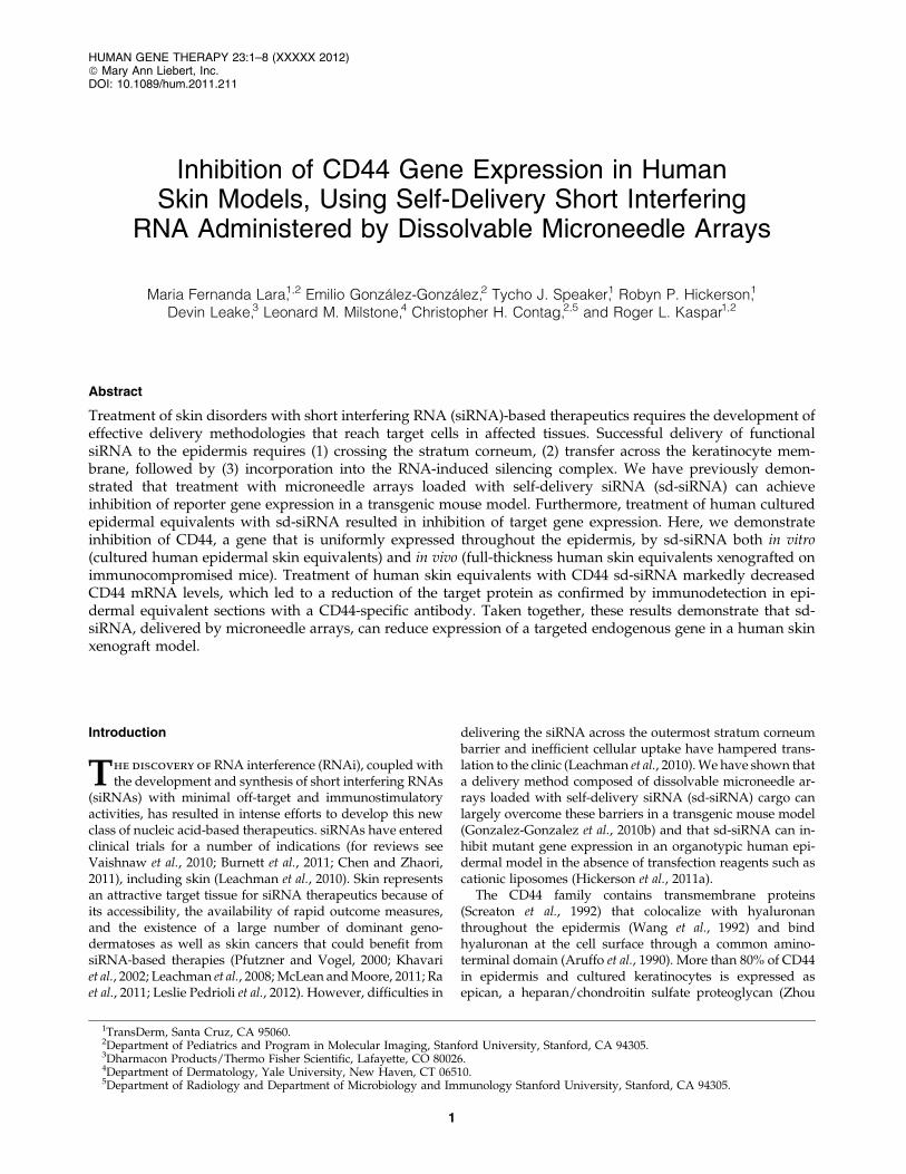
To confirm that CD44 expression is uniform throughoutthe epidermis and easily detected, skin sections were pre-pared from freshly obtained human abdominoplasty skinand analyzed by immunohistochemistry with an antibodythat detects CD44 epican (see Materials and Methods). Figure 1shows strong and uniform CD44 expression through thelive strata with little or no detection in the stratum corneumor dermal skin compartments, confirming that CD44 may bean appropriate target gene for studying functional siRNA
siRNA-MEDIATED GENE TARGETING IN HUMAN SKIN 3
delivery to live epidermal strata. CD44 is similarly expressedin human epidermal skin equivalents (in vitro) and humanskin xenografts (in vivo) (compare Fig. 1, Fig. 2C, and Sup-plementary Fig. 4A), suggesting these skin models maybe useful intermediates to study functional CD44 siRNAdelivery.
In vitro inhibition of CD44 gene expression in humanepidermal equivalents using self-delivery siRNA
Five independent sd-siRNAs that target CD44 gene ex-pression were prepared and comparatively evaluated inhuman HaCaT keratinocytes for their ability to inhibit CD44expression in the absence of transfection reagents (Supple-mentary Fig. S1). CD44 sd-siRNA-2 (CD44 sd-siRNA) waschosen, based on its in vitro activity, for further investigationin the skin equivalent models. The target site for CD44 sd-siRNA spans the exon 1/exon 2 border (see SupplementaryFig. S3) of the CD44 isoform 1 coding region (NCBI Re-ference Sequence NM_000610.3; www.ncbi.nlm.gov). Bothexon 1 and exon 2 are present in all CD44 isoforms (Naoret al., 1997); thus, this siRNA should target all CD44 mRNAs.
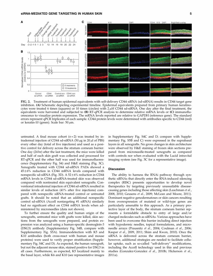
Epidermal equivalents were treated every other day withCD44 sd-siRNA over a span of 12 days, with treatmentscommencing on the twelfth day after initiation of keratino-cyte stratification (Fig. 2A). The epidermal equivalents wereharvested 24 hr after the last treatment; half of each epider-
mal equivalent was collected and processed for RNA anal-ysis (RT-qPCR) and the other half for detection of CD44 viaimmunohistochemistry. Untreated epidermal equivalents, orthose treated with nonspecific (K6a_513a.12) or non-selfdelivery (CD44) siRNAs, served as negative controls. CD44sd-siRNA treatment of epidermal equivalents resulted inreduction of CD44 mRNA levels by 37 – 7% ( p < 0.001)compared with controls treated with nonspecific sd-siRNA(Fig. 2B) and 45 – 7% when compared with untreated con-trols. Levels of CD44 mRNA in tissues treated with non-specific control sd-siRNA samples were not different fromthose in untreated samples ( p > 0.05). Furthermore, no sta-tistically significant differences in keratin-10 mRNA levels(nontargeted mRNA) were observed with the various treat-ments analyzed (data not shown). Similar results were ob-tained when the skin equivalents were treated 10 times,every other day over a 20-day period (36 – 9% reductioncompared with controls similarly treated with nonspecificsd-siRNA; Fig. 2B).
After siRNA treatment, representative samples of humanepidermal equivalents from each treatment group werecryosectioned and analyzed for CD44 protein expression.Consistent with the RT-qPCR results, epidermal equivalentstreated with CD44 sd-siRNA exhibited decreased CD44 im-munofluorescence after 6 and 10 treatments. Representativeimages are shown in Fig. 2C for the epidermal equivalentsreceiving six treatments. Reductions in CD44 protein levelswere not observed in epidermal equivalents treated withcontrol sd-siRNAs and were similar to untreated tissue sec-tions (Fig. 2C). Importantly, siRNA treatment did not no-ticeably alter keratin-10 (K10), K5, or K6 expression patterns(see Fig. 2C and Supplementary Fig. S4 for representativeimages), which localized as expected to the basal (K5) orsuprabasal (K10 and K6) layers.
In vivo inhibition of CD44 in human skinequivalent xenografts
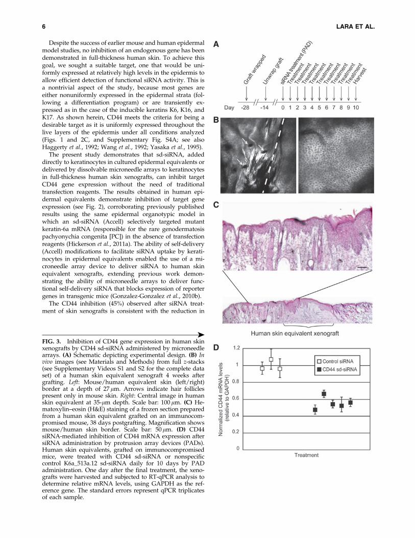
To analyze the ability of sd-siRNA to target an epidermalendogenous gene in vivo, full-thickness 3D human skinequivalents were grafted on immunocompromised mice andtreated with sd-siRNA delivered with microneedle arrays asoutlined in Fig. 3A. Four weeks after the xenograft procedure(before siRNA treatment), the nature and quality of thehuman skin equivalent grafts were assessed by intravitalimaging (Fig. 3B and Supplementary Videos S1 and S2). Thereflectance images revealed well-defined cellular structure atvarious depths for both human skin equivalent xenograftsand mouse skin; these images were consistent with healthyhuman or mouse skin morphology (Gonzalez and Gilaberte-Calzada, 2008; Nehal et al., 2008; Gonzalez-Gonzalez et al.,2011). Figure 3B (left) shows the border of the recipientmouse skin (left of dashed line) with the xenograft (right ofdashed line). Note the uniformity of cells and the absence ofhair follicles in the xenograft (compare with Fig. 3B, right,showing human skin).
The human skin equivalent grafts were treated with sd-siRNA-loaded PADs (three PADs per day) for 10 consecutivedays (Fig. 3A). Daily treatment consisted of application ofthree 5 · 5 microneedle arrays loaded with CD44 (n = 5 mice)or nonspecific control K6a_513a.12 (n = 3) sd-siRNA. As anadditional negative control, a mouse cohort (n = 3) was left
FIG. 1. CD44 is uniformly distributed throughout the liveepidermal strata of human skin. Frozen skin sections wereprepared from freshly obtained abdominal skin and reactedwith anti-CD44 antibody (red; see Materials and Methods).Blue: DAPI-stained nuclei. Scale bar: 50 lm.
4 LARA ET AL.
untreated. A final mouse cohort (n = 2) was treated by in-tradermal injection of CD44 sd-siRNA (50 lg in 20 ll of PBS)every other day (total of five injections) and used as a posi-tive control for delivery across the stratum corneum barrier.One day (24 hr) after the last treatment, the mice were killedand half of each skin graft was collected and processed forRT-qPCR and the other half was used for immunofluores-cence (Supplementary Fig. S4) and H&E staining (Fig. 3C).Xenografts treated with CD44 sd-siRNA PADs showed a45 – 6% reduction in CD44 mRNA levels compared withnonspecific sd-siRNA (Fig. 3D). A 52 – 6% reduction in CD44mRNA levels in CD44 sd-siRNA-treated skin was observedcompared with nontreated skin equivalent xenografts. Con-ventional intradermal injection of CD44 sd-siRNA resulted insimilar levels of reduction (41% after five injections) com-pared with nonspecific siRNA-treated control or untreatedgrafts. It should be noted that an additional nonspecificcontrol sd-siRNA (Accell nontargeting #1 siRNA) similarlyhad no significant effect on CD44 mRNA levels when ad-ministered by microneedles (data not shown).
To further ensure the quality and human origin of thexenografts, untreated mice with grafts were killed, skin sec-tions from the xenografts were prepared, and protein ex-pression was analyzed using a human-specific desmoglein-3(DSG3) antibody (Supplementary Fig. S4B, compare withSupplementary Fig. S5A). Immunodetection with K5 andK10 antibodies (both cross-react with human and mouseproteins) were used to verify proper stratification (Supple-mentary Fig. S4C and D). As expected, the human xenograft,but not the adjacent mouse skin, stained positive for DSG3 inall cases. Furthermore, as expected, K5 localized mainly tothe basal layer, while K6 and K10 (see representative images
in Supplementary Fig. S4C and D; compare with Supple-mentary Fig. S5B and C) were expressed in the suprabasallayers in all xenografts. No gross changes in skin architecturewere observed by H&E staining of frozen skin sections pre-pared from microneedle-treated xenografts as comparedwith controls nor when evaluated with the Lucid intravitalimaging system (see Fig. 3C for a representative image).
Discussion
The ability to harness the RNAi pathway through syn-thetic siRNAs that directly enter the RNA-induced silencingcomplex (RISC) presents opportunities to develop noveltherapeutics by targeting previously unassailable disease-causing genes including those affecting skin (Leachman et al.,2008, 2010; Geusens et al., 2009; McLean and Moore, 2011).Dominant negative genodermatoses or skin cancers resultingfrom overexpression of mutated or wild-type genes areparticularly amenable to this approach. As a primary pro-tective layer of the body, the stratum corneum barrier rep-resents a formidable obstacle to entry of large and/orcharged molecules such as siRNAs. Various approaches havebeen used to overcome this barrier including direct injectionwith hypodermic needles, topical formulations, and micro-needle arrays (Prausnitz et al., 2004; Coulman et al., 2006;Kaspar et al., 2009, 2011; Shim and Kwon, 2010). Once thesiRNA is delivered across the stratum corneum barrier,however, additional strategies are needed to facilitate cellu-lar uptake, such as so-called ‘‘self-delivery’’ modifications,including the Accell technology used in this and previousstudies (Gonzalez-Gonzalez et al., 2010b; Hickerson et al.,2011a).
FIG. 2. Treatment of human epidermal equivalents with self-delivery CD44 siRNA (sd-siRNA) results in CD44 target geneinhibition. (A) Schematic depicting experimental timeline. Epidermal equivalents prepared from primary human keratino-cytes were treated 6 times (squares) or 10 times (circles) with 2 lM CD44 sd-siRNA. One day after the final treatment, theequivalents were harvested and subjected to (B) RT-qPCR analysis to determine relative mRNA levels or (C) immunoflu-orescence to visualize protein expression. The mRNA levels reported are relative to GAPDH (reference gene). The standarderrors represent qPCR triplicates of each sample. CD44 protein levels were determined with antibodies specific to CD44 (red)or keratin-10 (green). Scale bar: 50 lm.
siRNA-MEDIATED GENE TARGETING IN HUMAN SKIN 5
Despite the success of earlier mouse and human epidermalmodel studies, no inhibition of an endogenous gene has beendemonstrated in full-thickness human skin. To achieve thisgoal, we sought a suitable target, one that would be uni-formly expressed at relatively high levels in the epidermis toallow efficient detection of functional siRNA activity. This isa nontrivial aspect of the study, because most genes areeither nonuniformly expressed in the epidermal strata (fol-lowing a differentiation program) or are transiently ex-pressed as in the case of the inducible keratins K6, K16, andK17. As shown herein, CD44 meets the criteria for being adesirable target as it is uniformly expressed throughout thelive layers of the epidermis under all conditions analyzed(Figs. 1 and 2C, and Supplementary Fig. S4A; see alsoHaggerty et al., 1992; Wang et al., 1992; Yasaka et al., 1995).
The present study demonstrates that sd-siRNA, addeddirectly to keratinocytes in cultured epidermal equivalents ordelivered by dissolvable microneedle arrays to keratinocytesin full-thickness human skin xenografts, can inhibit targetCD44 gene expression without the need of traditionaltransfection reagents. The results obtained in human epi-dermal equivalents demonstrate inhibition of target geneexpression (see Fig. 2), corroborating previously publishedresults using the same epidermal organotypic model inwhich an sd-siRNA (Accell) selectively targeted mutantkeratin-6a mRNA (responsible for the rare genodermatosispachyonychia congenita [PC]) in the absence of transfectionreagents (Hickerson et al., 2011a). The ability of self-delivery(Accell) modifications to facilitate siRNA uptake by kerati-nocytes in epidermal equivalents enabled the use of a mi-croneedle array device to deliver siRNA to human skinequivalent xenografts, extending previous work demon-strating the ability of microneedle arrays to deliver func-tional self-delivery siRNA that blocks expression of reportergenes in transgenic mice (Gonzalez-Gonzalez et al., 2010b).
The CD44 inhibition (45%) observed after siRNA treat-ment of skin xenografts is consistent with the reduction in
FIG. 3. Inhibition of CD44 gene expression in human skinxenografts by CD44 sd-siRNA administered by microneedlearrays. (A) Schematic depicting experimental design. (B) Invivo images (see Materials and Methods) from full z-stacks(see Supplementary Videos S1 and S2 for the complete dataset) of a human skin equivalent xenograft 4 weeks aftergrafting. Left: Mouse/human equivalent skin (left/right)border at a depth of 27 lm. Arrows indicate hair folliclespresent only in mouse skin. Right: Central image in humanskin equivalent at 35-lm depth. Scale bar: 100 lm. (C) He-matoxylin–eosin (H&E) staining of a frozen section preparedfrom a human skin equivalent grafted on an immunocom-promised mouse, 38 days postgrafting. Magnification showsmouse/human skin border. Scale bar: 50 lm. (D) CD44siRNA-mediated inhibition of CD44 mRNA expression aftersiRNA administration by protrusion array devices (PADs).Human skin equivalents, grafted on immunocompromisedmice, were treated with CD44 sd-siRNA or nonspecificcontrol K6a_513a.12 sd-siRNA daily for 10 days by PADadministration. One day after the final treatment, the xeno-grafts were harvested and subjected to RT-qPCR analysis todetermine relative mRNA levels, using GAPDH as the ref-erence gene. The standard errors represent qPCR triplicatesof each sample.
‰
6 LARA ET AL.
gene expression observed in other skin systems using in-tradermal injection, topical administration, or microneedlearrays (Gonzalez-Gonzalez et al., 2009, 2010b; Hsu andMitragotri, 2011). The reason that more inhibition is notachieved is not clear, but similar results have been observedin nonskin tissues including eye (Huang et al., 2011), brain(Kuwahara et al., 2011), and heart (Guido et al., 2011), whilehigher levels of inhibition (up to 95%) have been reported inliver (Zimmermann et al., 2006; Frank-Kamenetsky et al.,2008; Tadin-Strapps et al., 2011). Although incomplete inhi-bition of gene expression was observed after treatment withsd-CD44 siRNA, complete silencing of a gene target may notbe necessary to achieve a therapeutic effect. For example, inan inducible keratin-14 transgenic mouse model for thedominant disorder epidermolysis bullosa simplex (EBS),low-level expression of the mutant keratin-14 allele (1:2,mutant to wild type) produced no epidermal fragility phe-notype (Cao et al., 2001). These observations suggest thatstrategies resulting in partial inhibition of the targeted genemay result in a therapeutic benefit for patients suffering fromdominant keratin disorders (Chen and Roop, 2005). The in-tent of this study, therefore, was not to show a functionaleffect, but rather to demonstrate that sd-siRNA delivery viamicroneedle arrays can efficiently inhibit an endogenousgene that is uniformly distributed throughout the epidermalstrata. Many, if not most, genes (including the genes in-volved in PC—KRT6a/b, KRT16, and KRT17) have alteredepidermal expression patterns as keratinocytes undergodifferentiation. In our preliminary experiments, as well aspublished studies, epidermal CD44 appeared to be quiteuniformly expressed, making this the most appropriate epi-dermal target we could identify that is readily detected atboth the protein and mRNA levels. The downside to the useof CD44 is that it is a large family of genes that may com-pensate for one another, masking potential phenotypicchanges.
siRNA therapeutics have the potential to transform treat-ment of many skin disorders if delivery obstacles can beovercome. The present work demonstrates that administra-tion of dissolvable microneedle arrays, loaded with sd-siRNA cargo, can inhibit expression of an endogenous genein human skin models. The amount of sd-siRNA needed formicroneedle administration in a clinical setting is potentiallyless than what was required for the initial phase 1b PCclinical trial (Leachman et al., 2010). In that study, no thera-peutic effects were observed until doses of up to 17 mg ofunmodified siRNA were delivered by intralesional injection(hypodermic needle). In contrast, only 150 lg (100-fold less)of sd-siRNA (delivered by the dissolvable microneedle ar-rays) would be required to treat a surface area of a sizeequivalent to the area (*10 cm2) estimated to be reached byhypodermic needle injection. The successful translation ofthis delivery platform to the clinic will enable developmentof therapeutics for many heretofore untreatable skin disor-ders, particularly dominant genodermatoses, potentiallytransforming the way patients are treated. On the basis ofthis and other ongoing studies, microneedle array-mediateddelivery of siRNA appears to be a viable and patient-friendly(i.e., involving little or no pain Gill et al., 2008) alternative tosiRNA delivery by intradermal hypodermic needle injec-tions, offering a more patient-acceptable path forward forfuture clinical applications.
Acknowledgments
The authors sincerely thank Dr. Christine Collin-Djangone(L’Oreal) for insightful discussions, suggesting the use ofCD44 as a model endogenous target and identifying theCD44 target site sequence used for the siRNA experimentsdescribed herein. The authors thank Xiaomin Bao (PaulKhavari’s laboratory, Stanford University) for providing theDSG3 human-specific antibody. The authors also thank StellaChang, Manny Flores, and Andrea Burgon for technicalsupport and the participants of the GO Delivery! Project forinput and support. This project was supported by NIAMS/NIH grants RC2AR058955-02 (R.L.K., L.M.M., and C.H.C.)and R43AR059474 (R.L.K.).
Author Disclosure Statement
Roger Kaspar, Robyn Hickerson, Tycho Speaker, andMaria Fernanda Lara are employees of TransDerm, whichhas a patent pending for use of microneedle arrays to delivernucleic acids. Devin Leake is an employee of Thermo FisherScientific.
References
Aruffo, A., Stamenkovic, I., Melnick, M., et al. (1990). CD44 is theprincipal cell surface receptor for hyaluronate. Cell 61, 1303–1313.
Burnett, J.C., Rossi, J.J., and Tiemann, K. (2011). Current prog-ress of siRNA/shRNA therapeutics in clinical trials. Bio-technol. J. 6, 1130–1146.
Cao, T., Longley, M.A., Wang, X.J., and Roop, D.R. (2001). Aninducible mouse model for epidermolysis bullosa simplex:implications for gene therapy. J. Cell Biol. 152, 651–656.
Chen, J., and Roop, D.R. (2005). Mouse models in preclinicalstudies for pachyonychia congenita. J. Investig. Dermatol.Symp. Proc. 10, 37–46.
Chen, S.H., and Zhaori, G. (2011). Potential clinical applicationsof siRNA technique: benefits and limitations. Eur. J. Clin. In-vest. 41, 221–232.
Coulman, S., Allender, C., and Birchall, J. (2006). Microneedlesand other physical methods for overcoming the stratumcorneum barrier for cutaneous gene therapy. Crit. Rev. Ther.Drug Carrier Syst. 23, 205–258.
Frank-Kamenetsky, M., Grefhorst, A., Anderson, N.N., et al.(2008). Therapeutic RNAi targeting PCSK9 acutely lowersplasma cholesterol in rodents and LDL cholesterol in non-human primates. Proc. Natl. Acad. Sci. U.S.A. 105, 11915–11920.
Geusens, B., Sanders, N., Prow, T., et al. (2009). Cutaneous short-interfering RNA therapy. Expert Opin. Drug Deliv. 6, 1333–1349.
Gill, H.S., Denson, D.D., Burris, B.A., and Prausnitz, M.R. (2008).Effect of microneedle design on pain in human volunteers.Clin. J. Pain 24, 585–594.
Gonzalez, S., and Gilaberte-Calzada, Y. (2008). In vivo reflectance-mode confocal microscopy in clinical dermatology and cos-metology. Int. J. Cosmet. Sci. 30, 1–17.
Gonzalez-Gonzalez, E., Ra, H., Hickerson, R.P., et al. (2009).siRNA silencing of keratinocyte-specific GFP expression in atransgenic mouse skin model. Gene Ther. 16, 963–972.
Gonzalez-Gonzalez, E., Speaker, T.J., Hickerson, R.P., et al.(2010b). Silencing of reporter gene expression in skin usingsiRNAs and expression of plasmid DNA delivered by a sol-uble protrusion array device (PAD). Mol. Ther. 18, 1667–1674.
siRNA-MEDIATED GENE TARGETING IN HUMAN SKIN 7
Gonzalez-Gonzalez, E., Kim, Y.C., Speaker, T.J., et al. (2011).Visualization of plasmid delivery to keratinocytes in mouseand human epidermis. Sci. Rep. 1, 158.
Guido, M.C., Clemente, C.F., Moretti, A.I., et al. (2011). Smallinterfering RNA targeting focal adhesion kinase preventscardiac dysfunction in endotoxemia. Shock 37, 77–84.
Haggerty, J.G., Bretton, R.H., and Milstone, L.M. (1992). Identi-fication and characterization of a cell surface proteoglycan onkeratinocytes. J. Invest. Dermatol. 99, 374–380.
Hickerson, R.P., Smith, F.J., Reeves, R.E., et al. (2008). Single-nucleotide specific siRNA targeting in a dominant-negativeskin model. J. Invest. Dermatol. 128, 594–605.
Hickerson, R.P., Flores, M.A., Leake, D., et al. (2011a). Use of self-delivery siRNAs to inhibit gene expression in an organotypicpachyonychia congenita model. J. Invest. Dermatol. 131, 1037–1044.
Hickerson, R.P., Leachman, S.A., Pho, L.N., et al. (2011b). De-velopment of quantitative molecular clinical end points forsiRNA clinical trials. J. Invest. Dermatol. 131, 1029–1036.
Hsu, T., and Mitragotri, S. (2011). Delivery of siRNA and othermacromolecules into skin and cells using a peptide enhancer.Proc. Natl. Acad. Sci. U.S.A. 108, 15816–15821.
Huang, W.R., Fan, X.X., and Tang, X. (2011). SiRNA targetingEGFR effectively prevents posterior capsular opacificationafter cataract surgery. Mol. Vis. 17, 2349–2355.
Kaspar, R.L., McLean, W.H., and Schwartz, M.E. (2009).Achieving successful delivery of nucleic acids to skin: 6thAnnual Meeting of the International Pachyonychia CongenitaConsortium. J. Invest. Dermatol. 129, 2085–2087.
Kaspar, R.L., Leachman, S.A., McLean, W.H., and Schwartz,M.E. (2011). Toward a treatment for pachyonychia congenita:Report on the 7th Annual International Pachyonychia Con-genita Consortium meeting. J. Invest. Dermatol. 131, 1011–1014.
Khavari, P.A., Rollman, O., and Vahlquist, A. (2002). Cutaneousgene transfer for skin and systemic diseases. J. Intern. Med.252, 1–10.
Kugelman, L.C., Ganguly, S., Haggerty, J.G., et al. (1992). Thecore protein of epican, a heparan sulfate proteoglycan onkeratinocytes, is an alternative form of CD44. J. Invest. Der-matol. 99, 886–891.
Kuwahara, H., Nishina, K., Yoshida, K., et al. (2011). Efficientin vivo delivery of siRNA into brain capillary endothelial cellsalong with endogenous lipoprotein. Mol. Ther. 19, 2213–2221.
Leachman, S.A., Hickerson, R.P., Hull, P.R., et al. (2008). Ther-apeutic siRNAs for dominant genetic skin disorders includingpachyonychia congenita. J. Dermatol. Sci. 51, 151–157.
Leachman, S.A., Hickerson, R.P., Schwartz, M.E., et al. (2010).First-in-human mutation-targeted siRNA phase Ib trial of aninherited skin disorder. Mol. Ther. 18, 442–446.
Leslie Pedrioli, D.M., Fu, D.J., Gonzalez-Gonzalez, E., et al.(2012). Generic and personalized RNAi therapeutics for adominant-negative epidermal fragility disorder. J. Invest.Dermatol. (in press).
McLean, W.H., and Moore, C.B. (2011). Keratin disorders: Fromgene to therapy. Hum. Mol. Genet. 20, R189–R197.
Naor, D., Sionov, R.V., and Ish-Shalom, D. (1997). CD44:Structure, function, and association with the malignant pro-cess. Adv. Cancer Res. 71, 241–319.
Nehal, K.S., Gareau, D., and Rajadhyaksha, M. (2008). Skinimaging with reflectance confocal microscopy. Semin. Cutan.Med. Surg. 27, 37–43.
Pfutzner, W., and Vogel, J.C. (2000). Advances in skin genetherapy. Expert Opin. Investig. Drugs 9, 2069–2083.
Prausnit, M.R., Mitragotri, S., and Langer, R. (2004). Currentstatus and future potential of transdermal drug delivery. Nat.Rev. Drug Discov. 3, 115–124.
Ra, H., Piyawattanametha, W., Gonzalez-Gonzalez, E., et al.(2011). In vivo imaging of human and mouse skin with ahandheld dual-axis confocal fluorescence microscope. J. In-vest. Dermatol. 131, 1061–1066.
Schurr, M.J., Foster, K.N., Centanni, J.M., et al. (2009). Phase I/IIclinical evaluation of StrataGraft: A consistent, pathogen-freehuman skin substitute. J. Trauma 66, 866–873; discussion 873–874.
Screaton, G.R., Bell, M.V., Jackson, D.G., et al. (1992). Genomicstructure of DNA encoding the lymphocyte homing receptorCD44 reveals at least 12 alternatively spliced exons. Proc. Natl.Acad. Sci. U.S.A. 89, 12160–12164.
Shim, M.S., and Kwon, Y.J. (2010). Efficient and targeted deliv-ery of siRNA in vivo. FEBS J. 277, 4814–4827.
Tadin-Strapps, M., Peterson, L.B., Cumiskey, A.M., et al. (2011).siRNA-induced liver ApoB knockdown lowers serum LDL-cholesterol in a mouse model with human-like serum lipids. J.Lipid Res. 52, 1084–1097.
Vaishnaw, A.K., Gollob, J., Gamba-Vitalo, C., et al. (2010). Astatus report on RNAi therapeutics. Silence 1, 14.
Wang, C., Tammi, M., and Tammi, R. (1992). Distribution ofhyaluronan and its CD44 receptor in the epithelia of humanskin appendages. Histochemistry 98, 105–112.
Yasaka, N., Furue, M., and Tamaki, K. (1995). CD44 expres-sion in normal human skin and skin tumors. J. Dermatol. 22,88–94.
Zhou, J., Haggerty, J.G., and Milstone, L.M. (1999). Growth anddifferentiation regulate CD44 expression on human keratino-cytes. In Vitro Cell Dev. Biol. Anim. 35, 228–235.
Zimmermann, T.S., Lee, A.C., Akinc, A., et al. (2006). RNAi-mediated gene silencing in non-human primates. Nature 441,111–114.
Address correspondence to:Dr. Roger L. Kaspar
2161 Delaware Ave., Suite DSanta Cruz, CA 95060
E-mail: [email protected]
Received for publication November 23, 2011;accepted after revision March 7, 2012.
Published online: April 5, 2012.
8 LARA ET AL.