inhibition of an aquatic rhabdovirus demonstrates promise ...jung/pdfs/343.pdf · inhibition of an...
TRANSCRIPT

Inhibition of an Aquatic RhabdovirusDemonstrates Promise of a Broad-Spectrum Antiviral for Use inAquaculture
Bethany F. Balmer,a Rachel L. Powers,b Ting-Hu Zhang,c Jihye Lee,c
Frederic Vigant,d Benhur Lee,d Michael E. Jung,c Maureen K. Purcell,b
Kevin Snekvik,a,e Hector C. Aguilara,f
Department of Veterinary Microbiology and Pathology, College of Veterinary Medicine, Washington StateUniversity, Pullman, Washington, USAa; U.S. Geological Survey, Western Fisheries Research Center, Seattle,Washington, USAb; Department of Chemistry and Biochemistry, University of California, Los Angeles, LosAngeles, California, USAc; Department of Microbiology, Icahn School of Medicine at Mount Sinai, New York,New York, USAd; Washington Animal Disease Diagnostic Laboratory, College of Veterinary Medicine,Washington State University, Pullman, Washington, USAe; Paul G. Allen School for Global Animal Health,College of Veterinary Medicine, Washington State University, Pullman, Washington, USAf
ABSTRACT Many enveloped viruses cause devastating disease in aquaculture, result-ing in significant economic impact. LJ001 is a broad-spectrum antiviral compoundthat inhibits enveloped virus infections by specifically targeting phospholipids in thelipid bilayer via the production of singlet oxygen (1O2). This stabilizes positive curva-ture and decreases membrane fluidity, which inhibits virus-cell membrane fusionduring viral entry. Based on data from previous mammalian studies and the require-ment of light for the activation of LJ001, we hypothesized that LJ001 may be usefulas a preventative and/or therapeutic agent for infections by enveloped viruses inaquaculture. Here, we report that LJ001 was more stable with a prolonged inhibitoryhalf-life at relevant aquaculture temperatures (15°C), than in mammalian studies at37°C. When LJ001 was preincubated with our model virus, infectious hematopoieticnecrosis virus (IHNV), infectivity was significantly inhibited in vitro (using the epitheli-oma papulosum cyprini [EPC] fish cell line) and in vivo (using rainbow trout fry) in adose-dependent and time-dependent manner. While horizontal transmission of IHNVin a static cohabitation challenge model was reduced by LJ001, transmission wasnot completely blocked at established antiviral doses. Therefore, LJ001 may be bestsuited as a therapeutic for aquaculture settings that include viral infections withlower virus-shedding rates than IHNV or where higher viral titers are required to ini-tiate infection of naive fish. Importantly, our data also suggest that LJ001-inactivatedIHNV elicited an innate immune response in the rainbow trout host, making LJ001potentially useful for future vaccination approaches.
IMPORTANCE Viral diseases in aquaculture are challenging because there are fewpreventative measures and/or treatments. Broad-spectrum antivirals are highlysought after and studied because they target common components of viruses. Inour studies, we used LJ001, a broad-spectrum antiviral compound that specificallyinhibits enveloped viruses. We used the fish rhabdovirus infectious hematopoieticnecrosis virus (IHNV) as a model to study aquatic enveloped virus diseases and theirinhibition. We demonstrated inhibition of IHNV by LJ001 both in cell culture as wellas in live fish. Additionally, we showed that LJ001 inhibited the transmission of IHNVfrom infected fish to healthy fish, which lays the groundwork for using LJ001 as apossible therapeutic for aquatic viruses. Our results also suggest that virus inacti-vated by LJ001 induces an immune response, showing potential for future preventa-tive (e.g., vaccine) applications.
Received 2 November 2016 Accepted 22November 2016
Accepted manuscript posted online 30November 2016
Citation Balmer BF, Powers RL, Zhang T-H, LeeJ, Vigant F, Lee B, Jung ME, Purcell MK, SnekvikK, Aguilar HC. 2017. Inhibition of an aquaticrhabdovirus demonstrates promise of a broad-spectrum antiviral for use in aquaculture. J Virol91:e02181-16. https://doi.org/10.1128/JVI.02181-16.
Editor Douglas S. Lyles, Wake Forest University
Copyright © 2017 American Society forMicrobiology. All Rights Reserved.
Address correspondence to Kevin Snekvik,[email protected], or Hector C. Aguilar,[email protected].
M.K.P., K.S., and H.C.A. contributed equally.
VACCINES AND ANTIVIRAL AGENTS
crossm
February 2017 Volume 91 Issue 4 e02181-16 jvi.asm.org 1Journal of Virology
on June 22, 2017 by UC
LA B
IOM
ED
ICA
L LIB/S
ER
IALS
http://jvi.asm.org/
Dow
nloaded from

KEYWORDS antiviral, aquaculture, aquatic virus, broad spectrum, enveloped,membrane fusion, rhabdovirus, virus
Aquaculture is considered the fastest-growing food-producing sector, increasing atan average annual rate of 6.1%, from 36.8 million tons in 2002 to 66.6 million tons
in 2012 (1). Aquaculture is estimated to account for more than 30% of the world’s foodfish, and this is projected to increase to 50% by 2030 (2). As fish and shellfish demandsincrease, it is critical to reduce losses from diseases that economically devastate theaquaculture industry. Viral pathogens, in particular, are challenging since there are fewefficacious treatments or preventatives (3). A number of viruses are associated withsignificant economic losses for the aquaculture industry, including 11 envelopedviruses that cause diseases so severe that they are reportable to the World Organizationfor Animal Health (OIE) according to the 2016 aquatic animal health code (http://www.oie.int/en/). Examples of OIE-reportable viruses include infectious hematopoieticnecrosis virus (IHNV) in the trout industry (4, 5), salmonid alphavirus in the salmonindustry (3, 5), spring viremia of carp virus (SVCV) in the koi and carp industries (5, 6),and white spot syndrome virus (WSSV) in the shrimp industry (7, 8).
We used IHNV as a model to evaluate the broad-spectrum antiviral LJ001 forpotential use in aquaculture. IHNV belongs to the family Rhabdoviridae and genusNovirhabdovirus (http://ictvonline.org/virusTaxonomy.asp) and infects a range of sal-monid fish species (9) in addition to some nonsalmonid fish species (demonstratedexperimentally) (10, 11). IHNV is an enveloped, bullet-shaped RNA virus encoding anegative-sense, single-stranded genome. The virus is enzootic in river systems through-out western North America and has spread to Asia and Europe, likely by movement ofinfected fish and eggs (12). Fry are typically most susceptible to disease, with cumu-lative mortality rates reported to be as high as 90 to 95% during epizootic outbreaks.Horizontal transmission of IHNV occurs when the virus is shed in feces, urine, sexualfluids, and external mucus (13), and the virus can survive in freshwater for up to 7 weeks(14). The only licensed vaccine is a DNA vaccine that encodes the IHNV glycoprotein (2,15, 16) and is delivered by intramuscular injection. The vaccine elicits both early,nonspecific, and cross-protective antiviral immunity mediated by interferon as well asa long-term, specific adaptive response (17) and is considered effective (18). However,the vaccine can be cost-prohibitive and has delivery constraints (19), which makes itimpractical for large numbers of susceptible fry. There is still a significant problem withmany aquatic enveloped viruses, including IHNV, and new therapy options are needed.
A novel small-molecule antiviral rhodanine derivative, LJ001, has been shown toinhibit infections by numerous enveloped viruses at micromolar concentrations, withno effect on nonenveloped viruses (20). The antiviral activity of LJ001 is light dependentand requires the presence of molecular oxygen. The mechanism of action for LJ001 isbased on the production of singlet oxygen (1O2) molecules that modify phospholipidsin the lipid bilayer, which results in the stabilization of positive spontaneous curvature,decreased membrane fluidity, and, ultimately, inhibition of virus-cell membrane fusionand viral entry (21). Unlike viruses, host cells possess a “biogenic” membrane thatactively repairs its defects by lipid synthesis and replacement (22). In contrast, thehost-derived viral envelope cannot be repaired and is a susceptible target for LJ001. Ithas been suggested that there is limited opportunity for viruses to evolve resistance toLJ001 based on its mechanism of action and specific target of the membrane envelope(20). LJ001 has been shown to have no overt toxicity in Vero cells or mouse models ateffective antiviral doses (up to 10 �M), but as predicted, toxicity can be induced in thepresence of the fatty acid synthesis inhibitor 5-(tetradecyloxy)-2-furoic acid (TOFA) (20).
The use of LJ001 in mammalian models is impractical due to a reported biologicalhalf-life of �4 h at 37°C (20) and a lack of exposure to light in most tissues to activatethe compound in vivo. Aquaculture environments are unique because pathogens,including IHNV, are transmitted through water, which is transparent to light. In thisstudy, we determined that IHNV infection and horizontal transmission were inhibited
Balmer et al. Journal of Virology
February 2017 Volume 91 Issue 4 e02181-16 jvi.asm.org 2
on June 22, 2017 by UC
LA B
IOM
ED
ICA
L LIB/S
ER
IALS
http://jvi.asm.org/
Dow
nloaded from

by LJ001 and that LJ001 had a prolonged inhibitory half-life in aquaculture environ-ments. We also preliminarily investigated the ability of LJ001-inactivated IHNV toinduce an innate immune response in the rainbow trout host.
RESULTSLJ001 is not cytotoxic at antiviral concentrations. We first evaluated the potential
cytotoxicity of LJ001 via in vitro experiments in fish-derived epithelioma papulosumcyprini (EPC) cells. Microscopically, there were no changes in cellular morphology orgrowth at LJ001 concentrations of up to 10 �M for up to 7 days. Additionally, cellviability was quantitatively assessed by using a cell counting kit (CCK-8), which is basedon dehydrogenase enzyme activity. EPC cells were exposed to increasing concentra-tions of LJ001, and no cytotoxicity was evident with up to 7 days of exposure to 10 �MLJ001 (Fig. 1A). Cytotoxicity was reached at 7 days of exposure to 20 �M LJ001 (P �
0.001) and at all time points when cells were exposed to 50 �M LJ001. There was nocytotoxicity of the vehicle control (dimethyl sulfoxide [DMSO]) at concentrations of upto 0.1%, but cytotoxicity was established at 3 days (P � 0.05) and 7 days (P � 0.001) ofexposure to 0.5% DMSO (Fig. 1B). In Fig. 1A, cells exposed to 10 �M LJ001 weresimultaneously exposed to 0.1% DMSO (50 �M LJ001 in 0.5% DMSO).
Clinical parameters used to assess the potential toxicity of LJ001 during rainbowtrout fry experiments included prolonged lethargy, circling, swimming slowly, prefer-ence for the bottom of challenge containers, and overt death, none of which wereobserved. Cytotoxic effects were also assessed via histopathology of whole, fixed fry
FIG 1 LJ001 is not cytotoxic at antiviral concentrations. EPC cells were exposed to increasing concen-trations of LJ001 (A) or the vehicle control (DMSO) (B) for 1, 3, and 7 days in the presence of light at thetime of addition. Formazan dye absorbance was measured at 450 nm. Positive-control wells wereexposed to hydrogen peroxide (40 mM final concentration) for 1 day. The negative control (�) is no drugexposure. (A) Exposure to 0 to 50 �M LJ001 (50 �M LJ001 with 0.5% DMSO, 10 �M LJ001 with 0.1%DMSO, and 1.0 �M LJ001 with 0.01% DMSO). There was no cytotoxicity to EPC cells at up to 10 �M LJ001.Toxicity occurred when cells were exposed to 20 �M LJ001 for 7 days and 50 �M LJ001 at all time points.Data represent mean cell viabilities � standard errors (n � 3) normalized to values for no treatment(negative control). **, P � 0.01; ***, P � 0.001. (B) Exposure to 0 to 0.5% DMSO. No cytotoxic effects wereobserved at concentrations of up to 0.1%. Cytotoxic effects were detected after exposure to 0.5% DMSOat 3 and 7 days. Data represent mean cell viabilities � standard errors (n � 3) normalized to values forno treatment. *, P � 0.05; ***, P � 0.001.
Broad-Spectrum Aquaculture Antiviral Journal of Virology
February 2017 Volume 91 Issue 4 e02181-16 jvi.asm.org 3
on June 22, 2017 by UC
LA B
IOM
ED
ICA
L LIB/S
ER
IALS
http://jvi.asm.org/
Dow
nloaded from
(transverse sections). There was no histologic difference between three negative-control fish (histologic scores of 1, 2, and 1) and three fish immersed in 10 �M LJ001(histologic scores of 1, 2, and 2). The scores were based on mild to moderate hyper-plastic changes in the gills and skin (associated with various numbers of Ichthyobodonecator [“Costia”] parasites) with no internal organ abnormalities (data not shown) (P �
0.52 by an unpaired Student t test). Our results confirmed that LJ001 was not cytotoxicin vitro and in vivo at antiviral doses.
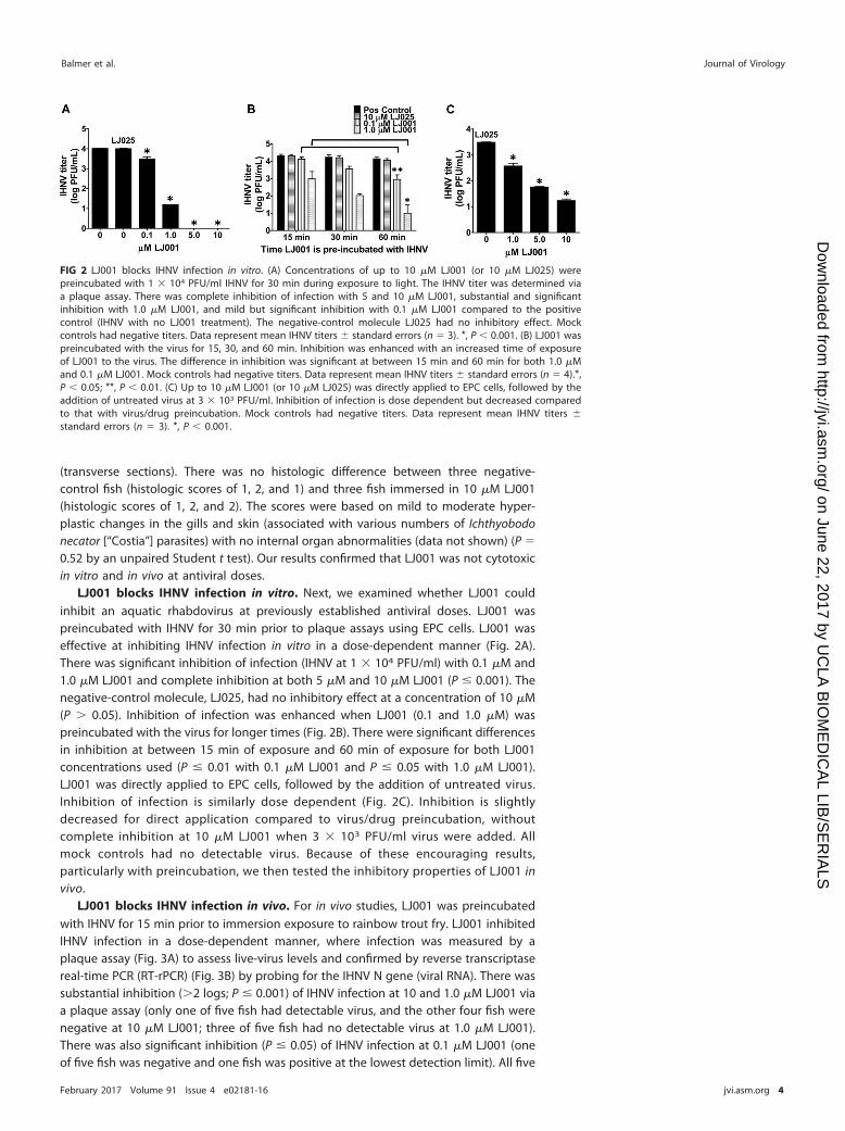
LJ001 blocks IHNV infection in vitro. Next, we examined whether LJ001 couldinhibit an aquatic rhabdovirus at previously established antiviral doses. LJ001 waspreincubated with IHNV for 30 min prior to plaque assays using EPC cells. LJ001 waseffective at inhibiting IHNV infection in vitro in a dose-dependent manner (Fig. 2A).There was significant inhibition of infection (IHNV at 1 � 104 PFU/ml) with 0.1 �M and1.0 �M LJ001 and complete inhibition at both 5 �M and 10 �M LJ001 (P � 0.001). Thenegative-control molecule, LJ025, had no inhibitory effect at a concentration of 10 �M(P � 0.05). Inhibition of infection was enhanced when LJ001 (0.1 and 1.0 �M) waspreincubated with the virus for longer times (Fig. 2B). There were significant differencesin inhibition at between 15 min of exposure and 60 min of exposure for both LJ001concentrations used (P � 0.01 with 0.1 �M LJ001 and P � 0.05 with 1.0 �M LJ001).LJ001 was directly applied to EPC cells, followed by the addition of untreated virus.Inhibition of infection is similarly dose dependent (Fig. 2C). Inhibition is slightlydecreased for direct application compared to virus/drug preincubation, withoutcomplete inhibition at 10 �M LJ001 when 3 � 103 PFU/ml virus were added. Allmock controls had no detectable virus. Because of these encouraging results,particularly with preincubation, we then tested the inhibitory properties of LJ001 invivo.
LJ001 blocks IHNV infection in vivo. For in vivo studies, LJ001 was preincubatedwith IHNV for 15 min prior to immersion exposure to rainbow trout fry. LJ001 inhibitedIHNV infection in a dose-dependent manner, where infection was measured by aplaque assay (Fig. 3A) to assess live-virus levels and confirmed by reverse transcriptasereal-time PCR (RT-rPCR) (Fig. 3B) by probing for the IHNV N gene (viral RNA). There wassubstantial inhibition (�2 logs; P � 0.001) of IHNV infection at 10 and 1.0 �M LJ001 viaa plaque assay (only one of five fish had detectable virus, and the other four fish werenegative at 10 �M LJ001; three of five fish had no detectable virus at 1.0 �M LJ001).There was also significant inhibition (P � 0.05) of IHNV infection at 0.1 �M LJ001 (oneof five fish was negative and one fish was positive at the lowest detection limit). All five
FIG 2 LJ001 blocks IHNV infection in vitro. (A) Concentrations of up to 10 �M LJ001 (or 10 �M LJ025) werepreincubated with 1 � 104 PFU/ml IHNV for 30 min during exposure to light. The IHNV titer was determined viaa plaque assay. There was complete inhibition of infection with 5 and 10 �M LJ001, substantial and significantinhibition with 1.0 �M LJ001, and mild but significant inhibition with 0.1 �M LJ001 compared to the positivecontrol (IHNV with no LJ001 treatment). The negative-control molecule LJ025 had no inhibitory effect. Mockcontrols had negative titers. Data represent mean IHNV titers � standard errors (n � 3). *, P � 0.001. (B) LJ001 waspreincubated with the virus for 15, 30, and 60 min. Inhibition was enhanced with an increased time of exposureof LJ001 to the virus. The difference in inhibition was significant at between 15 min and 60 min for both 1.0 �Mand 0.1 �M LJ001. Mock controls had negative titers. Data represent mean IHNV titers � standard errors (n � 4).*,P � 0.05; **, P � 0.01. (C) Up to 10 �M LJ001 (or 10 �M LJ025) was directly applied to EPC cells, followed by theaddition of untreated virus at 3 � 103 PFU/ml. Inhibition of infection is dose dependent but decreased comparedto that with virus/drug preincubation. Mock controls had negative titers. Data represent mean IHNV titers �standard errors (n � 3). *, P � 0.001.
Balmer et al. Journal of Virology
February 2017 Volume 91 Issue 4 e02181-16 jvi.asm.org 4
on June 22, 2017 by UC
LA B
IOM
ED
ICA
L LIB/S
ER
IALS
http://jvi.asm.org/
Dow
nloaded from

fish in both the group receiving 0.01 �M LJ001 treatment and the group receiving viruswith no treatment had positive viral titers. All mock-infected fish had negative titers.Our results demonstrated that IHNV infection can be inhibited in vivo by preincubationwith low micromolar concentrations of LJ001.
LJ001 is relatively stable in aquaculture environments. We then tested thestability of LJ001 in aqueous environments. LJ001 was added to water samples (river,hatchery, or deionized water, with hatchery water at 15°C being the most aquaculture-relevant environment containing a medium level of organic material). The sampleswere placed for up to 7 days at 15°C or 4°C (with exposure to light) and finallypreincubated with 1 � 104 PFU/ml IHNV, followed by a plaque assay. LJ001 was fairlystable in hatchery water at 15°C (Fig. 4A) and was less efficacious with increasedamounts of organic material present (the river water had abundant particulate matter).LJ001 added to hatchery water at 15°C had a calculated inhibitory half-life of 1.9 days(R2 � 0.83) when a nonlinear regression, best-of-fit line was applied (data not shown).Additionally, the stability of LJ001 was further enhanced at even lower temperatures(4°C) (Fig. 4B). The calculated inhibitory half-lives of LJ001 at 4°C were 2.7 days (R2 �
0.76) in river water and 5.6 days (R2 � 0.72) in hatchery water. These results indicatethat LJ001 is a relatively stable compound under aquaculture conditions.
LJ001 inhibits horizontal transmission of IHNV. Based on our in vivo pretreat-ment inhibition data, we explored the therapeutic applications of LJ001 and its abilityto inhibit horizontal transmission of IHNV. Donor fish were immersion infected withIHNV, followed by the addition of LJ001 (5 �M final concentration), and finally, naiverecipient fish were added after 15 min in a cohabitation experiment (see Fig. 5A forexperimental groups). IHNV recipient fish from LJ001-treated groups had significantlylower (P � 0.002) viral titers than did DMSO-treated (control) IHNV recipient fish (Fig.5B). None of the 36 LJ001-treated IHNV recipient fish had negative titers, but 19/36 fish
FIG 3 LJ001 blocks IHNV infection in vivo. Naive rainbow trout fry were immersed as a group in 1 � 104
PFU/ml IHNV that was preincubated for 15 min with 0 to 10 �M LJ001 while being exposed to light.Following 12 h, fish were separated into isolation beakers, and the virus was allowed to replicate for 72h. The homogenate supernatant from each fish was used for a plaque assay (A), and RNA was isolatedand quantified by RT-rPCR (B) to determine the IHNV titer or quantity, respectively. The positive controlwas IHNV and the vehicle control only (0 �M LJ001 and 0.01% DMSO, final concentration). The negativecontrol was MEM and 0.01% DMSO. (A) There was a highly significant inhibition of infection with 10 �Mand 1.0 �M LJ001 and significant inhibition with 0.1 �M LJ001. Data represent mean IHNV titers �standard errors (n � 5 fish per group). *, P � 0.05; ***, P � 0.001. (B) RT-rPCR probing for the IHNV N geneconfirmed the plaque assay results. Data represent mean numbers of IHNV N gene copies � standarderrors (n � 5). *, P � 0.05; ***, P � 0.001.
Broad-Spectrum Aquaculture Antiviral Journal of Virology
February 2017 Volume 91 Issue 4 e02181-16 jvi.asm.org 5
on June 22, 2017 by UC
LA B
IOM
ED
ICA
L LIB/S
ER
IALS
http://jvi.asm.org/
Dow
nloaded from

had titers of �3.5 log PFU/g, whereas only 4/36 DMSO control IHNV recipients hadtiters of �3.5 log PFU/g. IHNV-infected donor fish from LJ001-treated groups hadslightly lower, but not significantly lower (P � 0.38), viral titers. Our results revealed asignificant reduction, but not complete blockage, of horizontal transmission of IHNVwhen fish were treated with LJ001.
Mx-1 gene expression levels are elevated with LJ001-inactivated IHNV. Todetermine whether LJ001-inactivated virus may elicit an immune response and thusmay be promising as a vaccine vehicle, we measured Mx-1 gene expression levels inrainbow trout fry exposed to LJ001-inactivated IHNV. Up to 10 �M LJ001 was prein-cubated with IHNV for 15 min prior to immersion exposure of rainbow trout fry.Pectoral fins were bead homogenized to isolate RNA for Mx-1 gene expression viaquantitative PCR (qPCR) and for IHNV N gene copy numbers via RT-rPCR. As expected,Mx-1 gene expression was significantly upregulated in fin tissues of fish immersed inuntreated virus (IHNV and vehicle control only [0.0001% DMSO, final concentration])compared to the negative-control group (no virus and DMSO) (Fig. 6). Similarly, Mx-1expression was significantly upregulated in the 0.01 �M LJ001 treatment group. Fishimmersed in IHNV pretreated with higher doses of LJ001 (0.1, 1.0, and 10 �M) hadhigher Mx-1 gene expression levels than did those in the negative-control group,although upregulation was not statistically significant when the data were subjected toa multiple-group statistical comparison. At 10 �M LJ001, the Mx-1 expression level waselevated 7.5-fold over the that of negative-control group, despite a lack of detectableviral RNA in the fin tissues. Our results suggest that LJ001-inactivated virus may elicit an
FIG 4 LJ001 is relatively stable in aquaculture environments. LJ001 was added to sterilized deionized,hatchery, or river water and placed at either 4°C or 15°C for 0 to 8 days (a new sample was created eachday; not additive). On day 0 (final day), 1 � 104 PFU/ml IHNV (final concentration) was preincubated witheach treated water sample (10 �M LJ001, final concentration), followed by a plaque assay to determinethe IHNV titer. There was complete inhibition of infection by LJ001 for all water and temperatureconditions on day 0. The dotted line represents the inhibitory half-life titer (�50% inhibition of thepositive control). (A) In 15°C water, LJ001 had an inhibitory half-life of between 1 and 2 days in hatcherywater. After day 0, LJ001 in river water had no inhibitory effect. The inhibitory half-life of LJ001 in DI waterat 15°C was �5 to 6 days. (B) When placed in 4°C water, the inhibitory half-life of LJ001 was prolongedunder all water conditions. Data represent mean IHNV titers � standard errors (n � 2).
Balmer et al. Journal of Virology
February 2017 Volume 91 Issue 4 e02181-16 jvi.asm.org 6
on June 22, 2017 by UC
LA B
IOM
ED
ICA
L LIB/S
ER
IALS
http://jvi.asm.org/
Dow
nloaded from

innate immune response that could provide protection to the host. Future studies areaimed at testing this potential prophylactic approach.
DISCUSSION
LJ001 is established as a broad-spectrum antiviral against enveloped viruses inmammalian systems (20). The present study revealed that LJ001 was efficacious againsta fish virus in vitro (Fig. 2) and in vivo (Fig. 3). Additionally, LJ001 was not cytotoxic tofish cells (Fig. 1A) or juvenile rainbow trout (histopathology data not shown) at antiviralconcentrations. High concentrations of LJ001 (�50 �M), however, overwhelm thesystem and establish cytotoxic effects, which is compounded by the cytotoxic effects ofDMSO at concentrations of �0.1% (Fig. 1B). The main limitations for the use of LJ001in mammalian systems are the requirement of light for activation and a relatively shortbiological half-life at 37°C (reported to be �90 min) (20). Generally, compoundsdegrade more slowly at lower temperatures, and the normal physiological temperaturerange for salmonids and their viral pathogens includes 15°C (23). LJ001 also binds toany organic material that has a lipid membrane (20, 21, 24), which likely accounts forthe decrease in efficacy shown in Fig. 2C, where some LJ001 was presumed toimmediately bind to the cells before the addition of untreated virus. Pathogens inaquaculture are transmitted in water, and water is typically transparent to light. Thus,water is a more favorable medium for LJ001 applications during daylight hours or
FIG 5 LJ001 inhibits horizontal transmission of IHNV. Rainbow trout were immersion infected with 2 �105 PFU/ml IHNV (donor fish) or MEM (mock) and remained in the flowthrough for 24 h. (A) Experimentalgroups. Three IHNV- or mock-infected donor fish were placed into static challenge containers, followedby the addition of LJ001 (5 �M final concentration) or 0.005% DMSO (vehicle control); after 15 min, ninenaive recipient fish were added to each challenge container for cohabitation (n � 1 challenge containerper mock-infected group, and n � 4 challenge containers per IHNV-infected group). Water exchangesand fresh LJ001 or DMSO dosing occurred every 24 h. (B) For donor fish that were immersion infectedwith IHNV, there was a nonsignificant (P � 0.38, as determined by Student’s t test) decrease in viral loadsfor the LJ001-treated donor fish (12 fish) compared to DMSO-treated (vehicle control) donor fish (12 fish).There was a significant decrease in viral loads for LJ001-treated IHNV recipient fish (36 fish) compared toDMSO-treated (control) IHNV recipient fish (36 fish) (P � 0.002). All mock-infected fish had negative titers.Data represents mean IHNV titers � standard errors (n � 12 for mock groups, n � 12 for IHNV donors,and n � 36 for IHNV recipients).
Broad-Spectrum Aquaculture Antiviral Journal of Virology
February 2017 Volume 91 Issue 4 e02181-16 jvi.asm.org 7
on June 22, 2017 by UC
LA B
IOM
ED
ICA
L LIB/S
ER
IALS
http://jvi.asm.org/
Dow
nloaded from

under artificial illumination. Our model virus, IHNV, preferentially infects trout at 15°C(25), and we demonstrated that LJ001 was more stable at these temperatures (Fig. 4A),with an inhibitory half-life of �1.9 days (R2 � 0.83). Additionally, the amount of organicmaterial present in natural water environments will vary. We showed that increasedamounts of organic material in the water decreased the efficacy of LJ001 but that theefficacy of LJ001 remained relatively high in the tested hatchery water.
Horizontal transmission of IHNV occurs during epizootic outbreaks, in which a smallproportion of fish become infected initially and begin shedding virus in the water,which perpetuates the epizootic outbreak (26, 27). Often, two peaks of mortality areobserved following acute rhabdovirus infection, which indicates that horizontal trans-mission plays an important role in outbreaks. Our data demonstrated that LJ001significantly inhibited horizontal transmission from IHNV-infected donor fish to naiverecipient fish in a cohabitation challenge model (Fig. 5), which was likely due to directLJ001 inactivation of the virus being shed by donor fish. The amount of virus shed bymultiple fish (three infected fish were used in each experimental group) is expected tobe massive and should mimic horizontal transmission in a natural setting. Peak shed-ding rates in Atlantic salmon are upwards of 3.2 � 107 PFU/fish/h (27), and horizontaltransmission to naive fish requires only 10 PFU/ml (multiplicity of infection [MOI]).Therefore, small amounts of virus in the water can infect fish, followed by rapid viralreplication in the host. Although longer exposures and higher doses of LJ001 wouldlikely enhance the inhibition of horizontal transmission, inhibition below the MOI(essentially complete inhibition) is unlikely to be achieved for this virus and antiviralcombination. We chose this difficult model system (high shedding levels and low MOIneeded for infection) to demonstrate the potential of LJ001 as a therapeutic, and wesuspect that there is great promise for the compound in systems with lower levels ofvirus shedding and higher MOIs required for infection. Real-world conditions of variableamounts of organic material and high water flow rates/flowthrough systems are alsochallenges for therapeutic applications in aquaculture. For example, LJ001 might besuitable to inhibit viral transmission in pond aquaculture settings or when fish arehoused short-term under static conditions, as occurs when fish are transported, tagged,or vaccinated, and in tanks for ornamental fish trade.
We showed that LJ001 directly inhibits IHNV infection in rainbow trout (Fig. 3),where 4/5 fish in the 10 �M LJ001 treatment group and 3/5 fish in the 1.0 �M LJ001treatment group had negative viral titers (complete inhibition). We also suspect thatvirus inactivated by LJ001 may elicit an innate immune response that could beprotective to the host, which would potentially decrease viral loads and shedding or
FIG 6 Mx-1 gene expression levels are elevated with LJ001-inactivated IHNV (10 �M). Naive rainbowtrout fry (n � 5 fish per treatment group) were exposed in batch by immersion delivery of 1 � 104
PFU/ml IHNV preincubated with 0 to 10 �M LJ001. Following 12 h, fish were separated into isolationbeakers, and the virus was allowed to replicate for 72 h. Pectoral fins were sampled for Mx-1 geneexpression and corresponding IHNV N gene expression levels. The positive control was IHNV and thevehicle control only (0.01% DMSO, final concentration). The negative control was MEM (mock) and 0.01%DMSO. Mx-1 gene expression was significantly upregulated in the positive-control and 0.01 �M LJ001treatment groups compared to the negative control. *, P � 0.05. Data represent means � standard errorsfor Mx-1 expression and mean IHNV titers.
Balmer et al. Journal of Virology
February 2017 Volume 91 Issue 4 e02181-16 jvi.asm.org 8
on June 22, 2017 by UC
LA B
IOM
ED
ICA
L LIB/S
ER
IALS
http://jvi.asm.org/
Dow
nloaded from

block infection completely. Natural IHNV infection stimulates a robust early innateresponse followed by a long-term specific immune response (28). The speed andmagnitude of the response are dependent on a number of variables, including watertemperature, virus dose, strain, and other host/virus factors. The innate immuneresponse to IHNV is mediated largely by type I interferon and interferon-inducedproteins (including Mx-1) (29), which can be induced by the G protein alone (30).Studies have shown that the G protein elicits a strong early and nonspecific/cross-protective innate immune response (31, 32). Later, a specific immune response isassociated with detectable serum neutralizing antibodies (33, 34). Wild-type IHNV (notreatment) stimulates the upregulation of Mx-1, as shown in Fig. 6. Similarly, IHNVtreated with 0.01 �M LJ001 also significantly upregulates Mx-1 gene expression due toa lack of virus inhibition at this dose. The 10 �M LJ001 treatment group was the mostnoteworthy because Mx-1 gene expression was upregulated albeit not significantly dueto the variability of the sampled fish in this group. Virus in this treatment group wasinterpreted as being inactivated by LJ001 (as judged by the extremely low titer), andour data for Mx-1 suggest that inactivated virus may upregulate innate immune geneexpression.
The G protein has been shown to be the only IHNV protein necessary to inducelong-term specific immunity (35, 36), which is the basis for the DNA vaccine. However,the exact mechanism and pathway by which the G protein induces interferon are notknown. Hypothetically, the G protein could induce interferon at the cell surface(binding), within the endosome (endocytosis), within the cytoplasm (translation), wheninserted into the surface membrane for assembly (recognized by immune functioncells), or at a combination of these locations (29, 37). Additionally, degraded RNA withinthe endosome may activate Toll-like receptors 7 and 8 (38), with subsequent inductionof interferon. LJ001-inactivated virus should induce a protective (innate and adaptive)immune response based on evidence that the glycoproteins are left unaltered (20),which theoretically still allows binding and endocytosis (internalization) of the virus byhost cells. Conformation, glycosylation, and epitopes of G must be preserved to inducea protective immune response, and altered/modified viral proteins have been a pitfallfor previously explored inactivated IHNV vaccines (39). The advantages of using LJ001to inactivate viruses may include the possibility for ease of immersion administration(versus injection), lower costs and less labor associated with dosing mass numbers offish/shellfish at one time, and the broad range of viruses and hosts that could betargeted.
The use of LJ001 as a therapeutic for IHNV at the tested concentrations may belimited, but LJ001 could be useful as a therapeutic against other enveloped viruses withlower shedding rates and higher MOIs and those transmitted under static-waterconditions (e.g., ponds). Future studies will be focused on using LJ001-inactivated IHNVas a preventative measure by determining its ability to induce an immune response andits potential as a vaccine. An additional focus will be the study of the properties ofenhanced derivatives of LJ001 as both aquaculture therapeutic inhibitors as well asinactivating compounds for vaccine development.
MATERIALS AND METHODSLJ001. LJ001 and a negative-control molecule, LJ025 (20, 21), were produced at the University of
California, Los Angeles (UCLA), by Michael Jung’s group. LJ001 was reconstituted in 100% DMSO,protected from light, stored at room temperature, and used within 6 months of reconstitution.
Cells and cell viability assay (cytotoxicity). EPC cells (40, 41) are an IHNV-permissive fish cell lineobtained from the Washington Animal Disease Diagnostic Laboratory (WADDL) aquaculture section(ATCC CRL 2872). Cells were seeded into a 96-well plate and incubated at 22°C until they reachedconfluence. Concentrations of up to 50 �M LJ001 or up to 0.5% DMSO were added to wells in triplicatefor 1, 3, and 7 days. Cytotoxicity was assessed by using a cell counting kit (CCK-8; Dojindo MolecularTechnologies) according to the manufacturer’s instructions. Reaction plates were incubated for 90 min,and the absorbance was read at 450 nm by using a Tecan microplate reader (Infinite M-1000). Thequantity of formazan dye produced, when [2-(2-methoxy-4-nitrophenyl)-3-(4-nitrophenyl)-5-(2,4-disulfophenyl)-2H-tetrazolium, monosodium salt] (WST-8) is reduced by dehydrogenases, is directlyproportional to the number of living cells (i.e., cell viability).
Broad-Spectrum Aquaculture Antiviral Journal of Virology
February 2017 Volume 91 Issue 4 e02181-16 jvi.asm.org 9
on June 22, 2017 by UC
LA B
IOM
ED
ICA
L LIB/S
ER
IALS
http://jvi.asm.org/
Dow
nloaded from

IHNV propagation. For in vitro studies, stock concentrations of IHNV (ATCC VR1392 [039-82 {WRACstrain}]; American Type Culture Collection, Rockville, MD) were obtained from the WADDL aquaculturelaboratory. The virus was amplified by using Chinook salmon embryo 214 (CHSE-214) cells.
For in vivo studies, IHNV (strain 220-90) was propagated in EPC cells as previously described (42, 43).In vitro inhibition. For preincubation, EPC cells were seeded into a 24-well plate at 1 � 106 cells/ml
in Roswell Park Memorial Institute medium with 10% fetal bovine serum (RPMI-10). Cells were 95 to 100%confluent in 24 h. IHNV (final viral titer of 1 � 104 PFU/ml) and LJ001 (0 to 10 �M) or LJ025 (10 �M) werepreincubated together in RPMI-0 medium (without fetal bovine serum) in the presence of white light for30 min. Mock negative controls (0.1% DMSO in RPMI-0) were used. The viral titer was determined by aplaque assay as previously described (42).
For time exposure, IHNV (final titer, 1 � 104 PFU/ml) was preincubated with 0.1 and 1.0 �M LJ001 or10 �M LJ025 for 15, 30, and 60 min, followed by a plaque assay with mock controls.
For direct application, LJ001 (0 to 10 �M) or LJ025 (10 �M) was directly applied to EPC cells, followedby the addition of untreated virus at a final viral titer of 3 � 103 PFU/ml (plaque assay with mockcontrols).
Stability experiments. Hatchery water was obtained from the end of the final raceway at a rainbowtrout farm (Troutlodge, Inc., Sumner, WA). River water was obtained from a stream in Pullman, WA(downtown area). LJ001 (10 �M final concentration) was added to sterilized deionized (DI), hatchery, orriver water samples in the presence of light; LJ001 was added to a new water sample each day andplaced at either 4°C or 15°C (LJ001 remained in the water sample for 0 to 8 days). On day 8 (final day),1 � 104 PFU/ml IHNV (final concentration) were preincubated with each LJ001-treated water sample for30 min, followed by a plaque assay (described above) to determine the IHNV titer. To prevent osmoticlysis, 1 part LJ001 in water was added to 2 parts virus in medium to attain the final concentration/titer.
Fish experiments. Live rainbow trout exposure studies were performed at the Western FisheriesResearch Center (WFRC); all experiments were approved under WFRC IACUC protocol 2008-28. Fish weredonated as fry by Troutlodge, Inc. (Sumner, WA).
Immersion. IHNV was preincubated with LJ001 to a final viral titer of 1 � 104 PFU/ml and up to 10�M LJ001 or the vehicle control (0.01% DMSO, final concentration) in challenge containers (500 ml ofwater and a continuous air supply) in the presence of light. After 15 min, naive rainbow trout fry wereadded to the challenge container. The fish remained together for 12 h, at which time the fish wereseparated into individual beakers (to prevent fish-to-fish transmission) containing 400 ml of laboratorywater until 72 h postinfection. Mock negative controls (0.01% DMSO in minimal essential medium [MEM])were used. The entire exposure was conducted in static water with aeration. The fish were humanelyeuthanized with tricaine methanesulfonate (MS-222) at a final concentration of 240 mg/liter. Fish werefrozen at �80°C until processing.
Cohabitation. Donor fish were immersed in 2 � 105 PFU/ml IHNV or MEM only (mock infected). At24 h postexposure, three donor fish were placed into each challenge container (700 ml of water and acontinuous air supply), and LJ001 (5 �M final concentration) or a carrier reagent (0.005% DMSO, finalconcentration) was added to challenge containers in the presence of light. After 15 min, nine naiverecipient fish were added to each challenge container. Every 24 h, the donor and recipient fish werenetted into a new challenge container with fresh water (700 ml) and a new dose of LJ001 (5 �M) or DMSO(0.005%). Following 72 h of cohabitation, fish were euthanized with MS-222 and frozen at �80°C.
Histopathology. Three fish were exposed to 10 �M LJ001 (or no treatment [negative control]) for 72h and euthanized as described above. Fish were fixed whole in Davidson’s fixative. Seven transversesections of the head and abdominal cavity and a single longitudinal section of the tail were processed,embedded in paraffin, sectioned at 4 �m, and examined by hematoxylin and eosin (H&E) staining usingstandard methods (44). Scores defined prior to evaluation were as follows: 0 for no histologicalabnormalities; 1 for mild, multifocal epithelial hypertrophy and/or hyperplasia in the gills and/or skin; 2for moderate epithelial hypertrophy and hyperplasia with or without mild inflammation; 3 for moderateepithelial hyperplasia, evident epithelial degeneration or individual-cell necrosis, and/or mild to mod-erate inflammation; 4 for severe epithelial hyperplasia, individual-cell necrosis, and/or moderate to severeinflammation; and 5 for significant necrosis in any organs or skin ulceration. Histopathologic evaluationand scoring were performed by two veterinary pathologists who were blind to the treatment groups.
Whole-fish processing. Fish were processed and analyzed as previously described (43). Briefly, fishwere thawed and homogenized in MEM by using a stomacher or manual homogenization, and thehomogenate was subjected to low-speed centrifugation (1,000 � g). The supernatant was serially dilutedfor plaque assays as described above. Total RNA was also extracted from the homogenized supernatant(1:4 dilution) by using the Qiagen RNeasy minikit according to the manufacturer’s instructions, aspreviously described (43). Total RNA was eluted in 60 �l of nuclease-free water and stored at �80°C untiluse. Total RNA was quantified by using a NanoDrop ND-1000 instrument (Thermo Scientific). cDNA wasconstructed by reverse transcription using random primers from the High Capacity cDNA reversetranscription kit (Applied Biosystems). IHNV N gene quantification was performed via RT-rPCR (43).
Mx-1 expression. Pectoral fin clips were taken from each fish in the immersion experiment(described above) and stabilized in RNAlater (Qiagen). RNAlater was removed, and magnetic lysing matrixbeads (MP Biomedicals) were added to the samples. Tissues were bead homogenized (FastPrep-24instrument; MP Biomedicals) for 20 s. The homogenate was spun (centrifugation for 10 min at 8,000 rpm),and the supernatant was used for RNA extraction (RNeasy minikit; Qiagen). Total RNA was quantified byusing a NanoDrop ND-1000 instrument (Thermo Scientific). cDNA was constructed by reverse transcrip-tion as mentioned above. Gene expression levels were measured via qPCR, using probes for the Mx-1gene, and normalized to the expression levels of the ARP (acidic ribosomal phosphoprotein P0)
Balmer et al. Journal of Virology
February 2017 Volume 91 Issue 4 e02181-16 jvi.asm.org 10
on June 22, 2017 by UC
LA B
IOM
ED
ICA
L LIB/S
ER
IALS
http://jvi.asm.org/
Dow
nloaded from

housekeeping gene (45). Quantification of IHNV N gene copy numbers in fin tissue samples wasconducted by RT-rPCR as described above.
Statistical analyses. Statistical analyses were performed by using GraphPad PRISM 5. One-wayanalysis of variance (ANOVA) and Dunnett’s posttest with comparison to the negative control were usedfor the cell viability assay (in triplicate). Virus titers and N gene copy numbers were log10 transformedprior to statistical analyses for the remaining assays. ANOVA and Dunnett’s posttest with comparison tothe positive control were used for the in vitro (n � 3) preincubation and direct application data and fordata from the in vivo (5 fish per group) immersion inhibition studies. For time exposure experiments,ANOVA and Tukey’s multiple-comparison posttest were used to compare time points for LJ001-treatedcolumns (n � 4). Best-of-fit quadratic curves were applied to data from stability experiments, from whichthe inhibitory half-life was calculated via the quadratic formula and coordinate plots, and R2 values weregenerated. Student’s t test was applied to data from studies of the inhibition of horizontal transmission(12 donors and 36 recipients per treatment). A nonparametric Kruskal-Wallis test and Dunn’s multiple-comparison post hoc test for the negative control were used on data for the upregulation of Mx-1 geneexpression (5 fish per group).
ACKNOWLEDGMENTSWe thank Kyle Martin, Doug Dixon, and David Rockefeller (Troutlodge, Inc.) for
providing rainbow trout and hatchery water. We are grateful to the WADDL aquacul-ture section, particularly Katie McMenamin-Snekvik and Andrew Vo, for providing EPCcells and media for in vitro experiments. We also thank Bhadra Murthy Vemulapati fromWashington State University (now at Koneru Lakshmaiah University) for his technicalassistance in the early stages of the project.
This work was supported by the McCleary Endowment (WSU-VMP, to K.S.) andNational Institutes of Health/NIAID grant RO1 AI109022 (to H.C.A.). Use of any trade,firm, or product names is for descriptive purposes only and does not imply endorse-ment by the U.S. Government.
We declare that we have no conflicts of interest to disclose.
REFERENCES1. Food and Agriculture Organization of the United Nations. 2014. FAO
Yearbook 2012, fishery and aquaculture statistics. Food and AgricultureOrganization of the United Nations, Rome, Italy.
2. Alonso M, Leong JA. 2013. Licensed DNA vaccines against infectioushematopoietic necrosis virus (IHNV). Recent Pat DNA Gene Seq 7:62– 65.https://doi.org/10.2174/1872215611307010009.
3. Crane M, Hyatt A. 2011. Viruses of fish: an overview of significantpathogens. Viruses 3:2025–2046. https://doi.org/10.3390/v3112025.
4. Amend DF. 1975. Detection and transmission of infectious hematopoi-etic necrosis virus in rainbow trout. J Wildl Dis 11:471– 478. https://doi.org/10.7589/0090-3558-11.4.471.
5. Woo PTK, Leatherland JF, Bruno DW. 2011. Fish diseases and disorders.CABI Publishing, Oxford, United Kingdom.
6. Phelps NB, Armien AG, Mor SK, Goyal SM, Warg JV, Bhagyam R, MonahanT. 2012. Spring viremia of carp virus in Minnehaha Creek, Minnesota. JAquat Anim Health 24:232–237. https://doi.org/10.1080/08997659.2012.711267.
7. Dutta S, Chakrabarty U, Mallik A, Mandal N. 2015. White spot syndromevirus (WSSV) prevalence associated with disease resistance among wildpopulations of black tiger shrimp, Penaeus monodon (Fabricius). Aqua-cult Res 46:453– 461. https://doi.org/10.1111/are.12193.
8. Lo CF, Ho CH, Peng SE, Chen CH, Hsu HC, Chiu YL, Chang CF, Liu KF, SuMS, Wang CH, Kou GH. 1996. White spot syndrome baculovirus (WSBV)detected in cultured and captured shrimp, crabs and other arthropods.Dis Aquat Organ 27:215–225. https://doi.org/10.3354/dao027215.
9. Wagner R. 1987. Rhabdovirus biology and infection, p 9 –74. In WagnerR (ed), The rhabdoviruses. Springer, New York, NY. https://doi.org/10.1007/978-1-4684-7032-1_2.
10. Lapatra SE, Jones GR, Lauda KA, McDowell TS, Schneider R, Hedrick RP.1995. White sturgeon as a potential vector of infectious hematopoieticnecrosis virus. J Aquat Anim Health 7:225–230. https://doi.org/10.1577/1548-8667(1995)007�0225:WSAAPV�2.3.CO;2.
11. Ludwig M, Palha N, Torhy C, Briolat V, Colucci-Guyon E, Bremont M,Herbomel P, Boudinot P, Levraud JP. 2011. Whole-body analysis of a viralinfection: vascular endothelium is a primary target of infectious hema-topoietic necrosis virus in zebrafish larvae. PLoS Pathog 7:e1001269.https://doi.org/10.1371/journal.ppat.1001269.
12. Winton JR. 1991. Recent advances in detection and control of infectioushematopoietic necrosis virus in aquaculture. Annu Rev Fish Dis 1:83–93.https://doi.org/10.1016/0959-8030(91)90024-E.
13. Wolf K. 1988. Fish viruses and fish viral diseases. Comstock PublishingAssociates, Ithaca, NY.
14. Wedemeyer GA, Nelson NC, Smith CA. 1978. Survival of the salmonidviruses infectious hematopoietic necrosis (IHNV) and infectious pancre-atic necrosis (IPNV) in ozonated, chlorinated, and untreated waters. JFish Res Board Can 35:875– 879. https://doi.org/10.1139/f78-140.
15. Anderson ED, Mourich DV, Fahrenkrug SC, LaPatra S, Shepherd J, LeongJA. 1996. Genetic immunization of rainbow trout (Oncorhynchus mykiss)against infectious hematopoietic necrosis virus. Mol Mar Biol Biotechnol5:114 –122.
16. Heppell J, Davis HL. 2000. Intramuscular injection of DNA vaccines infish. Methods Mol Med 29:99 –103.
17. Kim CH, Johnson MC, Drennan JD, Simon BE, Thomann E, Leong JA.2000. DNA vaccines encoding viral glycoproteins induce nonspecificimmunity and Mx protein synthesis in fish. J Virol 74:7048 –7054. https://doi.org/10.1128/JVI.74.15.7048-7054.2000.
18. Kurath G, Garver KA, Corbeil S, Elliott DG, Anderson ED, LaPatra SE. 2006.Protective immunity and lack of histopathological damage two yearsafter DNA vaccination against infectious hematopoietic necrosis virus introut. Vaccine 24:345–354. https://doi.org/10.1016/j.vaccine.2005.07.068.
19. Plant KP, LaPatra SE. 2011. Advances in fish vaccine delivery. Dev CompImmunol 35:1256 –1262. https://doi.org/10.1016/j.dci.2011.03.007.
20. Wolf MC, Freiberg AN, Zhang T, Akyol-Ataman Z, Grock A, Hong PW, LiJ, Watson NF, Fang AQ, Aguilar HC, Porotto M, Honko AN, DamoiseauxR, Miller JP, Woodson SE, Chantasirivisal S, Fontanes V, Negrete OA,Krogstad P, Dasgupta A, Moscona A, Hensley LE, Whelan SP, Faull KF,Holbrook MR, Jung ME, Lee B. 2010. A broad-spectrum antiviral targetingentry of enveloped viruses. Proc Natl Acad Sci U S A 107:3157–3162.https://doi.org/10.1073/pnas.0909587107.
21. Vigant F, Lee J, Hollmann A, Tanner LB, Akyol Ataman Z, Yun T, Shui G,Aguilar HC, Zhang D, Meriwether D, Roman-Sosa G, Robinson LR, JuelichTL, Buczkowski H, Chou S, Castanho MA, Wolf MC, Smith JK, Banyard A,Kielian M, Reddy S, Wenk MR, Selke M, Santos NC, Freiberg AN, Jung ME,Lee B. 2013. A mechanistic paradigm for broad-spectrum antivirals that
Broad-Spectrum Aquaculture Antiviral Journal of Virology
February 2017 Volume 91 Issue 4 e02181-16 jvi.asm.org 11
on June 22, 2017 by UC
LA B
IOM
ED
ICA
L LIB/S
ER
IALS
http://jvi.asm.org/
Dow
nloaded from

target virus-cell fusion. PLoS Pathog 9:e1003297. https://doi.org/10.1371/journal.ppat.1003297.
22. Holthuis JC, Levine TP. 2005. Lipid traffic: floppy drives and a superhigh-way. Nat Rev Mol Cell Biol 6:209 –220. https://doi.org/10.1038/nrm1591.
23. Mulcahy D, Pascho R, Jenes CK. 1984. Comparison of in vitro growthcharacteristics of ten isolates of infectious haematopoietic necrosis virus.J Gen Virol 65(Part 12):2199 –2207. https://doi.org/10.1099/0022-1317-65-12-2199.
24. Hollmann A, Castanho MA, Lee B, Santos NC. 2014. Singlet oxygeneffects on lipid membranes: implications for the mechanism of action ofbroad-spectrum viral fusion inhibitors. Biochem J 459:161–170. https://doi.org/10.1042/BJ20131058.
25. Garver KA, Batts WN, Kurath G. 2006. Virulence comparisons of infectioushematopoietic necrosis virus U and M genogroups in sockeye salmonand rainbow trout. J Aquat Anim Health 18:232–243. https://doi.org/10.1577/H05-038.1.
26. Hershberger PK, Gregg JL, Grady CA, Hart LM, Roon SR, Winton JR. 2011.Factors controlling the early stages of viral haemorrhagic septicaemiaepizootics: low exposure levels, virus amplification and fish-to-fish trans-mission. J Fish Dis 34:893– 899. https://doi.org/10.1111/j.1365-2761.2011.01305.x.
27. Garver KA, Mahony AA, Stucchi D, Richard J, Van Woensel C, ForemanM. 2013. Estimation of parameters influencing waterborne transmis-sion of infectious hematopoietic necrosis virus (IHNV) in Atlanticsalmon (Salmo salar). PLoS One 8:e82296. https://doi.org/10.1371/journal.pone.0082296.
28. Purcell MK, Laing KJ, Winton JR. 2012. Immunity to fish rhabdoviruses.Viruses 4:140 –166. https://doi.org/10.3390/v4010140.
29. Collet B. 2014. Innate immune responses of salmonid fish to viral infec-tions. Dev Comp Immunol 43:160 –173. https://doi.org/10.1016/j.dci.2013.08.017.
30. Verjan N, Ooi EL, Nochi T, Kondo H, Hirono I, Aoki T, Kiyono H, YukiY. 2008. A soluble nonglycosylated recombinant infectious hemato-poietic necrosis virus (IHNV) G-protein induces IFNs in rainbow trout(Oncorhynchus mykiss). Fish Shellfish Immunol 25:170 –180. https://doi.org/10.1016/j.fsi.2008.04.004.
31. LaPatra SE, Corbeil S, Jones GR, Shewmaker WD, Lorenzen N, AndersonED, Kurath G. 2001. Protection of rainbow trout against infectious he-matopoietic necrosis virus four days after specific or semi-specific DNAvaccination. Vaccine 19:4011– 4019. https://doi.org/10.1016/S0264-410X(01)00113-X.
32. Lorenzen N, Lorenzen E, Einer-Jensen K, LaPatra SE. 2002. Immunityinduced shortly after DNA vaccination of rainbow trout against rhabdo-viruses protects against heterologous virus but not against bacterialpathogens. Dev Comp Immunol 26:173–179. https://doi.org/10.1016/S0145-305X(01)00059-3.
33. LaPatra SE, Corbeil S, Jones GR, Shewmaker WD, Kurath G. 2000. Thedose-dependent effect on protection and humoral response to a DNA
vaccine against infectious hematopoietic necrosis (IHN) virus in subyear-ling rainbow trout. J Aquat Anim Health 12:181–188. https://doi.org/10.1577/1548-8667(2000)012�0181:FDDEOP�2.0.CO;2.
34. Lorenzen N, LaPatra SE. 2005. DNA vaccines for aquacultured fish. RevSci Tech 24:201–213.
35. Engelking HM, Leong J-AC. 1989. The glycoprotein of infectioushematopoietic necrosis virus elicits neutralizing antibody and pro-tective responses. Virus Res 13:213–230. https://doi.org/10.1016/0168-1702(89)90017-8.
36. Xu L, Mourich DV, Engelking HM, Ristow S, Arnzen J, Leong JC. 1991.Epitope mapping and characterization of the infectious hematopoieticnecrosis virus glycoprotein, using fusion proteins synthesized in Esche-richia coli. J Virol 65:1611–1615.
37. Albertini AA, Baquero E, Ferlin A, Gaudin Y. 2012. Molecular and cellularaspects of rhabdovirus entry. Viruses 4:117–139. https://doi.org/10.3390/v4010117.
38. Palti Y, Gahr SA, Purcell MK, Hadidi S, Rexroad CE, III, Wiens GD. 2010.Identification, characterization and genetic mapping of TLR7, TLR8a1and TLR8a2 genes in rainbow trout (Oncorhynchus mykiss). Dev CompImmunol 34:219 –233. https://doi.org/10.1016/j.dci.2009.10.002.
39. Anderson E, Clouthier S, Shewmaker W, Weighall A, LaPatra S. 2008.Inactivated infectious haematopoietic necrosis virus (IHNV) vaccines. JFish Dis 31:729 –745. https://doi.org/10.1111/j.1365-2761.2008.00960.x.
40. Fijan N, Sulimanovic D, Bearzotti M, Muzinic D, Zwillenberg LO, Chilmon-czyk S, Vautherot JF, de Kinkelin P. 1983. Some properties of theepithelioma papulosum cyprini (EPC) cell line from carp Cyprinus carpio.Ann Inst Pasteur Virol 134:207–220. https://doi.org/10.1016/S0769-2617(83)80060-4.
41. Winton J, Batts W, deKinkelin P, LeBerre M, Bremont M, Fijan N. 2010.Current lineages of the epithelioma papulosum cyprini (EPC) cell line arecontaminated with fathead minnow, Pimephales promelas, cells. J FishDis 33:701–704. https://doi.org/10.1111/j.1365-2761.2010.01165.x.
42. Batts WN, Winton JR. 1989. Enhanced detection of infectious hemato-poietic necrosis virus and other fish viruses by pretreatment of cellmonolayers with polyethylene glycol. J Aquat Anim Health 1:284 –290.https://doi.org/10.1577/1548-8667(1989)001�0284:EDOIHN�2.3.CO;2.
43. Purcell MK, Thompson RL, Garver KA, Hawley LM, Batts WN, Sprague L,Sampson C, Winton JR. 2013. Universal reverse-transcriptase real-timePCR for infectious hematopoietic necrosis virus (IHNV). Dis Aquat Organ106:103–115. https://doi.org/10.3354/dao02644.
44. Carson FL. 2015. Histotechnology: a self instructional text. AmericanSociety of Clinical Pathology, Chicago, IL.
45. Purcell MK, Kurath G, Garver KA, Herwig RP, Winton JR. 2004. Quantita-tive expression profiling of immune response genes in rainbow troutfollowing infectious haematopoietic necrosis virus (IHNV) infection orDNA vaccination. Fish Shellfish Immunol 17:447– 462. https://doi.org/10.1016/j.fsi.2004.04.017.
Balmer et al. Journal of Virology
February 2017 Volume 91 Issue 4 e02181-16 jvi.asm.org 12
on June 22, 2017 by UC
LA B
IOM
ED
ICA
L LIB/S
ER
IALS
http://jvi.asm.org/
Dow
nloaded from