influenza virus infection induces the nuclear ... virus infection induces the nuclear relocalization...
TRANSCRIPT

Influenza Virus Infection Induces the NuclearRelocalization of the Hsp90 Co-Chaperone p23 andInhibits the Glucocorticoid Receptor ResponseXingyi Ge1,2,3., Marie-Anne Rameix-Welti1,2,3,4., Elyanne Gault1,2,3,4., Geoffrey Chase5, Emmanuel dos
Santos Afonso1,2,3¤, Didier Picard6, Martin Schwemmle5, Nadia Naffakh1,2,3*
1 Institut Pasteur, Unite de Genetique Moleculaire des Virus a ARN, Departement de Virologie, Paris, France, 2 CNRS, URA3015, Paris, France, 3 Universite Paris Diderot,
Sorbonne Paris Cite, Unite de Genetique Moleculaire des Virus a ARN, Paris, France, 4 Universite Versailles Saint-Quentin-en-Yvelines, Guyancourt, France, 5 Department of
Virology, Institute for Medical Microbiology and Hygiene, University of Freiburg, Germany, 6 Departement de Biologie Cellulaire, Universite de Geneve, Geneve,
Switzerland
Abstract
The genomic RNAs of influenza A viruses are associated with the viral polymerase subunits (PB1, PB2, PA) and nucleoprotein(NP), forming ribonucleoprotein complexes (RNPs). Transcription/replication of the viral genome occurs in the nucleus ofinfected cells. A role for Hsp90 in nuclear import and assembly of newly synthetized RNA-polymerase subunits has beenproposed. Here we report that the p23 cochaperone of Hsp90, which plays a major role in glucocorticoid receptor foldingand function, associates with influenza virus polymerase. We show that p23 is not essential for viral multiplication incultured cells but relocalizes to the nucleus in influenza virus-infected cells, which may alter some functions of p23 andHsp90. Moreover, we show that influenza virus infection inhibits glucocorticoid receptor-mediated gene transactivation,and that this negative effect can occur through a p23-independent pathway. Viral-induced inhibition of the glucocorticoidreceptor response might be of significant importance regarding the physiopathology of influenza infections in vivo.
Citation: Ge X, Rameix-Welti M-A, Gault E, Chase G, dos Santos Afonso E, et al. (2011) Influenza Virus Infection Induces the Nuclear Relocalization of the Hsp90Co-Chaperone p23 and Inhibits the Glucocorticoid Receptor Response. PLoS ONE 6(8): e23368. doi:10.1371/journal.pone.0023368
Editor: Paul Digard, University of Cambridge, United Kingdom
Received February 12, 2011; Accepted July 15, 2011; Published August 10, 2011
Copyright: � 2011 Ge et al. This is an open-access article distributed under the terms of the Creative Commons Attribution License, which permits unrestricteduse, distribution, and reproduction in any medium, provided the original author and source are credited.
Funding: Institut Pasteur (http://www.pasteur.fr), FLUINNATE SP5B-CT-2006-044161 EU program (http://www.fluinnate.org/), and FLUPHARM FP7-INFLUENZA-2010-259751 (http://flupharm.eu). X.G. was supported by a fellowship from the Chinese Academy of Sciences. The funders had no role in study design, datacollection and analysis, decision to publish, or preparation of the manuscript.
Competing Interests: The authors have declared that no competing interests exist.
* E-mail: [email protected]
¤ Current address: Promega France, Charbonnieres-les-Bains, France
. These authors contributed equally to this work.
Introduction
The genome of influenza A viruses consists of eight molecules of
single-stranded RNA of negative polarity. The viral RNAs
(vRNAs) are associated with the nucleoprotein (NP) and with the
three subunits of the polymerase complex (PB1, PB2 and PA) to
form viral ribonucleoproteins (vRNPs) (reviewed in [1]). Once in
the infected cells, the vRNPs are transported to the nucleus, where
they undergo transcription and replication. Newly synthetised NP
and polymerase subunits are imported from the cytoplasm into the
nucleus to form new vRNPs. At late stages in infection, vRNPs are
exported from the nucleus to the cytoplasm, and assembly with the
other viral proteins occurs at the plasma membrane. There are
evidence for physical and functional association between the
vRNP components and the cellular machineries for transcription,
nuclear import and nuclear export. A model for the import of
newly synthetised polymerase has been proposed, based on the
findings that the RanBP5 importin interacts with the PB1-PA
dimer [2], and importins a interacts with PB2 [3,4]. The Hsp90
protein was found to interact with both PB1 and PB2 and to
undergo nuclear relocalization in infected cells [5], suggesting that
it could also be involved in nuclear import of newly synthetised
viral polymerase subunits. Nuclear proteins are clearly involved in
the production of viral RNAs. In particular, the synthesis and
processing of viral messenger RNAs (mRNAs) depends on cellular
mRNAs transcription [1,6,7,8,9,10], splicing [11] and export
[12,13] machineries. Nuclear export of the newly synthetized
vRNPs is promoted by the M1 and NEP viral proteins and
mediated by molecular interactions with the cellular CRM1 export
pathway [14,15]. Further characterization of the interplay
between vRNPs and host factors is needed for a better
understanding of the molecular mechanisms of viral RNAs
synthesis and trafficking in the host cell, and the role of the
RNA polymerase as a determinant of influenza virus host range
and pathogenicity. In the longer term, it could provide a rationale
for the development of antivirals targeting essential interactions
between vRNPs and host factors.
Here we used a recombinant influenza virus expressing a PB2
protein fused to a purification tag to identify vRNP-associated host
factors. We report that the p23 cochaperone of Hsp90, which
plays a major role in the folding and function of glucocorticoid
receptors [16], associates with the viral polymerase and relocalizes
to the nucleus in influenza virus-infected cells. We show that p23 is
not essential for viral multiplication in cultured cells, and that
PLoS ONE | www.plosone.org 1 August 2011 | Volume 6 | Issue 8 | e23368

glucocorticoid receptor-mediated signalling is impaired in influ-
enza virus-infected cells.
Materials and Methods
PlasmidsThe series of eight pPolI plasmids containing the sequences
corresponding to the genomic segments of WSN virus, and the four
recombinant pcDNA3.1 plasmids for the expression of WSN-PB1, -
PB2, -PA and -NP proteins [17] were kindly provided by G.
Brownlee (Sir William Dunn School of Pathology, Oxford, UK). In
order to insert the Strep-tag sequence downstream the PB2-ORF
into the pPolI-PB2 plasmid, two PCR reactions were performed in
parallel, using pPolI-PB2 or pEXPR-IBA103 (IBA GmbH) as a
template, and oligonucleotides designed so that the amplified
products contained an overlapping sequence corresponding to the
junction between the PB2 and Strep-tag coding sequences 59-
TGGATTATCAGAAACTGGGAAAC-39 and 59GTCGTCA
TCGTCTTTGTAGTCAGCTGCATTGATGGCCATCCGAA
TTCTTTTGGTCG-39 on the one hand, 59-CATCAATGCAGC
TGACTACAAAGACGATGACGACAAATAGTGTCGAATA
GTTTAAAAACGACCTTG-39 and 59-CAGCTGGCGAAAG
GGGGATGTGC-39 on the other hand). An equimolar mix of
the amplified products was used as a template for a third PCR
reaction, and the resulting amplicon was cloned between the NheI
and BstXI sites of plasmid pPolI-PB2-Flag-143 [18]. The same
protocol was used for insertion of the HA tag sequence downstream
the PB2-ORF into the pPolI-PB2 plasmid. The pPR7-FluA-Luc
plasmid was constructed by replacing the sequences encoding CAT
by the sequences encoding the Renilla luciferase in the pPR7-FluA-
CAT plasmid [19], using a standard PCR-based protocol.
The GR expression vector and GR luciferase reporter plasmid
were described previously [20].
The p23 and EF1a coding sequences were amplified from a
human spleen cDNA library (kindly provided by Y. Jacob, Institut
Pasteur, Paris) with the oligonucleotides 59-GGGGACAACTTTG
TACAAAAAAGTTGGCATGCAGCCTGCTTCTGCAAAGT
GGTACGATC-39 and 59-GGGGACAACTTTGTACAAAAA
AGTTGGCAGTTACTCCAGATCTGGCATTTTTTCATCA
TCAC-39 for p23, and 59-GGGGACAACTTTGTACAAAAAAG
TTGGCGGAAAGGAAAAGACTCATATCAACATTGTCG-39
and 59-GGGACAACTTTGTACAAAAAAGTTGTTAGTTAT
TTAGCCTTCTGAGCTTTCTGGGCAG-39 for EF1a). The
resulting amplicons were subcloned using the Gateway technology
downstream the GST coding sequences into a pCMV-GST plasmid
(Y. Jacob, Institut Pasteur, Paris), for construction of the pCMV-
GST-p23 and pCMV-GST-EF1a plasmids.
All constructs were verified by the sequencing of positive clones
using a Big Dye terminator sequencing kit and an automated
sequencer (Perkin Elmer). The sequences of the oligonucleotides
used for amplification and sequencing can be obtained upon
request.
Cell and virusesWild-type and p232/2 MEFs [21], 293T (ATCC, CRL-11268)
and A549 (ATCC, CCL-185) cells were grown in complete
Dulbecco’s modified Eagle’s medium (DMEM) supplemented with
10% fetal calf serum (FCS). MDCK cells were grown in modified
Eagle’s medium supplemented with 5% FCS.
The method used for the production of the WSN-PB2-Strep
and P908-WSN-PB2-Strep recombinant influenza viruses by
reverse genetics was adapted from previously described procedures
[17,18]. Briefly, the eight pPolI and 4 pcDNA3.1 plasmids (0.5 mg
of each) were co-transfected into a subclonfluent monolayer of
cocultivated 293T and MDCK cells (46105 and 36105 cells,
respectively, in a 35-mm dish), using 10 ml of the Fugene 6
transfection reagent (Roche). After 24 hours of incubation at
35uC, the supernatant was removed and replaced with DMEM
supplemented with 2% FCS, and the cells were incubated at 35uCfor two more days. The efficiency of reverse genetics was evaluated
by titrating the supernatant on MDCK cells, in a standard plaque
assay using an agarose overlay in complete MEM with 2% FCS.
Viral stocks were produced by infecting MDCK cells at a m.o.i. of
0.001 and collecting the supernatant after an incubation of 2 days
at 35uC in DMEM supplemented with 2% FCS. Experimental
infections were performed at 37uC, unless otherwise indicated.
PB2-strep complex purification on Strep-tactin columsAt 6 hours following infection of 293T cells (46108 cells per
150 mm dishes) with the PB2-wt or PB2-Strep viruses at a m.o.i. of
5 pfu/cell, cells were washed twice with PBS, collected with a cell
scraper and centrifuged at 450 g for 5 min. The packed cell
volume (PCV) was estimated. Cells were resuspended in 56PCV
of a hypotonic lysis buffer (Hepes 100 mM, MgCl2 1.5 mM, KCl
100 mM, DTT 1 mM, Protease Inhibitor Cocktail-Sigma) and
kept on ice for 15 mn. NP40 was added at a final concentration of
0.3%, and the lysate was centrifuged at 11,000 g for 2 mn at
+4uC. The supernatant corresponding to the cytoplasmic fraction
was transferred to a fresh tube, 1:20 of the volume was frozen at
280uC. The remaining volume was loaded on a 1 ml Strep-tactin
column following the recommendations of the supplier (IBA
GmbH). Following elution with 66200 ml of a desthiobiotin
solution (IBA GmbH), elution fractions nu 2 to 5 were pooled and
concentrated approximately 15-fold using a 10,000 MWCO
Vivaspin tube (Sartorius). One third of the resulting sample was
subjected to electrophoresis on a 4–15% Tris-Glycine-SDS
polyacrylamide gel (Biorad). Following overnight staining of the
gel with SYPRO-Ruby (Invitrogen), or western-blotting as
described below, the proteins were vizualized using the G-Box
(Syngene).
Mass spectrometry analysisDestaining of SYPRO-Ruby-stained gel slices, reduction,
alkylation, trypsin digestion of the proteins followed by peptide
extraction were carried out with the Progest Investigator (Genomic
Solutions). Peptides were eluted directly using the ProMS
Investigator, (Genomic Solutions) onto a 96-well stainless steel
MALDI (Matrix Assisted Laser Desorption Ionisation) target plate
(Applied Biosystems) with 0.5 mL of CHCA (alpha-cyano-4-
hydroxy cinnamic acid) matrix (2,5 mg/ml in 70% Acetonitrile,
30% H2O, 0.1% Trifluoroacetic acid).
Raw data for protein identification were obtained on the 4800
Proteomics Analyzer (Applied Biosystems) and analyzed by GPS
Explorer 2.0 software (Applied Biosystems/MDS SCIEX). For
positive-ion reflector mode spectra 3000 laser shots were averaged.
For MS calibration, autolysis peaks of trypsin ([M+H]+ = 842.5100
and 2211.1046) were used as internal calibrates. Monoisotopic
peak masses were automatically determined within the mass range
800–4000 Da with a signal to noise ratio minimum set to 20. Up
to 10 of the most intense ion signals were selected as precursors for
Tandem Mass Spectrometry (MS/MS) acquisition excluding
common trypsin autolysis peaks and matrix ion signals. In MS/
MS positive ion mode, 4000 spectra were averaged, collision
energy was 2 kV, collision gas was air and default calibration was
set using the Glu1-Fibrino-peptide B ([M+H]+ = 1570.6696)
spotted onto fourteen positions of the MALDI target. Combined
Peptide Mass Fingerprinting (PMF) and MS/MS queries were
performed using the MASCOT search engine 2.1 (Matrix Science
p23 Association with Influenza Virus Polymerase
PLoS ONE | www.plosone.org 2 August 2011 | Volume 6 | Issue 8 | e23368
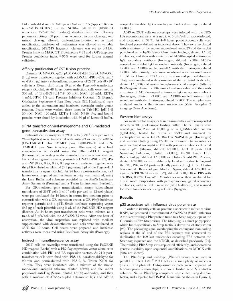
Ltd.) embedded into GPS-Explorer Software 3.5 (Applied Biosys-
tems/MDS SCIEX,) on the NCBInr [20100119 (10348164
sequences; 3529470745 residues)] database with the following
parameter settings: 50 ppm mass accuracy, trypsin cleavage, one
missed cleavage allowed, carbamidomethylation set as fixed
modification, oxidation of methionines was allowed as variable
modification, MS/MS fragment tolerance was set to 0.3 Da.
Protein hits with MASCOT Protein score $83 and a GPS Explorer
Protein confidence index $95% were used for further manual
validation.
Affinity purification of GST-fusion proteinsPlasmids pCMV-GST-p23, pCMV-GST-EF1a or pCMV-GST
(1 mg) were transfected together with pcDNA3.1-PB1, -PB2, and/
or -PA (1 mg) into a subconfluent monolayer of 293T cells (86105
cells in a 35-mm dish) using 10 ml of the Fugene-6 transfection
reagent (Roche). At 48 hours post-transfection, cells were lysed in
300 mL of Tris-HCl [pH 7.4] 50 mM, NaCl 120 mM, EDTA
1 mM, NP40 1% and Protease Inhibitor Cocktail 1X (Sigma).
Gluthation Sepharose 4 Fast Flow beads (GE Healthcare) were
added to the supernatant and incubated overnight under gentle
rotation. Beads were washed three times in Tris-HCl [pH 7.4]
50 mM, NaCl 120 mM, EDTA 1 mM, NP40 1%, and bound
proteins were eluted by incubation with 30 ml of Laemmli buffer.
siRNA transfection,viral-minigenome and GR-mediatedgene transactivation assay
Subconfluent monolayers of 293T cells (26105 cells per well in
24-well-plates) were transfected with anti-p23 or control siRNAs
(ON-TARGET plus SMART pool L-004496-00 and ON-
TARGET plus Non targeting pool, Dharmacon) at a final
concentration of 25 nM using the DharmaFECT reagent
(Dharmacon) according to the manufacturer’s recommendations.
For viral minigenome assays, plasmids pcDNA3.1-PB1, -PB2, -PA
and -NP (0.25, 0.25, 0.25, 0.5 mg) were transfected together with
the pPR7-FluA-Luc plasmid (0.1 mg) using 5 ml of the Fugene-HD
transfection reagent (Roche). At 24 hours post-transfection, cell
lysates were prepared and luciferase activity was measured, using
the Lysis Buffer and substrate provided in the Renilla Luciferase
Assay System kit (Promega) and a Tecan luminometer (Berthold).
For GR-mediated gene transactivation assays, subconfluent
monolayers of 293T cells (46105 cells per well in 12-well-plates)
were pre-incubated for 16 hours in serum free medium prior to
cotransfection with a GR expression vector, a GR-Firefly luciferase
reporter plasmid and a pTK-Renilla luciferase expressing vector
(0.5 mg of each plasmid) using 5 mL of the FuGENE HD reagent
(Roche). At 24 hours post-transfection cells were infected at a
m.o.i. of 5 pfu/cell with the A/WSN/33 virus. After one hour of
adsorption, the viral suspension was replaced with medium
supplemented with dexamethasone and cells were incubated at
35uC for 10 hours. Cell lysates were prepared and luciferase
activities were measured using Luciferase Assay kits (Promega).
Indirect immunofluorescence assay293T cells on coverslips were transfected using the FuGENE
HD reagent (Roche) with a PB2-flag expression vector alone or in
combination with PB1 and PA expression vectors. At 24 h post-
transfection cells were fixed with PBS-4% paraformaldehyde for
20 min and permeabilized with PBS-0.1% Triton X100 for
15 min. They were incubated with a mixture of the mouse
monoclonal anti-p23 (Abcam, diluted 1/250) and the rabbit
polyclonal anti-Flag (Sigma, diluted 1/400) antibodies, and then
with a mixture of AF555-coupled anti-mouse IgG and AF488
coupled anti-rabbit IgG secondary antibodies (Invitrogen, diluted
1/1000).
A549 or 293T cells on coverslips were infected with the PB2-
HA recombinant virus at a m.o.i. of 5 pfu/cell or mock-infected,
and incubated at 37uC. At 4–8 hours post-infection, cells were
fixed and permeabilized as indicated above. They were incubated
with a mixture of the mouse monoclonal anti-p23 and the rabbit
polyclonal anti-Hsp90 (Santa Cruz Biotechnology, diluted 1/100)
antibodies, and then with a mixture of AF640-coupled anti-mouse
IgG secondary antibody (Invitrogen, diluted 1/500), AF555-
coupled anti-rabbit IgG secondary antibody (Invitrogen, diluted
1/500), and AF488-coupled anti-HA antibody (Invitrogen, diluted
1/200). Alternatively, cells were incubated with dexamethasone
10 nM for 1 hour at 37uC prior to fixation and permeabilization.
They were incubated with a mixture of the rat anti-HA (Roche,
diluted 1/1,000) and mouse anti-glucocorticoid receptor (Affinity
BioReagents, diluted 1/300) monoclonal antibodies, and then with
a mixture of AF555-coupled anti-mouse IgG secondary antibody
(Invitrogen, diluted 1/1,000) and AF488-coupled anti-rat IgG
secondary antibody (Invitrogen, diluted 1/500). The samples were
analyzed under a fluorescence microscope (Zeiss Axioplan 2
imaging- Zeiss ApoTome).
Western-blot assaysFor western blot assays, cells in 35-mm dishes were resuspended
directly in 300 ml of sample loading buffer. The cell lysates were
centrifuged for 2 mn at 16,000 g on a QIAShredder column
(QIAGEN), heated for 3 min at 95uC and analyzed by
electrophoresis on a 4–12% Bis-Tris NuPAGE gel (Invitrogen)
and western blotting using PVDF membranes. The membranes
were incubated overnight at 4uC with primary antibodies directed
against p23 (Abcam, diluted 1/1,000), GST (Upstate Cell
Signalling Solutions, diluted 1/4,000), Hsp90 (Santa Cruz
Biotechnology, diluted 1/1,000) or Histone3 (ab1791, Abcam,
diluted 1/3,000), or with rabbit polyclonal serum directed against
the PB1, PB2, or PA proteins (kindly provided by J. Ortin, Centro
Nacional de Biotecnologia, Madrid, Spain, diluted 1/5,000) or
against A/PR/8/34 virions ([22], diluted 1/10,000) in PBS with
1% BSA, 0.25% Tween20. Membranes were then incubated for
1 h at room temperature with peroxydase-conjugated secondary
antibodies, with the ECL+ substrate (GE Healthcare), and scanned
for chemiluminescence using a G-Box (Syngene).
Results
p23 associates with influenza virus polymeraseIn order to identify cellular proteins associated to influenza virus
RNPs, we produced a recombinant A/WSN/33 (WSN) influenza
A virus expressing a PB2 protein fused to a Strep-tag epitope at the
C-terminus (PB2-Strep virus). The Strep-tag is a short polypeptide
which binds specifically to Strep-tactin, a derivative of streptavidin
[23]. The packaging signal overlapping the coding and non-coding
regions at the 59 end of the PB2 segment was conserved by
duplicating the 109 last nucleotides encoding PB2 between the
Strep-tag sequence and the 59NCR, as described previously [24].
The resulting PB2-Strep virus replicated efficiently, and showed no
genetic instability upon sequential amplifications on MDCK cells
(data not shown).
The PB2-Strep and wild-type (PB2-wt) viruses were used in
parallel to infect 46109 293T cells at a multiplicity of infection
(m.o.i.) of 5 pfu/cell. Cytoplasmic extracts were prepared at
6 hours post-infection (hpi), and were loaded onto Strep-tactin
columns. Native PB2-Strep complexes were eluted using desthio-
biotin, and subjected to SDS-PAGE analysis. As shown in Figure 1,
p23 Association with Influenza Virus Polymerase
PLoS ONE | www.plosone.org 3 August 2011 | Volume 6 | Issue 8 | e23368

Sypro-Ruby staining of the gel revealed a number of bands which
were present in the sample derived from cells infected with the
PB2-Strep virus, but not from cells infected with the PB2-wt virus.
Slices of the gel were sent to Institut Pasteur Proteomics Facility.
As expected, slices corresponding to the major bands correspond-
ed to the PB2, PB1, PA and NP viral proteins (Figure 1), which
was in agreement with our previous observations using PB2-Strep
viruses [24,25]. The slice containing PB2 and PB1 was also found
to contain the Hsp90 protein, whereas a band of lower molecular
weight corresponded to the p23 co-chaperone of Hsp90. Western-
blot analysis using anti-Hsp90 and anti-p23 antibodies confirmed
that Hsp90 and p23 were specifically co-purified with PB2-Strep
complexes (Figure 1).
Co-purification assays were then performed using a GST-p23
fusion protein transiently expressed in 293T cells. A pCMV-GST-
p23 expression vector was transfected in 293T cells together with
pcDNA3.1 expression vectors for the WSN-PB1, -PB2 or -PA
proteins, separately or in combination. Similar to p23, EF1-a is an
abundant, constitutively expressed, cytoplasmic protein, and
therefore a GST-EF1a construct was used in parallel as a control.
At 48 h post-transfection, total cell extracts were prepared and
incubated with glutathion beads as described under the Materials
and Methods section. The GST-p23 complexes were washed,
eluted using Laemmli buffer and subjected to western-blot
analysis. As expected the Hsp90 protein was co-purified with the
GST-p23 protein, but not with the GST-EF1a protein (Figure 2).
Each of the PB1, PB2 and PA subunits of the polymerase complex
bound specifically to the GST-p23 protein, whether expressed
alone, or in combination with one or both of the other subunits
(Figure 2).
Viral replication is not impaired in p23-deficient culturedcells
To investigate the functional relevance of p23 interaction with
the influenza polymerase complex in the replication cycle of
influenza viruses, we compared viral growth on mouse embryonic
fibroblasts either derived from p23-deficient mice (p232/2) or
from control wild-type mice (wt) [21]. Following infection at a
m.o.i. of 1023 pfu/cell, viral titers in the culture supernatants were
determined at different times post-infection by plaque assay on
MDCK cells. The WSN virus replicated at a similar rate on p232/2
and wt cells, titers of 107–108 pfu/ml being observed at 48 hpi
(Figure 3A). The same observations were made when the cells were
incubated at 33uC or 39uC instead of 35uC upon infection, or when
a recombinant virus expressing the PB1, PB2, PA and NP proteins
derived from the A/Paris/908/97 (H3N2) human isolate was used
instead of the A/WSN/33 virus (data not shown).
To further document the effect of p23 depletion on influenza
virus replication, we performed single-cycle growth assays on
Figure 1. Co-purification of influenza virus polymerase andp23 upon infection of 293T cells with a PB2-Strep-WSN virus.293T cells were infected with the WSN (WT) or WSN-PB2-Strep (Strep)virus at a m.o.i. of 5 for 6 hours. Cell lysates were loaded onto a Strep-tactin column, and bound proteins were eluted in desthiobiotineelution buffer. Eluted proteins were analysed on a 4–15% polyacryl-amide gel and stained with Sypro-Ruby. Proteins from individual gelslices were analysed by mass spectrometry (MS). For confirmation,western blot analysis of eluted proteins was performed usingantibodies specific for the PB2, Hsp90 and p23 proteins.doi:10.1371/journal.pone.0023368.g001
Figure 2. Co-purification of a GST-p23 fusion protein and the influenza virus polymerase subunits upon co-expression in 293Tcells. 293T cells were co-transfected with expression plasmids for GST-p23 or GST-EF1a fusion proteins, together with expression plasmids for theWSN-PB1, WSN-PB2, and/or WSN-PA proteins. At 48 hours post-transfection, cell lysates were incubated with Glutathion Sepharose beads overnight,and bound proteins were eluted in Laemmli buffer. Eluted proteins were analysed by western blot using antibodies specific for the GST, Hsp90, p23,PB1, PB2 and PA proteins.doi:10.1371/journal.pone.0023368.g002
p23 Association with Influenza Virus Polymerase
PLoS ONE | www.plosone.org 4 August 2011 | Volume 6 | Issue 8 | e23368

p232/2 and wt cells. Following infection with the WSN virus at a
high m.o.i. of 10 pfu/cell, total cell extracts were prepared at
various times post-infection and analyzed by western-blot using a
polyclonal serum to detect the viral NP and M1 viral proteins, or
cells were fixed and analyzed by immunofluorescence to detect the
PB2 protein. As shown in Figure 3B the NP and M1 proteins
steady-state levels were similar in p232/2 and wt cells. The
nuclear accumulation of the PB2 viral polymerase subunit was not
delayed in p232/2 cells (Figure S1). The viral titers in the
supernatants of p232/2 and wt cells were in the same range, and
reached 106–107 pfu/ml at 48 hpi (data not shown).
Single-cycle growth assays were also performed in 293T cells
transiently transfected with anti-p23 siRNAs. The steady-state
level of p23, as evaluated by western-blot analysis of serial dilutions
of total cell extracts, showed a significant reduction in 293T
transfected with the anti-p23 siRNAs as compared to the control
siRNAs (Figure 3C, middle panel). Following infection with the
WSN virus at a high m.o.i. of 10 pfu/cell, accumulation of the NP
and M1 proteins occurred at the same rate in anti-p23 and control
siRNA-treated cells (Figure 3C, upper panel).
The viral polymerase activity was assayed in anti-p23 and
control siRNA-treated 293T cells, by co-expressing transiently the
PB1, PB2, PA and NP proteins of WSN together with an
influenza-like RNA containing the luciferase reporter gene. The
efficiency with which the influenza-like RNA underwent tran-
scription/replication, as monitored by the levels of luciferase
activity in transfected cell extracts, was similar in both types of cells
(data not shown).
p23 relocalizes to the nucleus in influenza virus-infectedcells
The subcellular localization of p23 in influenza virus- and
mock-infected A549 and 293T cells was compared. A recombi-
nant WSN virus expressing a PB2 protein fused with the HA tag at
the C-terminus was used in these experiments, to allow
simultaneous labeling of the viral PB2 and cellular p23 and
Figure 3. Influenza virus replication on p232/2 and wild-type mouse embyonic fibroblasts. A. Subconfluent monolayers of p232/2 andwild-type mouse embryonic fibroblasts (MEFs) were infected at a m.o.i. of 1023 pfu/cell with the WSN virus and incubated for 72 h at 37uC. At theindicated time-points, supernatants were harvested and viral titers were determined by plaque assays on MDCK cells. The mean values 6 SD from 3independent experiments are shown. The horizontal dotted line represents the limit of detection in the plaque assays. B. Subconfluent monolayers ofp232/2 and wild-type MEFs were infected at a m.o.i. of 10 pfu/cell with the WSN virus and incubated for 24 h at 37uC. Total cell lysates prepared atthe indicated time-points were analysed by western blot, using an anti-p23 antibody, a polyclonal serum directed against the A/PR/8/34 virus whichenables detection of the NP and M1 proteins of WSN, and a polyclonal anti-Histone3 antibody. Data are representative of two independentexperiments. C. 293T cells were transfected with anti-p23 or control siRNAs. At 48 hours post-transfection, they were infected at a m.o.i. of 10 pfu/cellwith the WSN virus and incubated for 24 h at 37uC. Total cell lysates prepared at the indicated time-points were analysed by western blot, as in B.doi:10.1371/journal.pone.0023368.g003
p23 Association with Influenza Virus Polymerase
PLoS ONE | www.plosone.org 5 August 2011 | Volume 6 | Issue 8 | e23368

Hsp90 proteins. The anti-HA antibody did not recognize the
WSN hemagglutinin, as expected from the fact that the
YPYDVPDY sequence of the HA tag is not present in this
hemagglutinin, and as demonstrated by the background levels of
fluorescence measured on control cells infected with the wild-type
WSN virus (Figure S2). In uninfected A549 and 293T cells, p23
and Hsp90 were predominantly detected in the cytoplasm
(Figure 4A and 4B, respectively, lower panels). In infected cells,
relocalisation of p23 to the nucleus became detectable at 4 hpi
(data not shown) and was very obvious at 6–8 hpi (Figure 4A and
4B, upper panels). Relocalisation of Hsp90 was also observed, in
agreement with previously published data [5], but was less
pronounced as compared to p23 relocalisation (Figure 4A and
4B, upper panels).
The subcellular localization of p23 was then examined in
transfected cells transiently expressing the viral polymerase
subunits. Relocalisation of p23 to the nucleus was clearly observed
in A549 or 293T cells transiently expressing the PB2 subunit,
either alone or in combination with the PB1 and PA subunits
(Figure 5A and 5B, respectively). Nuclear relocalisation of p23 was
also observed in 293T cells expressing the PA subunit alone but
not in cells expressing the PB1 subunit alone (data not shown),
which was consistent with the fact that unlike PA, PB1 is not
efficiently imported in the nucleus when expressed on its own [26].
Nuclear relocalisation of p23 was not observed in cells expressing
the viral nucleoprotein, although the nucleoprotein was detected
in the nucleus as well as in the cytoplasm of transfected cells
(Figure S3). Overall, these observations supported the hypothesis
that the specific association of p23 with the viral polymerase
subunits is driving p23 relocalisation to the nucleus in infected
cells.
Glucocorticoid-receptor mediated signalling is impairedin influenza virus infected cells
As a component of the Hsp90 chaperone complex, p23 plays a
complex role in the maturation and function of the glucocorticoid
receptor (GR) (reviewed in [16]). In p23-deficient cells, the GR-
mediated gene transactivation is impaired, which coincides with a
delayed nuclear translocation of the GR in response to
dexamethasone [21]. We hypothetized that the strong relocalisa-
Figure 4. Relocalization of p23 to the nucleus in influenza virus-infected cells. A549 (A) or 293T (B) cells were infected at a m.o.i. of 10 pfu/cell with the WSN-PB2-HA virus (upper panels) or mock-infected (lower panels). At 8 hpi (A) or 6 hpi (B), cells were fixed, permeabilized, and stainedwith antibodies specific for the HA tag (PB2), and for the p23 and the Hsp90 proteins. Samples were analyzed under a fluorescence microscope (ZeissAxioplan 2 Imaging - Zeiss ApoTome). A merge of the signals corresponding to DAPI (blue), HA (green) and p23 (red) is shown. Data arerepresentative of three independent experiments.doi:10.1371/journal.pone.0023368.g004
p23 Association with Influenza Virus Polymerase
PLoS ONE | www.plosone.org 6 August 2011 | Volume 6 | Issue 8 | e23368

tion of p23 observed in influenza-virus infected cells might have an
impact on GR signalling. To evaluate GR-mediated gene
transactivation in influenza-infected vs mock-infected cells, 293T
cells were cotransfected with a GR expressing plasmid, a reporter
plasmid containing the Firefly-luciferase gene under the control of a
minimal TK promoter and GR-responsive elements, and a Renilla-
luciferase expressing construct. At 48 hours post-transfection, they
were infected with WSN at a m.o.i. of 5 pfu/ml, or mock-infected.
After one hour of adsorption, the viral inoculum was removed, and
replaced with fresh medium either supplemented with dexameth-
asone or not. After 10 hours of stimulation, cell lysates were
prepared and Firefly-luciferase activities were determined and
normalized with respect to Renilla-luciferase activities. As shown in
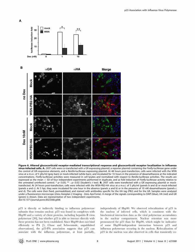
Figure 6A, the dexamethasone-induced transactivation of the
Firefly-luciferase gene in infected cells (12- and 19-fold at 1 nM and
10 nM dexamethasone, respectively) was reduced as compared to
in mock-infected cells (28- and 25-fold at 1 nM and 10 nM
dexamethasone, p,0.02 and p,0.05, respectively). The subcel-
lular localisation of the GR was examined in WSN-PB2-HA- and
in mock-infected 293T cells, upon incubation in the absence or in
the presence of 10 nM dexamethasone. An indirect immunoassay
was performed at 5 hpi, using a mixture of an anti-HA and an
anti-GR antibody. Uninfected cells generally showed cytoplasmic
or cytoplasmic and nuclear GR staining in the absence of
dexamethasone (Figure 6B, panel a), and predominantly nuclear
GR upon stimulation with dexamethasone (Figure 6B, panel c).
Strikingly, infected cells showed nuclear relocalisation of the GR in
the absence as well as in the presence of dexamethasone
(Figure 6B, panels b and d, respectively). Sequestration of the
GR in the nucleus in its hormone-free conformation could
contribute to the impairment of GR-mediated signalling by
reducing the pool of cytoplasmic GR available for hormone
recognition.
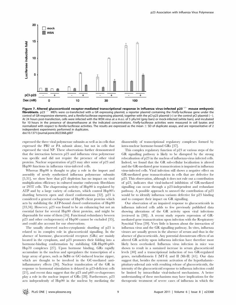
We finally asked whether the observed reduction of GR-
mediated gene transactivation in influenza-infected vs mock-
infected cells was mediated by p23. To this end, the luciferase
reporter assay described above was repeated using p232/2 MEFs,
either transfected with a p23 expression plasmid or mock-
transfected. Upon infection, cells were incubated in the presence
of 10 to 100 nM dexamethasone. In mock-infected cells,
transactivation of the Firefly-luciferase gene increased with the
dose of dexamethasone and was stronger in the presence of the
p23 expression plasmid (Figure 7, white bars), which was in
agreement with the known functions of p23 in GR-mediated
signalling [21,27,28,29]. A negative effect of viral infection on
transactivation of the Firefly-luciferase gene in response to
dexamethasone was clearly observed, in the presence as well as
in the absence of the p23 expression plasmid (Figure 7, grey bars).
These data indicated that the observed viral effect could occur
through a p23-independent pathway.
Discussion
We used a recombinant influenza virus expressing a PB2
protein fused to a Strep-tag for the co-purification and
identification of interacting host factors during the course of viral
infection. The Strep-tag has been described as an appropriate tag
to purify protein complexes from crude extracts under mild elution
conditions and with good yields [23]. Indeed, PB2-Strep viruses
allowed co-purification of PB2 and PB2-associated viral proteins
such as PB1, PA and NP (this study, and [24]), as well as a number
of cellular proteins that have already been identified as cellular
interactors of the viral polymerase or nucleoprotein, such as the
RNA polymerase II large subunit [10,24], Hsp90 (this study),
Hsp70, and actin (data not shown). The PB2-Strep virus is
potentially a useful tool to explore the spatial and temporal
dynamics of these protein interactions.
We found that the p23 cochaperone of Hsp90 was associated
with PB2 in cytoplasmic extracts from 293T cells infected with the
WSN-PB2-Strep virus. A transient co-expression/purification
assay confirmed that a GST-p23 fusion protein, but not a control
GST-EF1a fusion protein, bound to each of the PB1, PB2 and PA
polymerase subunits. In both experimental systems, the elution
samples containing the p23 and polymerase subunit(s) also
contained the Hsp90 chaperone, in agreement with existing data
showing PB2-Hsp90 and p23-Hsp90 interactions [5,16]. Whether
Figure 5. Relocalization of p23 to the nucleus of cellstransiently expressing influenza virus polymerase subunits.A549 (A) or 293T (B) cells were transfected with a WSN-PB2-Flagexpression plasmid alone or in combination with PB1 and PA expressionplasmids, or mock-transfected. At 24 hours post-transfection, cells werefixed, permeabilized, and stained with antibodies specific for the Flagtag (PB2) and for the p23 protein. Samples were analyzed under afluorescence microscope (Zeiss Axioplan 2 Imaging - Zeiss ApoTome). Amerge of the signals corresponding to DAPI (blue), Flag (green) and p23(red) is shown. Data are representative of two independent experi-ments.doi:10.1371/journal.pone.0023368.g005
p23 Association with Influenza Virus Polymerase
PLoS ONE | www.plosone.org 7 August 2011 | Volume 6 | Issue 8 | e23368
p23 is directly or indirectly binding to influenza polymerase
subunits thus remains unclear. p23 was found in complexes with
Hsp90 and a variety of client proteins, including hepatitis B virus
polymerase [30], but whether p23 is able to interact directly with
these proteins has not been established. Since Hsp90 does not bind
efficiently to PA [5, Chase and Schwemmle, unpublished
observations], the p23-PA association suggests that p23 can
associate with the influenza polymerase, at least partially,
independently of Hsp90. We observed relocalization of p23 in
the nucleus of infected cells, which is consistent with the
biochemical interaction data as the viral polymerase accumulates
in the nuclear compartment. Nuclear retention was more
pronounced for p23 than for Hsp90, which might be indicative
of some Hsp90-independent interaction between p23 and
influenza polymerase occuring in the nucleus. Relocalization of
p23 in the nucleus was also observed in cells that transiently co-
Figure 6. Altered glucocorticoid receptor-mediated transcriptional response and glucocorticoid receptor localization in influenzavirus-infected cells. A. 293T cells were co-transfected with a GR expressing plasmid, a reporter plasmid containing the Firefly-luciferase gene underthe control of GR-responsive elements, and a Renilla-luciferase expressing plasmid. At 48 hours post-transfection, cells were infected with the WSNvirus at a m.o.i. of 5 pfu/ml (grey bars) or mock-infected (white bars), and incubated for 10 hours in the presence of dexamethasone at the indicatedconcentrations. Firefly-luciferase activities were measured in cell lysates and normalized with respect to Renilla-luciferase activities. The results areexpressed as the mean 6 SD of four independent experiments performed in duplicate, and as fold induction of Firefly-luciferase activity relative tothe untreated uninfected control. * p,0.05; ** : p,0.02 (Student’s t test). B. 293T cells were transfected with a GR expressing plasmid or mock-transfected. At 24 hours post-transfection, cells were infected with the WSN-PB2-HA virus at a m.o.i. of 5 pfu/ml (panels b and d) or mock-infected(panels a and c). At 5 hpi, they were incubated for one hour in the absence (panels a and b) or in the presence of 10 nM dexamethasone (panels cand d). The cells were then fixed, permeabilized, and stained with antibodies specific for the HA tag (PB2) and for the GR. Samples were analyzedunder a fluorescence microscope (Zeiss Axioplan 2 Imaging - Zeiss ApoTome). A merge of the signals corresponding to DAPI (blue), HA (red) and GR(green) is shown. Data are representative of two independent experiments.doi:10.1371/journal.pone.0023368.g006
p23 Association with Influenza Virus Polymerase
PLoS ONE | www.plosone.org 8 August 2011 | Volume 6 | Issue 8 | e23368
expressed the three viral polymerase subunits as well as in cells that
expressed the PB2 or PA subunit alone, but not in cells that
expressed the viral NP. These observations further demonstrated
that the interaction between p23 and influenza virus polymerase
was specific and did not require the presence of other viral
proteins. Nuclear sequestration of p23 may alter some of p23 and
Hsp90 functions in influenza virus-infected cells.
Whereas Hsp90 is thought to play a role in the import and
assembly of newly synthetized influenza polymerase subunits
[5,31], we show here that p23 depletion has no impact on viral
multiplication efficiency in cultured murine embryonic fibroblasts
or 293T cells. The chaperoning activity of Hsp90 is regulated by
ATP and by a large variety of cofactors, which control Hsp90’s
shuttling between open and closed conformations [32]. p23 is
considered a general cochaperone of Hsp90 client proteins which
acts by stabilizing the ATP-bound closed conformation of Hsp90
[33,34]. However, p23 was found to be an enhancing but not an
essential factor for several Hsp90 client proteins, and might be
dispensable for some of them [16]. Functional redundancy beween
p23 and other cochaperone(s) of Hsp90 cannot be excluded [35],
and could also account for our observations.
The usually observed nucleo-cytoplasmic shuttling of p23 is
related to its complex role in glucocorticoid signalling. In the
absence of hormone, glucocorticoid receptors (GRs) are mainly
located in the cytoplasm, and p23 promotes their folding into a
hormone-binding conformation by stabilizing GR-Hsp90-p60-
Hsp70 complexes [27]. Upon hormone binding, GRs rapidly
accumulate in the nucleus and upregulates the transcription of a
large array of genes, such as IkBa or GC-induced leucine zipper,
which are thought to be involved in the GC-mediated anti-
inflammatory effects [36]. Nuclear translocation of the GR in
response to hormonal stimulation is delayed in p23-deficient cells
[21], and recent data suggest that the p23 and p60 co-chaperones
play a role in the nuclear import of GRs [28]. Furthermore, p23
acts independently of Hsp90 in the nucleus by mediating the
disassembly of transcriptional regulatory complexes formed by
intra-nuclear hormone-bound GRs [37].
This complex regulatory function of p23 at various steps of the
GR signalling pathway is likely to be disrupted by the strong
relocalisation of p23 in the nucleus of influenza-virus infected cells.
Indeed, we found that the GR sub-cellular localisation is altered
and the GR-mediated gene transactivation is impaired in influenza
virus-infected cells. Viral infection still shows a negative effect on
GR-mediated gene transactivation in cells that are defective for
p23. This observation, although it does not rule out a contribution
of p23, indicates that viral-induced inhibition of GR-mediated
signalling can occur through a p23-independent and redundant
pathway. A possible approach to unravel the contribution of p23
would be to identify influenza variants defective for p23 binding
and to compare their impact on GR signalling.
Our observation of an impaired response to glucocorticoids in
influenza infected cells adds to few previously published data
showing alterations of the GR activity upon viral infections
(reviewed in [38]). A recent study reports repression of GR-
mediated gene transactivation upon infection with the Respiratory
Syncitial Virus [39]. Very little is known about the interaction of
influenza virus and the GR signalling pathway. In vitro, influenza
viruses are usually grown in the absence of serum and thus in the
absence of glucocorticoids. Any potential downstream effects of an
altered GR activity upon influenza infection have therefore most
likely been overlooked. Influenza virus infection in mice was
shown to result in a sustained increase in serum glucocorticoid
levels [40] and a transcriptional induction of two GR-regulated
genes, metallothionein I (MT-I) and II (Mt-II) [41]. Our data
suggest that, besides the systemic activation of the hypothalamic-
pituitary-adrenal axis with resulting release of glucocorticoids, the
intensity of the glucocorticoid response to influenza infection could
be limited by intracellular viral-induced mechanisms. A better
understanding of these mechanisms may have implications for the
therapeutic treatment of severe cases of influenza in which the
Figure 7. Altered glucocorticoid receptor-mediated transcriptional response in influenza virus-infected p232/2 mouse embyonicfibroblasts. p232/2 MEFs were co-transfected with a GR expressing plasmid, a reporter plasmid containing the Firefly-luciferase gene under thecontrol of GR-responsive elements, and a Renilla-luciferase expressing plasmid, together with the pCI-p23 plasmid (+) or the control pCI plasmid (2).At 24 hours post-transfection, cells were infected with the WSN virus at a m.o.i. of 5 pfu/ml (grey bars) or mock-infected (white bars), and incubatedfor 10 hours in the presence of dexamethasone at the indicated concentrations. Firefly-luciferase activities were measured in cell lysates andnormalized with respect to Renilla-luciferase activities. The results are expressed as the mean 6 SD of duplicate assays, and are representative of 2independent experiments performed in duplicates.doi:10.1371/journal.pone.0023368.g007
p23 Association with Influenza Virus Polymerase
PLoS ONE | www.plosone.org 9 August 2011 | Volume 6 | Issue 8 | e23368

balance between pro-inflammatory and anti-inflammatory cyto-
kines is a major physiopathological parameter [42].
Supporting Information
Figure S1 Subcellular localisation of the PB2 proteinupon influenza virus infection of p232/2 and wild-typemouse embyonic fibroblasts. p232/2 and wild-type p23+/+
mouse embryonic fibroblasts (MEFs) were infected at a m.o.i. of
10 pfu/cell with the WSN-PB2-HA virus. At 5 hpi, cells were
fixed, permeabilized and stained with antibodies specific for the
HA tag (PB2) and for the Hsp90 protein. Samples were analyzed
under a fluorescence microscope (Zeiss Axioplan 2 Imaging - Zeiss
ApoTome). A merge of the signals corresponding to DAPI (blue),
HA (red) and Hsp90 (green) is shown.
(TIF)
Figure S2 Specific recognition of the HA-tag and not theWSN virus hemagglutinin by the anti-HA antibody. A549
(A) or 293T (B) cells were infected at a m.o.i. of 10 pfu/cell with
the WSN-PB2-HA virus (upper panels) or the WSN wild-type
virus (lower panels). At 8 hpi (A) or 6 hpi (B), cells were fixed,
permeabilized, and stained with antibodies specific for the HA tag
(PB2) and for the p23 protein. Samples were analyzed under a
fluorescence microscope (Zeiss Axioplan 2 Imaging - Zeiss
ApoTome). A merge of the signals corresponding to DAPI (blue),
HA (green) and p23 (red) is shown.
(TIF)
Figure S3 Subcellular localisation of p23 in 293T cellstransiently expressing the viral PB2 or NP protein. 293T
cells were transfected with a plasmid encoding NP (middle pannel),
PB2-Flag (lower panel) or mock-transfected (upper panel). At
24 hpi cells were fixed, permeabilized, and stained with antibodies
specific for the Flag tag (PB2) or the NP protein, together with an
anti-p23 antibody. Samples were analyzed under a fluorescence
microscope (Zeiss Axioplan 2 Imaging - Zeiss ApoTome). A merge
of the signals corresponding to DAPI (blue), Flag or NP (green)
and p23 (red) is shown.
(TIF)
Acknowledgments
We are grateful to G. Brownlee (Oxford University, UK), J. Ortin (Centro
Nacional de Biotecnologia, Madrid, Spain), Y. Jacob (Institut Pasteur,
Paris, France) and D. Toft (Mayo Clinic, Rochester, USA) for providing
plasmids and reagents. We thank P. Lenormand at the Plateforme de
Proteomique of Institut Pasteur for mass spectrometry analysis, the
Plateforme de sequencage and Plateforme d’Imagerie Dynamique at
Institut Pasteur for their expertise, and S. van der Werf for her support.
Author Contributions
Conceived and designed the experiments: M-AR-W EG GC ESA NN.
Performed the experiments: XG M-AR-W EG GC ESA NN. Analyzed the
data: XG M-AR-W EG GC EdSA DP MS NN. Contributed reagents/
materials/analysis tools: DP MS NN. Wrote the paper: NN M-AR-W.
References
1. Engelhardt OG, Fodor E (2006) Functional association between viral andcellular transcription during influenza virus infection. Rev Med Virol 16:
329–345.
2. Deng T, Engelhardt OG, Thomas B, Akoulitchev AV, Brownlee GG, et al.(2006) Role of ran binding protein 5 in nuclear import and assembly of the
influenza virus RNA polymerase complex. J Virol 80: 11911–11919.
3. Tarendeau F, Boudet J, Guilligay D, Mas PJ, Bougault CM, et al. (2007)
Structure and nuclear import function of the C-terminal domain of influenzavirus polymerase PB2 subunit. Nat Struct Mol Biol 14: 229–233.
4. Gabriel G, Herwig A, Klenk HD (2008) Interaction of polymerase subunit PB2
and NP with importin alpha1 is a determinant of host range of influenza A virus.PLoS Pathog 4: e11.
5. Naito T, Momose F, Kawaguchi A, Nagata K (2007) Involvement of Hsp90 in
assembly and nuclear import of influenza virus RNA polymerase subunits. J Virol81: 1339–1349.
6. Plotch SJ, Bouloy M, Ulmanen I, Krug RM (1981) A unique cap(m7GpppXm)-
dependent influenza virion endonuclease cleaves capped RNAs to generate the
primers that initiate viral RNA transcription. Cell 23: 847–858.
7. Dias A, Bouvier D, Crepin T, McCarthy AA, Hart DJ, et al. (2009) The cap-
snatching endonuclease of influenza virus polymerase resides in the PA subunit.
Nature 458: 914–918.
8. Guilligay D, Tarendeau F, Resa-Infante P, Coloma R, Crepin T, et al. (2008)The structural basis for cap binding by influenza virus polymerase subunit PB2.
Nat Struct Mol Biol 15: 500–506.
9. Yuan P, Bartlam M, Lou Z, Chen S, Zhou J, et al. (2009) Crystal structure of anavian influenza polymerase PA(N) reveals an endonuclease active site. Nature
458: 909–913.
10. Engelhardt OG, Smith M, Fodor E (2005) Association of the influenza A virusRNA-dependent RNA polymerase with cellular RNA polymerase II. J Virol 79:
5812–5818.
11. Shih SR, Krug RM (1996) Novel exploitation of a nuclear function by influenza
virus: the cellular SF2/ASF splicing factor controls the amount of the essentialviral M2 ion channel protein in infected cells. Embo J 15: 5415–5427.
12. Read EK, Digard P (2010) Individual influenza A virus mRNAs show
differential dependence on cellular NXF1/TAP for their nuclear export. J GenVirol 91: 1290–1301.
13. Schneider J, Wolff T (2009) Nuclear functions of the influenza A and B viruses
NS1 proteins: do they play a role in viral mRNA export? Vaccine 27:6312–6316.
14. Elton D, Simpson-Holley M, Archer K, Medcalf L, Hallam R, et al. (2001)
Interaction of the influenza virus nucleoprotein with the cellular CRM1-mediated nuclear export pathway. J Virol 75: 408–419.
15. Neumann G, Hughes MT, Kawaoka Y (2000) Influenza A virus NS2 protein
mediates vRNP nuclear export through NES-independent interaction with
hCRM1. Embo J 19: 6751–6758.
16. Felts SJ, Toft DO (2003) p23, a simple protein with complex activities. Cell
Stress Chaperones 8: 108–113.
17. Fodor E, Devenish L, Engelhardt OG, Palese P, Brownlee GG, et al. (1999)Rescue of influenza A virus from recombinant DNA. J Virol 73: 9679–9682.
18. Dos Santos Afonso E, Escriou N, Leclercq I, van der Werf S, Naffakh N (2005)The generation of recombinant influenza A viruses expressing a PB2 fusion
protein requires the conservation of a packaging signal overlapping the codingand noncoding regions at the 59 end of the PB2 segment. Virology 341: 34–46.
19. Crescenzo-Chaigne B, Naffakh N, van der Werf S (1999) Comparative analysisof the ability of the polymerase complexes of influenza viruses type A, B and C to
assemble into functional RNPs that allow expression and replication ofheterotypic model RNA templates in vivo. Virology 265: 342–353.
20. Bunone G, Briand PA, Miksicek RJ, Picard D (1996) Activation of theunliganded estrogen receptor by EGF involves the MAP kinase pathway and
direct phosphorylation. Embo J 15: 2174–2183.
21. Grad I, McKee TA, Ludwig SM, Hoyle GW, Ruiz P, et al. (2006) The Hsp90
cochaperone p23 is essential for perinatal survival. Mol Cell Biol 26: 8976–8983.
22. Vignuzzi M, Gerbaud S, van der Werf S, Escriou N (2001) Naked RNA
immunization with replicons derived from poliovirus and Semliki Forest virusgenomes for the generation of a cytotoxic T cell response against the influenza A
virus nucleoprotein. J Gen Virol 82: 1737–1747.
23. Schmidt GM, Skerra A (2007) The Strep-tag system for one-step purification
and high-affinity detection or capturing of proteins. Nature Protocols 2:1528–1535.
24. Rameix-Welti MA, Tomoiu A, Dos Santos Afonso E, van der Werf S, Naffakh N
(2009) Avian Influenza A virus polymerase association with nucleoprotein, but
not polymerase assembly, is impaired in human cells during the course ofinfection. J Virol 83: 1320–1331.
25. Robb NC, Chase G, Bier K, Vreede FT, Shaw PC, et al. (2011) The influenza A
virus NS1 protein interacts with the nucleoprotein of viral ribonucleoprotein
complexes. J Virol 85: 5228–5231.
26. Fodor E, Smith M (2004) The PA subunit is required for efficient nuclearaccumulation of the PB1 subunit of the influenza A virus RNA polymerase
complex. J Virol 78: 9144–9153.
27. Dittmar KD, Demady DR, Stancato LF, Krishna P, Pratt WB (1997) Folding of
the glucocorticoid receptor by the heat shock protein (hsp) 90-based chaperonemachinery. The role of p23 is to stabilize receptor.hsp90 heterocomplexes
formed by hsp90.p60.hsp70. J Biol Chem 272: 21213–21220.
28. Echeverria PC, Mazaira G, Erlejman A, Gomez-Sanchez C, Piwien Pilipuk G,
et al. (2009) Nuclear import of the glucocorticoid receptor-hsp90 complexthrough the nuclear pore complex is mediated by its interaction with Nup62 and
importin beta. Mol Cell Biol 29: 4788–4797.
29. Lovgren AK, Kovarova M, Koller BH (2007) cPGES/p23 is required for
glucocorticoid receptor function and embryonic growth but not prostaglandin
E2 synthesis. Mol Cell Biol 27: 4416–4430.
p23 Association with Influenza Virus Polymerase
PLoS ONE | www.plosone.org 10 August 2011 | Volume 6 | Issue 8 | e23368

30. Hu J, Toft D, Anselmo D, Wang X (2002) In vitro reconstitution of functional
hepadnavirus reverse transcriptase with cellular chaperone proteins. J Virol 76:
269–279.
31. Chase G, Deng T, Fodor E, Leung BW, Mayer D, et al. (2008) Hsp90 inhibitors
reduce influenza virus replication in cell culture. Virology 377: 431–439.
32. Pearl LH, Prodromou C (2006) Structure and mechanism of the Hsp90
molecular chaperone machinery. Annu Rev Biochem 75: 271–294.
33. Ali MM, Roe SM, Vaughan CK, Meyer P, Panaretou B, et al. (2006) Crystal
structure of an Hsp90-nucleotide-p23/Sba1 closed chaperone complex. Nature
440: 1013–1017.
34. Karagoz GE, Duarte AM, Ippel H, Uetrecht C, Sinnige T, et al. (2011) N-
terminal domain of human Hsp90 triggers binding to the cochaperone p23. Proc
Natl Acad Sci U S A 108: 580–585.
35. Caplan AJ (2003) What is a co-chaperone? Cell Stress Chaperones 8: 105–107.
36. Clark AR (2007) Anti-inflammatory functions of glucocorticoid-induced genes.
Mol Cell Endocrinol 275: 79–97.
37. Freeman BC, Yamamoto KR (2002) Disassembly of transcriptional regulatory
complexes by molecular chaperones. Science 296: 2232–2235.38. Webster JI, Sternberg EM (2004) Role of the hypothalamic-pituitary-adrenal
axis, glucocorticoids and glucocorticoid receptors in toxic sequelae of exposure
to bacterial and viral products. J Endocrinol 181: 207–221.39. Hinzey A, Alexander J, Corry J, Adams KM, Claggett AM, et al. (2010)
Respiratory Syncytial Virus Represses Glucocorticoid Receptor-Mediated GeneActivation. Endocrinology.
40. Jamieson AM, Yu S, Annicelli CH, Medzhitov R (2010) Influenza virus-induced
glucocorticoids compromise innate host defense against a secondary bacterialinfection. Cell Host Microbe 7: 103–114.
41. Ghoshal K, Majumder S, Zhu Q, Hunzeker J, Datta J, et al. (2001) Influenzavirus infection induces metallothionein gene expression in the mouse liver and
lung by overlapping but distinct molecular mechanisms. Mol Cell Biol 21:8301–8317.
42. Peiris JS, Hui KP, Yen HL (2010) Host response to influenza virus: protection
versus immunopathology. Curr Opin Immunol 22: 475–481.
p23 Association with Influenza Virus Polymerase
PLoS ONE | www.plosone.org 11 August 2011 | Volume 6 | Issue 8 | e23368