influenza virus-host interactions and their modulation by ... · influenza virus-host interactions...
TRANSCRIPT
Institute for Molecular Medicine Finland – FIMM
and
Department of Bacteriology and Immunology
Haartman Institute, Faculty of Medicine
University of Helsinki
Finland
INFLUENZA VIRUS-HOST INTERACTIONS AND THEIR
MODULATION BY SMALL MOLECULES
Oxana Denisova
ACADEMIC DISSERTATION
To be presented with the permission of the Faculty of Medicine of the University of
Helsinki, for public examination in the Auditorium XII of the University Main building,
Fabianinkatu 33, on 9th May 2014, at 12 o’clock noon.
Helsinki 2014

SUPERVISOR Docent Denis Kainov
Institute for Molecular Medicine Finland
University of Helsinki
Finland
REVIEWERS Docent Maria Söderlund-Venermo
Department of Virology
Haartman Institute
University of Helsinki
Finland
Docent Eugene Makeyev
School of Biological Sciences
Nanyang Technological University
Singapore
OPPONENT Professor Stephan Ludwig
Institute of Molecular Virology
Centre for Molecular Biology of Inflammation
University of Münster
Germany
ISBN 978-952-10-9848-2 (paperback)
ISBN 978-952-10-9849-9 (PDF)
Picaset Oy
http://ethesis.helsinki.fi
Helsinki 2014

To my family

CONTENTS
ABBREVIATIONS ......................................................................................................... i
ORIGINAL PUBLICATIONS ....................................................................................... ii
ABSTRACT ................................................................................................................... iii
1. REVIEW OF THE LITERATURE ........................................................................ 1
1.1. Introduction ........................................................................................................ 1
1.2. Influenza A virus ................................................................................................ 1
1.2.1. Structure and classification................................................................................ 1
1.2.2. The life cycle of influenza virus ........................................................................ 3
1.2.3. Evolution of influenza viruses ........................................................................... 4
1.3. Prevention and treatment of influenza................................................................. 5
1.3.1. Novel virus-directed antiviral agents ................................................................. 7
1.3.1.1. M2 ion channel blockers .............................................................................. 7
1.3.1.2. Neuraminidase inhibitors.............................................................................. 8
1.3.1.3. Hemagglutinin inhibitors .............................................................................. 8
1.3.1.4. Polymerase inhibitors ................................................................................. 13
1.3.1.5. Non-structural protein 1 inhibitors .............................................................. 14
1.3.1.6. Nucleoprotein inhibitors ............................................................................. 15
1.3.1.7. Nucleic acid-based antiviral drugs .............................................................. 15
1.3.2. Host-directed antiviral agents .......................................................................... 16
1.3.2.1. Agents cleaving sialic acid ......................................................................... 17
1.3.2.2. Protease inhibitors ...................................................................................... 17
1.3.2.3. Vacuolar proton-ATPase inhibitors ............................................................ 19
1.3.2.4. Inhibitors of signaling pathways ................................................................. 20
1.3.2.5. Inhibitors of lipid metabolism .................................................................... 26
1.3.2.6. Inhibitors of nucleotide metabolism............................................................ 27
1.4. Combination antiviral therapy .......................................................................... 28
1.5. Drug repositioning and new formulations ......................................................... 29

2. AIMS OF THE STUDY ........................................................................................ 31
3. MATERIALS AND METHODS .......................................................................... 32
3.1. Cells, compounds and viruses (I – IV) .............................................................. 32
3.2. THCPSi nanoparticles loaded with SaliPhe (III) ............................................... 35
3.3. In vitro experiments ......................................................................................... 35
3.4. In vivo experiments .......................................................................................... 41
3.5. Ethics ............................................................................................................... 41
4. RESULTS AND DISCUSSION ............................................................................ 42
4.1. Identification of host factors involved in influenza virus replication ..................... 42
4.1.1. Assembly of library of compounds targeting host factors (I, II, IV) ................. 42
4.1.2. Method for searching potential antiviral agents (I, II, IV) ................................ 43
4.1.3. Identification of hit compounds (I, II, IV)........................................................ 44
4.2. Potential mechanisms of action of promising compounds (I, II, IV) ..................... 46
4.3. Effect of compounds on influenza virus-mediated cellular antiviral and pro-
inflammatory responses (I, II, IV) ............................................................................... 50
4.4. Spectrum of antiviral activities of compounds (I, IV, unpublished) ...................... 51
4.5. Development of resistance (unpublished) ............................................................. 53
4.6. Optimization of SaliPhe clinical utility (III) ......................................................... 54
5. FUTURE DIRECTIONS ...................................................................................... 56
6. CONCLUSIONS ................................................................................................... 57
ACKNOWLEDGEMENTS ......................................................................................... 58
REFERENCES ............................................................................................................. 60

i
ABBREVIATIONS
APAF1 apoptotic protease-activating
factor 1 Bad Bcl-2 antagonist of cell death
Bak Bcl-2 antagonist killer
Bax Bcl-2-associated X protein
Bcl-2 B-cell lymphoma-2 Bcl-w Bcl-2-like protein 2
Bcl-xL B-cell lymphoma-extra large
Bid Bcl-2 homology domain 3 (BH3)-interacting domain death
agonist
Bim Bcl-2-interacting mediator of cell death
BUNV Bunyamwera virus
CCL chemokine (C-C motif) ligand
CPE cytopathic effect CPSF30 cleavage and polyadenylation
specificity factor
CXCL chemokine (C-X-C motif) ligand GM-SCF granulocyte-macrophage colony-
stimulating factor
DENV Dengue virus
DHODH dihydroorotate dehydrogenase DHX9 ATP-dependent RNA
helicase A
EC50 half-maximal effective concentration
Echo6 human echovirus 6
FLII protein flightless-1 homologue H2B histone H2B
HA hemagglutinin
HCV hepatitis C virus
HIV human immunodeficiency virus HSV herpes simplex virus
IC50 half-maximal inhibitory
concentration IFN interferon
IL interleukin
IP immunoprecipitation IP-10 IFN-γ-induced protein 10
LRRFIP2 leucine-rich repeat flightless-
interacting protein 2
M1 matrix protein 1 M2 proton channel
MAPK mitogen-activated protein kinases
Mcl-1 myeloid cell leukemia-1
MeV Measles virus
mTOR mammalian target of
rapamycin
NA neuraminidase
NEP nuclear export protein
NF-κB nuclear factor kappa-light-
chain-enhancer of activated B
cells
NP nucleoprotein
NS1 non-structural protein 1
PA polymerase acid protein
PB1 N10 N-terminally truncated version
of the polypeptide from PB1
codon 40
PB1 polymerase basic protein 1
PB1-F2 protein encoded the +1 reading
frame of the PB1 gene
PB2 polymerase basic protein 2
PBMC peripheral blood mononuclear
cells
PI3K phosphoinositide 3-kinase
PSi porous silicon
SFV Semliki forest virus
SINV Sindbis virus
siRNA small interfering RNA
TBEV tick-borne encephalitis virus
THCPSi thermally hydrocarbonized
porous silicon
TLR4 Toll-like receptor 4
TNF-α tumor necrosis factor alpha
TOLLIP Toll-interacting protein
UACA uveal autoantigen with coiled-
coil domains and ankyrin
repeats
VACV Vaccinia virus
v-ATPase vacuolar proton-ATPase

ii
ORIGINAL PUBLICATIONS
This thesis is based on the following publications that are referred to in the text by
their Roman numerals. Additional unpublished data will be presented.
I. Denisova O.V., Kakkola L., Feng L., Stenman J., Nagaraj A., Lampe J., Yadav B.,
Aittokallio T., Kaukinen P., Ahola T., Kuivanen S., Vapalahti O., Kantele A.,
Tynell J., Julkunen I., Kallio-Kokko H., Paavilainen H., Hukkanen V., Elliott R.M.,
De Brabander J.K., Saelens X., Kainov D.E. (2012) Obatoclax, saliphenylhalamide
and gemcitabine inhibit influenza A virus infection. J Biol Chem. 287(42): 35324–32.
II. Kakkola L.*, Denisova O.V.*, Tynell J., Viiliäinen J., Ysenbaert T., Matos R.C.,
Nagaraj A., Öhman T., Kuivanen S., Paavilainen H., Feng L., Yadav B., Julkunen I.,
Vapalahti O., Hukkanen V., Stenman J., Aittokallio T., Verschuren E.W., Ojala P.M.,
Nyman T., Saelens X., Dzeyk K., Kainov D.E. (2013) Anticancer compound ABT-
263 accelerates apoptosis in virus infected cells and imbalances cytokine production
and lowers survival rates of infected mice. Cell Death Dis. 4: e742.
III. Bimbo L.M., Denisova O.V., Mäkilä E., Kaasalainen M., De Brabande J.K.,
Hirvonen J., Salonen J., Kakkola L., Kainov D.E., Santos H.A. (2013) Inhibition of
influenza A virus infection in vitro by saliphenylhalamide loaded porous silicon
nanoparticles. ASC Nano. 7(8): 6884-93.
IV. Denisova O.V., Virtanen S., Von Schantz-Fant C., Bychkov D., Desloovere J.,
Soderholm S., Theisen L., Tynell J., Ikonen N., Vashchinkina E., Nyman T.,
Matikainen S., Kallioniemi O., Julkunen I., Muller C.P., Saelens X., Verkhusha V.V.,
Kainov D.E. Akt inhibitor MK2206 prevents influenza A(H1N1)pdm09 virus
infection in vitro. Submitted.
* equal contribution

iii
ABSTRACT
Influenza viruses cause annual epidemics and pandemics which have serious
consequences for public health and global economy. The severity of infections with
influenza viruses can vary from asymptomatic to life-threatening viral pneumonias
frequently complicated by multi-organ failure and exacerbation of other underlying
conditions. Currently, four licensed anti-influenza drugs are available for the prevention
and treatment of influenza virus infections. However, resistance to the licensed
antivirals develops rapidly. Therefore, there is a need for next-generation antiviral
agents to combat influenza virus infections.
Recent advances in understanding influenza virus-host interactions have revealed
a number of host targets for potential antiviral interventions. In particular, basic cellular
functions, metabolic and biosynthesis pathways as well as the signaling cascades
essential for virus replication could be modulated by small-molecule inhibitors to block
virus infection. Moreover, temporal inhibition of these host functions will be less likely
to induce viral drug resistance. In addition, many of the inhibitors of cellular functions
are already approved or in clinical development for other diseases. Drug repurposing
will facilitate their introduction for treatment of viral infections, since the
pharmacokinetics and toxicity profile of these drugs are already known.
In this work, a library of small-molecule inhibitors targeting host factors and
potentially interfering with influenza virus infection was built and screened. Inhibitors
of vacuolar proton-ATPase (v-ATPase), Akt kinase, ribonucleotide reductase and the
anti-apoptotic B-cell lymphoma-2 (Bcl-2) family proteins showed antiviral activity in
vitro. However, ABT-263, an inhibitor of Bcl-2 family proteins, was ineffective in vivo.
SaliPhe, an inhibitor of v-ATPase, was the most potent antiviral agent and it was
effective against a broad range of influenza viruses and some other RNA viruses in
vitro, and against a mouse adapted influenza strain in vivo. In order to overcome the low
water solubility and high toxicity of SaliPhe, it was needed to optimize the drug’s
bioavailability using a porous silicon particle-based delivery system prior to embarking
on future clinical trials. The results presented in this study expand the understanding of
influenza virus-host interactions, and are important for rational approaches of
discovering new antiviral agents.

1
1. REVIEW OF THE LITERATURE
1.1. Introduction
Influenza viruses cause annual epidemics and pandemics with serious
consequences for public health and global economy. During the annual epidemics,
influenza viruses infect 5 – 15% of the human population resulting in 3 – 5 million
cases of severe illness and 250,000 – 500,000 death each year all around the world
(Shirey et al, 2013). In addition, occasionally strains to which humans are naïve appear
and cause pandemic outbreaks. During the past 100 years, five pandemic influenza
outbreaks have occurred: “Spanish flu” (H1N1) in 1918, “Asian flu” (H2N2) in 1957,
“Hong Kong flu” (H3N2) in 1968, “Russian flu” (H1N1) 1977, and “swine flu” (H1N1)
in 2009. In particular, the 1918 influenza pandemic affected almost 30% of the global
population and is believed to have killed over 50 million people (Johnson & Mueller,
2002).
Influenza viruses usually invade the epithelial cells of the upper respiratory tract
and cause acute respiratory disease. The typical influenza symptoms are fever,
headaches, chills, nasal congestion, sore throat and body aches. However, the highly-
pathogenic influenza A (H5N1) virus infects the lower respiratory tract and causes viral
pneumonia and acute respiratory distress syndrome. There may be multiple organ
failure, caused by intense induction of proinflammatory cytokines. Recently, a new
avian influenza A (H7N9) strain has been identified in China. The influenza H7N9 virus
was characterized by rapidly progressive pneumonia, respiratory failure, acute
respiratory distress syndrome, and fatal outcomes (39 deaths in 132 confirmed cases)
(Li et al, 2013).
1.2. Influenza A virus
1.2.1. Structure and classification
Influenza A viruses belong to the Orthomyxoviridae family which also includes
the influenza B and C types. The influenza viruses differ in several ways, i.e. genome
organization, variability of the surface glycoproteins, host range and pathogenicity.
Both influenza A and B viruses contain eight segments of negative-strand RNA,
whereas influenza C virus contains only seven segments (Cheung & Poon, 2007).

2
Influenza A virus has a wide range of hosts including domestic and wild birds, pigs,
horses, mink, seals, bats, as well as humans, while influenza virus types B and C infect
predominantly humans (Tong et al, 2013; Webster et al, 1992). Every year, influenza
virus types A and B cause epidemics in human population. However, only influenza A
viruses have been responsible for pandemic outbreaks.
Influenza A virus is an enveloped virus of 80-120 nm in diameter with eight
segments of negative-strand RNA encoding up to 15 proteins depending on the strain.
The proteins include hemagglutinin (HA), neuraminidase (NA), matrix protein 1 (M1),
proton channel protein (M2), nucleoprotein (NP), polymerase acid protein (PA),
polymerase basic protein 1 and 2 (PB1 and PB2), non-structural protein 1 (NS1) and
nuclear export protein (NEP, also known as NS2) (Medina & Garcia-Sastre, 2011). In
some strains, there are additional proteins, such as PB1-F2, PB1-N40, and PA-X (Chen
et al, 2001; Jagger et al, 2012; Wise et al, 2009). Recently, two novel proteins expressed
from the PA segment, PA-N155 and PA-N182, have been identified (Muramoto et al,
2013).
Three viral proteins HA, NA and M1 are embedded in a lipid envelope which is
derived from the plasma membrane of the infected cell (Figure 1). HA and NA are
glycoproteins that form trimers and tetramers on the virion surface, respectively. Each
RNA segment is associated with multiple copies of NP and with three polymerase
subunits, PB1, PB2 and PA (Moeller et al, 2012). This large complex, called viral
ribonucleoprotein (vRNP), is surrounded by a layer of M1 and stabilized by NEP. Other
proteins, such as NS1, PB1-F2, PB1-N40, PA-X, PA-N155, and PA-N182, are non-
structural proteins that are synthesized during virus replication in the cell (Muramoto et
al, 2013).
Figure 1. Schematic representation of influenza A virus.

3
Depending on the differences in the surface glycoproteins, influenza A viruses are
classified into 162 subtypes with at least 18 HA (H1 – H18) and 11 NA (N1 – N11)
subtypes having been documented to date (Tong et al, 2013). For example, influenza
virus subtypes H1, H2, and H3 are found in humans, subtypes H5, H7 and H9 in birds
(Medina & Garcia-Sastre, 2011).
1.2.2. The life cycle of influenza virus
The influenza virus life cycle (Figure 2) can be divided into the following stages:
(i) attachment to the cell surface; (ii) virus entry by endocytosis; (iii) release of vRNPs
into the cytoplasm; (iv) migration of vRNPs into the nucleus; (v) transcription and
replication of the viral RNAs; (vi) translation of viral proteins; (vii) export of the
vRNPs from the nucleus; (viii) assembly and budding at the host cell plasma membrane
(Nayak et al, 2009; Samji, 2009; Sun & Whittaker, 2013).
Figure 2. Influenza virus replication cycle.
HA binds to a sialic acid receptor on the plasma membrane of the infected cell.
Interestingly, human influenza A strains bind to α2,6-linked sialic acid receptors, avian
influenza strains bind to α2,3-linked sialic acids, and swine influenza strains can
recognize both types of receptors (Skehel & Wiley, 2000; Suzuki et al, 2000). However,
it has been recently shown that the avian influenza A (H7N9) virus binds to both
human-like α2,6- and avian-like α2,3-linked sialic acids (van Riel et al, 2013). After
binding, the virus particle enters the host cell by clathrin-dependent receptor-mediated
endocytosis (Samji, 2009). The low pH (5 – 6) in the endosome induces conformational
4
changes in HA that trigger the fusion of the viral envelop and the endosomal membrane
(Han et al, 2001). At the same time, the M2 ion channel transports protons into the virus
particle interior. This evokes conformational changes in M1 resulting in the disruption
of M1-vRNPs interactions (Pinto & Lamb, 2006). When fusion of the viral envelope
and the endosomal membrane occurs, vRNPs are released into the cytoplasm and they
migrate to the nucleus. In the nucleus, the site of influenza virus replication and
transduction, vRNPs serve as templates for the production of mRNA and cRNA. The
RNA polymerase formed by PB1, PB2 and PA subunits catalyzes the synthesis of
mRNAs and cRNAs (Amorim & Digard, 2006). The mRNAs are exported from the
nucleus to allow their translation in the cellular ribosomes. The synthesized viral
proteins NP, M1, NS2 and polymerase subunits are then imported into the nucleus for
vRNP assembly. The newly formed vRNPs are exported from the nucleus and directed
to the apical plasma membrane where assembly of progeny virions occurs (Nayak et al,
2009). At this point, NA removes sialic acid residues from glycoproteins and
glycolipids, thus allowing the new virus particles to bud off from the host cell surface
and to repeat the infection cycle.
1.2.3. Evolution of influenza viruses
It is well known that influenza genes HA and NA mutate frequently. This process,
referred to as antigenic drift, leads to the emergence of new virus strains with different
antigenic profiles (Figure 3A). These new strains are not recognized by the antibodies
that were produced against previous strains, and as a result an annual influenza
epidemic can occur (Dixit et al, 2013; Medina & Garcia-Sastre, 2011). In addition, the
segmented organization of influenza genome allows genetic reassortment between two
or more viruses infecting the same cell. This process is called antigenic shift and it leads
to the emergence of new subtypes (Figure 3B). In the case of influenza A viruses with a
broad host range, reassortment events sometimes result in the emergence of new
influenza subtypes to which the human population is naïve. Most of the pandemic-
causing viruses over the last 100 years as well as the new avian influenza A (H7N9)
virus identified in China in 2013, are thought to have emerged through genome
reassortant of viruses from human, avian and/or swine hosts (Li et al, 2013; Medina &
Garcia-Sastre, 2011; Shin & Seong, 2013).

5
Figure 3. Evolution of influenza A viruses.
1.3. Prevention and treatment of influenza
Currently, two types of influenza virus vaccines and four directly acting antiviral
drugs are approved by FDA for the prevention and treatment of influenza virus
infections. Vaccination is the primary method for prophylactic protection from
influenza virus infection. Routine annual influenza vaccination is recommended for
persons under the age of 6 months, persons over the age of 50 years, health care
workers, pregnant women and people of all ages with chronic respiratory diseases, heart
or renal diseases, diabetes or immunosuppression due to disease or treatment (Baguelin
et al, 2012). Due to the high mutation rate of influenza surface glycoproteins new
seasonal influenza vaccines need to be produced every year, a major task since the
production of influenza vaccines takes 6 to 8 months. Moreover, the vaccine should be
administered about 4 weeks before the start of the next epidemic season (in the
Northern Hemisphere the influenza season starts in December, in the Southern
Hemisphere in May). WHO Global Influenza Surveillance and Response System issues
a recommendation about the composition of influenza virus vaccines. Usually seasonal
influenza vaccines contain two influenza A (H1N1 and H3N2) subtypes, and one
influenza B (trivalent vaccine) or two influenza B strain (quadrivalent vaccine) (Centers
for Disease & Prevention, 2013). There are two types of licensed influenza vaccines.
Inactivated virus containing vaccines are available in an injectable form, whereas live

6
attenuated virus containing vaccines are administered as an intranasal spray (FluMist)
(Lee et al, 2014; Luksic et al, 2013). In practice, the seasonal influenza vaccines will be
effective in prevention and control of seasonal epidemics if they are produced and
delivered in time. However, the effectiveness can differ from season to season and it
will depend on the circulating influenza strains (Centers for Disease & Prevention,
2013; Gefenaite et al, 2014; Valenciano et al, 2011). Moreover, seasonal influenza
vaccines are completely ineffective in the prevention of the occasional influenza
pandemics caused by new emerging strains.
Currently, four licensed anti-influenza drugs are available to cure disease and
shorten the infection period. These antivirals target two major surface glycoproteins M2
(amantadine and rimantadine) and NA (oseltamivir and zanamivir) (Table 1).
Amantadine and rimantadine target the M2 ion channel blocking transport of protons
into the virion interior and are effective against influenza virus type A, but not B and C
(McKimm-Breschkin, 2013b). Both antivirals are approved for adults, amantadine in
addition for children older than one year old. In 1999, FDA approved two NA
inhibitors, oseltamivir (GS4104, Tamiflu®) and zanamivir (GG167, Relenza®), for
treatment and prevention of acute infections caused by influenza virus types A and B.
These agents act by blocking the functions of NA, release and spreading of progeny
virions at the late stage of virus replication is prevented. Oseltamivir is approved for
adults, whereas zanamivir is approved for adults and for children over 7 years of age.
Resistance to the licensed virus-targeted antivirals has developed rapidly.
Amantadine resistance caused by a S31N mutation in the M2 protein has been described
in all human (H1N1, H3N2, and H1N1pdm09) and avian (H5N1 and H7N9) influenza
A strains circulating globally (Bright et al, 2005; Hayden & de Jong, 2011; Nelson et al,
2009; Zhou et al, 2013). The S31N mutation does not decrease viral replication or
transmissibility (Hayden & de Jong, 2011). In addition, other amantadine resistance-
conferring mutations such as A30T, L26F, and V27A have been detected (Deyde et al,
2007). Oseltamivir resistance caused by the H275Y mutation in NA has been identified
among the clinical isolates of human (H1N1, H3N2, and H1N1pdm09) and avian
(H5N1) influenza A viruses (Dharan et al, 2009; Hayden & de Jong, 2011; Le et al,
2005; Meijer et al, 2009). It has been demonstrated that the H275Y mutation leads to
reduction of viral infectivity and virulence in seasonal influenza H1N1 viruses (Hayden
& de Jong, 2011). In addition, other oseltamivir-resistance mutations in NA, such as
E119V, N294S, R292K, D198N/E, I222T/V/M, and R292K have been described

7
(McKimm-Breschkin, 2013b; Moscona, 2009). During the 2009 pandemic, patients
infected by viruses with the H275Y mutation were successfully treated with zanamivir
(Uyeki, 2009).
Thus, the applicability of M2- and NA-directed licensed antiviral drugs is limited
because the globally circulating influenza virus strains have acquired resistance to
amantadine and/or oseltamivir (Hayden & de Jong, 2011; McKimm-Breschkin, 2013a).
Nowadays, there are ongoing investigations of new potential agents targeting both virus
proteins and cellular host factors. Many of these are in advanced stages of clinical
development and are intended to be used in the treatment of influenza virus infection in
combination therapy with licensed M2 and NA inhibitors (Haasbach et al, 2013a; Tarbet
et al, 2012).
Table 1. FDA approved anti-influenza drugs.
Name Target Chemical structure EC50
Amantadine M2
A: 0.15 – 1.6 µMa
Rimantadine M2
A: 14 nMb
Oseltamivir
(GS4104, Tamiflu®)
NA
A: 1.34 nMc
B: 10.3 nMc
Zanamivir
(GG167, Relenza®)
NA
A: 1.64 nMc
B: 6.49 nMc
Note: EC50 represents effective concentration required to inhibit infectious viral yield by 50%; a (Shin & Seong, 2013); b (Savinova et al, 2009); c (Yamashita et al, 2009).
1.3.1. Novel virus-directed antiviral agents
1.3.1.1. M2 ion channel blockers
Two novel compounds, I5 and I9 (Table 2), have been discovered by virtual
screening by docking and pharmacophore modeling against influenza A (H3N2 and
H1N1pdm09) viruses (Tran et al, 2011). However, detailed studies of their efficacy in
vitro and in vivo are lacking.

8
Table 2. M2 ion channel blockers.
Name Chemical structure EC50 Clinical development
I5
n.a. Only virtual screen
I9
n.a. Only virtual screen
n.a., not available.
1.3.1.2. Neuraminidase inhibitors
In addition to oseltamivir and zanamivir, there are other NA inhibitors, peramivir
(BCX-1812, RWJ-270201) and laninamivir (CS-8958, Inavir®) (Table 3) that have
been used in the prophylaxis and treatment of influenza virus infection in several
countries (Sidwell & Smee, 2002; Yamashita et al, 2009). Peramivir and laninamivir are
already approved in Japan, and peramivir also in South Korea (Hayden, 2013).
However, it has been reported that oseltamivir-resistant strains with the H275Y
mutation were insensitive to peramivir treatment (McKimm-Breschkin, 2013b). At the
same time, several pyrrolidine derivatives such as A-192558 and A-315675 (Table 3)
have been shown to possess antiviral activity against influenza virus types A and B
(Kati et al, 2002; Wang et al, 2001). Importantly, there is a report that A-315675 retains
activity against oseltamivir- and zanamivir-resistant influenza types A and B (De
Clercq, 2006).
1.3.1.3. Hemagglutinin inhibitors
HA is a critical viral protein that can potentially be targeted to treat influenza
infections caused by M2 and/or NA inhibitor-resistant strains. Taking into account the
fact that there are 18 HA subtypes, selection and rational drug design of broad-spectrum
influenza virus inhibitors targeting HA is a challenging task. A number of
investigational protein-based peptides have been described (Table 4). For instance,
Jones and colleagues identified a 20-amino-acid peptide (EB, as an entry blocker) with
a broad-spectrum antiviral activity against influenza A (H5N1) and B viruses in vivo
and in vitro (Jones et al, 2006). This peptide specifically binds to HA and inhibits its
attachment to the cellular receptor.

9
Table 3. NA inhibitors and their antiviral activity against influenza virus infection.
Name Chemical structure EC50 Clinical development
Peramivir
(BCX-1812,
RWJ-270201)
A: 0.2 nMa
B: 8.5 nMa
Licensed in Japan and South
Korea; phase III for treatment
of influenza A and B
Laninamivir
(CS-8958,
Inavir®)
A: 2.5 nMb
B: 18.9 nMb
Licensed in Japan; phase III
for treatment of influenza A
and B
A-192558
A: 0.2 nMc
B: 8 nMc
Not in clinical development
A-315675
A: 0.4 nMb
B: 5.9 nMb
Not in clinical development
a (Kati et al, 2002); b (Yamashita et al, 2009); c (Wang et al, 2001).
Another study has described a number of N-stearoyl peptides that mimic sialic acid and
inhibit virus attachment to the cell (Matsubara et al, 2010). Peptides C18-s2(1-8) and
C18-s2 (1-5) have exhibited broad-spectrum antiviral activity against influenza A
(H1N1 and H3N2) strains in vitro. Based on a docking simulation, the authors
demonstrated that the peptides were recognized by a receptor-binding site in HA
(Matsubara et al, 2010). A 16-amino-acid peptide (Flufirvitide) derived from a fusion
initiation region of HA has been demonstrated to block influenza A virus infection
(Badani et al., 2011). Currently, flufirvitide is in phase I clinical trials. Recently, 12-20
amino acid peptides containing highly conserved sequences of HA1 and HA2 subunits
have been designed in silico and nine peptides have been tested against influenza A
(H1N1 and H5H1) strains in vitro (Jesus et al, 2012). Based on the docking results, the
authors proposed that the peptides bind to the HA stalk and prevent the HA
conformational changes required for membrane fusion events (Lopez-Martinez et al,
2013).
Moreover, a number of small-molecule inhibitors that suppress influenza virus
infection by preventing low pH-mediated conformation changes of HA and HA
maturation have been described (Table 5). For instance, Bodian and colleagues identified

10
benzoquinone and hydroquinone compounds that bind to HA and stabilize its non-
fusogenic conformation (Bodian et al, 1993).
Table 4. Peptides blocking HA and their antiviral activity against influenza virus infection.
Name Chemical structure EC50 Clinical development
EB NH2-
RRKKAAVALLPAVLLALLAP-
COOH
4.5 μMa Not in clinical
development
C18-s2(1-8) C17H35CO-ARLPRTMV-NH2 3 – 4.2 μMb Not in clinical
development
C18-s2(1-5) C17H35CO-ARLPR-NH2 1.6 – 1.9 μMb Not in clinical
development
Flufirvitide n.a. n.a. Phase I a (Jones et al, 2006); b (Matsubara et al, 2010); n.a., not available.
Recent research has revealed that a widely used food preservative tert-butyl
hydroquinone (TBHQ) is a very promising lead compound for the development of
antivirals targeting HA (Antanasijevic et al, 2013). It was demonstrated that TBHQ
inhibits HA-mediated entry of influenza A (H7N7 and H3N2) viruses. Based on the
limited proteolysis assay data, the authors claimed that TBHQ could bind to a specific
stem-loop element and block the pH-induced conformation of HA necessary for the
fusion of viral and endosomal membranes (Antanasijevic et al, 2013). Other
compounds, BMY-27709, CL-61917 (N-substituted piperidine), and CL-62554,
blocking the conformational changes of HA specifically inhibited replication of
influenza A (H1N1 and H2N2) subtypes but not the H3N2 (Luo et al, 1996; Plotch et al,
1999). Analysis of mutant viruses resistant to these compounds revealed mutations
clustered in the stem region of the HA homotrimer near to the HA2 fusion peptide part.
Similarly, the antiviral drug arbidol (Umifenovir) which is widely used in Russia and
China, inhibits the early membrane fusion events in influenza A and B virus infections
and mutations associated with resistance to this compound have been mapped to HA2
(Boriskin et al, 2008; Leneva et al, 2009). Recently, two novel compounds, MBX2329
and MBX2546, binding in the stem region of the HA trimer and inhibiting HA mediated
fusion have been identified (Basu et al, 2014).
Interestingly, salicylanilides have a wide range of biological activities including
antiviral properties (Kratky & Vinsova, 2011). The nitrothiazole derivative of
salicylamide, nitazoxanide (Alinia®) (Table 5) is an FDA-approved orally administered
antiprotozoal drug used in the treatment of diarrhea in children and adults caused by
Cryptosporidium or Giardia. This compound and its active circulating metabolite

11
tizoxanide are also known to be effective against influenza A (H1N1 and H5N9) viruses
in vitro (Rossignol et al, 2009). The authors clearly demonstrated that during virus
infection, nitazoxanide acts at the post-translational level by blocking the HA
maturation therefore impairing intracellular trafficking of HA and preventing insertion
into the cellular membrane. Currently, nitazoxanide is undergoing phase III clinical
trials for the treatment of acute uncomplicated influenza virus infections as well as in
phase II/III clinical trials for the treatment of chronic hepatitis C virus (HCV) infection.
Table 5. HA inhibitors and their antiviral activity against influenza virus infection.
Name Chemical structure EC50 Clinical development
TBHQ
6 µMa Not in clinical development
BMY-27709
3 – 8 µMb Not in clinical development
CL-61917
6 µMc Not in clinical development
CL-62554
25 µMc Not in clinical development
Arbidol (Umifenovir)
5.6 – 23 µMd Approved in Russia and
China; phase IV for treatment
of influenza and common cold
Nitazoxanide
(Alinia®)
1.5 – 3 µMe Approved as antiprotozoal
agent; phase III for influenza
treatment
Tizoxanide
1.5 – 3 µMe Not in clinical development
MBX2329
0.3 – 5.9 µMf Not in clinical development
MBX2546
0.5 – 5.8 µMf Not in clinical development
a (Antanasijevic et al, 2013); b (Luo et al, 1996); c (Plotch et al, 1999); d (Boriskin et al, 2008); e (Rossignol et al, 2009); f (Basu et al, 2014).
Finally, a number of natural compounds interact with the HA protein and possess
a broad spectrum anti-influenza activity (Table 6). For example, curcumin, a natural
ingredient in curry, a commonly used coloring agent and spice in food, has been found
to block influenza A (H1N1 and H6N1) virus entry targeting HA in vitro (Chen et al,

12
2013). Moreover, curcumin was shown to be active against coxsackievirus B3, herpes
simplex virus (HSV), hepatitis B virus (HBV), and human immunodeficiency virus
(HIV) (Kutluay et al, 2008; Rechtman et al, 2010; Si et al, 2007; Sui et al, 1993).
Recently, a number of curcumin analogues with anti-influenza activities,
tetrahydrocurcumin and petasiphenol, have been described (Ou et al, 2013).
Table 6. Natural compounds blocking of HA and their antiviral activity against influenza virus
infection.
Name Chemical structure EC50 Clinical development
Curcumin
0.17 µMa Phase II/III against
cancer
Tetrahydro-
curcumin
15 μMa Not in clinical
development
Petasiphenol
14.65 μMa Not in clinical
development
EGCG
22 – 28 µMb Phase II/III as antiviral
ECG
22 – 40 µMb Phase II/III against
Alzheimer’s disease
AL-1
7 – 15 µMc Not in clinical
development
a (Ou et al, 2013); b (Song et al, 2005); c (Chen et al, 2009).
Moreover, it has been shown that catechins, bioactive ingredients in green tea, are able
to inhibit HA. For example, epigallocatechin gallate (EGCG) and epicatechin gallate
(ECG) displayed significant activity against influenza A (H1N1 and H3N2) and B
viruses in vitro (Song et al, 2005). Recently Kim and colleagues found that the
conformational changes in HA result from EGCG-mediated viral lipid membrane
damage rather than from any direct interaction between EGCG and viral HA (Kim et al,
2013). In addition, andrographolides from a herb Andrographis paniculata which is

13
widely used in Asian countries, such as 14-α–lipoyl andrographolide (AL-1), were
shown to be active against avian influenza A (H9N2 and H5N1) and human influenza A
(H1N1) viruses in vitro (Chen et al, 2009).
1.3.1.4. Polymerase inhibitors
The viral RNA polymerase consisting of PB1, PB2 and PA subunits is a key
enzyme responsible for the synthesis of viral mRNA and cRNA. A number of
compounds blocking the functions of viral polymerase have been identified (Table 7),
some of which are already in clinical use. Ribavirin (Virazole®) is a guanosine analog
which inhibits a broad range of RNA and DNA viruses (Jonsson et al, 2008; Khan et al,
2008; Markland et al, 2000). The synthesis and antiviral activity of ribavirin were
reported more than 40 years ago. Three mechanisms of action have been proposed for
this compound: (i) ribavirin may target inosine 5’-monophosphate dehydrogenase, a key
cellular enzyme involved in the biosynthesis of GTP, or (ii) ribavirin triphosphate may
inhibit the function of a viral RNA polymerase or (iii) ribavirin may inhibit a guanine
pyrophosphate “cap” on the 5’-end of viral mRNA (De Clercq, 2006; Ilyushina et al,
2008; Markland et al, 2000). An aerosol form of ribavirin has been approved for the
treatment of respiratory syncytial virus infection, and in combination with pegylated
interferon (IFN)-α for the treatment of chronic HCV infection (De Clercq, 2004).
Viramidine (Taribavirin) is a 3-carboxamidine prodrug of ribavirin that has lower
toxicity and a shorter life time in vivo (Wu et al, 2006). Ribavirin and viramidine were
shown to exert antiviral effects against a range of influenza A (H1N1, H3N2, and
H5N1) and B viruses in vitro and in vivo (Sidwell et al, 2005). Currently, viramidine is
undergoing evaluation in phase II/III clinical trials for treatment of influenza virus
infection and in phase III for HCV treatment (Hayden, 2013).
Favipiravir (T-705) has been shown to exert antiviral effect against influenza
virus and some other RNA viruses including arenaviruses, bunyaviruses, West Nile
virus, yellow fever virus, and foot-and-mouth disease virus (Baranovich et al, 2013;
Furuta et al, 2009). There are experimental findings indicating that the favipiravir
metabolite inhibits RNA polymerase of influenza virus without affecting the synthesis
of host cellular DNA and RNA, in the way of ribavirin (Furuta et al, 2005). The authors
suggested that the high-error rate viral RNA polymerase false-recognizes nucleotides
more frequently compared to the cellular RNA polymerase (Furuta et al, 2009). To date,
no favipiravir-resistant virus variants have been reported (Hayden, 2013). Favipiravir is

14
currently undergoing clinical evaluation in Japan for treatment of influenza infections
with virus types A and B.
A number of other molecules targeting the activity of influenza virus RNA
polymerase, have been identified by high-throughput screening and of these,
compounds 367 and ASN2 are thought to act through the PB1 subunit since mutations
in this protein (H456P and Y499H, respectively) have been associated with elevated
resistance to these drugs (Ortigoza et al, 2012; Su et al, 2010).
Table 7. Polymerase inhibitors and their antiviral activity against influenza virus infection.
Name Chemical structure EC50 Clinical development
Ribavirin (Virazole®)
2 – 22 µMa Phase II/III for treatment of
influenza; approved for
treatment of respiratory
syncytial virus and HCV
Viramidine
(Taribavirin)
8 – 131 µMa Phase II/III for treatment of
influenza
Favipiravir
(T-705)
11.4 – 17 µMb Phase III for treatment of
influenza
Compound 367
n.a. Not in clinical development
ASN2
3 µMc Not in clinical development
a (Sidwell et al, 2005); b (Baranovich et al, 2013); c (Ortigoza et al, 2012); n.a., not available.
1.3.1.5. Non-structural protein 1 inhibitors
The multifunctional NS1 protein is a virulence factor of influenza virus. It has
been shown that NS1 can inhibit the production of antiviral mRNAs particularly IFN-β
mRNA by interacting with the cleavage and polyadenylation specificity factor
(CPSF30) required for the 3’-end processing of cellular pre-mRNAs (Das et al, 2008).
NS1 interacts with the second and third zinc finger domains of CPSF30 (F2F3) and in
vitro the expression of the F2F3 fragment has been shown to inhibit viral replication by
blocking the CPSF30-binding site of NS1 (Twu et al, 2006). Indeed, virus yield in
F2F3-expressing cells was reduced by 35- to 60-fold in comparison to control cells. The
X-ray crystal structure of the C-terminal domain of the influenza A virus NS1 protein in

15
complex with the CPSF30 F2F3 fragment solved at the 1.95-Å resolution suggested
possible antiviral approaches aimed at disrupting this interaction (Das et al, 2008).
However, as far as is known no compounds targeting the CPSF30-binding pocket of
NS1 have been developed.
1.3.1.6. Nucleoprotein inhibitors
NP is the most abundant viral protein expressed during influenza virus infection.
NP is a multifunctional protein, which accumulates in the nucleus at an early stage of
infection and migrates to the cytoplasm during virus assembly and maturation (Kao et
al, 2010). There are recent findings that NP may be a druggable target for influenza
treatment and prophylaxis. For instance, Kao and colleagues identified nucleozin (FA-4;
Table 8) as a potent inhibitor of NP (Kao et al, 2010). Nucleozin triggers the
aggregation of NP and inhibits its nuclear accumulation. Nucleozin has inhibited
infection of influenza A (H1N1, H3N2, and H5N1) in vitro and protected mice infected
with avian influenza A (H5N1) virus (Kao et al, 2010). The Y289H mutation in NP was
shown to be crucial in the development of nucleozin resistance. In addition, a nucleozin
analog, compound 3061 (FA-2), has inhibited replication of influenza A (H1N1) virus
in both cell culture and a mouse model, with resistance to this drug appearing as a result
of Y52H mutation in NP (Su et al, 2010).
Table 8. NP inhibitors and their antiviral activity against influenza virus infection.
Name Chemical structure EC50 Clinical development
Nucleozin (FA-4)
70 – 333 nMa Not in clinical
development
3061 (FA-2)
n.a. Not in clinical
development
a (Kao et al, 2010); n.a., not available.
1.3.1.7. Nucleic acid-based antiviral drugs
Nucleic acid-based antiviral drugs including antisense oligonucleotides and small
interfering RNA (siRNA) were found to exert potent antiviral effects against influenza
and other viruses (DeVincenzo, 2012; Zhang et al, 2011; Zhou et al, 2007). They
specifically target viral genes but have no impact on host gene expression, avoiding
potential side effects (Zhang et al, 2011). Antisense oligonucleotides targeting PA, PB1,
16
PB2, NP and NS1 genes were shown to be able to inhibit the replication of influenza A
virus (H1N1 and H5N1) in vitro and in vivo (Wu et al, 2008; Zhang et al, 2011).
Similarly, M2-specific and NP-specific siRNAs have been tested to be effective against
influenza A virus (H5N1, H1N1, and H9N2) in vitro and in vivo (Zhou et al, 2007). In
addition, a phosphorodiamidate morpholino oligomer AVI-7100 targeting expression of
the M1 and M2 genes has been designed (Iversen et al., 2012). AVI-7100 is effective
against influenza A (H1N1 and H3N2) in both mice and ferrets. Currently, AVI-7100 is
in phase I clinical trials as a candidate agent for treatment of influenza infections.
1.3.2. Host-directed antiviral agents
The progress in our understanding of virus-host interactions made over the last
decade has inspired alternative strategies of controlling and treating viral infection. Of
these, targeting host factors essential for virus biology is an especially attractive
approach since it is associated with two potential advantages. First, resistance against
host-directed antivirals is expected to emerge substantially more slowly than against
drugs targeting highly evolvable viral proteins. Second, compounds interacting with
host factors might possess a broad-spectrum antiviral potential and, as a result, higher
clinical value. Genome-wide siRNA screens have identified multiple host genes and
molecular networks crucial for influenza virus (Karlas et al, 2010; Konig et al, 2010)
and several other RNA virus replication processes (Li et al, 2009; Sessions et al, 2009;
Zhou et al, 2008). In addition, a number of quantitative proteomic techniques have been
applied for evaluating virus-host cell interactions (Dove et al, 2012; Lietzen et al, 2011;
Munday et al, 2012). In view of the fact that genome-wide screens could contain false
positives, Watanabe and colleagues analyzed 1449 candidate genes identified in six
recently published independent genome-wide screens. They found 128 human genes
affected by influenza virus that were present in at least two of the screens (Watanabe et
al, 2010). In the past decade the roles of signaling pathways, including Ras-dependent
Raf/MEK/ERK, PI3K/Akt (phosphatidylinositol 3-kinase), NF-κB (nuclear factor
kappa-light-chain-enhancer of activated B cells) signaling and protein kinase C (PCK)
cascades, in efficient influenza virus replication have also been well established
(Ludwig et al, 2003; Planz, 2013). Importantly, signaling pathways have proved to be
ideal targets in cancer therapy, and a large number of specific and highly effective
inhibitors have been generated aimed at those targets (Engelman, 2009; Fresno Vara et
al, 2004; Wong et al, 2010).

17
This chapter summarizes our knowledge about anti-influenza virus activity of
molecules targeting host proteins and host components of signaling pathways. Some of
these molecules, for example DAS181 and aprotinin, are undergoing clinical trials,
some are approved for treatment of influenza virus infections in some countries. The
other types of host-directed antivirals have been originally developed as anticancer
therapeutics and a subset of these molecules are either approved by FDA or in the
clinical development stage. Given that de novo drug development is a notoriously
lengthy and costly endeavor, repurposing cancer drugs for antiviral use opens up an
exciting new avenue of clinical research and, as will be discussed in detail, it provides
an innovative experimental approach to understanding influenza virus biology.
1.3.2.1. Agents cleaving sialic acid
Influenza viruses require sialic acid-containing cell receptors for entry, therefore
inhibitors of virus interaction with sialic acid have potential uses in therapeutic
intervention. One promising candidate for influenza treatment is DAS181 (Fludase®),
which is currently in phase II clinical trials (Moss et al, 2012). DAS181 is a
recombinant fusion protein (46 kDa) composed of a catalytic domain from Actinomyces
vircosus sialidase linked with a cell surface-anchoring domain of human amphiregulin
(Malakhov et al, 2006). DAS181 becomes attached to a respiratory epithelium cell and
cleaves off the sialic acid from the cell surface, thus preventing viral adsorption.
DAS181 removes both human-like α2,6- and avian-like α2,3-linked sialic acids from
host cells and it has been shown to be active against human (H3N2 and H1N1pdm09),
including oseltamivir-resistant strains and avian (H5N1) influenza A viruses in vitro
with EC50 values in a range of 0.04 – 0.9 nM and it is also effective in vivo (mice,
ferrets) (Belser et al, 2007; Moss et al, 2012; Triana-Baltzer et al, 2009). These findings
support the belief that DAS181 may be able to exert antiviral activity against a broad
range of influenza viruses. Moreover, an inhaled administration of DAS181 was shown
to protect against parainfluenza virus (Chen et al, 2011). Virus resistance to DAS181
was reported to be minimal and unstable (Triana-Baltzer et al, 2009).
1.3.2.2. Protease inhibitors
Cleavage of HA0 to HA1 and HA2 by cellular proteases is a crucial step in
influenza virus infection since it is necessary for the fusion of the viral envelop and the
endosomal membrane. Therefore HA-cleaving cellular trypsin-like proteases represent

18
potential targets for antiviral drug development. It was already demonstrated that a
number of protease inhibitors widely used for the treatment of pancreatitis, including
leupeptin, camostat, nafamostat (Futhan), gabexate and aprotinin (Trasylol®) (Table 9),
are active against influenza virus types A and B in vitro and in vivo (Hosoya et al, 1992;
Lee et al, 1996; Noma et al, 1998; Tashiro et al, 1987; Zhirnov et al, 2011).
Interestingly, none of the protease inhibitors possessed activity against measles virus
(MeV), respiratory syncytial virus or parainfluenza virus type 3 (Hosoya et al, 1992).
Table 9. Protease inhibitors and their antiviral activity against influenza virus infection.
Name Chemical structure EC50 Clinical development
Leupeptin
n.a. Not in clinical development
Camostat
(Fiopan)
A: 4 μMa
B: 11 μMa
Not in clinical development
Nafamostat (Futhan)
A: 1.2 μMa
B: 4.3 μMa
Approved as anticoagulant
Gabexate
(FOY)
A: 738 μMa
B: 417 μMa
Phase IV for anticoagulation
treatment
Aprotinin
(Trasylol®) 58-amino acid polypeptide A: 26 μM
a
B: 39 μMa
Approved in Russia for
treatment of influenza; phase
III in vascular surgery a (Hosoya et al, 1992); n.a., not available.
Leupeptin is a small peptide produced by actinomycetes capable of inhibiting serine,
cysteine and threonine proteases. Tashiro and colleagues showed that leupeptin
suppresses virus replication in mice co-infected with influenza virus and
Staphylococcus aureus (Tashiro et al, 1987). Nafamostat and gabexate are synthetic
serine protease inhibitors used as anticoagulant drugs (Maruyama et al, 2011; Yuksel et
al, 2003). Aprotinin is a 58-amino acid polypeptide purified from bovine lungs. It has
been shown that aprotinin can suppress proinflammatory cytokines, such as interleukin
(IL)-6, IL-1b and tumor necrosis factor (TNF)-α, in vitro and in vivo (Pan et al, 2011).
Aerosol administration of aprotinin is now approved as an anti-influenza treatment in
Russia (Zhirnov et al, 2011).

19
1.3.2.3. Vacuolar proton-ATPase inhibitors
The v-ATPase is a multisubunit complex with a molecular mass exceeding 850
kDa and responsible for the acidification of endosomes. v-ATPase consists of a
cytosolic V1-domain, containing eight subunits (A, B, C, D, E, F, G, and H), and a
trans-membrane proton translocation V0-domain, containing five subunits (a, d, c, c′,
and c″) (Kane & Smardon, 2003). Human v-ATPase plays important roles in normal
physiological processes of the cell as well as in some severe diseases and cancers.
Therefore, v-ATPase has proved to be a potential therapeutic target in cancer therapy
(Dreisigacker et al, 2012; Lebreton et al, 2008; Wiedmann et al, 2012). Several studies
have identified a role of v-ATPase in influenza virus infection (Drose & Altendorf,
1997; Guinea & Carrasco, 1995; Marjuki et al, 2011; Müller et al, 2011). Genome-wide
siRNA screening revealed a number of v-ATPase genes necessary for influenza virus
entry, including ATP6V1A, ATP6V1B2, ATP6V0B, ATP6V0C, ATP60D1 and ATP6AP1
(Karlas et al, 2010; Konig et al, 2010), and for other RNA viruses such as West Nile
virus, Dengue virus (DENV), and HIV (Konig et al, 2008; Krishnan et al, 2008;
Sessions et al, 2009).
There are several groups of v-ATPase inhibitors differing in their structural
characteristics and mechanism of action (Table 10). A group of plecomacrolide
antibiotics such as bafilomycin A1 and concanamycin A block the functions of v-
ATPases and P-ATPases but not mitochondrial F-ATPase (Drose & Altendorf, 1997;
Guinea & Carrasco, 1995). A second group of v-ATPase inhibitors is archazolides,
which specifically inhibit the functions of v-ATPases (Huss et al, 2005). It has been
shown that archazolid A and B can hinder the migration of invasive cancer cells in vitro
and in vivo (Wiedmann et al, 2012). Benzolactone enamides represent a third group of
v-ATPase inhibitors, these compounds include salicylihalamides A and B that
specifically block mammalian v-ATPase (Boyd et al, 2001). It has been reported that
salicylihalamide A binds irreversibly to the trans-membrane V0-domain of v-ATPase
(Xie et al, 2004). Lebreton and colleagues synthesized saliphenylhalamide (SaliPhe), a
more stable phenyl derivative of salicylihalamide A (Lebreton et al, 2008). Moreover,
SaliPhe selectively inhibited the growth of tumorigenic human mammary epithelial
cells (Herbert et al, 2005). Müller and colleagues investigated antiviral activity of all
three classes of v-ATPase inhibitors against influenza A (H1N1, H1N1pdm09, and
H5N1) strains in vitro and compared antiviral activities of SaliPhe and bafilomycin A1
in mice. Unlike bafilomycin A1, SaliPhe provided partial protection for mice against a

20
lethal challenge with the mouse-adapted influenza strain (Müller et al, 2011).
Additionally, v-ATPase inhibitors, bafilomycin A1 and concanamycin A, display broad-
spectrum antiviral activity against influenza viruses, vesicular stomatitis virus and
Semliki Forest virus (SFV) (Drose & Altendorf, 1997; Guinea & Carrasco, 1995; Perez
& Carrasco, 1994).
Table 10. v-ATPase inhibitors and their antiviral activity against influenza virus infection.
Name Chemical structure EC50 Clinical
development
Concanamycin A
0.26 – 3.1 nMa Not in clinical
development
Bafilomycin A1
0.36 – 1.3 nMa Not in clinical
development
Archazolid B
0.43 – 1.9 nMa Not in clinical
development
SaliPhe
28 – 206 nMa Not in clinical
development
a (Müller et al, 2011).
1.3.2.4. Inhibitors of signaling pathways
Many cellular signaling pathways such as PI3K/Akt/mTOR, PCK, NF-κB and
Raf/MEK/ERK have been demonstrated to play important roles during influenza virus
replication (Figure 4) (Ludwig et al, 2003; Planz, 2013). Several studies have proposed
a role of PI3K and its downstream effector Akt, also known as protein kinase B, in
influenza virus infection (Ehrhardt & Ludwig, 2009; Ehrhardt et al, 2006; Zhirnov &
Klenk, 2007). It was reported that anti-apoptotic PI3K/Akt signaling is activated by
NS1 in the early/middle phases of influenza infection, therefore being able to suppress
premature apoptosis during the later stages of infection (Ehrhardt & Ludwig, 2009;
Zhirnov & Klenk, 2007). Moreover, Marjuki and colleagues showed that inhibition of

21
influenza virus-induced early activation of extracellular signal-regulated kinase (ERK)
and PI3K reduce v-ATPase activity and the acidification of endosomes in infected cells
(Marjuki et al, 2011). In addition, several studies have demonstrated a role of PKC in
influenza virus entry (Arora & Gasse, 1998; Root et al, 2000; Sieczkarski et al, 2003).
For example, PKCβII was shown to regulate the endocytic trafficking needed for
influenza virus entry and infection (Sieczkarski et al, 2003).
Figure 4. Signaling pathways activated during influenza virus infection.
Moreover, influenza virus has been shown to induce the NF-κB pathway to support its
own replication processes via TNF-related apoptosis-induction ligand (TRAIL) and
FasL-induced caspase activation promoting vRNP export (Mazur et al, 2007; Wurzer et
al, 2004). In cells, NF-κB is present in a latent, inactive dimer form through association
with inhibitor-of-kappa-B protein (IκK) in the cytoplasm. Many different stimuli,
including TNF-α, IL-1 or some pathogens, like viruses, activate the IκB kinase (IKK)
complex, which mediates phosphorylation-induced degradation of IκB in the 26S
proteasome (Scheidereit, 2006). This leads to the release of the NF-κB dimer, which is
transferred to the nucleus where it activates expression of its specific target genes such
as IFN-β, TRAIL and Fas (Wurzer et al, 2004). Additionally, influenza virus activates
the Raf/MEK/ERK signaling pathway for efficient export of vRNPs from the nucleus to
the cytoplasm (Ludwig et al, 2004; Planz, 2013; Pleschka et al, 2001). It is notable that
cells with activated Raf/MEK/ERK signaling pathway have been found to yield high
influenza virus titers (Ludwig et al, 2004).
Two pan-PI3K inhibitors, LY294002 and wortmannin (Table 11), have been used
in attempts to clarify the role of the PI3K/Akt pathway in influenza virus infection

22
(Ehrhardt et al, 2006; Zhirnov & Klenk, 2007). Treatment with LY294002 or
wortmannin inhibited virus production and reduced virus titer in vitro (Ehrhardt et al,
2006). Moreover, LY294002 was shown to inhibit phosphorylation of Akt and to
accelerate apoptosis in influenza virus-infected cells (Zhirnov & Klenk, 2007).
LY294002 is a morpholino derivative of quercetin, which inhibits PI3Kα/β/γ/δ with an
IC50 of 0.73 μM/0.31 μM/6.6 μM/1.06 μM, respectively (Chaussade et al, 2007).
LY294002 inhibits cell proliferation and induces apoptosis in human colon cancer cells
in vitro and in vivo (Semba et al, 2002). Wortmannin is a steroid metabolite isolated
from Penicillium wortmanni, which inhibits PI3K and many other PI3K-related
molecules (Ferby et al, 1996). Neither LY294002 nor wortmannin have progressed to
clinical development due to their unfavourable pharmacological properties (Markman et
al, 2010). However, a wortmannin derivative, PX-866, has better pharmacological
properties and is currently being evaluated in phase II clinical trials for the treatment of
solid tumors. Moreover, second-generation PI3K inhibitors, NVP-BKM120, XL765
(SAR245409), NVP-BEZ235, and NVP-BGT226, are available for oral administration
and have better pharmacological properties than LY294002 and wortmannin. Currently,
those compounds are in phase I/II clinical trials for the treatment of advanced solid
tumors (Courtney et al, 2010; Markman et al, 2010; Mukherjee et al, 2012). Clearly, an
evaluation of their antiviral potential against influenza virus infection is needed.
A genome-wide siRNA screen identified mammalian target of rapamycin
complex1 (mTORC1) as being involved in influenza virus replication (Konig et al,
2010). Mata and colleagues examined naphthalimides (Table 11) that inhibited
replication of influenza virus and vesicular stomatitis virus. Naphthalimides functioned
by increasing expression of REDD1, a major negative regulator of the mTORC1 (Mata
et al, 2011). Recently, Keating and colleagues demonstrated that treatment with
sirolimus (also known as rapamycin) during primary infection of influenza A (H3N2)
virus could achive cross-strain protection of mice against secondary infection by
influenza A (H5N1, H7N9, and H1N1) viruses (Keating et al, 2013). The authors also
showed that mTORC1 was required for antibody class switching and that inhibition of
this pathway during vaccination promoted an antibody repertoire (Keating et al, 2013).
Hoffmann and colleagues used a cell-based high-throughput screening approach
to identify distinct groups of inhibitors and enhancers of PKCs. These authors
demonstrated that treatment by the commercially available PKC inhibitor rottlerin
(Table 11) reduced replication of influenza virus in vitro. In contrast, activation of PKC

23
leads to enhanced virus production in vitro (Hoffmann et al, 2008). In their study
investigating the role of PKCβII in endocytic trafficking, Sieczkarski and colleagues
showed that chemical inhibition of PKC by Gö6976 and calphostin C could prevent
influenza virus entry (Sieczkarski et al, 2003). Moreover, another specific PKC
inhibitor bisindolylmaleimide I prevented replication of influenza virus types A and B
in vitro (Root et al, 2000). Treatment with bisindolylmaleimide I at a micromolar
concentration reduced virus yields by more than 3 orders of magnitude.
Inhibition of the NF-κB pathway has resulted in blockade of vRNP export (Mazur
et al, 2007). The authors demonstrated the acetylsalicylic acid could exert an antiviral
effect against several influenza viruses, including H5N1, in vitro and in vivo (H7N7 in
mice). Acetylsalicylic acid (Aspirin™) (Table 11) is an FDA-approved analgesic and
anti-inflammatory drug inhibiting cyclooxygenase (COX)-1/-2. Moreover, the
acetylsalicylic acid blocks the IκB kinase (IKK) complex and therefore the
phosphorylation-induced degradation of IκB. Survival rates of mice infected with
influenza were significantly increased when acetylsalicylic acid and other NF-κB-
inhibiting agents such as the radical scavenger pyrrolidine dithiocarbamate (PDTC) and
a proteasome inhibitor MG132 were applied as an aerosol (Mazur et al, 2007). Other
NF-κB inhibitors, BAY11-7085 and BAY11-7082, were also able to block influenza
virus infection (Nimmerjahn et al, 2004). Recently, another NF-κB inhibitor, SC75741,
was shown to prevent influenza virus propagation and to protect mice against highly
pathogenic avian influenza A (H5N1 and H7N7) viruses in vivo (Ehrhardt et al, 2013;
Haasbach et al, 2013b).
Proteasome inhibitors stabilizing IκB suppress of activation of the NF-κB
signaling pathway (Haasbach et al, 2011; Russo et al, 2010; Widjaja et al, 2010).
MG132 (Table 11) is a natural triterpene derived from a Chinese medicinal plant which
binds to the active site of 20S proteasome to inhibit the proteolytic activity of the 26S
proteasome complex (Guo & Peng, 2013). MG132 is widely used in basic research, but
is not undergoing clinical development. Widjaja and colleagues showed that synthesis
of viral RNAs depends on an ubiquitin-proteasome system, and that inhibition of
proteasome activity by MG132 could modify virus replication at a post-fusion step
(Widjaja et al, 2010). Another 26S proteasome inhibitor, PS-341 (Bortezomib,
Velcade®), an FDA approved drug for treatment of multiple myeloma and several solid
tumor types, could potently inhibit replication of influenza A virus and vesicular
stomatitis virus (Dudek et al, 2010). The authors claimed that treatment of infected cells
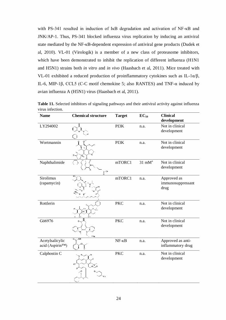
24
with PS-341 resulted in induction of IκB degradation and activation of NF-κB and
JNK/AP-1. Thus, PS-341 blocked influenza virus replication by inducing an antiviral
state mediated by the NF-κB-dependent expression of antiviral gene products (Dudek et
al, 2010). VL-01 (Virologik) is a member of a new class of proteasome inhibitors,
which have been demonstrated to inhibit the replication of different influenza (H1N1
and H5N1) strains both in vitro and in vivo (Haasbach et al, 2011). Mice treated with
VL-01 exhibited a reduced production of proinflammatory cytokines such as IL-1α/β,
IL-6, MIP-1β, CCL5 (C-C motif chemokine 5; also RANTES) and TNF-α induced by
avian influenza A (H5N1) virus (Haasbach et al, 2011).
Table 11. Selected inhibitors of signaling pathways and their antiviral activity against influenza
virus infection.
Name Chemical structure Target EC50 Clinical
development
LY294002
PI3K n.a. Not in clinical
development
Wortmannin
PI3K n.a. Not in clinical
development
Naphthalimide
mTORC1 31 mMa Not in clinical
development
Sirolimus
(rapamycin)
mTORC1 n.a. Approved as
immunosuppressant
drug
Rottlerin
PKC n.a. Not in clinical
development
Gö6976
PKC n.a. Not in clinical development
Acetylsalicylic
acid (Aspirin™)
NF-κB n.a. Approved as anti-
inflammatory drug
Calphostin C
PKC n.a. Not in clinical
development

25
Bisindolylma-
leimide I
PKC n.a. Not in clinical
development
BAY11-7085
NF-κB n.a. Not in clinical
development
SC75741
NF-κB 53 nMb* Not in clinical
development
MG132
26 S proteasome
n.a. Not in clinical development
PS-341
(Bortezomib, Velcade®)
26 S
proteasome n.a. Approved for cancer
treatment
VL-01
(Virologik)
20S and
26S
proteasome
0.8 - 2.4
µMc
Not in clinical
development
U0126
MEK 1.2 - 82
µMd*
Not in clinical
development
PD-0325901
MEK 5 nMe* Phase II against
cancer. Stopped in
2008.
AZD-6244
(Selumetinib)
MEK 750 nMe* Phase II against
cancer
AZD-8330
MEK 40 nMe* Phase I against
cancer
RDEA-119
(BAY 869766)
MEK 60 nMe* Phase I/II against
cancer
* values obtained on A549 cells; a (Mata et al, 2011); b (Haasbach et al, 2013b); c (Haasbach et al, 2011); d (Droebner et al, 2011); e (Haasbach et al, 2013a); n.a., not available.
There are several studies demonstrating that chemical inhibition of mitogen-
activated protein kinase (MAPK) kinases (MEK) by U0126 (Table 11) can exert
antiviral activity against influenza A (H1N1, H5N1, and H7N7) and B viruses in vitro
and in vivo (Droebner et al, 2011; Ludwig et al, 2004; Pleschka et al, 2001). In one
study, inhibition of MEK resulted in nuclear retention of vRNP at a late stage of virus
replication cycle (Pleschka et al, 2001). U0126 is a highly selective inhibitor of both
MEK1 and MEK2 with IC50 values of 72 nM and 58 nM, respectively. Due to

26
unfavourable pharmaceutical properties, U0126 did not proceed to clinical evaluation
for cancer therapy and was examined only in research (Fremin & Meloche, 2010). A
recent study demonstrated that four orally available MEK inhibitors, PD-0325901,
AZD-6244, AZD-8330 and RDEA-119 (Table 11), possessed antiviral effects against
influenza A (H1N1)pdm09 in vitro with EC50 values in nanomolar range (Haasbach et
al, 2013a). Moreover, a combination of these inhibitors and oseltamivir has been shown
to induce a strong synergistic antiviral effect against influenza. However, further
investigations of the best compound combinations are needed (Haasbach et al, 2013a).
It would be interesting to test an antiviral potential of two other MEK inhibitors,
trametinib and PD184352, against influenza virus as single agents or in combination
with oseltamivir. Trametinib (GSK1120212) is currently approved for the treatment of
patients with unresectable or metastatic melanoma with V600E or V600K mutations in
serine/threonine-protein kinase B-Raf. PD184352 (CI-1040) is available for oral
administration and is currently being evaluated in phase II clinical trials in cancer
patients.
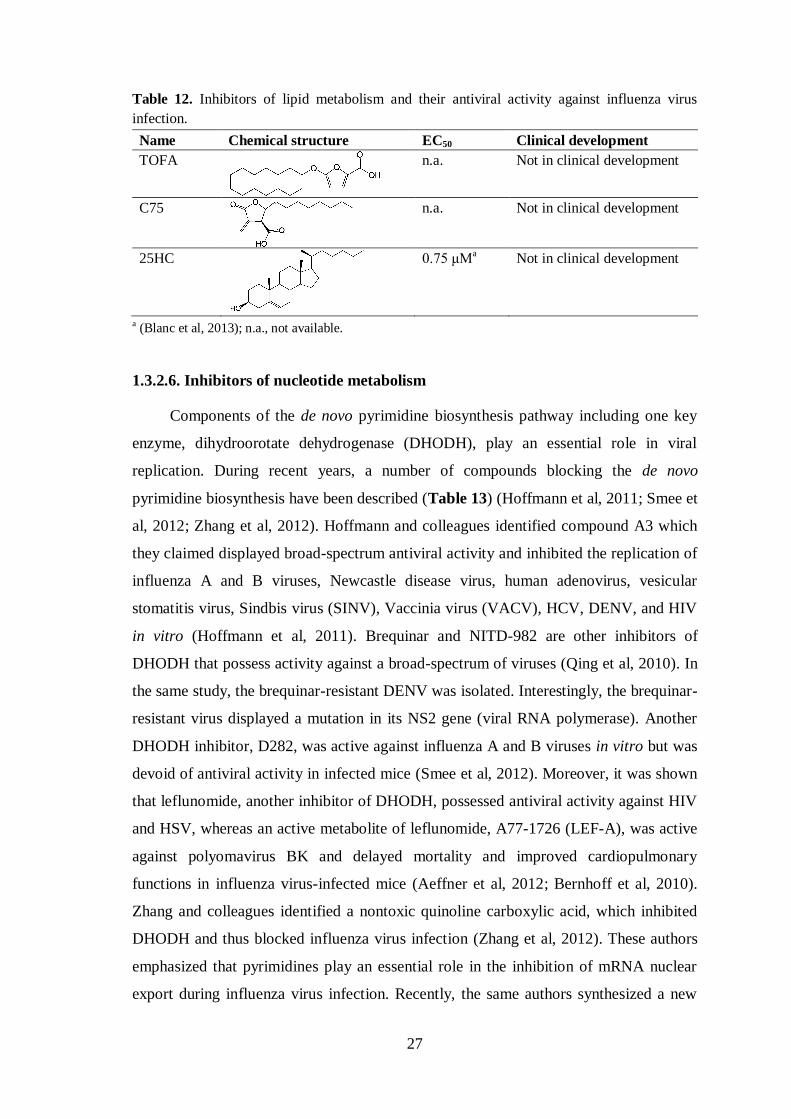
1.3.2.5. Inhibitors of lipid metabolism
It is known that viruses also alter cellular lipid metabolism in order that they can
achieve efficient replication. Thus it has been reported that inhibition of cholesterol and
fatty acid biosynthesis could disturb virus replication, maturation and budding (Blanc et
al, 2013; Munger et al, 2008; Spencer et al, 2011; Taylor et al, 2011). Munger and
colleagues demonstrated that chemical inhibition of acetyl-CoA carboxylase and fatty
acid synthase by TOFA (5-tetradecyloxy-2-furoic acid) and C75 (trans-4-carboxy-5-
octyl-3-methylene-butyrolactone) (Table 12), respectively, dramatically reduced
influenza virus particle production (Munger et al, 2008). Recent studies revealed the
role for the sterol metabolic network in the IFN-mediated antiviral response (Blanc et
al, 2013; Liu et al, 2013b). It was shown that macrophage-synthesized and -secreted
oxysterol, 25-hydroxycholesterol (25HC), inhibited viral growth by blocking fusion of
virus and cell membranes (Liu et al, 2013b). In vitro 25HC blocked a broad range of
enveloped viruses, including influenza A (H1N1), herpes simplex virus type 1 (HSV-1),
murine gamma herpes virus 68, varicella zoster virus, HIV, Ebola, Rift Valley Fever
Virus, Russian Spring-Summer Encephalitis, and Nipah viruses (Blanc et al, 2013; Liu
et al, 2013b). Moreover, administration of 25HC reduces HIV infection in humanized
mice (Liu et al, 2013b).
27
Table 12. Inhibitors of lipid metabolism and their antiviral activity against influenza virus
infection.
Name Chemical structure EC50 Clinical development
TOFA
n.a. Not in clinical development
C75
n.a. Not in clinical development
25HC
0.75 μMa Not in clinical development
a (Blanc et al, 2013); n.a., not available.
1.3.2.6. Inhibitors of nucleotide metabolism
Components of the de novo pyrimidine biosynthesis pathway including one key
enzyme, dihydroorotate dehydrogenase (DHODH), play an essential role in viral
replication. During recent years, a number of compounds blocking the de novo
pyrimidine biosynthesis have been described (Table 13) (Hoffmann et al, 2011; Smee et
al, 2012; Zhang et al, 2012). Hoffmann and colleagues identified compound A3 which
they claimed displayed broad-spectrum antiviral activity and inhibited the replication of
influenza A and B viruses, Newcastle disease virus, human adenovirus, vesicular
stomatitis virus, Sindbis virus (SINV), Vaccinia virus (VACV), HCV, DENV, and HIV
in vitro (Hoffmann et al, 2011). Brequinar and NITD-982 are other inhibitors of
DHODH that possess activity against a broad-spectrum of viruses (Qing et al, 2010). In
the same study, the brequinar-resistant DENV was isolated. Interestingly, the brequinar-
resistant virus displayed a mutation in its NS2 gene (viral RNA polymerase). Another
DHODH inhibitor, D282, was active against influenza A and B viruses in vitro but was
devoid of antiviral activity in infected mice (Smee et al, 2012). Moreover, it was shown
that leflunomide, another inhibitor of DHODH, possessed antiviral activity against HIV
and HSV, whereas an active metabolite of leflunomide, A77-1726 (LEF-A), was active
against polyomavirus BK and delayed mortality and improved cardiopulmonary
functions in influenza virus-infected mice (Aeffner et al, 2012; Bernhoff et al, 2010).
Zhang and colleagues identified a nontoxic quinoline carboxylic acid, which inhibited
DHODH and thus blocked influenza virus infection (Zhang et al, 2012). These authors
emphasized that pyrimidines play an essential role in the inhibition of mRNA nuclear
export during influenza virus infection. Recently, the same authors synthesized a new

28
derivative of a quinoline carboxylic acid named C44, which was reported to inhibit
replication of influenza virus and vesicular stomatitis virus in vitro (Das et al, 2013).
Table 13. Inhibitors of the de novo pyrimidine biosynthesis and their antiviral activity against
influenza virus infection.
Name Chemical structure EC50 Clinical development
A3
0.178 μMa Not in clinical development
D282 n.a. 6-31 μM Not in clinical development
Leflunomide
(AVARA®)
n.a. Approved as
immunomodulatory drug
C44 n.a. 41 nMb Not in clinical development
a (Hoffmann et al, 2011); b (Das et al, 2013); n.a., not available.
1.4. Combination antiviral therapy
Combination therapy, i.e. simultaneous usage of antiviral drugs with different
mechanisms of action, is a well established approach for the treatment of rapidly
mutating viruses such as HCV and HIV (Arts & Hazuda, 2012; Casey & Lee, 2013).
Combination therapy is required for maximal control of virus replication and prevention
of the emergence of drug-resistance especially in immunocompromised and seriously ill
patients (Hayden & de Jong, 2011; Ilyushina et al, 2006; Perelson et al, 2012).
Combination therapy might minimize the adverse effects of a single-agent therapy (De
Clercq, 2006; Hayden, 2013). It has been shown that combination therapy is also
effective in the treatment of influenza virus infection (Hayden et al, 1984; Ilyushina et
al, 2006; Ilyushina et al, 2008; Nguyen et al, 2010). As early as 1984, Hayden and
colleagues showed that human IFN-α2 in combination with rimantadine and ribavirine
exerted a synergistic antiviral effect against influenza A (H3N2 and H1N1) and B
viruses (Hayden et al, 1984). Exogenous IFN (alone or in combination) is approved for
the treatment cancer as well as some chronic viral diseases (Finter et al, 1991; Rong &
Perelson, 2010). Moreover, the feasibility and efficacy of IFN as influenza prophylaxis
have been evaluated (Kugel et al, 2009). However, side effects after long term therapy
and repeated administration prevented the clinical evaluation of IFNs for the treatment
of respiratory diseases. The triple combination of amantadine, oseltamivir and ribavirin
was shown to possess synergistic and broad-spectrum activity against drug resistant
influenza A strains in vitro (Nguyen et al, 2010). Additionally, the triple combination
therapy with amantadine, oseltamivir and ribavirin demonstrated good efficacy in vivo

29
(Seo et al, 2013). The effectiveness of a combination of oseltamivir and ribavirin has
been assessed against highly pathogenic avian influenza A (H5N1) virus in vivo
(Ilyushina et al, 2008).
Several studies examining possible pharmacokinetic interactions between
currently available antivirals have been performed in healthy adults (Atiee et al, 2012;
Pukrittayakamee et al, 2011). The authors reported that drug combinations (oral
oseltamivir combined with intravenous zanamivir therapy; oral oseltamivir combined
with intravenous peramivir therapy) were well tolerated without causing any
pharmacokinetic interactions.
In addition, a number of promising antiviral agents targeting viral proteins have
considerable potential alone or in combination with licensed antivirals. It was shown
that favipiravir (influenza virus RNA polymerase inhibitor) combined with oseltamivir
exerted a strong synergistic effect over monotherapy in mice infected with influenza A
(H1N1, H3N2, and H5N1) viruses (Smee et al, 2010). In another study, the effect of
combination therapy using favipiravir and peramivir was evaluated in a murine model
of influenza A (H1N1)pdm09 infection (Tarbet et al, 2012).
Additionally, inhibitors targeting components of cellular signaling pathways as
well as other host proteins may provide better option for combination therapy in
conjunction with already approved M2 and NA inhibitors for influenza treatment and
prophylaxis. For example, recent in vitro studies demonstrated that a combination of
oseltamivir with four different orally available MEK inhibitors that are currently in
phase I/II clinical trials against cancer had antiviral activity exhibiting a strong
synergism (Haasbach et al, 2013a). Moreover, this supports adopting a drug repurposing
strategy to achieve the development for new antivirals.
1.5. Drug repositioning and new formulations
Increased knowledge of networks in virus-host interactions has opened new
horizons for alternative drug development strategies. Drug development and
pharmaceutical companies are already searching for new therapeutic uses of existing
drugs or lead compounds. An increasing number of small-molecule inhibitors targeting
host proteins or components of cellular signaling pathways are in advanced clinical
studies or already approved for treatment of cancer, diabetes, atherosclerosis and
cardiovascular diseases. Several recent reports have described the problems but also the
30
advantages of repurposing anticancer drugs to reveal their antiviral potential (de
Chassey et al, 2012; Planz, 2013). In particular, current anticancer compounds are
second-generation inhibitors, meaning that they have high specificity, better
pharmacological properties, less side effects and are capable of oral administration.
Most of them have safety data from phase I/II clinical trials that could be helpful in
antiviral drug development. However, as the authors have pointed out, anticancer and
antiviral therapies differ in dosage and duration of treatment and therefore most of the
side effects described for cancer patients might be irrelevant for antiviral treatment.
Therefore, more clinical data is needed. One example of successful drug repurposing is
the FDA-approved antiprotozoal agent nitazoxanide (Alinia®), which has broad
antiviral potency against influenza virus, HCV and rotaviruses (Kratky & Vinsova,
2011).
Another way to find new therapeutic uses of existing drugs is to probe alternative
administration routes. For example, it has been demonstrated that aerosol, but not oral
administration of acetylsalicylic acid could reduce virus titers and promote the survival
of mice infected with influenza virus (Mazur et al, 2007).
In the past decade, in addition to conventional oral or intravenous drug
administration, alternative routes have been intensively studied. Micro- and
nanomaterial- based drug delivery systems have a great potential for further application
of drugs that do not have clinical potential due to their unfavourable bioavailability.
There are several reports demonstrating that mesoporous materials and porous silicon
(PSi)-based materials can be promising tools to increase dissolution and stability of
drugs loaded within their mesopores (Salonen et al, 2008; Santos et al, 2011; Wang et
al, 2013). These materials have a good stability and biocompatibility properties as well
as a high drug loading capacity (Liu et al, 2013a; Wang et al, 2013). PSi nanoparticles
loaded with anticancer agents could be additionally peptide-functionalized to achieve
both targeted and effective cellular uptake and drug delivery (Wang et al, 2013).
Moreover, some PSi-based materials are in widespread use in brachytherapy. For
example, BrachysilTM
which is composed of 30 microne PSi particles encapsulating
isotope [32
P] is currently undergoing phase II clinical trials for the treatment of solid
tumors (Salonen et al, 2008).

31
2. AIMS OF THE STUDY
The purpose of the present study was to identify druggable host factors
involved in influenza virus infection and to develop antiviral agents to be investigated in
future clinical trials.
The specific aims were:
1. To develop a screening approach to search for druggable cellular factors involved
in influenza virus infection;
2. To understand the molecular mechanisms of action of promising antiviral agents
directed against host cell factors;
3. To begin to optimize the clinical utility of host-directed antiviral agents.

32
3. MATERIALS AND METHODS
This chapter briefly summarizes the materials and methods used in this study.
More detailed descriptions are found in the original publications (I - IV).
3.1. Cells, compounds and viruses (I – IV)
Cell types, compounds and viruses used in this study are summarized in Tables
14, 15 and 16. All cells used in this study were cultured in the respectively
recommended growth media and maintained at +37°C in 5% CO2. Cells were regularly
checked by PCR to ensure they were free of mycoplasma contamination. The human
macrophages were obtained from peripheral blood mononuclear cells (PBMCs) isolated
from healthy individuals and buffy coats of the voluntary blood donors (Finnish Red
Cross) by using lymphocyte separation medium (PAA) and cultured for seven days in
Macrophage Serum-free Medium (Life Technologies) supplemented with 10 ng/ml
human granulocyte macrophage-colony-stimulating factor (GM-CSF; Sigma-Aldrich).
Table 14. Cells types used in the study.
Name Description Study
hTERT RPE Telomerase-expressing human retinal pigment epithelial cells I - III
MDCK Madin-Darby canine kidney cells I - IV
A549 Human lung cancer cells I, II, IV
VERO-E6 African green monkey kidney cells I, II
BHK-21 Syrian hamster kidney cells I, II
iSLK.219 Doxycycline-inducible human endothelial cells latently infected
with Kaposi's sarcoma-associated herpes virus
II
Human
macrophages
PBMC-derived macrophages from healthy donors I, II
NCI-H1666 Human lung cancer cells IV
NCI-H1703 Human lung cancer cells IV
NCI-1437 Human lung cancer cells IV
NCI-460 Human lung cancer cells IV
NCI-H2126 Human lung cancer cells IV
Calu-1 Human lung cancer cells IV
All compounds were dissolved in DMSO (Sigma-Aldrich) to a concentration of
10 mM and stored at −80°C until use.

33
Table 15. Compounds used in the study.
Name Chemical structure Molecular
weight
Source Study
Obatoclax
(GX15-070MS)
413.49 Selleck Chemicals I, II, IV
SaliPhe
459.53 * I - IV
Gemcitabine
(Gemzar®)
263.198 Selleck Chemicals I, II
ABT-263
(Navitoclax)
974.61 Selleck Chemicals II, IV
ABT-737
813.43 Selleck Chemicals II
ABT-199
(GDC-0199)
868.44 Active Biochem II
MK-2206
480.39 ChemieTek IV
* provided by Professor Jef De Brabander, Department of Biochemistry, University of Texas
Southwestern Center at Dallas, Dallas, TX, USA.
The influenza A (H1N1, H3N2, and H1N1pdm09) strains were propagated in
MDCK cells, whereas monkey Vero-E6 cells were used for propagation of A/PR8-
NS116-GFP. Virus stocks were titrated by a plaque assay on MDCK cells. The
following virus stocks were obtained from collaborators: A/Udorn/72 from Sampsa
Matikainen, Finland; A/Brussels/BB/2009 from Xavier Saelens, Belgium;
B/Shandong/7/97 from Ilkka Julkunen, Finland; SINV and SFV from Tero Ahola,

34
Finland; HSV-1 and HSV-2 from Veijo Hukkanen, Finland; Bunyamwera virus
(BUNV) from Richard M. Elliott, UK.
Table 16. Viruses used in the study.
Name Group Family Study
A/WSN/33 (H1N1) (-)ssRNA Orthomyxoviridae I - IV
A/PR/8/34 (H1N1) (-)ssRNA Orthomyxoviridae I, III
A/PR/8/34 (H1N1) (mouse-adapted) (-)ssRNA Orthomyxoviridae II
A/PR8-NS116-GFP (H1N1) (-)ssRNA Orthomyxoviridae I, III
A/Helsinki/P14/2009 (H1N1pdm09) (-)ssRNA Orthomyxoviridae IV
A/Helsinki/P15/2009 (H1N1pdm09) (-)ssRNA Orthomyxoviridae IV
A/Helsinki/P18/2009 (H1N1pdm09) (-)ssRNA Orthomyxoviridae IV
A/Helsinki/Vi1/2009 (H1N1pdm09) (-)ssRNA Orthomyxoviridae IV
A/Helsinki/Vi2/2009 (H1N1pdm09) (-)ssRNA Orthomyxoviridae IV
A/Helsinki/Vi3/2009 (H1N1pdm09) (-)ssRNA Orthomyxoviridae IV
A/Helsinki/543/2013 (H1N1pdm09) (-)ssRNA Orthomyxoviridae IV
A/Helsinki/629/2013 (H1N1pdm09) (-)ssRNA Orthomyxoviridae IV
A/Helsinki/551/2013 (H1N1pdm09) (-)ssRNA Orthomyxoviridae IV
A/Helsinki/628/2013 (H1N1pdm09) (-)ssRNA Orthomyxoviridae IV
A/Helsinki/552/2013 (H1N1pdm09) (-)ssRNA Orthomyxoviridae IV
A/Helsinki/568/2013 (H1N1pdm09) (-)ssRNA Orthomyxoviridae IV
A/Helsinki/604/2013 (H1N1pdm09) (-)ssRNA Orthomyxoviridae IV
A/Helsinki/668/2013 (H1N1pdm09) (-)ssRNA Orthomyxoviridae IV
A/Helsinki/598/2013 (H1N1pdm09) (-)ssRNA Orthomyxoviridae IV
A/Helsinki/526/2013 (H1N1pdm09) (-)ssRNA Orthomyxoviridae IV
A/Helsinki/678/2013 (H1N1pdm09) (-)ssRNA Orthomyxoviridae IV
A/Helsinki/552/2013 (H1N1pdm09) (-)ssRNA Orthomyxoviridae IV
A/Brussels/BB/2009 (H1N1pdm09) (-)ssRNA Orthomyxoviridae IV
A/Sydney/5/1997(H3N2) (-)ssRNA Orthomyxoviridae I, IV
A/Udorn/307/1972 (H3N2) (-)ssRNA Orthomyxoviridae IV
A/Chicken/Nigeria/BA211/2006 (H5N1) (-)ssRNA Orthomyxoviridae IV
A/Anhui/01/2013 (H7N9) (-)ssRNA Orthomyxoviridae IV
B/Shandong/7/97 (-)ssRNA Orthomyxoviridae I, II
Bunyamwera virus (BUNV) (-)ssRNA Bunyaviridae I, II
Measles virus (MeV) (-)ssRNA Paramyxoviridae I, II
Sindbis virus (SINV) (+)ssRNA Togaviridae I, II
Semliki forest virus (SFV) (+)ssRNA Togaviridae I, II
Human echovirus 6 (Echo6) (+)ssRNA Picornaviridae I, II
Tick-borne encephalitis virus (TBEV) (+)ssRNA Flaviviridae II
Dengue virus (DENV) (+)ssRNA Flaviviridae II
Vaccinia virus (VACV) dsDNA Poxviridae I, II
Herpes simplex virus type 1 (HSV-1) dsDNA Herpesviridae I, II
Herpes simplex virus type 2 (HSV-2) dsDNA Herpesviridae II
Kaposi's sarcoma-associated herpes virus dsDNA Herpesviridae II

35
3.2. THCPSi nanoparticles loaded with SaliPhe (III)
In an attempt to analyze alternative options to DMSO to dissolve and deliver the
compounds, SaliPhe was loaded into nanoparticles. The thermally hydrocarbonized PSi
(THCPSi) nanoparticles were prepared from PSi films (III). The loading degree of
SaliPhe in the mesopores of THCPSi nanoparticles was 2.88±0.11 wt %. The SaliPhe
loaded nanoparticles were tested in in vitro experiments.
3.3. In vitro experiments
Drug screens (I, unpublished)
Drug screens were performed in cooperation with the High Throughput
Biomedicine Unit, FIMM Technology Centre, Finland. A 384-well plate-based assay
was optimized to identify the compounds that could influence influenza infection. The
toxicity assay was optimized to identify inhibitors that influence cell viability. The drug
screens were performed against the influenza A/PR8-NS116-GFP strain in human
hTERT RPE cells (I), and influenza A/WSN/33 and B/Shandong/7/97 viruses in A549
cells (unpublished). Approximately, 2,500 cells were seeded in 25 µl of an appropriate
growth using a Multidrop 384 (Thermo). After 24 h, the growth medium was changed
to the virus growth medium. The drug library comprising either 201 compounds
targeting host factors (I, Suppl. table 1) or a library with 337 compounds (including the
201 drugs and the HTB compound collection (Pemovska et al, 2013)) was added to the
cells using 10-fold serial dilutions at five different concentrations starting from 10 µM
with an Echo acoustic dispenser (Labsite). DMSO and benzethonium chloride were
added to the control wells. The cells were infected with influenza virus at a multiplicity
of infection (MOI) of 3 or left uninfected (mock). At 24 h post-infection (in hTERT
RPE cells), virus-mediated GFP fluorescence was measured using a PHERAstar FS
plate reader (BMG Labtech). The effective concentrations (EC50) were calculated using
SigmaPlot 11 software (Systat Software GmbH, Germany). At 24 h (in hTERT RPE
cells) or 48h post-infection (in A549 cells), cell viability was measured using a Cell
Titer Glo viability assay (CTG; Promega) according to the manufacturer’s instructions.
Luminescence was read using the PHERAstar FS plate reader, and inhibitory
concentrations (IC50) were calculated. Selectivity indices (SI=IC50/EC50) were
calculated to distinguish the antiviral effect from toxic side effects of the compounds.
Those compounds with SI greater than 10 were validated further.

36
Compound efficacy testing (I-IV)
Compound efficacy testing was performed in 96-well plates with the different cell
lines listed in Table 14. Typically, 40,000 cells were seeded in 100 µl of the appropriate
growth medium. After 24 h, the growth medium was changed and compounds listed in
Table 15 or SaliPhe-loaded THCPsi nanoparticles were added to the cells at 3-fold
serial dilutions at seven different concentrations starting from 10 µM. DMSO was used
as a control. Then, the cells were mock-infected or infected with different influenza
strains (MOI of 3 or 10) or other viruses (Table 16). Cell viability was measured using
the CTG assay at 24, 48 or 72 h post-infection when virus-induced cytopathic effect
(CPE) was observed.
Testing of compound efficacy against Kaposi's sarcoma-associated herpes virus,
MeV, VACV, TBEV, DENV, and influenza A strains H5N1 and H7N9 infections was
done by collaborators based on the optimized and established protocol under biosafety
level (BSL)-2 and BSL-3 conditions (I, II, unpublished).
Activity and toxicity scores (ATS) were calculated in order to quantitatively
profile the compounds that rescued the infected cells (I, IV). ATS varies between -100
and +100, where negative values indicate excessive toxicity whereas the highest
positive values refer to the most potent compounds. Drug sensitivity scores (DSS) were
calculated to quantitatively profile those compounds that accelerated virus-induced cell
death (II). DSS summarizes the area under the dose-response curve relative to the total
area between 10% threshold and 100% inhibition (Pemovska et al, 2013).
Virus titration (I-IV)
Antiviral efficacies of compounds (Table 15) or SaliPhe-loaded THCPsi
nanoparticles were validated by the plaque assay. Briefly, cells were non- or compound-
treated and virus-infected (MOI of 0.1). Supernatants were collected at 24, 48 or 72 h
post-infection. The supernatants were 10-fold diluted in virus growth media and added
to MDCK cells on 6-well plates. After 1 h, the cells were overlaid with Avicel-based
medium and incubated for two days. The cells were fixed with 4% formaldehyde and
stained with crystal violet. Plaque forming units were calculated.
For the other viruses the titration procedure slightly differed from that described
above. SINV, SFV, VACV and BUNV were titered on Vero-E6 cells, whereas A549
cells were used for titration of Echo6 virus. Other viruses were titered by collaborators
based on their established protocols (I). The degree of inhibition mediated by a
37
compound was calculated as the ratio between virus titers in non- and compound-treated
infected cells.
Time-of-compound-addition experiment (I, II, IV)
The cells were grown in 96-well plates and were mock- or influenza virus-
infected (MOI of 3 or 10). Compounds at non-toxic concentrations were added every
hour. Cell viability was measured using the CTG assay at 24 h post-infection.
Compound-competition experiment (II, IV)
hTERT RPE or NCI-H1666 cells were grown in 96-well plates. Cells were treated
with obatoclax (1 µM), SaliPhe (0.4 µM), gemcitabine (10 µM) or MK-2206 (10 µM)
and with increasing concentrations of ABT-263. The cells were also treated with ABT-
263 (0.4 µM) and with increasing concentrations of obatoclax, SaliPhe, gemcitabine or
MK-2206. Treated cells were mock- or influenza virus-infected (MOI of 3 or 10), and
cell viability was measured using the CTG assay at 24 h post-infection.
Caspase assay (II)
hTERT RPE cells were grown in 96-well plates and were non- or ABT-263-
treated and mock- or influenza virus-infected or RNA transfected. At 24 h post-
infection or transfection, caspase 8, 9 and 3/7 activities were measured with Caspase-
Glo-8, -9 and -3/7 assays (Promega) according to the manufacturer’s instructions.
Metabolic labeling of proteins with [35
S]-Methionine
hTERT RPE cells were grown in 96-well plates and were non- or ABT-263-
treated and mock- or influenza virus-infected (MOI of 3). At different time points post-
infection, cells were washed twice with PBS and were incubated with methionine-free
DMEM (Sigma-Aldrich) supplemented with 10% BSA and 0.11 µCi/ml [35
S]-labeled
methionine (EasyTag EXPRESS35
S protein labeling mix, specific activity 1175
Ci/mmol; PerkinElmer) for 30 minutes at +37°C. Cells were washed twice with PBS
and lysed in 2x SDS-sample buffer. Proteins were resolved by electrophoresis on 4-20%
SDS-PAGE (BioRad) and newly synthesized [35
S]-labeled proteins were monitored by
autoradiography using a Typhoon 9400 scanner (Amersham).
Fluorimetry (IV)
The fluorescence spectra of MK-2206 were recorded (IV).

38
RNA isolation and quantitative RT-PCR (I, II, IV)
hTERT RPE or NCI-H1666 cells were grown in 6-well plates and were non- or
compound-treated and mock- or influenza virus-infected (MOI of 3). Total RNA was
isolated from cells with the RNeasy Plus Mini Kit (Qiagen) and used in further
quantitative RT-PCRs with the primers indicated in Table 17 (I, II, IV).
Table 17. Primers used in the study
Target 5′→3′ sequence Study
PB2 Sequence-modifying RT:
(+) strand: TCAGTCCCATTGCAGCCTTGCccgccgCCACG (-) strand: GCAGAACCCAACAGAAGAGCAAtttaTttATATTTG
PCR: Fwd: GCAGAACCCAACAGAAGAGC
Rev: TCAGTCCCATTGCAGCCT
I, II
β-actin Fwd: CCGACAGGATGCAGAAGG Rew: AGGAAAGACACCCACCTTGA
I, II
NS1 Fwd: GAAATGTCAMGAGACT Rew: AGAAAGCTCTTATCTCTTG
IV
huGAPDH Fwd: GGCTGGGGCTCATTTGCAGGG Rew: TGACCTTGGCCAGGGGTGCT
IV
18S Hs99999901_s1* IV
IFN-β Hs01077958_s1* IV
IL-29 Hs00601677_g1* IV * TaqMan primers and probes from Applied Biosystems.
RNA transfection (I, II)
In the viral RNA transfection experiment, A/WNS/33 virus was grown in MDCK
cells. Supernatants were collected at 24 h post-infection, and the virus was pelleted
using ultracentrifugation at 285,000 g for 4 h. Viral RNA was purified and transfected
into hTERT RPE cells using Lipofectamine 2000 (Life Technologies) according to the
manufacturer’s instructions. Six hours after transfection, the medium was replaced with
growth medium with or without ABT-263. After 18 h, the cell viability and caspase
activities were measured.
Knockdown of the myeloid cell leukemia-1 (Mcl-1) protein was done in hTERT
RPE cells using Hs_MCL1_12 Flexi-Tube siRNA (SI04949721; Qiagen) to suppress
the expression of Mcl-1 (I).
Transcription profiling (IV)
NCI-H1666 cells were grown in 6-well plates and were non- or MK-2206-treated
and mock- or influenza virus-infected (MOI of 3). After 8 h post-infection, the cells
were collected, total RNA was isolated and used in the subsequent whole genome gene
expression analysis (IV).

39
Immunoprecipitation and mass spectrometry (II)
Bcl-xL-associated factors were immunoprecipitated from non- or ABT-263-
treated and mock- or influenza-infected hTERT RPE cells and analyzed using liquid
chromatography-tandem mass spectrometry (II).
Immunoblotting (I, II, IV)
Cells were grown in 96-well plates and were non- or compound-treated and mock-
or influenza virus-infected (MOI of 3 or 10). At different time points post-infection, the
cells were lysed in 2x SDS-sample buffer. Proteins were resolved by electrophoresis on
4-20% SDS-PAGE and transferred to polyvinylidine fluoride membranes (GE
Healthcare). The membranes were blocked with 5% milk in Tris-buffered saline (TBS)
and incubated with primary guinea pig anti-NS1, rabbit anti-M1 or rabbit anti-NP
(1:2000, 1:2000, and 1:500 dilutions, respectively; from Ilkka Julkunen, Finland),
mouse anti-LRP130 (1:100; clone G-10; Santa Cruz Biotechnology), rabbit anti-Bcl-xL
(1:1000; Cell Signaling Technology), rabbit anti-Bid (1:1000; Cell Signaling
Technology), rabbit anti-Bax (1:100; Santa Cruz Biotechnology), mouse anti-Bad
(1:200; Santa Cruz Biotechnology), rabbit anti-UACA (1:250; Sigma-Aldrich) or mouse
anti-β-actin (1:2000; Termo Fisher Scientific) antibody overnight at +4°C. After three
washes, membranes were incubated for 4 h at 4°C with the representative secondary
antibodies conjugated to infrared dyes 680LT or 800CW (1:20,000; Li-Cor
Biosciences). The membranes were scanned with an Odyssey scanner (Li-Cor
Biosciences).
Immunofluorescence assay (I-IV)
hTERT RPE or NCI-H1666 cells were grown on cover glasses in 6-well plates.
Cells were non- or compound-treated and mock- or influenza virus-infected (MOI of 3
or 30) on ice for 1 h. Cells were washed twice with ice-cold virus growth media,
overlaid with pre-warmed media with or without compound, and incubated at +37°C in
5% CO2. At 1, 2, 4 or 18h post-infection cells were fixed with 4% paraformaldehyde
and blocked and permeabilized with PBS with 1% BSA and 0.1% Triton X-100. The
following antibodies were used in the detection of proteins, rabbit anti-Bcl-xL (1:200;
clone 54H6; Cell Signaling Technology), mouse anti-Bad (1:100; clone c-7; Santa Cruz
Biotechnology), rabbit anti-Tom20 (1:300; clone FL-145; Santa Cruz Biotechnology),
human anti-Mcl-1 (1:100; clone 22/Mcl-1; BD Transduction Laboratories) or rabbit
anti-NP and anti-M1 (1:1000; from Ilkka Julkunen, Finland). Secondary antibodies

40
were anti-mouse or anti-rabbit Alexa Fluor 594 or anti-rabbit Alexa Fluor 488-
conjugated antibodies (1:2000 or 1:1000; Life Technologies). Nuclei were
counterstained with DAPI. Images were captured with a Nikon 90i microscope and
processed with NIS Elements AR software.
Phospho-protein profiling (IV)
NCI-H1666 cells were grown in 6-well plates and were non- or MK-2206-treated
and mock- or influenza virus-infected (MOI of 3). After 0.5, 4 and 12 h post-infection,
the cells were lysed and the phosphorylation profiles of 43 kinases and 2 related
substrates were analyzed using a human phospho-kinase array (R&D Systems)
according to the manufacturer’s instructions. The membranes were placed on films and
were exposed to X-rays and then the films were scanned. Each image was analyzed with
ImageJ software (NIH, USA).
Cytokine profiling (I, II, IV)
Cytokine profiling was performed using mouse lung homogenates or supernatants
of non- or compound-treated and mock- or influenza virus-infected (MOI of 3 or 10) or
vRNA-transfected cells. Cytokine levels were measured using the human or mouse
cytokine array panel A (R&D Systems) according to the manufacturer’s instructions.
The membranes were exposed onto X-ray films and the films were scanned. The
scanned image was analyzed with ImageJ software. In addition, the levels of TNF-α,
IFN-β and IFN-λ in the cell supernatants were assayed with ELISA (PBL Interferon
Source) (I, II).
Automated image acquisition and image analysis (IV)
MDCK cells were grown in 96-well plates and treated with different
concentrations of MK-2206. In a subsequent experiment, MDCK cells were treated with
MK-2206 (10 μM), and in combinations with NH4Cl (20 mM), SaliPhe (0.4 μM) or
obatoclax (0.4 μM). Cells were fixed with 4% paraformaldehyde and imaged with a
modular epifluorescence microscope ScanR (Olympus) to analyze the number and the
mean intensity of the objects (IV).
Serial-passage experiment (IV, unpublished)
Human NCI-H1666 cells were grown in 6-well plates and were non- or
compound-treated (MK-2206 or SaliPhe) and infected with A/Helsinki/P14/2009 (MOI

41
of 0.1). At 48 h post-infection, 20 μl of medium was passaged to fresh cells. During
passaging, the concentration of compounds was gradually increased (MK-2206, from
0.1 to 10 μM; SaliPhe, from 0.01 to 1 µM). A total of 15 passages were performed and
the virus titers were determined with plaque assay.
3.4. In vivo experiments
Compound efficacy testing in vivo (II)
Pathogen-free 6-7-week old female BALB/c mice were used for ABT-263
efficacy testing in vivo. Mice were inoculated intranasally with 50 µl PBS with or
without two median lethal doses (LD50) of the mouse-adapted influenza A/PR8/8/34
strain. ABT-263 (50 mg/kg in carrier) was administered orally, and then the mice were
monitored for 14 days for weight loss (II). Mice were sacrificed on day 6 and lung
tissue dissected. Virus titers, caspase 3/7 activities and cytokine levels were measured
from lungs (II).
Histochemistry (II)
In the experiment monitoring the degree of inflammation, the left lobes of the
mouse lung slices were processed for histochemistry and the tissue slides were stained
with hematoxylin and eosin (II).
3.5. Ethics
Virus experiments were carried out under BSL-2+ conditions and in compliance
with regulations of the University of Helsinki (permit 21/M/09) and under BSL-3
conditions and in compliance with regulations of the Centre de Recherche Public de la
Sante/Laboratoire National de Santé. Mouse influenza virus infection experiments were
carried out in the Ghent University under BSL-2+ conditions and were authorized by
the Institutional Ethics Committee on Experimental Animal of the VIB Department for
Molecular Biomedical Research of Ghent University. Buffy coat preparations of
voluntary blood donors were obtained with permission from the Finnish Red Cross
Blood Transfusion Service and their use was approved by the ethics review committee
of the University of Helsinki Central Hospital, Finland (165/13/03/00/2011). Collection
of blood samples from volunteers was approved by the ethics review committee of the
University of Helsinki Central Hospital, Finland (244/13/03/01/2012).

42
4. RESULTS AND DISCUSSION
4.1. Identification of host factors involved in influenza virus replication
4.1.1. Assembly of library of compounds targeting host factors (I, II, IV)
There are two main systemic approaches which can be used when searching of
host factors essential for virus replication. One approach is to conduct a genome-wide
siRNA screening to identify the host genes that are crucial for virus replication (Karlas
et al, 2010; Konig et al, 2010; Li et al, 2009; Sessions et al, 2009). The other approach
is screening of libraries of chemical compounds capable of blocking host factors for
molecules that inhibit virus replication without evoking toxicity in the host cell (Zhang
et al, 2012). Both approaches are widely used for understanding viral biology as well as
developing alternative antiviral drugs.
To identify host factors essential for influenza virus replication it was decided to
assemble a library of small-molecule inhibitors targeting cellular proteins and
components of signaling pathways. The primary focus was on commercially available
drugs with well described targets that were approved or under clinical development for
treatment of cancer or other diseases. Small-molecule inhibitors that target the cell cycle
control, apoptotic pathways, lipid metabolism, and other cellular functions that could be
essential for influenza infection in the host cell were included. Moreover, the screen
also contained some small-molecule inhibitors with known anti-influenza activity such
as gemfibrozil and TOFA (lipid metabolism), 17-AAG and bortezomib (protein quality
control), fluorouracil and cytarabine (de novo nucleotide biosynthesis), 3-methyladenine
(autophagy), PD153035 (EGFR) and wortmannin (PI3K) (Chase et al, 2008; Dudek et
al, 2010; Eierhoff et al, 2010; Meneghesso et al, 2012; Müller et al, 2012; Munger et al,
2008; Zhou et al, 2009). In addition, functional analogues of known influenza
inhibitors, such as erlotinib and gefitinib (analogues of PD153035), 17-DMAG (analog
of 17-AAG), carfilzomib (analog of bortezomib), and leflunomide and gemcitabine
(analogues of fluorouracil) were examined. The control was a v-ATPase inhibitor,
SaliPhe, which was shown to block influenza virus replication in vitro and in vivo
(Müller et al, 2011). A total of 201 small-molecule inhibitors were collected into the
library (I, Suppl. table 1). Since most of these inhibitors are approved or are under
clinical development for treatment of human malignancies, it was decided to screen the
library in nonmalignant cells in order to avoid cancer-specific effects of drugs. A human

43
telomerase-expressing retinal pigment epithelial (hTERT RPE) cell line was selected
since this line has been shown to be appropriate for influenza A virus replication
(Michaelis et al, 2009). Additionally, two specific drug subsets were assembled
targeting the anti-apoptotic signals, especially the Akt kinase and the B-cell lymphoma
2 (Bcl-2) family members. It is well established that influenza virus activates the
PI3K/Akt/mTOR signaling pathway for efficient viral replication (Ehrhardt et al, 2006;
Zhirnov & Klenk, 2007). However, none of Akt inhibitors had been tested in influenza
virus-infected cells. Therefore, the first set to be examined contained inhibitors targeting
Akt such as MK-2206, Akt inhibitor VIII, GDC-0068, and perifosine (IV). The second
set consisted of ABT-263, ABT-737 and ABT-199, gossypol and TW-37 that target
predominantly Bcl-2, B-cell lymphoma-extra large (Bcl-xL), and Bcl-2-like protein 2
(Bcl-w) proteins (II). It was hypothesized that chemical inhibition of Bcl-xL, Bcl-2 and
Bcl-w could have an effect on the survival of virus-infected cells.
Our library and the drug subsets comprised highly specific small-molecule
inhibitors targeting several well-described host factors and components of signaling
pathways. The evaluation of this library and the subsets against influenza virus infection
could provide information about the importance of these host factors both for virus
infection and for cell survival. Moreover, this library and subsets could be tested against
other virus infections to clarify the broad-spectrum antiviral properties of those
compounds. Additionally, since the library included multiple drugs that are approved or
are under clinical development for treatment of cancer or other diseases, it was deemed
to verify the potential of these drugs for repositioning as antiviral agents.
4.1.2. Method for searching potential antiviral agents (I, II, IV)
After assembly of the library and the drug subsets a cell-based approach was
developed which allowed identification of chemical probes to study virus-host
interactions. In this method, increasing concentrations of small-molecule inhibitors were
tested for their ability (i) to attenuate or accelerate death of infected cells, and (ii) to
suppress or induce virus replication at concentrations that would not be toxic for
noninfected cells (mock control). Cell survival and virus replication were measured by
cell viability and plaque assays, respectively. Importantly, these techniques are suitable
for virus infections that result in virus-induced CPE. In order to obtain high CPE values,
MOI of 3 or higher was used. This approach was successfully used and its validity
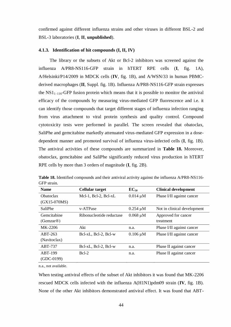
44
confirmed against different influenza strains and other viruses in different BSL-2 and
BSL-3 laboratories (I, II, unpublished).
4.1.3. Identification of hit compounds (I, II, IV)
The library or the subsets of Akt or Bcl-2 inhibitors was screened against the
influenza A/PR8-NS116-GFP strain in hTERT RPE cells (I, fig. 1A),
A/Helsinki/P14/2009 in MDCK cells (IV, fig. 1B), and A/WSN/33 in human PBMC-
derived macrophages (II, Suppl. fig. 1B). Influenza A/PR8-NS116-GFP strain expresses
the NS11–116-GFP fusion protein which means that it is possible to monitor the antiviral
efficacy of the compounds by measuring virus-mediated GFP fluorescence and i.e. it
can identify those compounds that target different stages of influenza infection ranging
from virus attachment to viral protein synthesis and quality control. Compound
cytotoxicity tests were performed in parallel. The screen revealed that obatoclax,
SaliPhe and gemcitabine markedly attenuated virus-mediated GFP expression in a dose-
dependent manner and promoted survival of influenza virus-infected cells (I, fig. 1B).
The antiviral activities of these compounds are summarized in Table 18. Moreover,
obatoclax, gemcitabine and SaliPhe significantly reduced virus production in hTERT
RPE cells by more than 3 orders of magnitude (I, fig. 2B).
Table 18. Identified compounds and their antiviral activity against the influenza A/PR8-NS116-
GFP strain.
Name Cellular target EC50 Clinical development
Obatoclax
(GX15-070MS)
Mcl-1, Bcl-2, Bcl-xL 0.014 µM Phase I/II against cancer
SaliPhe v-ATPase 0.254 µM Not in clinical development
Gemcitabine
(Gemzar®)
Ribonucleotide reductase 0.068 µM Approved for cancer
treatment
MK-2206 Akt n.a. Phase I/II against cancer
ABT-263
(Navitoclax)
Bcl-xL, Bcl-2, Bcl-w 0.106 µM Phase I/II against cancer
ABT-737 Bcl-xL, Bcl-2, Bcl-w n.a. Phase II against cancer
ABT-199
(GDC-0199)
Bcl-2 n.a. Phase II against cancer
n.a., not available.
When testing antiviral effects of the subset of Akt inhibitors it was found that MK-2206
rescued MDCK cells infected with the influenza A(H1N1)pdm09 strain (IV, fig. 1B).
None of the other Akt inhibitors demonstrated antiviral effect. It was found that ABT-

45
263 and its structural analogues ABT-737 and ABT-199 induced apoptosis in influenza
virus-infected cells in a dose-dependent manner (II, Suppl. fig. 1). In contrast, gossypol
and TW-37 exerted no effect on the survival of influenza virus-infected cells. A non-
toxic concentration of ABT-263 was then selected and the survival of mock- or
influenza virus-infected cells was monitored at different time points post-infection. The
results revealed that ABT-263 promoted premature apoptosis in influenza virus-infected
cells and interfered with virus replication (II, fig. 1). Additionally, the apoptotic cell
death was dependent on MOI.
Thus, two different sets of compounds were identified: (i) obatoclax, SaliPhe,
gemcitabine and MK-2206 that rescued influenza virus-infected cells and reduced virus
production, and (ii) ABT-263, ABT-737 and ABT-199 that promoted premature
apoptosis in influenza virus-infected cells and also reduced virus titers. Obatoclax
(GX15-070) is a BH3 mimetic which inhibits Mcl-1 in addition to blocking Bcl-2 and
Bcl-xL (Nguyen et al, 2007). Obatoclax overcomes Mcl-1-mediated resistance to
apoptosis and currently is in phase I/II trials for the treatment of chronic lymphocytic
leukemia. SaliPhe is an investigational anticancer and anti-influenza compound, which
has been reported to inhibit v-ATPase activity (Lebreton et al, 2008; Müller et al, 2011).
Gemcitabine (Gemzar®) is a nucleoside analog, which is FDA approved for cancer
treatment. There is a report that gemcitabine blocks cellular ribonucleotide reductase
required for DNA replication and repair (Cerqueira et al, 2007). MK-2206 is a potent,
oral allosteric inhibitor of Akt kinases, which inhibits auto-phosphorylation of both Akt
T308 and S473 (Hirai et al, 2010). Currently MK-2206 is in phase I/II trials for cancer
treatment. The two BH3 mimetics, ABT-263 (Navitoclax) and ABT-737, target the anti-
apoptotic proteins Bcl-xL, Bcl-2 and Bcl-w but not Mcl-1 or A1 (Tse et al, 2008). A
recently discovered BH3 mimetic, ABT-199 (GDC-0199), has been claimed to
selectively inhibit Bcl-2 but not Bcl-xL (Vandenberg & Cory, 2013; Vogler et al, 2013).
Currently ABT-263 and its structural analogues are undergoing phase I/II trials for the
treatment of chronic lymphocytic leukemia.
Taken together, these results suggest that cellular v-ATPase, Akt kinase,
ribonucleotide reductase and anti-apoptotic Bcl-2 family members, such as Mcl-1, Bcl-
xL, Bcl-2 and Bcl-w, represent essential host factors for influenza virus infection.

46
4.2. Potential mechanisms of action of promising compounds (I, II, IV)
The mechanism of action of obatoclax, SaliPhe, gemcitabine, MK-2206 and ABT-
263 was investigated by testing these inhibitors in a time-of-compound-addition
experiment. Appropriate cells were infected with influenza virus, and the inhibitors
were added every hour. It was found that obatoclax, SaliPhe and MK-2206 added at the
time of infection could inhibit early steps of influenza virus infection (I, fig. 3A; IV, fig.
2D). It has been shown previously that SaliPhe inhibited v-ATPase that is needed for
acidification of endosomes and release of vRNPs into the cytoplasm (Müller et al,
2011). Thus, one could postulate that obatoclax and MK-2206 also inhibit entry stages
of influenza virus infection, whereas gemcitabine which targets cellular ribonucleotide
reductase blocks influenza virus at the stage of viral RNA transcription and replication.
Interestingly, ABT-263 induced apoptosis in influenza virus-infected cells
independently of the time of its addition (II, fig. 3C). To address the stage of influenza
virus infection when ABT-263 triggers the cell death a compound competition
experiment was conducted with obatoclax, SaliPhe, gemcitabine and MK-2206. It was
found that obatoclax, SaliPhe and MK-2206, but not gemcitabine, rescued ABT-263-
treated cells from influenza virus-mediated death (II, fig. 3A and B; IV, fig. 2E). These
results indicate that ABT-263 sensitized cells to undergo premature apoptosis at
multiple stages of influenza virus infection following viral endocytic uptake.
Viral NP or M1 proteins were then monitored in an immunofluorescence assay at
different time points. Immunofluorescence experiments revealed that obatoclax, SaliPhe
and MK-2206 prevented accumulation of NP in the nucleus or M1 in the cytoplasm
compared to non-treated or gemcitabine-treated influenza virus-infected cells (I, fig.
3B; IV, fig. 4G and H).
Next the production of viral RNAs and the synthesis of viral proteins were
compared at different time points post-infection. The results demonstrated that
obatoclax, SaliPhe, gemcitabine and MK-2206 substantially affected viral RNA
transcription and replication and subsequent synthesis of viral proteins (I, fig. 3C and D;
IV, fig. 2F). Since ABT-263 was able to induce premature apoptosis in influenza virus-
infected cells already at 8 h post-infection, production of viral RNAs and proteins
declined at the same time (II, Suppl. fig. 3).
Based on these results it was concluded that obatoclax, SaliPhe and MK-2206
block virus entry at a stage preceding vRNA uncoating, while ABT-263 sensitizes the

47
release of vRNPs from the endosome to the cytoplasm, and gemcitabine inhibits
transcription of viral RNAs (summarized in Figure 5). Thus, host factors such as Mcl-1,
v-ATPase and Akt kinase are necessary for endocytic trafficking and release of vRNPs
into the cytoplasm, while Bcl-xL, Bcl-2 and Bcl-w sensitize the cell to viral RNA after
virus release from the endosome, and ribonucleotide reductase is essential for viral
RNA transcription and replication in the nucleus of the infected cell.
Figure 5. Schematic representation showing the stages of influenza virus replication cycle that could be blocked by obatoclax, SaliPhe, gemcitabine, MK-2206 or accelerated by ABT-263 and its structural analogues. Compounds marked with green rescue infected cells; those marked with
red accelerate virus-induced apoptosis.
Obatoclax is a novel anti-influenza agent, whereas SaliPhe and gemcitabine
analogues have been shown to possess antiviral activity (Meneghesso et al, 2012;
Müller et al, 2011). Recently MK-2206 was demonstrated to exert an antiviral effect
against HSV in vitro (Cheshenko et al, 2013). In order to prove that obatoclax target
Mcl-1 is an essential host factor for influenza virus infection Mcl-1 was partially
silenced in hTERT RPE cells by Mcl-1-specific siRNA and these cells were infected
with the influenza A/PR8-NS116-GFP strain. At 24 h post-infection, the levels of
influenza virus-mediated GFP expression and cell viability were analyzed. The results
revealed that silencing of Mcl-1 substantially reduced influenza virus-mediated GFP
expression and slightly affected the viability of the infected cells (I, fig. 4B). Taken
together, these data suggest that cellular Mcl-1 is involved in both influenza virus
infection and cellular apoptosis. In addition, it was demonstrated that Mcl-1 is

48
upregulated during the first hours of influenza virus infection (I, fig. 4C). Data from the
immunofluorescence experiment revealed that Mcl-1 has a localization pattern similar
to that of viral M1 (I, fig. 4D), indicating that Mcl-1 could be involved in virus
recognition.
It has been shown that ABT-263 targets Bcl-xL, Bcl-2 and Bcl-w proteins at
mitochondria in the cytoplasm and disrupts their interactions with Bcl-2 antagonist of
cell death (Bad), Bcl-2-associated X protein (Bax) and Bcl-2 antagonist killer (Bak)
proteins to initiate apoptosis in cancer cells (Tse et al, 2008). It was tested whether
ABT-263 could exert these interactions in nonmalignant human cells infected with
influenza virus. The immunofluorescence and immunoprecipitation (IP) experiments
revealed that at non-toxic concentrations ABT-263 displaced Bad from Bcl-xL and
mitochondria, and influenza virus facilitated this process (II, fig. 4 and 5).
Interactions of Bcl-xL, Bcl-2 and Bcl-w proteins are not limited to Bad, Bax and
Bak. Bcl-xL has also been shown to interact with VDAC, Bim, DMN1L, Becn1,
PGAM5, PUMA, p53, IKZF3, HEBP2, whereas Bcl-2 also interacts with APAF1
(apoptotic protease-activated factor 1), BBC3, BNIPL, MRPL41, TP53BP2, FKBP8,
BAG1, RAF1, EGLN3 and G0S2 (Follis et al, 2013; Renault & Chipuk, 2013). In
addition, the composition of Bcl-xL/Bcl-2 interactions could differ in different cell
types. Based on IP and mass-spectrometry data it was confirmed that in hTERT RPE
cells Bcl-xL could interact with Bad, Bax and Bak and also it was found to have novel
interacting partners, such as UACA (uveal autoantigen with coiled-coil domains and
ankyrin repeats), FLII (protein flightless-1 homologue), LRRFIP2 (leucine-rich repeat
flightless-interacting protein 2), TOLLIP (Toll-interacting protein), TRIM21, H2B
(histone H2B), DHX9 (ATP-dependent RNA helicase A), 14-3-3, PAWR, NUAK1,
DAPK1, various cytoskeleton proteins and viral HA, M1, NS1, NP proteins (II, Suppl.
table 1). The Bcl-xL interactions with pattern recognition receptors, such as LRRFIP2,
H2B and DHX9, indicate that Bcl-xL is potentially involved in the sensing of viral
nucleic acids, and the Bcl-xL interactions with FLII, TOLLIP and LRRFIP2 indicate
that it could be also involved in TLR4 (Toll-like receptor 4) signaling cascade. Thus,
novel interacting partners for Bcl-xL, and possibly for Bcl-2 and Bcl-w, were
investigated.
The disruption of Bcl-xL, Bcl-2 and Bcl-w interactions with newly identified
partners might contribute to ABT-263-mediated death of infected cells. It was examined
whether ABT-263 had any effect on the interactions between Bcl-xL and its newly

49
identified protein partners in mock- and influenza virus-infected cells. SDS-PAGE
analysis of immunoprecipitated Bcl-xL-interacting proteins detected differences in
protein composition of IPs (II, fig. 5B). Mass-spectrometry and immunoblot analysis of
protein candidates showed that ABT-263 in combination with influenza virus could
displace UACA, Bax and Bad from Bcl-xL (II, fig. 5A and B). Thus, it is concluded
that ABT-263 alters the composition of Bcl-xL, and perhaps Bcl-2 and Bcl-w,
interactions in influenza virus-infected cells.
Since Bcl-2 proteins regulate apoptosis, the activation of caspase-8, -9, and -3/7 in
response to ABT-263 treatment and influenza virus infection was monitored in hTERT
RPE cells. It is well known that apoptosis can be activated by extrinsic and intrinsic
stimuli that lead to activation of caspase cascades. Extrinsic stimuli activate pro-
caspase-8, which in turn activates pro-caspase-3. The intrinsic pathway (also known as
the mitochondrial pathway) is triggered when Bax is relocated to the mitochondria
leading to cytochrome c release. Cytosolic cytochrome c interacts with APAF1 which
activates pro-caspase-9 and pro-caspase-3. Caspase-3 is the terminal enzyme in both
extrinsic and intrinsic pathways (Cullen & Martin, 2009).
It was observed that a combination of ABT-263 and influenza virus infection
resulted in enhancement of caspase-8, -9, -3 and -7 activities, and this phenomenon
coincided with the decline of cell viability (II, fig. 6A and B). In influenza virus-
infected cells activated caspases cleaved their substrates, such as Bid, and disrupted
basic cellular pathways and functions, that resulted in inhibition of transcription and
translation of cellular and viral mRNAs and reduction in production of infectious
virions (II, Suppl. fig. 3). Thus, ABT-263 is able to modulate Bcl-xL, and perhaps Bcl-
2 and Bcl-w interactions, and this activates caspase-8, -9 and -3/7 to trigger apoptosis in
influenza virus-infected cells. Interestingly, in hTERT RPE cells transfected with
influenza virus genomic RNA, ABT-263 was also able to stimulate caspase-9 and -3/7
activity and weakly caspase-8 (II, fig. 6C). However, the latter effect was insufficient to
trigger cell death (II, fig. 6D). Thus, these results indicate that ABT-263 promotes
premature apoptosis only in response to replicating virus.

50
4.3. Effect of compounds on influenza virus-mediated cellular antiviral
and pro-inflammatory responses (I, II, IV)
The innate immune response is crucial in antiviral defence and is associated with
production of cytokines by the infected cell; the immune response is important in
intercellular signaling and communication. The cytokines are a diverse group of small
proteins which include interferons (IFN-α, IFN-β, IFN-γ, and IFN-λ), interleukins (e.g.
IL-1α, IL-1β, IL-18), chemokines (about 44 members), colony-stimulating factors
(CSF; GM-CSF, G-CSF) and TNF (Tisoncik et al, 2012). The cytokines are responsible
for development of pro-inflammatory (IL-1, TNF, IL-6 and IL-12) and anti-
inflammatory (IL-10, IL-4 and TNF-β) responses in virus-infected and in neighboring
cells. Several studies have described a number of drugs that could reduce the production
of pro-inflammatory cytokines, and thus these compounds could potentially be used for
influenza treatment either on their own or in combination with licensed antivirals. For
instance, gemfibrozil has been shown to inhibit the production of pro-inflammatory
cytokines and to increase the survival of mice infected with influenza A (H2N2) virus
(Budd et al, 2007). Combinations of zanamivir and inhibitors of inflammation, such as
celexoxib and mesalasine, also increased the survival of mice infected with influenza A
(H5N2) virus (Carey et al, 2010).
The next question asked was whether the identified inhibitors could suppress
influenza virus-mediated antiviral and pro-inflammatory responses. It was found that
obatoclax, SaliPhe and gemcitabine suppressed influenza virus-mediated production of
pro-inflammatory cytokines including IP-10 (IFN-γ-induced protein 10; also CXCL10),
CXCL1 (C-X-C motif chemokine 1; also GROα), IL-6, IL-8, CCL2 (also MCP-1), and
CCL5 (also RANTES) (I, fig 5). Surprisingly, obatoclax inhibited, whereas SaliPhe and
gemcitabine stimulated, the production of antiviral IFN-β and IFN-λ. These results
indicate that obatoclax, in contrast to SaliPhe and gemcitabine, could prevent activation
of cellular antiviral responses, most probably, via suppression on the Mcl-1-type I IFN
signaling axis.
The effect of MK-2206 on cellular signaling pathways was also investigated.
Phospho-proteome profiling revealed that MK-2206 treatment abrogated influenza
virus-mediated decrease phosphorylation of Akt (T308), HSP60 and p53 (S392), and
increase phosphorylation of Akt (S473), HSP27 (S78/S82), and c-Jun (S63) at 12 h
post-infection (IV, fig. 4B). A whole-genome expression analysis and cytokine/

51
chemokine profiling showed that MK-2206 reduced influenza virus-mediated
transcription of 23 out of 70 antiviral and pro-inflammatory genes (log fold change cut
off > 2 and < -2; IV, fig. 5) and production of cytokines, including IP-10 (also
CXCL10), IL-6, IL-8, GM-SCF (granulocyte-macrophage colony-stimulating factor;
also CSF-2), IL-1α, IL-1ra (interleukin-1 receptor antagonist), and CCL5 (IV, fig. 6).
Thus, MK-2206 prevents the development of cellular immune responses in influenza
virus-infected cells. It is essential to note, that MK-2206 regulates expression of LPIN1,
RASD1, DHCR7, HMGCR, SC4MOL, HMGCS1, SQLE, and ABCA1 genes (log fold
change cut off > 1 and < -1) involved in lipid biosynthesis pathways. Thus, MK-2206
could modulate lipid metabolism.
The analysis of cytokine production in ABT-263 treated cells revealed that ABT-
263 attenuated the influenza virus-mediated production of IP-10, IL-6, IL-8, CXCL1,
CCL2, CCL5, IFN-β and IFN-λ, but stimulated the production of IL-1ra. Similar results
were obtained with ABT-737 and ABT-199 (II, Suppl. fig. 4). Interestingly, ABT-263
treatment was ineffective in inhibition of CCL5, IL-6, IL-8, IFN-β and IFN-λ
production in cells transfected with influenza virus genomic RNAs. Thus, it is
concluded that ABT-263 induces premature apoptosis which limits the development of
cellular antiviral and pro-inflammatory responses. Importantly, ABT-263 lowered the
survival rate of influenza virus-infected mice by disrupting the host innate response (II,
fig. 7). It is proposed that the imbalance likely resulted from an inability of the immune
system to clear the virus, eventually leading to the death of the influenza virus-infected
and ABT-263-treated mice. Therefore, ABT-263 was excluded from further studies.
4.4. Spectrum of antiviral activities of compounds (I, IV, unpublished)
Since different viruses use the same host factors for efficient replication,
development of host-directed antivirals with broad-spectrum potential is a very
promising strategy. For instance, it has been demonstrated that the protease inhibitors
camostat, nafamostat, gabexate and aprotinin are active against negative-stranded RNA
viruses including influenza types A and B, MeV, respiratory syncytial virus and
parainfluenza virus type 3 (Hosoya et al, 1992). Moreover, the sterol metabolite 25HC
was reported to inhibit a broad range of enveloped RNA and DNA viruses by blocking
the membrane fusion between the virus and the cell (Liu et al, 2013b).
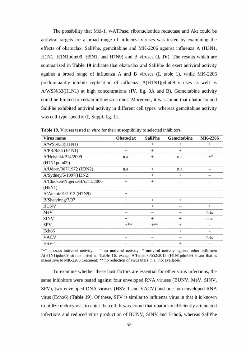
52
The possibility that Mcl-1, v-ATPase, ribonucleotide reductase and Akt could be
antiviral targets for a broad range of influenza viruses was tested by examining the
effects of obatoclax, SaliPhe, gemcitabine and MK-2206 against influenza A (H3N1,
H1N1, H1N1pdm09, H5N1, and H7N9) and B viruses (I, IV). The results which are
summarized in Table 19 indicate that obatoclax and SaliPhe do exert antiviral activity
against a broad range of influenza A and B viruses (I, table 1), while MK-2206
predominantly inhibits replication of influenza A(H1N1)pdm09 viruses as well as
A/WSN/33(H1N1) at high concentrations (IV, fig. 3A and B). Gemcitabine activity
could be limited to certain influenza strains. Moreover, it was found that obatoclax and
SaliPhe exhibited antiviral activity in different cell types, whereas gemcitabine activity
was cell-type specific (I, Suppl. fig. 1).
Table 19. Viruses tested in vitro for their susceptibility to selected inhibitors.
Virus name Obatoclax SaliPhe Gemcitabine MK-2206
A/WSN/33(H1N1) + + + +
A/PR/8/34 (H1N1) + + + –
A/Helsinki/P14/2009
(H1N1pdm09)
n.a. + n.a. +*
A/Udorn/307/1972 (H3N2) n.a. + n.a. –
A/Sydney/5/1997(H3N2) + + + –
A/Chicken/Nigeria/BA211/2006
(H5N1)
+ + – –
A/Anhui/01/2013 (H7N9) + – – –
B/Shandong/7/97 + + + –
BUNV + + – +
MeV – – – n.a.
SINV + + + n.a.
SFV +** +** + –
Echo6 + – + -
VACV – – – n.a.
HSV-1 – – + –
“+” possess antiviral activity, “–” no antiviral activity; * antiviral activity against other influenza
A(H1N1)pdm09 strains listed in Table 16, except A/Helsinki/552/2013 (H1N1pdm09) strain that is
insensitive to MK-2206 treatment; ** no reduction of virus titers; n.a., not available.
To examine whether these host factors are essential for other virus infections, the
same inhibitors were tested against four enveloped RNA viruses (BUNV, MeV, SINV,
SFV), two enveloped DNA viruses (HSV-1 and VACV) and one non-enveloped RNA
virus (Echo6) (Table 19). Of these, SFV is similar to influenza virus in that it is known
to utilize endocytosis to enter the cell. It was found that obatoclax efficiently attenuated
infections and reduced virus production of BUNV, SINV and Echo6, whereas SaliPhe

53
suppressed infection of BUNV and SINV. Obatoclax and SaliPhe were able to rescue
SFV-infected cells, however no reduction in the numbers of infectious virus particles
was observed. Moreover, gemcitabine suppressed HSV-1, SINV, SFV and Echo6 virus
infections.
In summary, SaliPhe and obatoclax exerted antiviral activity against a broad range
of influenza and some other RNA viruses. Obatoclax was toxic to cells during
prolonged incubation periods and, for this reason, was excluded from further studies.
Thus, subsequent studies focused on SaliPhe as the most potential antiviral agent.
However, it will be interesting to test the antiviral potential of another selective
inhibitor of Mcl-1, maronopyrrole A (maritoclax), which has higher specificity and
better pharmacological properties in comparison to obatoclax (Doi et al, 2012).
4.5. Development of resistance (unpublished)
The increasing clinical use of licensed antivirals has been associated with the
global emergence of amantadine- and/or oseltamivir-resistant influenza strains (Hayden
& de Jong, 2011; Zhou et al, 2013). Resistance results from both antigenic drift and
antigenic shift events specific for influenza viruses. Therefore, current prevention
options for influenza infections are limited and there is a need for developing next-
generation antiviral agents. Although, it was emphasized above that targeting host
factors essential for virus biology would be associated with a decreased likelihood of
resistance emergence, examples of virus resistance to potential host-directed antivirals
have been described in the literature (Qing et al, 2010; Triana-Baltzer et al, 2009).
The next experiment assessed whether continued inhibition of cellular v-ATPase
by SaliPhe could lead to the emergence of resistant influenza virus variants by
conducting a serial-passage experiment. During passaging the concentration of SaliPhe
was gradually increased. It is argued that this strategy does provide enough time for
virus resistance to emerge. A total of 15 passages were performed and the virus titer
determined after each passage. The results showed that the virus titer in the presence of
SaliPhe (0.35 µM, passage #8) decreased, and was not restored in further passages
(Figure 6). These results confirm the hypothesis that the influenza A/Helsinki/P14/2009
strain cannot efficiently adapt to selective pressure associated with the reduction of v-
ATPase activity. Thus, it is concluded that SaliPhe is a promising antiviral agent
exhibiting a high-barrier to case resistant virus variants.

54
Figure 6. Influenza A/Helsinki/P14/2009 virus titers from the serial-passage experiment on
NCI-H1666 cells.
4.6. Optimization of SaliPhe clinical utility (III)
SaliPhe is a small-molecule inhibitor of v-ATPase, a potential target for antitumor
chemotherapy (Lebreton et al, 2008). Moreover, on particle-induced osteolysis in vivo
SaliPhe was shown to inhibit bone destruction and suppress inflammatory response (Qin
et al, 2012). In addition, SaliPhe is a potential antiviral drug. It has been shown
previously that SaliPhe increases the survival rates of mice infected with lethal doses of
a mouse-adapted influenza strain (Müller et al, 2011). However, SaliPhe has a low
water solubility and high toxicity that are major bottlenecks for the evaluation of this
compound in the clinic (Müller et al, 2011).
In an attempt to increase the solubility and bioavailability of SaliPhe and to
reduce side-effects, it was decided to evaluate the PSi nanoparticle-based delivery
system that encompasses several advantageous properties for drug delivery. It has been
shown that PSi-based materials increase dissolution of otherwise poorly water-soluble
drugs loaded within its mesopores (Bimbo et al, 2011; Salonen et al, 2008). SaliPhe was
loaded into thermally hydrocarbonized porous silicon (THCPSi) nanoparticles with a
loading degree 2.88±0.11 wt% and then the drug releasing properties of SaliPhe-loaded
THCPSi nanoparticles were investigated. It was observed that at pH 7.4 THCPSi
nanoparticles released approximately 30% of the loaded SaliPhe during the first 15
minutes (III, fig. 3). In contrast, the pure form of SaliPhe could achieve only a 5%
dissolution within the same time period. Importantly, the THCPSi nanoparticles allowed
the delivery of SaliPhe in the absence of DMSO, and they increased the drug dissolution
rates in comparison to the pure drug.

55
The SaliPhe-loaded THCPSi nanoparticles were further investigated for their
ability to inhibit the influenza A/PR8-NS116-GFP virus infection in vitro. It was
observed that SaliPhe-loaded THCPSi nanoparticles like SaliPhe dissolved in DMSO
efficiently inhibited influenza virus-mediated GFP expression and reduced virus titers
by more than two orders of magnitude (III, fig. 5 and fig. 7). In addition, SaliPhe-
loaded THCPSi nanoparticles exerted less cytotoxic effects than SaliPhe dissolved in
DMSO (III, fig. 4 and fig. 5). Thus, the therapeutic window of SaliPhe-loaded THCPSi
nanoparticles was wider in comparison with SaliPhe dissolved in DMSO.
This data provides the first proof-of-principle of the potential of THCPSi
nanoparticles to delivery antiviral agent to virus-infected cells and to inhibit the
infection. Optimization of administration routes, such as inhalation or intravenous
injection, is currently envisioned, and further preclinical evaluation is warranted for the
development of PSi nanoparticles-based delivery systems as a potential platform for
targeted delivery of antiviral compounds in vivo.

56
5. FUTURE DIRECTIONS
Currently, the usage of licensed anti-influenza drugs is limited due to the globally
circulating drug-resistant influenza strains. It is essential to develop of new potential
antivirals targeting cellular host factors or components of signaling pathways in order to
control and treat future influenza virus epidemics and pandemics. Host-directed
antiviral agents represent a very attractive approach because the likelihood of resistance
emergence is very low and host-directed antivirals might possess broad-spectrum
antiviral properties.
The drug screening described in Study I focused on the cellular requirements for
virus entry, uncoating, nuclear import, and viral RNA transcription/translation. Another
screen was performed against wild type influenza A and B viruses in order to identify
host factors involved in other stages of influenza virus in A549 cells. The screen
revealed compounds identified in the previous screen (I) and a number of potential
antiviral-agents (unpublished). The hit list and EC50 values are presented in Table 20.
Additionally, their antiviral activity was tested against avian influenza A (H5N1 and
H7N9) strains. It is clear that further investigation of their mechanism of action and
their antiviral efficacy in vivo is needed.
Table 20. Identified compounds and their antiviral activity against influenza infection.
Name Target EC50 (µM)*
A(H1N1)a A (H5N1)
b A(H7N9)
c B
d
Flavopiridol
(Alvocidib)
CDK 0.60 0.34
0.47 0.50
SNS-032
(BMS-387032)
CDK 0.32 0.38
1.00 0.50
Dinaciclib
(SCH727965)
CDK 0.12 0.04
0.04 0.04
DCC-2036
(Rebastinib)
Abl 0.04 –
0.04 2.50
PIK-75 PI3K 0.08 0.08 – 0.13
Homoharring-
tonine
60S ribosome – –
– 0.12
* values obtained on A549 cells; “–” no antiviral activity; a A/WSN/33; b A/Chicken/Nigeria/BA211/2006; c A/Anhui/01/2013; d B/Shandong/7/97.

57
6. CONCLUSIONS
A screening approach was developed to identify druggable host factors involved
in influenza virus replication and it was efficiently used to reveal cellular Mcl-1, v-
ATPase, ribonucleotide reductase, Akt kinase and Bcl-xL. The detailed molecular
mechanisms of action of potential antiviral agents, such as obatoclax, SaliPhe,
gemcitabine, MK-2206 and ABT-263, targeting these host proteins, have been
elucidated. These potential antiviral agents demonstrated a broad spectrum of antiviral
activities in vitro. However, ABT-263, an inhibitor of Bcl-2 family proteins, was
ineffective in vivo. SaliPhe showed the greatest as an antiviral agent with activity
against a broad range of influenza A and B viruses and some other RNA viruses in vitro
and against a mouse adapted influenza strain in vivo. In order to overcome a low water
solubility and high toxicity of SaliPhe, its bioavailability was improved using a porous
silicon particle-based delivery system for use in future clinical trials. The results
presented in this thesis expand the understanding of virus-host interactions, and provide
a novel perspective for ways to adopt a rational approach in the discovery of new
antiviral agents.

58
ACKNOWLEDGEMENTS
This work was carried out at the Institute for Molecular Medicine Finland
(FIMM) during the years 2011-2013. I thank the Director of FIMM, Professor Olli
Kallioniemi, for providing excellent research facilities. This work was financially
supported by the Centre for International Mobility (CIMO), the University of Helsinki,
and the Finnish Foundation for Research on Viral Diseases.
I express my deepest appreciation to my thesis supervisor Docent Denis Kainov
for accepting me into his lab and for his trust and support throughout this project.
My warmest gratitude goes also to Dr. Laura Kakkola for introducing me to the
world of virology, and offer my sincere thanks for her wisdom and supportive attitude,
and for her continued interest in my work.
I warmly thank the reviewers of this thesis, Docent Maria Söderlund-Venermo
and Docent Eugene Makeyev, for their constructive and valuable comments, and for
completing the review in such a really tight time schedule. I would like to express my
great appreciation to Professor Stephan Ludwig who accepted the invitation to be the
opponent.
Scientific research would be nearly impossible without collaborators. I want to
express my deep gratitude to my collaborators and co-authors: Professor Jef De
Brabander for providing SaliPhe, Professor Ilkka Julkunen, Professor Olli Vapalahti,
Professor Veijo Hukkanen, Docent, Research Director Päivi Ojala, Docent Tero Ahola,
Docent Sampsa Matikainen, Dr. Pasi Kaukinen, Professor Richard Elliott, Professor
Claude Muller, Dr. Linda Theisen, Dr. Johanna Lampe, MSc Henrik Paavilainen, MSc
Niina Ikonen, MSc Johanna Viiliäinen, MSc Suvi Kuivanen, and MSc Ashwini Nagaraj
for providing virus stocks and testing of antiviral activities of hit compounds under
BSL-2 and BSL-3 conditions, Docent Anu Kantele and Docent Hannimari Kallio-
Kokko for coordinating and collecting the patients material, Docent Tuula Nyman, Dr.
Tiina Johanna Öhman and MSc Kristina Dzeyk for mass spectrometry analysis, Docent
Tero Aittokallio and MSc Yadav Bhagwan for developing methods to quantify the drug
response data, Professor Xavier Saelens and MSc Tine Ysenbaert for help with the mice
experiments, Professor Vladislav Verkhusha and MSc Salla Virtanen for fluorometric
analysis, Docent Emmy Verschuren and MSc Rita Matos for histochemistry, Dr. Jacob
Stenman, MSc Lin Feng and MSc Sandra Söderholm for RT-qPCR, MSc Janne Tynell
for ELISA, and MSc Dmitrii Bychkov for help with the bioinformatics analysis.
My special gratitude goes to Docent Helder Santos and Dr. Luis Bimbo for the
fruitful collaboration on the SaliPhe nanoparticles. I have greatly enjoyed discussing
that project.
I thank Docent Krister Wennerberg and his High Throughput Biomedicine Unit at
the FIMM Technology Centre, especially Dr. Jani Saarela, MSc Evgeny Kulesskiy,
MSc Ida Lindenschmidt, and BSc Anna Lehto, for excellent technical assistance and
59
help with the drug screens and also for providing aliquots of hit compounds. I would
like to thank Dr. Carina Von Schantz-Fant for her help and expertise in the automated
image acquisition and image analysis.
I also want to acknowledge all the people who voluntarily gave blood for some
parts of this study.
I thank the present and former members of Kainov’s research group, Masha,
Laura, Johanna, Minttu, Triin, Andrei, Petri, Maarten, and Jens, for creating such an
enjoyable atmosphere in the laboratory and for being such excellent colleagues. I
especially thank, Masha, Laura, Johanna, and Minttu, for their friendship and support,
and for outdoor activities such as the Extreme run and Nuuksio bicycle trip. I also
would like to thank all wonderful people of Kuznetsov’s and Verschuren’s research
teams, especially Daria, Pauliina, Katja, Ashwini, Jenni, Manuela, Annabrita, Dat,
Yuexi, and Sharif, for everyday support, lovely friendship and occasional cakes and
sparkling wine. I am grateful to have you in my life. I want to thank all FIMM
personnel for the stimulating atmosphere on FIMM coffee-break presentations and
annual retreats. I also thank girls from the faculty office, Katja and Tiina, for patiently
answering to thousands of questions especially in these last months. I thank Dr. Ewen
MacDonald for reviewing the language of my thesis. I also thank all my friends and
colleagues who have found time to read all or some parts of this manuscript.
Special thanks to my CIMO friends, Julia, Lena, Katya and Sergei, Mike,
Solomon, Boris, and Miiko, for the great times we had together and for the salsa. I also
thank my Finnish friends from the HePo bicycle club, Olli, Risto, and Satu, for the
interesting summer and winter trips. I also thank all my scientific friends and ultimate
Frisbee members from Pushchino, Russia, for emotional support and occasional warm
meetings around the world.
I warmly thank my school teachers from my native town Vyazniki, Vladimir
region, Russia, for giving me such a pleasant start in my life. I thank for your friendly
attitude, wisdom, and support during our meetings and phone conversations throughout
long years.
I send my thanks to my parents Nadezda and Valera, my sister Anna and her
family, my warmly loving grandparents, babushka Nina, babushka Tamara and
dedushka Volodya, and all my other relatives. I would not be here without your love
and support. Thank you for your trust in me.
Finally, my deepest gratitude goes to my husband Sergei. Thank you for
supporting my decision to start this PhD study in Finland, and for the following one and
half years of separation. I cannot imagine how I could finish my thesis without your
love, support, patience, dearest care in everyday life and the thousands of kilometres
that we have bicycled together.
Helsinki, March 2014

60
REFERENCES
Aeffner F, Bratasz A, Flano E, Powell KA, Davis IC (2012) Postinfection A77-1726 treatment improves
cardiopulmonary function in H1N1 influenza-infected mice. American journal of respiratory cell and
molecular biology 47: 543-551
Amorim MJ, Digard P (2006) Influenza A virus and the cell nucleus. Vaccine 24: 6651-6655
Antanasijevic A, Cheng H, Wardrop DJ, Rong L, Caffrey M (2013) Inhibition of influenza h7
hemagglutinin-mediated entry. PloS one 8: e76363
Arora DJ, Gasse N (1998) Influenza virus hemagglutinin stimulates the protein kinase C activity of
human polymorphonuclear leucocytes. Archives of virology 143: 2029-2037
Arts EJ, Hazuda DJ (2012) HIV-1 antiretroviral drug therapy. Cold Spring Harbor perspectives in
medicine 2: a007161 Atiee G, Lasseter K, Baughman S, McCullough A, Collis P, Hollister A, Hernandez J (2012) Absence of
pharmacokinetic interaction between intravenous peramivir and oral oseltamivir or rimantadine in
humans. Journal of clinical pharmacology 52: 1410-1419
Badani H, Garry RF, Wilson RB, Wimley WC (2011) Mechanism and action of flufirvitide, a peptide
inhibitor of influenza virus infection. Biophysical Journal 100: 216a
Baguelin M, Jit M, Miller E, Edmunds WJ (2012) Health and economic impact of the seasonal influenza
vaccination programme in England. Vaccine 30: 3459-3462
Baranovich T, Wong SS, Armstrong J, Marjuki H, Webby RJ, Webster RG, Govorkova EA (2013) T-705
(favipiravir) induces lethal mutagenesis in influenza A H1N1 viruses in vitro. Journal of virology 87:
3741-3751
Basu A, Antanasijevic A, Wang M, Li B, Mills DM, Ames JA, Nash PJ, Williams JD, Peet NP, Moir DT,
Prichard MN, Keith KA, Barnard DL, Caffrey M, Rong L, Bowlin TL (2014) New small molecule entry inhibitors targeting hemagglutinin-mediated influenza a virus fusion. Journal of virology 88:
1447-1460
Belser JA, Lu X, Szretter KJ, Jin X, Aschenbrenner LM, Lee A, Hawley S, Kim do H, Malakhov MP, Yu
M, Fang F, Katz JM (2007) DAS181, a novel sialidase fusion protein, protects mice from lethal avian
influenza H5N1 virus infection. The Journal of infectious diseases 196: 1493-1499
Bernhoff E, Tylden GD, Kjerpeseth LJ, Gutteberg TJ, Hirsch HH, Rinaldo CH (2010) Leflunomide
inhibition of BK virus replication in renal tubular epithelial cells. Journal of virology 84: 2150-2156
Bimbo LM, Makila E, Laaksonen T, Lehto VP, Salonen J, Hirvonen J, Santos HA (2011) Drug
permeation across intestinal epithelial cells using porous silicon nanoparticles. Biomaterials 32: 2625-
2633
Blanc M, Hsieh WY, Robertson KA, Kropp KA, Forster T, Shui G, Lacaze P, Watterson S, Griffiths SJ, Spann NJ, Meljon A, Talbot S, Krishnan K, Covey DF, Wenk MR, Craigon M, Ruzsics Z, Haas J,
Angulo A, Griffiths WJ, Glass CK, Wang Y, Ghazal P (2013) The transcription factor STAT-1
couples macrophage synthesis of 25-hydroxycholesterol to the interferon antiviral response. Immunity
38: 106-118
Bodian DL, Yamasaki RB, Buswell RL, Stearns JF, White JM, Kuntz ID (1993) Inhibition of the fusion-
inducing conformational change of influenza hemagglutinin by benzoquinones and hydroquinones.
Biochemistry 32: 2967-2978
Boriskin YS, Leneva IA, Pecheur EI, Polyak SJ (2008) Arbidol: a broad-spectrum antiviral compound
that blocks viral fusion. Current medicinal chemistry 15: 997-1005
Boyd MR, Farina C, Belfiore P, Gagliardi S, Kim JW, Hayakawa Y, Beutler JA, McKee TC, Bowman
BJ, Bowman EJ (2001) Discovery of a novel antitumor benzolactone enamide class that selectively
inhibits mammalian vacuolar-type (H+)-atpases. The Journal of pharmacology and experimental therapeutics 297: 114-120
Bright RA, Medina MJ, Xu X, Perez-Oronoz G, Wallis TR, Davis XM, Povinelli L, Cox NJ, Klimov AI
(2005) Incidence of adamantane resistance among influenza A (H3N2) viruses isolated worldwide
from 1994 to 2005: a cause for concern. Lancet 366: 1175-1181
Budd A, Alleva L, Alsharifi M, Koskinen A, Smythe V, Mullbacher A, Wood J, Clark I (2007) Increased
survival after gemfibrozil treatment of severe mouse influenza. Antimicrobial agents and
chemotherapy 51: 2965-2968
Carey MA, Bradbury JA, Rebolloso YD, Graves JP, Zeldin DC, Germolec DR (2010) Pharmacologic
inhibition of COX-1 and COX-2 in influenza A viral infection in mice. PloS one 5: e11610
Casey LC, Lee WM (2013) Hepatitis C virus therapy update 2013. Current opinion in gastroenterology
29: 243-249 Centers for Disease C, Prevention (2013) Prevention and control of seasonal influenza with vaccines.
Recommendations of the Advisory Committee on Immunization Practices--United States, 2013-2014.

61
MMWR Recommendations and reports : Morbidity and mortality weekly report Recommendations
and reports / Centers for Disease Control 62: 1-43
Cerqueira NM, Fernandes PA, Ramos MJ (2007) Understanding ribonucleotide reductase inactivation by
gemcitabine. Chemistry 13: 8507-8515
Chase G, Deng T, Fodor E, Leung BW, Mayer D, Schwemmle M, Brownlee G (2008) Hsp90 inhibitors
reduce influenza virus replication in cell culture. Virology 377: 431-439
Chaussade C, Rewcastle GW, Kendall JD, Denny WA, Cho K, Gronning LM, Chong ML, Anagnostou
SH, Jackson SP, Daniele N, Shepherd PR (2007) Evidence for functional redundancy of class IA
PI3K isoforms in insulin signalling. The Biochemical journal 404: 449-458
Chen JX, Xue HJ, Ye WC, Fang BH, Liu YH, Yuan SH, Yu P, Wang YQ (2009) Activity of
andrographolide and its derivatives against influenza virus in vivo and in vitro. Biological & pharmaceutical bulletin 32: 1385-1391
Chen TY, Chen DY, Wen HW, Ou JL, Chiou SS, Chen JM, Wong ML, Hsu WL (2013) Inhibition of
enveloped viruses infectivity by curcumin. PloS one 8: e62482
Chen W, Calvo PA, Malide D, Gibbs J, Schubert U, Bacik I, Basta S, O'Neill R, Schickli J, Palese P,
Henklein P, Bennink JR, Yewdell JW (2001) A novel influenza A virus mitochondrial protein that
induces cell death. Nature medicine 7: 1306-1312
Chen YB, Driscoll JP, McAfee SL, Spitzer TR, Rosenberg ES, Sanders R, Moss RB, Fang F, Marty FM
(2011) Treatment of parainfluenza 3 infection with DAS181 in a patient after allogeneic stem cell
transplantation. Clinical infectious diseases : an official publication of the Infectious Diseases Society
of America 53: e77-80
Cheshenko N, Trepanier JB, Stefanidou M, Buckley N, Gonzalez P, Jacobs W, Herold BC (2013) HSV activates Akt to trigger calcium release and promote viral entry: novel candidate target for treatment
and suppression. FASEB journal : official publication of the Federation of American Societies for
Experimental Biology 27: 2584-2599
Cheung TK, Poon LL (2007) Biology of influenza a virus. Annals of the New York Academy of Sciences
1102: 1-25
Courtney KD, Corcoran RB, Engelman JA (2010) The PI3K pathway as drug target in human cancer.
Journal of clinical oncology : official journal of the American Society of Clinical Oncology 28: 1075-
1083
Cullen SP, Martin SJ (2009) Caspase activation pathways: some recent progress. Cell death and
differentiation 16: 935-938
Das K, Ma LC, Xiao R, Radvansky B, Aramini J, Zhao L, Marklund J, Kuo RL, Twu KY, Arnold E, Krug RM, Montelione GT (2008) Structural basis for suppression of a host antiviral response by
influenza A virus. Proceedings of the National Academy of Sciences of the United States of America
105: 13093-13098
Das P, Deng X, Zhang L, Roth MG, Fontoura BM, Phillips MA, De Brabander JK (2013) SAR Based
Optimization of a 4-Quinoline Carboxylic Acid Analog with Potent Anti-Viral Activity. ACS
medicinal chemistry letters 4: 517-521
de Chassey B, Meyniel-Schicklin L, Aublin-Gex A, Andre P, Lotteau V (2012) New horizons for
antiviral drug discovery from virus-host protein interaction networks. Current opinion in virology 2:
606-613
De Clercq E (2004) Antiviral drugs in current clinical use. Journal of clinical virology : the official
publication of the Pan American Society for Clinical Virology 30: 115-133
De Clercq E (2006) Antiviral agents active against influenza A viruses. Nature reviews Drug discovery 5: 1015-1025
DeVincenzo JP (2012) The promise, pitfalls and progress of RNA-interference-based antiviral therapy for
respiratory viruses. Antiviral therapy 17: 213-225
Deyde VM, Xu X, Bright RA, Shaw M, Smith CB, Zhang Y, Shu Y, Gubareva LV, Cox NJ, Klimov AI
(2007) Surveillance of resistance to adamantanes among influenza A(H3N2) and A(H1N1) viruses
isolated worldwide. The Journal of infectious diseases 196: 249-257
Dharan NJ, Gubareva LV, Meyer JJ, Okomo-Adhiambo M, McClinton RC, Marshall SA, St George K,
Epperson S, Brammer L, Klimov AI, Bresee JS, Fry AM, Oseltamivir-Resistance Working G (2009)
Infections with oseltamivir-resistant influenza A(H1N1) virus in the United States. JAMA : the
journal of the American Medical Association 301: 1034-1041
Dixit R, Khandaker G, Ilgoutz S, Rashid H, Booy R (2013) Emergence of oseltamivir resistance: control and management of influenza before, during and after the pandemic. Infectious disorders drug targets
13: 34-45
Doi K, Li R, Sung SS, Wu H, Liu Y, Manieri W, Krishnegowda G, Awwad A, Dewey A, Liu X, Amin S,
Cheng C, Qin Y, Schonbrunn E, Daughdrill G, Loughran TP, Jr., Sebti S, Wang HG (2012) Discovery
of marinopyrrole A (maritoclax) as a selective Mcl-1 antagonist that overcomes ABT-737 resistance

62
by binding to and targeting Mcl-1 for proteasomal degradation. The Journal of biological chemistry
287: 10224-10235
Dove BK, Surtees R, Bean TJ, Munday D, Wise HM, Digard P, Carroll MW, Ajuh P, Barr JN, Hiscox JA
(2012) A quantitative proteomic analysis of lung epithelial (A549) cells infected with 2009 pandemic
influenza A virus using stable isotope labelling with amino acids in cell culture. Proteomics 12: 1431-
1436
Dreisigacker S, Latek D, Bockelmann S, Huss M, Wieczorek H, Filipek S, Gohlke H, Menche D,
Carlomagno T (2012) Understanding the inhibitory effect of highly potent and selective archazolides
binding to the vacuolar ATPase. Journal of chemical information and modeling 52: 2265-2272
Droebner K, Pleschka S, Ludwig S, Planz O (2011) Antiviral activity of the MEK-inhibitor U0126
against pandemic H1N1v and highly pathogenic avian influenza virus in vitro and in vivo. Antiviral research 92: 195-203
Drose S, Altendorf K (1997) Bafilomycins and concanamycins as inhibitors of V-ATPases and P-
ATPases. The Journal of experimental biology 200: 1-8
Dudek SE, Luig C, Pauli EK, Schubert U, Ludwig S (2010) The clinically approved proteasome inhibitor
PS-341 efficiently blocks influenza A virus and vesicular stomatitis virus propagation by establishing
an antiviral state. Journal of virology 84: 9439-9451
Ehrhardt C, Ludwig S (2009) A new player in a deadly game: influenza viruses and the PI3K/Akt
signalling pathway. Cellular microbiology 11: 863-871
Ehrhardt C, Marjuki H, Wolff T, Nurnberg B, Planz O, Pleschka S, Ludwig S (2006) Bivalent role of the
phosphatidylinositol-3-kinase (PI3K) during influenza virus infection and host cell defence. Cellular
microbiology 8: 1336-1348 Ehrhardt C, Ruckle A, Hrincius ER, Haasbach E, Anhlan D, Ahmann K, Banning C, Reiling SJ, Kuhn J,
Strobl S, Vitt D, Leban J, Planz O, Ludwig S (2013) The NF-kappaB inhibitor SC75741 efficiently
blocks influenza virus propagation and confers a high barrier for development of viral resistance.
Cellular microbiology 15: 1198-1211
Eierhoff T, Hrincius ER, Rescher U, Ludwig S, Ehrhardt C (2010) The epidermal growth factor receptor
(EGFR) promotes uptake of influenza A viruses (IAV) into host cells. PLoS pathogens 6: e1001099
Engelman JA (2009) Targeting PI3K signalling in cancer: opportunities, challenges and limitations.
Nature reviews Cancer 9: 550-562
Ferby I, Waga I, Kume K, Sakanaka C, Shimizu T (1996) PAF-induced MAPK activation is inhibited by
wortmannin in neutrophils and macrophages. Advances in experimental medicine and biology 416:
321-326 Finter NB, Chapman S, Dowd P, Johnston JM, Manna V, Sarantis N, Sheron N, Scott G, Phua S, Tatum
PB (1991) The use of interferon-alpha in virus infections. Drugs 42: 749-765
Follis AV, Chipuk JE, Fisher JC, Yun MK, Grace CR, Nourse A, Baran K, Ou L, Min L, White SW,
Green DR, Kriwacki RW (2013) PUMA binding induces partial unfolding within BCL-xL to disrupt
p53 binding and promote apoptosis. Nature chemical biology 9: 163-168
Fremin C, Meloche S (2010) From basic research to clinical development of MEK1/2 inhibitors for
cancer therapy. Journal of hematology & oncology 3: 8
Fresno Vara JA, Casado E, de Castro J, Cejas P, Belda-Iniesta C, Gonzalez-Baron M (2004) PI3K/Akt
signalling pathway and cancer. Cancer treatment reviews 30: 193-204
Furuta Y, Takahashi K, Kuno-Maekawa M, Sangawa H, Uehara S, Kozaki K, Nomura N, Egawa H,
Shiraki K (2005) Mechanism of action of T-705 against influenza virus. Antimicrobial agents and
chemotherapy 49: 981-986 Furuta Y, Takahashi K, Shiraki K, Sakamoto K, Smee DF, Barnard DL, Gowen BB, Julander JG, Morrey
JD (2009) T-705 (favipiravir) and related compounds: Novel broad-spectrum inhibitors of RNA viral
infections. Antiviral research 82: 95-102
Gefenaite G, Rahamat-Langendoen J, Ambrozaitis A, Mickiene A, Jancoriene L, Kuliese M, Velyvyte D,
Niesters H, Stolk RP, Zagminas K, Hak E (2014) Seasonal influenza vaccine effectiveness against
influenza in 2012-2013: A hospital-based case-control study in Lithuania. Vaccine 32: 857-863
Guinea R, Carrasco L (1995) Requirement for vacuolar proton-ATPase activity during entry of influenza
virus into cells. Journal of virology 69: 2306-2312
Guo N, Peng Z (2013) MG132, a proteasome inhibitor, induces apoptosis in tumor cells. Asia-Pacific
journal of clinical oncology 9: 6-11
Haasbach E, Hartmayer C, Planz O (2013a) Combination of MEK inhibitors and oseltamivir leads to synergistic antiviral effects after influenza A virus infection in vitro. Antiviral research 98: 319-324
Haasbach E, Pauli EK, Spranger R, Mitzner D, Schubert U, Kircheis R, Planz O (2011) Antiviral activity
of the proteasome inhibitor VL-01 against influenza A viruses. Antiviral research 91: 304-313

63
Haasbach E, Reiling SJ, Ehrhardt C, Droebner K, Ruckle A, Hrincius ER, Leban J, Strobl S, Vitt D,
Ludwig S, Planz O (2013b) The NF-kappaB inhibitor SC75741 protects mice against highly
pathogenic avian influenza A virus. Antiviral research 99: 336-344
Han X, Bushweller JH, Cafiso DS, Tamm LK (2001) Membrane structure and fusion-triggering
conformational change of the fusion domain from influenza hemagglutinin. Nature structural biology
8: 715-720
Hayden FG (2013) Newer influenza antivirals, biotherapeutics and combinations. Influenza and other
respiratory viruses 7 Suppl 1: 63-75
Hayden FG, de Jong MD (2011) Emerging influenza antiviral resistance threats. The Journal of infectious
diseases 203: 6-10
Hayden FG, Schlepushkin AN, Pushkarskaya NL (1984) Combined interferon-alpha 2, rimantadine hydrochloride, and ribavirin inhibition of influenza virus replication in vitro. Antimicrobial agents and
chemotherapy 25: 53-57
Herbert BS, Gellert GC, Hochreiter A, Pongracz K, Wright WE, Zielinska D, Chin AC, Harley CB, Shay
JW, Gryaznov SM (2005) Lipid modification of GRN163, an N3'-->P5' thio-phosphoramidate
oligonucleotide, enhances the potency of telomerase inhibition. Oncogene 24: 5262-5268
Hirai H, Sootome H, Nakatsuru Y, Miyama K, Taguchi S, Tsujioka K, Ueno Y, Hatch H, Majumder PK,
Pan BS, Kotani H (2010) MK-2206, an allosteric Akt inhibitor, enhances antitumor efficacy by
standard chemotherapeutic agents or molecular targeted drugs in vitro and in vivo. Molecular cancer
therapeutics 9: 1956-1967
Hoffmann HH, Kunz A, Simon VA, Palese P, Shaw ML (2011) Broad-spectrum antiviral that interferes
with de novo pyrimidine biosynthesis. Proceedings of the National Academy of Sciences of the United States of America 108: 5777-5782
Hoffmann HH, Palese P, Shaw ML (2008) Modulation of influenza virus replication by alteration of
sodium ion transport and protein kinase C activity. Antiviral research 80: 124-134
Hosoya M, Matsuyama S, Baba M, Suzuki H, Shigeta S (1992) Effects of protease inhibitors on
replication of various myxoviruses. Antimicrobial agents and chemotherapy 36: 1432-1436
Huss M, Sasse F, Kunze B, Jansen R, Steinmetz H, Ingenhorst G, Zeeck A, Wieczorek H (2005)
Archazolid and apicularen: novel specific V-ATPase inhibitors. BMC biochemistry 6: 13
Ilyushina NA, Bovin NV, Webster RG, Govorkova EA (2006) Combination chemotherapy, a potential
strategy for reducing the emergence of drug-resistant influenza A variants. Antiviral research 70: 121-
131
Ilyushina NA, Hay A, Yilmaz N, Boon AC, Webster RG, Govorkova EA (2008) Oseltamivir-ribavirin combination therapy for highly pathogenic H5N1 influenza virus infection in mice. Antimicrobial
agents and chemotherapy 52: 3889-3897
Iversen PL, Schnell F, Crumley S, Mourich D, Voss T (2012) Post exposure efficacy of AVI-7100
against influenza A in mouse and ferret infection models. Thesis on 22nd European Congress of
Clinical Microbiology and Infectious Diseases.
Jagger BW, Wise HM, Kash JC, Walters KA, Wills NM, Xiao YL, Dunfee RL, Schwartzman LM,
Ozinsky A, Bell GL, Dalton RM, Lo A, Efstathiou S, Atkins JF, Firth AE, Taubenberger JK, Digard P
(2012) An overlapping protein-coding region in influenza A virus segment 3 modulates the host
response. Science 337: 199-204
Jesus T, Rogelio L, Abraham C, Uriel L, G JD, Alfonso MT, Lilia BB (2012) Prediction of antiviral
peptides derived from viral fusion proteins potentially active against herpes simplex and influenza A
viruses. Bioinformation 8: 870-874 Johnson NP, Mueller J (2002) Updating the accounts: global mortality of the 1918-1920 "Spanish"
influenza pandemic. Bulletin of the history of medicine 76: 105-115
Jones JC, Turpin EA, Bultmann H, Brandt CR, Schultz-Cherry S (2006) Inhibition of influenza virus
infection by a novel antiviral peptide that targets viral attachment to cells. Journal of virology 80:
11960-11967
Jonsson CB, Hooper J, Mertz G (2008) Treatment of hantavirus pulmonary syndrome. Antiviral research
78: 162-169
Kane PM, Smardon AM (2003) Assembly and regulation of the yeast vacuolar H+-ATPase. Journal of
bioenergetics and biomembranes 35: 313-321
Kao RY, Yang D, Lau LS, Tsui WH, Hu L, Dai J, Chan MP, Chan CM, Wang P, Zheng BJ, Sun J, Huang
JD, Madar J, Chen G, Chen H, Guan Y, Yuen KY (2010) Identification of influenza A nucleoprotein as an antiviral target. Nature biotechnology 28: 600-605
Karlas A, Machuy N, Shin Y, Pleissner KP, Artarini A, Heuer D, Becker D, Khalil H, Ogilvie LA, Hess
S, Maurer AP, Muller E, Wolff T, Rudel T, Meyer TF (2010) Genome-wide RNAi screen identifies
human host factors crucial for influenza virus replication. Nature 463: 818-822

64
Kati WM, Montgomery D, Carrick R, Gubareva L, Maring C, McDaniel K, Steffy K, Molla A, Hayden F,
Kempf D, Kohlbrenner W (2002) In vitro characterization of A-315675, a highly potent inhibitor of A
and B strain influenza virus neuraminidases and influenza virus replication. Antimicrobial agents and
chemotherapy 46: 1014-1021
Keating R, Hertz T, Wehenkel M, Harris TL, Edwards BA, McClaren JL, Brown SA, Surman S, Wilson
ZS, Bradley P, Hurwitz J, Chi H, Doherty PC, Thomas PG, McGargill MA (2013) The kinase mTOR
modulates the antibody response to provide cross-protective immunity to lethal infection with
influenza virus. Nature immunology
Khan SH, Goba A, Chu M, Roth C, Healing T, Marx A, Fair J, Guttieri MC, Ferro P, Imes T, Monagin C,
Garry RF, Bausch DG, Mano River Union Lassa Fever N (2008) New opportunities for field research
on the pathogenesis and treatment of Lassa fever. Antiviral research 78: 103-115 Kim M, Kim SY, Lee HW, Shin JS, Kim P, Jung YS, Jeong HS, Hyun JK, Lee CK (2013) Inhibition of
influenza virus internalization by (-)-epigallocatechin-3-gallate. Antiviral research 100: 460-472
Konig R, Stertz S, Zhou Y, Inoue A, Hoffmann HH, Bhattacharyya S, Alamares JG, Tscherne DM,
Ortigoza MB, Liang Y, Gao Q, Andrews SE, Bandyopadhyay S, De Jesus P, Tu BP, Pache L, Shih C,
Orth A, Bonamy G, Miraglia L, Ideker T, Garcia-Sastre A, Young JA, Palese P, Shaw ML, Chanda
SK (2010) Human host factors required for influenza virus replication. Nature 463: 813-817
Konig R, Zhou Y, Elleder D, Diamond TL, Bonamy GM, Irelan JT, Chiang CY, Tu BP, De Jesus PD,
Lilley CE, Seidel S, Opaluch AM, Caldwell JS, Weitzman MD, Kuhen KL, Bandyopadhyay S, Ideker
T, Orth AP, Miraglia LJ, Bushman FD, Young JA, Chanda SK (2008) Global analysis of host-
pathogen interactions that regulate early-stage HIV-1 replication. Cell 135: 49-60
Kratky M, Vinsova J (2011) Antiviral activity of substituted salicylanilides--a review. Mini reviews in medicinal chemistry 11: 956-967
Krishnan MN, Ng A, Sukumaran B, Gilfoy FD, Uchil PD, Sultana H, Brass AL, Adametz R, Tsui M,
Qian F, Montgomery RR, Lev S, Mason PW, Koski RA, Elledge SJ, Xavier RJ, Agaisse H, Fikrig E
(2008) RNA interference screen for human genes associated with West Nile virus infection. Nature
455: 242-245
Kugel D, Kochs G, Obojes K, Roth J, Kobinger GP, Kobasa D, Haller O, Staeheli P, von Messling V
(2009) Intranasal administration of alpha interferon reduces seasonal influenza A virus morbidity in
ferrets. Journal of virology 83: 3843-3851
Kutluay SB, Doroghazi J, Roemer ME, Triezenberg SJ (2008) Curcumin inhibits herpes simplex virus
immediate-early gene expression by a mechanism independent of p300/CBP histone acetyltransferase
activity. Virology 373: 239-247 Le QM, Kiso M, Someya K, Sakai YT, Nguyen TH, Nguyen KH, Pham ND, Ngyen HH, Yamada S,
Muramoto Y, Horimoto T, Takada A, Goto H, Suzuki T, Suzuki Y, Kawaoka Y (2005) Avian flu:
isolation of drug-resistant H5N1 virus. Nature 437: 1108
Lebreton S, Jaunbergs J, Roth MG, Ferguson DA, De Brabander JK (2008) Evaluating the potential of
vacuolar ATPase inhibitors as anticancer agents and multigram synthesis of the potent
salicylihalamide analog saliphenylhalamide. Bioorganic & medicinal chemistry letters 18: 5879-5883
Lee MG, Kim KH, Park KY, Kim JS (1996) Evaluation of anti-influenza effects of camostat in mice
infected with non-adapted human influenza viruses. Archives of virology 141: 1979-1989
Lee YT, Kim KH, Ko EJ, Lee YN, Kim MC, Kwon YM, Tang Y, Cho MK, Lee YJ, Kang SM (2014)
New vaccines against influenza virus. Clinical and experimental vaccine research 3: 12-28
Leneva IA, Russell RJ, Boriskin YS, Hay AJ (2009) Characteristics of arbidol-resistant mutants of
influenza virus: implications for the mechanism of anti-influenza action of arbidol. Antiviral research 81: 132-140
Li Q, Brass AL, Ng A, Hu Z, Xavier RJ, Liang TJ, Elledge SJ (2009) A genome-wide genetic screen for
host factors required for hepatitis C virus propagation. Proceedings of the National Academy of
Sciences of the United States of America 106: 16410-16415
Li Q, Zhou L, Zhou M, Chen Z, Li F, Wu H, Xiang N, Chen E, Tang F, Wang D, Meng L, Hong Z, Tu
W, Cao Y, Li L, Ding F, Liu B, Wang M, Xie R, Gao R, Li X, Bai T, Zou S, He J, Hu J, Xu Y, Chai
C, Wang S, Gao Y, Jin L, Zhang Y, Luo H, Yu H, Gao L, Pang X, Liu G, Shu Y, Yang W, Uyeki TM,
Wang Y, Wu F, Feng Z (2013) Preliminary Report: Epidemiology of the Avian Influenza A (H7N9)
Outbreak in China. The New England journal of medicine
Lietzen N, Ohman T, Rintahaka J, Julkunen I, Aittokallio T, Matikainen S, Nyman TA (2011)
Quantitative subcellular proteome and secretome profiling of influenza A virus-infected human primary macrophages. PLoS pathogens 7: e1001340
Liu D, Herranz-Blanco B, Makila E, Arriaga LR, Mirza S, Weitz DA, Sandler N, Salonen J, Hirvonen J,
Santos HA (2013a) Microfluidic Templated Mesoporous Silicon-Solid Lipid Microcomposites for
Sustained Drug Delivery. ACS applied materials & interfaces

65
Liu SY, Aliyari R, Chikere K, Li G, Marsden MD, Smith JK, Pernet O, Guo H, Nusbaum R, Zack JA,
Freiberg AN, Su L, Lee B, Cheng G (2013b) Interferon-inducible cholesterol-25-hydroxylase broadly
inhibits viral entry by production of 25-hydroxycholesterol. Immunity 38: 92-105
Lopez-Martinez R, Ramirez-Salinas GL, Correa-Basurto J, Barron BL (2013) Inhibition of influenza a
virus infection in vitro by peptides designed in silico. PloS one 8: e76876
Ludwig S, Planz O, Pleschka S, Wolff T (2003) Influenza-virus-induced signaling cascades: targets for
antiviral therapy? Trends in molecular medicine 9: 46-52
Ludwig S, Wolff T, Ehrhardt C, Wurzer WJ, Reinhardt J, Planz O, Pleschka S (2004) MEK inhibition
impairs influenza B virus propagation without emergence of resistant variants. FEBS letters 561: 37-
43
Luksic I, Clay S, Falconer R, Pulanic D, Rudan I, Campbell H, Nair H (2013) Effectiveness of seasonal influenza vaccines in children -- a systematic review and meta-analysis. Croatian medical journal 54:
135-145
Luo G, Colonno R, Krystal M (1996) Characterization of a hemagglutinin-specific inhibitor of influenza
A virus. Virology 226: 66-76
Malakhov MP, Aschenbrenner LM, Smee DF, Wandersee MK, Sidwell RW, Gubareva LV, Mishin VP,
Hayden FG, Kim DH, Ing A, Campbell ER, Yu M, Fang F (2006) Sialidase fusion protein as a novel
broad-spectrum inhibitor of influenza virus infection. Antimicrobial agents and chemotherapy 50:
1470-1479
Marjuki H, Gornitzky A, Marathe BM, Ilyushina NA, Aldridge JR, Desai G, Webby RJ, Webster RG
(2011) Influenza A virus-induced early activation of ERK and PI3K mediates V-ATPase-dependent
intracellular pH change required for fusion. Cellular microbiology 13: 587-601 Markland W, McQuaid TJ, Jain J, Kwong AD (2000) Broad-spectrum antiviral activity of the IMP
dehydrogenase inhibitor VX-497: a comparison with ribavirin and demonstration of antiviral
additivity with alpha interferon. Antimicrobial agents and chemotherapy 44: 859-866
Markman B, Atzori F, Perez-Garcia J, Tabernero J, Baselga J (2010) Status of PI3K inhibition and
biomarker development in cancer therapeutics. Annals of oncology : official journal of the European
Society for Medical Oncology / ESMO 21: 683-691
Maruyama Y, Yoshida H, Uchino S, Yokoyama K, Yamamoto H, Takinami M, Hosoya T (2011)
Nafamostat mesilate as an anticoagulant during continuous veno-venous hemodialysis: a three-year
retrospective cohort study. The International journal of artificial organs 34: 571-576
Mata MA, Satterly N, Versteeg GA, Frantz D, Wei S, Williams N, Schmolke M, Pena-Llopis S,
Brugarolas J, Forst CV, White MA, Garcia-Sastre A, Roth MG, Fontoura BM (2011) Chemical inhibition of RNA viruses reveals REDD1 as a host defense factor. Nature chemical biology 7: 712-
719
Matsubara T, Onishi A, Saito T, Shimada A, Inoue H, Taki T, Nagata K, Okahata Y, Sato T (2010) Sialic
acid-mimic peptides as hemagglutinin inhibitors for anti-influenza therapy. Journal of medicinal
chemistry 53: 4441-4449
Mazur I, Wurzer WJ, Ehrhardt C, Pleschka S, Puthavathana P, Silberzahn T, Wolff T, Planz O, Ludwig S
(2007) Acetylsalicylic acid (ASA) blocks influenza virus propagation via its NF-kappaB-inhibiting
activity. Cellular microbiology 9: 1683-1694
McKimm-Breschkin JL (2013a) Influenza neuraminidase inhibitors: antiviral action and mechanisms of
resistance. Influenza and other respiratory viruses 7 Suppl 1: 25-36
McKimm-Breschkin JL (2013b) Influenza neuraminidase inhibitors: antiviral action and mechanisms of
resistance. Influenza and other respiratory viruses 7 Suppl 1: 25-36 Medina RA, Garcia-Sastre A (2011) Influenza A viruses: new research developments. Nature reviews
Microbiology 9: 590-603
Meijer A, Lackenby A, Hungnes O, Lina B, van-der-Werf S, Schweiger B, Opp M, Paget J, van-de-
Kassteele J, Hay A, Zambon M, European Influenza Surveillance S (2009) Oseltamivir-resistant
influenza virus A (H1N1), Europe, 2007-08 season. Emerging infectious diseases 15: 552-560
Meneghesso S, Vanderlinden E, Stevaert A, McGuigan C, Balzarini J, Naesens L (2012) Synthesis and
biological evaluation of pyrimidine nucleoside monophosphate prodrugs targeted against influenza
virus. Antiviral research 94: 35-43
Michaelis M, Geiler J, Klassert D, Doerr HW, Cinatl J, Jr. (2009) Infection of human retinal pigment
epithelial cells with influenza A viruses. Investigative ophthalmology & visual science 50: 5419-5425
Moeller A, Kirchdoerfer RN, Potter CS, Carragher B, Wilson IA (2012) Organization of the influenza virus replication machinery. Science 338: 1631-1634
Moscona A (2009) Global transmission of oseltamivir-resistant influenza. The New England journal of
medicine 360: 953-956

66
Moss RB, Hansen C, Sanders RL, Hawley S, Li T, Steigbigel RT (2012) A phase II study of DAS181, a
novel host directed antiviral for the treatment of influenza infection. The Journal of infectious diseases
206: 1844-1851
Mukherjee B, Tomimatsu N, Amancherla K, Camacho CV, Pichamoorthy N, Burma S (2012) The dual
PI3K/mTOR inhibitor NVP-BEZ235 is a potent inhibitor of ATM- and DNA-PKCs-mediated DNA
damage responses. Neoplasia 14: 34-43
Müller KH, Kainov DE, El Bakkouri K, Saelens X, De Brabander JK, Kittel C, Samm E, Muller CP
(2011) The proton translocation domain of cellular vacuolar ATPase provides a target for the
treatment of influenza A virus infections. British journal of pharmacology 164: 344-357
Müller KH, Kakkola L, Nagaraj AS, Cheltsov AV, Anastasina M, Kainov DE (2012) Emerging cellular
targets for influenza antiviral agents. Trends in pharmacological sciences 33: 89-99 Munday DC, Surtees R, Emmott E, Dove BK, Digard P, Barr JN, Whitehouse A, Matthews D, Hiscox JA
(2012) Using SILAC and quantitative proteomics to investigate the interactions between viral and host
proteomes. Proteomics 12: 666-672
Munger J, Bennett BD, Parikh A, Feng XJ, McArdle J, Rabitz HA, Shenk T, Rabinowitz JD (2008)
Systems-level metabolic flux profiling identifies fatty acid synthesis as a target for antiviral therapy.
Nature biotechnology 26: 1179-1186
Muramoto Y, Noda T, Kawakami E, Akkina R, Kawaoka Y (2013) Identification of novel influenza A
virus proteins translated from PA mRNA. Journal of virology 87: 2455-2462
Nayak DP, Balogun RA, Yamada H, Zhou ZH, Barman S (2009) Influenza virus morphogenesis and
budding. Virus research 143: 147-161
Nelson MI, Simonsen L, Viboud C, Miller MA, Holmes EC (2009) The origin and global emergence of adamantane resistant A/H3N2 influenza viruses. Virology 388: 270-278
Nguyen JT, Hoopes JD, Le MH, Smee DF, Patick AK, Faix DJ, Blair PJ, de Jong MD, Prichard MN,
Went GT (2010) Triple combination of amantadine, ribavirin, and oseltamivir is highly active and
synergistic against drug resistant influenza virus strains in vitro. PloS one 5: e9332
Nguyen M, Marcellus RC, Roulston A, Watson M, Serfass L, Murthy Madiraju SR, Goulet D, Viallet J,
Belec L, Billot X, Acoca S, Purisima E, Wiegmans A, Cluse L, Johnstone RW, Beauparlant P, Shore
GC (2007) Small molecule obatoclax (GX15-070) antagonizes MCL-1 and overcomes MCL-1-
mediated resistance to apoptosis. Proceedings of the National Academy of Sciences of the United
States of America 104: 19512-19517
Nimmerjahn F, Dudziak D, Dirmeier U, Hobom G, Riedel A, Schlee M, Staudt LM, Rosenwald A,
Behrends U, Bornkamm GW, Mautner J (2004) Active NF-kappaB signalling is a prerequisite for influenza virus infection. The Journal of general virology 85: 2347-2356
Noma K, Kiyotani K, Kouchi H, Fujii Y, Egi Y, Tanaka K, Yoshida T (1998) Endogenous protease-
dependent replication of human influenza viruses in two MDCK cell lines. Archives of virology 143:
1893-1909
Ortigoza MB, Dibben O, Maamary J, Martinez-Gil L, Leyva-Grado VH, Abreu P, Jr., Ayllon J, Palese P,
Shaw ML (2012) A novel small molecule inhibitor of influenza A viruses that targets polymerase
function and indirectly induces interferon. PLoS pathogens 8: e1002668
Ou JL, Mizushina Y, Wang SY, Chuang DY, Nadar M, Hsu WL (2013) Structure-activity relationship
analysis of curcumin analogues on anti-influenza virus activity. The FEBS journal 280: 5829-5840
Pan HY, Yano M, Kido H (2011) Effects of inhibitors of Toll-like receptors, protease-activated receptor-2
signalings and trypsin on influenza A virus replication and upregulation of cellular factors in
cardiomyocytes. The journal of medical investigation : JMI 58: 19-28 Pemovska T, Kontro M, Yadav B, Edgren H, Eldfors S, Szwajda A, Almusa H, Bespalov MM, Ellonen P,
Elonen E, Gjertsen BT, Karjalainen R, Kulesskiy E, Lagstrom S, Lehto A, Lepisto M, Lundan T,
Majumder MM, Marti JM, Mattila P, Murumagi A, Mustjoki S, Palva A, Parsons A, Pirttinen T,
Ramet ME, Suvela M, Turunen L, Vastrik I, Wolf M, Knowles J, Aittokallio T, Heckman CA, Porkka
K, Kallioniemi O, Wennerberg K (2013) Individualized systems medicine strategy to tailor treatments
for patients with chemorefractory acute myeloid leukemia. Cancer discovery 3: 1416-1429
Perelson AS, Rong L, Hayden FG (2012) Combination antiviral therapy for influenza: predictions from
modeling of human infections. The Journal of infectious diseases 205: 1642-1645
Perez L, Carrasco L (1994) Involvement of the vacuolar H(+)-ATPase in animal virus entry. The Journal
of general virology 75 ( Pt 10): 2595-2606
Pinto LH, Lamb RA (2006) The M2 proton channels of influenza A and B viruses. The Journal of biological chemistry 281: 8997-9000
Planz O (2013) Development of cellular signaling pathway inhibitors as new antivirals against influenza.
Antiviral research 98: 457-468

67
Pleschka S, Wolff T, Ehrhardt C, Hobom G, Planz O, Rapp UR, Ludwig S (2001) Influenza virus
propagation is impaired by inhibition of the Raf/MEK/ERK signalling cascade. Nature cell biology 3:
301-305
Plotch SJ, O'Hara B, Morin J, Palant O, LaRocque J, Bloom JD, Lang SA, Jr., DiGrandi MJ, Bradley M,
Nilakantan R, Gluzman Y (1999) Inhibition of influenza A virus replication by compounds interfering
with the fusogenic function of the viral hemagglutinin. Journal of virology 73: 140-151
Pukrittayakamee S, Jittamala P, Stepniewska K, Lindegardh N, Chueasuwanchai S, Leowattana W,
Phakdeeraj A, Permpunpanich S, Hanpithakpong W, Pan-Ngum W, Fukuda C, Panapipat S,
Singhasivanon P, White NJ, Day NP (2011) An open-label crossover study to evaluate potential
pharmacokinetic interactions between oral oseltamivir and intravenous zanamivir in healthy Thai
adults. Antimicrobial agents and chemotherapy 55: 4050-4057 Qin A, Cheng TS, Lin Z, Cao L, Chim SM, Pavlos NJ, Xu J, Zheng MH, Dai KR (2012) Prevention of
wear particle-induced osteolysis by a novel V-ATPase inhibitor saliphenylhalamide through inhibition
of osteoclast bone resorption. PloS one 7: e34132
Qing M, Zou G, Wang QY, Xu HY, Dong H, Yuan Z, Shi PY (2010) Characterization of dengue virus
resistance to brequinar in cell culture. Antimicrobial agents and chemotherapy 54: 3686-3695
Rechtman MM, Har-Noy O, Bar-Yishay I, Fishman S, Adamovich Y, Shaul Y, Halpern Z, Shlomai A
(2010) Curcumin inhibits hepatitis B virus via down-regulation of the metabolic coactivator PGC-
1alpha. FEBS letters 584: 2485-2490
Renault TT, Chipuk JE (2013) Getting away with murder: how does the BCL-2 family of proteins kill
with immunity? Annals of the New York Academy of Sciences 1285: 59-79
Rong L, Perelson AS (2010) Treatment of hepatitis C virus infection with interferon and small molecule direct antivirals: viral kinetics and modeling. Critical reviews in immunology 30: 131-148
Root CN, Wills EG, McNair LL, Whittaker GR (2000) Entry of influenza viruses into cells is inhibited by
a highly specific protein kinase C inhibitor. The Journal of general virology 81: 2697-2705
Rossignol JF, La Frazia S, Chiappa L, Ciucci A, Santoro MG (2009) Thiazolides, a new class of anti-
influenza molecules targeting viral hemagglutinin at the post-translational level. The Journal of
biological chemistry 284: 29798-29808
Russo A, Bronte G, Fulfaro F, Cicero G, Adamo V, Gebbia N, Rizzo S (2010) Bortezomib: a new pro-
apoptotic agent in cancer treatment. Current cancer drug targets 10: 55-67
Salonen J, Kaukonen AM, Hirvonen J, Lehto VP (2008) Mesoporous silicon in drug delivery
applications. Journal of pharmaceutical sciences 97: 632-653
Samji T (2009) Influenza A: understanding the viral life cycle. The Yale journal of biology and medicine 82: 153-159
Santos HA, Bimbo LM, Lehto VP, Airaksinen AJ, Salonen J, Hirvonen J (2011) Multifunctional porous
silicon for therapeutic drug delivery and imaging. Current drug discovery technologies 8: 228-249
Savinova OV, Pavlova NI, Boreko EI (2009) [New betulin derivatives in combination with rimantadine
for inhibition of influenza virus reproduction]. Antibiotiki i khimioterapiia = Antibiotics and
chemoterapy [sic] / Ministerstvo meditsinskoi i mikrobiologicheskoi promyshlennosti SSSR 54: 16-20
Scheidereit C (2006) IkappaB kinase complexes: gateways to NF-kappaB activation and transcription.
Oncogene 25: 6685-6705
Semba S, Itoh N, Ito M, Harada M, Yamakawa M (2002) The in vitro and in vivo effects of 2-(4-
morpholinyl)-8-phenyl-chromone (LY294002), a specific inhibitor of phosphatidylinositol 3'-kinase,
in human colon cancer cells. Clinical cancer research : an official journal of the American
Association for Cancer Research 8: 1957-1963 Seo S, Englund JA, Nguyen JT, Pukrittayakamee S, Lindegardh N, Tarning J, Tambyah PA, Renaud C,
Went GT, de Jong MD, Boeckh MJ (2013) Combination therapy with amantadine, oseltamivir and
ribavirin for influenza A infection: safety and pharmacokinetics. Antiviral therapy 18: 377-386
Sessions OM, Barrows NJ, Souza-Neto JA, Robinson TJ, Hershey CL, Rodgers MA, Ramirez JL,
Dimopoulos G, Yang PL, Pearson JL, Garcia-Blanco MA (2009) Discovery of insect and human
dengue virus host factors. Nature 458: 1047-1050
Shin WJ, Seong BL (2013) Recent advances in pharmacophore modeling and its application to anti-
influenza drug discovery. Expert opinion on drug discovery 8: 411-426
Shirey KA, Lai W, Scott AJ, Lipsky M, Mistry P, Pletneva LM, Karp CL, McAlees J, Gioannini TL,
Weiss J, Chen WH, Ernst RK, Rossignol DP, Gusovsky F, Blanco JC, Vogel SN (2013) The TLR4
antagonist Eritoran protects mice from lethal influenza infection. Nature 497: 498-502 Si X, Wang Y, Wong J, Zhang J, McManus BM, Luo H (2007) Dysregulation of the ubiquitin-
proteasome system by curcumin suppresses coxsackievirus B3 replication. Journal of virology 81:
3142-3150
Sidwell RW, Bailey KW, Wong MH, Barnard DL, Smee DF (2005) In vitro and in vivo influenza virus-
inhibitory effects of viramidine. Antiviral research 68: 10-17

68
Sidwell RW, Smee DF (2002) Peramivir (BCX-1812, RWJ-270201): potential new therapy for influenza.
Expert opinion on investigational drugs 11: 859-869
Sieczkarski SB, Brown HA, Whittaker GR (2003) Role of protein kinase C betaII in influenza virus entry
via late endosomes. Journal of virology 77: 460-469
Skehel JJ, Wiley DC (2000) Receptor binding and membrane fusion in virus entry: the influenza
hemagglutinin. Annual review of biochemistry 69: 531-569
Smee DF, Hurst BL, Day CW (2012) D282, a non-nucleoside inhibitor of influenza virus infection that
interferes with de novo pyrimidine biosynthesis. Antiviral chemistry & chemotherapy 22: 263-272
Smee DF, Hurst BL, Wong MH, Bailey KW, Tarbet EB, Morrey JD, Furuta Y (2010) Effects of the
combination of favipiravir (T-705) and oseltamivir on influenza A virus infections in mice.
Antimicrobial agents and chemotherapy 54: 126-133 Song JM, Lee KH, Seong BL (2005) Antiviral effect of catechins in green tea on influenza virus.
Antiviral research 68: 66-74
Spencer CM, Schafer XL, Moorman NJ, Munger J (2011) Human cytomegalovirus induces the activity
and expression of acetyl-coenzyme A carboxylase, a fatty acid biosynthetic enzyme whose inhibition
attenuates viral replication. Journal of virology 85: 5814-5824
Su CY, Cheng TJ, Lin MI, Wang SY, Huang WI, Lin-Chu SY, Chen YH, Wu CY, Lai MM, Cheng WC,
Wu YT, Tsai MD, Cheng YS, Wong CH (2010) High-throughput identification of compounds
targeting influenza RNA-dependent RNA polymerase activity. Proceedings of the National Academy
of Sciences of the United States of America 107: 19151-19156
Sui Z, Salto R, Li J, Craik C, Ortiz de Montellano PR (1993) Inhibition of the HIV-1 and HIV-2 proteases
by curcumin and curcumin boron complexes. Bioorganic & medicinal chemistry 1: 415-422 Sun X, Whittaker GR (2013) Entry of influenza virus. Advances in experimental medicine and biology
790: 72-82
Suzuki Y, Ito T, Suzuki T, Holland RE, Jr., Chambers TM, Kiso M, Ishida H, Kawaoka Y (2000) Sialic
acid species as a determinant of the host range of influenza A viruses. Journal of virology 74: 11825-
11831
Tarbet EB, Maekawa M, Furuta Y, Babu YS, Morrey JD, Smee DF (2012) Combinations of favipiravir
and peramivir for the treatment of pandemic influenza A/California/04/2009 (H1N1) virus infections
in mice. Antiviral research 94: 103-110
Tashiro M, Klenk HD, Rott R (1987) Inhibitory effect of a protease inhibitor, leupeptin, on the
development of influenza pneumonia, mediated by concomitant bacteria. The Journal of general
virology 68 ( Pt 7): 2039-2041 Taylor HE, Linde ME, Khatua AK, Popik W, Hildreth JE (2011) Sterol regulatory element-binding
protein 2 couples HIV-1 transcription to cholesterol homeostasis and T cell activation. Journal of
virology 85: 7699-7709
Tisoncik JR, Korth MJ, Simmons CP, Farrar J, Martin TR, Katze MG (2012) Into the eye of the cytokine
storm. Microbiology and molecular biology reviews : MMBR 76: 16-32
Tong S, Zhu X, Li Y, Shi M, Zhang J, Bourgeois M, Yang H, Chen X, Recuenco S, Gomez J, Chen LM,
Johnson A, Tao Y, Dreyfus C, Yu W, McBride R, Carney PJ, Gilbert AT, Chang J, Guo Z, Davis CT,
Paulson JC, Stevens J, Rupprecht CE, Holmes EC, Wilson IA, Donis RO (2013) New world bats
harbor diverse influenza A viruses. PLoS pathogens 9: e1003657
Tran L, Choi SB, Al-Najjar BO, Yusuf M, Wahab HA, Le L (2011) Discovery of potential M2 channel
inhibitors based on the amantadine scaffold via virtual screening and pharmacophore modeling.
Molecules 16: 10227-10255 Triana-Baltzer GB, Gubareva LV, Nicholls JM, Pearce MB, Mishin VP, Belser JA, Chen LM, Chan RW,
Chan MC, Hedlund M, Larson JL, Moss RB, Katz JM, Tumpey TM, Fang F (2009) Novel pandemic
influenza A(H1N1) viruses are potently inhibited by DAS181, a sialidase fusion protein. PloS one 4:
e7788
Tse C, Shoemaker AR, Adickes J, Anderson MG, Chen J, Jin S, Johnson EF, Marsh KC, Mitten MJ,
Nimmer P, Roberts L, Tahir SK, Xiao Y, Yang X, Zhang H, Fesik S, Rosenberg SH, Elmore SW
(2008) ABT-263: a potent and orally bioavailable Bcl-2 family inhibitor. Cancer research 68: 3421-
3428
Twu KY, Noah DL, Rao P, Kuo RL, Krug RM (2006) The CPSF30 binding site on the NS1A protein of
influenza A virus is a potential antiviral target. Journal of virology 80: 3957-3965
Uyeki T (2009) Antiviral treatment for patients hospitalized with 2009 pandemic influenza A (H1N1). The New England journal of medicine 361: e110
Valenciano M, Kissling E, Cohen JM, Oroszi B, Barret AS, Rizzo C, Nunes B, Pitigoi D, Larrauri
Camara A, Mosnier A, Horvath JK, O'Donnell J, Bella A, Guiomar R, Lupulescu E, Savulescu C,
Ciancio BC, Kramarz P, Moren A (2011) Estimates of pandemic influenza vaccine effectiveness in

69
Europe, 2009-2010: results of Influenza Monitoring Vaccine Effectiveness in Europe (I-MOVE)
multicentre case-control study. PLoS medicine 8: e1000388
van Riel D, Leijten LM, de Graaf M, Siegers JY, Short KR, Spronken MI, Schrauwen EJ, Fouchier RA,
Osterhaus AD, Kuiken T (2013) Novel avian-origin influenza A (H7N9) virus attaches to epithelium
in both upper and lower respiratory tract of humans. The American journal of pathology 183: 1137-
1143
Vandenberg CJ, Cory S (2013) ABT-199, a new Bcl-2-specific BH3 mimetic, has in vivo efficacy against
aggressive Myc-driven mouse lymphomas without provoking thrombocytopenia. Blood 121: 2285-
2288
Vogler M, Dinsdale D, Dyer MJ, Cohen GM (2013) ABT-199 selectively inhibits BCL2 but not BCL2L1
and efficiently induces apoptosis of chronic lymphocytic leukaemic cells but not platelets. British journal of haematology 163: 139-142
Wang CF, Makila EM, Kaasalainen MH, Liu D, Sarparanta MP, Airaksinen AJ, Salonen JJ, Hirvonen JT,
Santos HA (2013) Copper-free azide-alkyne cycloaddition of targeting peptides to porous silicon
nanoparticles for intracellular drug uptake. Biomaterials
Wang GT, Chen Y, Wang S, Gentles R, Sowin T, Kati W, Muchmore S, Giranda V, Stewart K, Sham H,
Kempf D, Laver WG (2001) Design, synthesis, and structural analysis of influenza neuraminidase
inhibitors containing pyrrolidine cores. Journal of medicinal chemistry 44: 1192-1201
Watanabe T, Watanabe S, Kawaoka Y (2010) Cellular networks involved in the influenza virus life cycle.
Cell host & microbe 7: 427-439
Webster RG, Bean WJ, Gorman OT, Chambers TM, Kawaoka Y (1992) Evolution and ecology of
influenza A viruses. Microbiological reviews 56: 152-179 Widjaja I, de Vries E, Tscherne DM, Garcia-Sastre A, Rottier PJ, de Haan CA (2010) Inhibition of the
ubiquitin-proteasome system affects influenza A virus infection at a postfusion step. Journal of
virology 84: 9625-9631
Wiedmann RM, von Schwarzenberg K, Palamidessi A, Schreiner L, Kubisch R, Liebl J, Schempp C,
Trauner D, Vereb G, Zahler S, Wagner E, Muller R, Scita G, Vollmar AM (2012) The V-ATPase-
inhibitor archazolid abrogates tumor metastasis via inhibition of endocytic activation of the Rho-
GTPase Rac1. Cancer research 72: 5976-5987
Wise HM, Foeglein A, Sun J, Dalton RM, Patel S, Howard W, Anderson EC, Barclay WS, Digard P
(2009) A complicated message: Identification of a novel PB1-related protein translated from influenza
A virus segment 2 mRNA. Journal of virology 83: 8021-8031
Wong KK, Engelman JA, Cantley LC (2010) Targeting the PI3K signaling pathway in cancer. Current opinion in genetics & development 20: 87-90
Wu JZ, Yeh LT, Lin CC, Hong Z (2006) Conversion of viramidine to ribavirin in vivo by adenosine
deaminase and its inhibition by 2'-deoxycoformycin. Antiviral chemistry & chemotherapy 17: 33-39
Wu Y, Zhang G, Li Y, Jin Y, Dale R, Sun LQ, Wang M (2008) Inhibition of highly pathogenic avian
H5N1 influenza virus replication by RNA oligonucleotides targeting NS1 gene. Biochemical and
biophysical research communications 365: 369-374
Wurzer WJ, Ehrhardt C, Pleschka S, Berberich-Siebelt F, Wolff T, Walczak H, Planz O, Ludwig S (2004)
NF-kappaB-dependent induction of tumor necrosis factor-related apoptosis-inducing ligand (TRAIL)
and Fas/FasL is crucial for efficient influenza virus propagation. The Journal of biological chemistry
279: 30931-30937
Xie XS, Padron D, Liao X, Wang J, Roth MG, De Brabander JK (2004) Salicylihalamide A inhibits the
V0 sector of the V-ATPase through a mechanism distinct from bafilomycin A1. The Journal of biological chemistry 279: 19755-19763
Yamashita M, Tomozawa T, Kakuta M, Tokumitsu A, Nasu H, Kubo S (2009) CS-8958, a prodrug of the
new neuraminidase inhibitor R-125489, shows long-acting anti-influenza virus activity. Antimicrobial
agents and chemotherapy 53: 186-192
Yuksel M, Okajima K, Uchiba M, Okabe H (2003) Gabexate mesilate, a synthetic protease inhibitor,
inhibits lipopolysaccharide-induced tumor necrosis factor-alpha production by inhibiting activation of
both nuclear factor-kappaB and activator protein-1 in human monocytes. The Journal of
pharmacology and experimental therapeutics 305: 298-305
Zhang L, Das P, Schmolke M, Manicassamy B, Wang Y, Deng X, Cai L, Tu BP, Forst CV, Roth MG,
Levy DE, Garcia-Sastre A, de Brabander J, Phillips MA, Fontoura BM (2012) Inhibition of
pyrimidine synthesis reverses viral virulence factor-mediated block of mRNA nuclear export. The Journal of cell biology 196: 315-326
Zhang T, Wang TC, Zhao PS, Liang M, Gao YW, Yang ST, Qin C, Wang CY, Xia XZ (2011) Antisense
oligonucleotides targeting the RNA binding region of the NP gene inhibit replication of highly
pathogenic avian influenza virus H5N1. International immunopharmacology 11: 2057-2061

70
Zhirnov OP, Klenk HD (2007) Control of apoptosis in influenza virus-infected cells by up-regulation of
Akt and p53 signaling. Apoptosis : an international journal on programmed cell death 12: 1419-1432
Zhirnov OP, Klenk HD, Wright PF (2011) Aprotinin and similar protease inhibitors as drugs against
influenza. Antiviral research 92: 27-36
Zhou H, Jin M, Yu Z, Xu X, Peng Y, Wu H, Liu J, Liu H, Cao S, Chen H (2007) Effective small
interfering RNAs targeting matrix and nucleocapsid protein gene inhibit influenza A virus replication
in cells and mice. Antiviral research 76: 186-193
Zhou H, Xu M, Huang Q, Gates AT, Zhang XD, Castle JC, Stec E, Ferrer M, Strulovici B, Hazuda DJ,
Espeseth AS (2008) Genome-scale RNAi screen for host factors required for HIV replication. Cell
host & microbe 4: 495-504
Zhou J, Wang D, Gao R, Zhao B, Song J, Qi X, Zhang Y, Shi Y, Yang L, Zhu W, Bai T, Qin K, Lan Y, Zou S, Guo J, Dong J, Dong L, Zhang Y, Wei H, Li X, Lu J, Liu L, Zhao X, Li X, Huang W, Wen L,
Bo H, Xin L, Chen Y, Xu C, Pei Y, Yang Y, Zhang X, Wang S, Feng Z, Han J, Yang W, Gao GF, Wu
G, Li D, Wang Y, Shu Y (2013) Biological features of novel avian influenza A (H7N9) virus. Nature
499: 500-503
Zhou Z, Jiang X, Liu D, Fan Z, Hu X, Yan J, Wang M, Gao GF (2009) Autophagy is involved in
influenza A virus replication. Autophagy 5: 321-328