influenza - ju medicine€¦ · classification and nomenclature •the standard nomenclature system...
TRANSCRIPT

INFLUENZA
By :Nader Alaridah MD, PhD

General Consideration
• Acute viral respiratory illnesses are among the most common of human diseases, accounting for one-half or more of all acute illnesses.
• Influenza is an acute respiratory illness caused by infection with influenza viruses.
• One of the most important Emerging and Reemerging infectious diseases. • The illness affects the upper and/or lower respiratory tract and is often
accompanied by systemic signs and symptoms such as fever, headache, myalgia, and weakness.
• Outbreaks of illness of variable extent and severity occur nearly every year. Such outbreaks result in significant morbidity rates in the general population and in increased mortality rates among certain high-risk patients, mainly as a result of pulmonary complications.

Myxoviruses
Orthomyxo
viruses
Paramyxo
viruses
Smaller Segmented RNA genome Liable to Agic variation
Larger Single piece of RNA Not liable to Agic variation
Influenza viruses
Parainfluenza
Mumps vairus
Measles virus
Respiratory syncytial virus
Myxo = affinity to mucin

Characteristics of Influenza VirusPleomorphic
Types A, B, C, D
Diameter 80 - 120 nm
Pleomorphic, spherical, filamentous particles
Single-stranded RNA
Segmented genome, 8 segments in A and B
Hemagglutinin andNeuraminidase on surface of
the virion

Influenza Structure• 8 segments of single-stranded RNA
• Segments combine with nucleoprotein (NP) to form the ribonucleoprotein core
• M1 matrix protein surrounds the core
• Lipid coat surrounds the matrix
• Embedded in the lipid membrane are 2 important viral proteins: hemaglutinin (HA) and neuraminidase (NA)
• RNA segments + nucleocapsid = a nucleocapsidwith helical symmetry

NS2
Lipid Bilayer
NA (Neuraminidase)
HA (Hemagglutinin)
M2 (Ion channel)
M1 (Matrix protein)
NP (Nucleocapsid)PB1, PB2, PA
(Transcriptase complex)
Infected cell protein
NS1
Influenza A Virus Structure

Antigenic structure& ClassificationI- Type Specific Ag ( core Ag):
Three serotypes: A,B & C according to internal structure ptns ( nucleocapsid& matrix). These ptns don’t cross react
II- Strain ( subtype) specific Ag:
Two surface glycoptns, HA & NA are used to subtype the virus
Influenza strains are named after their types of HA & NA surface ptns e.g. H1N1

Haemagglutinin (H)Binds to host cell surface
receptor
Neuraminidase (N)Cleaves neuraminic acid torelease virus progeny from
infected cells

Fusion with Host Membrane
The flu virus binds onto sugars on the surfaces of epithelial cells such as nose, throat, and lungs
of mammals and intestines of birds.

Influenza virus Replication cycle

Types of Influenza virus
I- Type A virus:
Infects humans as well as animals
Undergoes continuous Antigenic variations
Many animal species have their own influenza A virus
Pigs & birds are the reservoirs playing a role in occurrence of influenza epidemics

II- Type B virus:
Causes milder disease
Infects human only
Only undergo antigenic drift
Not known to undergo antigenic shift
III- Type C virus:
Agntigenically stable
Known to cause only minor respiratory disease; probably not involved in epidemics
Types of Influenza virus

Hemagglutinin
• Structure: trimer of “lollipops” with fibrous stem anchored in the membrane and globular protein sphere containing the sialic acid receptor site
• Function: Sialic acid receptor sites bind to host cell’s glycoproteins allowing for infection to occur

Neuraminidase
• Structure: Box-shaped tetramer with stalk that anchors it to the cellular membrane
• Function: Cleaves off sialicacid molecules from the surface of cells thereby preventing infected cells from “recapturing” budding virus molecules .

Haemagglutinin
Binds to host cell surface receptor
The target of neutralizing Abs
HaemagglutinatesRBCs from various animal species
Neuraminidase
Cleaves neuraminicacid to release virus progeny from infected cells
Degrades the protective layer of mucin in the respiratory tract
Plays a minimal role in immunity to influenza
Surface Antigens

Antigenic Variation
Ag Variations occurs only in infuenza A because it
has a wide host range, giving influenza A the
opportunity for a major reorganization of its
genome & hence its surface Ags
Pigs are susceptible to avian, human & swine
influenza viruses and they potentially may be
infected with influenza viruses from different
species. If this happens, it is possible for the
genes of these viruses to mix and create a new
virus

Antigenic Variation1- antigenic shift
It is the process in which the genetic segment
encoding for envelope glycoproteins (HA&NA)
is replaced by another one from a different
strain through genetic reassortment causing
replacement of the original HA or NA by a new
one
Major change, new subtype, May result in pandemic.
Genetic reassortment: the exchange of
genetic material between viruses inside a host
cell

Duck Influenza
Virus
Human Influenza
Virus
Human
Influenza
Virus with
Duck HA
Immune system
Has no recall for
Duck HA
Antigenic
Shift
event
This is responsible
for appearance of
completely new
strains to which no
one is immune &
not covered by
annual
vaccinations

Human H3N2Chicken H5N1
H5N2 influenza A
Example of antigenic shift
H2N2 virus circulated in 1957-1967H3N2 virus appeared in 1968 and completely replaced H2N2 virus

Antigenic Variation
2) Antigenic Drift• Minor change, same subtype
• Caused by point mutations in gene, minor change of an amino acid sequence of HA or NA. Occurs in influenza A & B produce new strains are referred to as antigenic shifts
• May result in epidemic
• Example of antigenic drift
• In 2003-2004, A/Fujian/411/2002-like (H3N2) virus was dominant
• A/California/7/2004 (H3N2) began to circulate and became the dominant virus in 2005


Classification and Nomenclature
• The standard nomenclature system for influenza virus isolates includes the following information: type, host of origin, geographic origin, strain number, and year of isolation. Antigenic descriptions of the HA and the NA are given in parentheses for type A.
• The host of origin is not indicated for human isolates, such as A/Hong Kong/03/68(H3N2), but it is indicated for others, such as A/swine/Iowa/15/30(H1N1).
• So far, 18 subtypes of HA (H1–H15) and eleven subtypes of NA (N1–N9), in many different combinations, have been recovered from birds, animals, or humans. Four HA (H1–H3, H5) and two NA (N1, N2) subtypes have been recovered from humans.

Pathogenesis
Epithelial cells ofrespiratory tract
Viral NA degrades the protective mucin layerAllowing the virus to enter the cells
Replication inside the cellsCilia damageEpithelial desquamation
The infection is limited to the respiratory tract
There are proteases there essential for HA to be active
Despite systemic symptoms, no viremia
Those symptoms are due to cytokines production

Mode of transmission
• Highly contagious disease with person to person transmission
• Three modes of transmission
Droplet
Contact
Air- Borne
Direct Indirect
Short Incubation Period 1-3 days

Duration of shedding
• In otherwise healthy adults with influenza infection, viral shedding can be detected 24 to 48 hours before illness onset, but is generally at much lower titers than during the symptomatic period
• In a review of 56 studies of 1280 healthy adults who were experimentally challenged with influenza virus, shedding of influenza virus increased sharply one-half to one day following exposure, peaked on the second day, and then rapidly declined
• The average duration of shedding was 4.8 days Shedding ceased after six or seven days in most studies but occurred for up to 10 days in some. Studies of natural infection in healthy adults have shown similar results

Clinical Findings
• High fever
• Non-productive as well as productive cough
• Shortness of breath
• Dyspnoea
• Hypoxia
• Evidence of lower respiratory tract disease with opacities, consolidation, and infiltrates noted on chest imaging
• More severe infections (i.e. pneumonia) are sometimes associated with Influenza because of the increased susceptibility to other infections as a result of a damaged airway
Pulmonary complications
Primary influenza pneumonia
• Primary influenza pneumonia occurs when influenza virus infection directly involves the lung, typically producing a severe pneumonia.
• Clinical suspicion for primary influenza pneumonia should be raised when symptoms persist and increase instead of resolving in a patient with acute influenza.
• High fever, dyspnea, and even progression to cyanosis can be seen.
Secondary bacterial pneumonia (Streptococcus pneumoniae, Staphylococcus aureus, and Haemophilus influenzae).
Mixed viral and bacterial pneumonia

Complications
• Septic shock,
• Respiratory failure,
• Acute respiratory distress syndrome,
• Refractory hypoxemia,
• Acute renal dysfunction,
• Multiple organ dysfunction,
• Rhabdomyolysis,
• Encephalopathy (Reye syndrome)
• Bacterial and fungal infections such as ventilator-associated pneumonia and blood-stream infection sometimes by multi-drug resistant bacteria
Groups at high risk for influenza complication
Children <2 years*
Adults ≥65 years of age
Persons with chronic pulmonary (including asthma), cardiovascular (except hypertension), renal, hepatic, hematologic (including sickle cell disease), metabolic (including diabetes mellitus), neurologic, neuromuscular, and neurodevelopmental disorders (including disorders of the brain, spinal cord, peripheral nerve and muscle such as cerebral palsy, epilepsy, stroke, intellectual disability [mental retardation], moderate to severe developmental delay, muscular dystrophy, or spinal cord injury)
Immunosuppression (including immunosuppression caused by medications or by human immunodeficiency virus)
Women who are pregnant or postpartum (within 2 weeks after delivery)
Children <19 years of age and receiving long-term aspirin therapy
Native Americans and Alaskan Natives
Morbidly obese (body mass index [BMI] ≥40 for adults or BMI >2.33 standard deviations above the mean for children)
Residents of nursing homes and other chronic care facilities

Laboratory Diagnosis
A. Polymerase Chain Reaction
• Rapid tests based on detection of influenza RNA in clinical specimens using reverse transcription polymerase chain reaction (RT-PCR) are preferred for diagnosis of influenza. RT-PCR is rapid (<1 day), sensitive, and specific.
B. Isolation and Identification of Virus
• Viral culture procedures take 3–10 days. Classically, embryonated eggs and primary monkey kidney cells have been the isolation methods of choice for influenza viruses, although some continuous cell lines may be used. in the presence of trypsin, which cleaves and activates the HA so that replicating virus will spread throughout the culture. Cell cultures can be tested for the presence of virus by hemadsorption 3–5 days after inoculation, or the culture fluid can be examined for virus after 5–7 days by hemagglutination.

C. Serology
• Antibodies to several viral proteins (hemagglutinin, neuraminidase, nucleoprotein, and matrix) are produced during infection with influenza virus. The immune response against the HA glycoprotein is associated with resistance to infection.
• Routine serodiagnostic tests in use are based on haemagglutinationinhibition (HI) and enzyme-linked immunosorbent assay. Paired acute and convalescent sera are necessary because normal individuals usually have influenza antibodies. A fourfold or greater increase in titer must occur to indicate influenza infection. Human sera often contain nonspecific mucoprotein inhibitors that must be destroyed before testing by HI.

Hemagglutinin Subtypes of Influenza A Virus
Subtype Human Swine Horse Bird
H1
H2
H3
H4
H5
H6
H7
H8
H9
H10
H11
H12
H13
H14
H15

History: Known Flu Pandemics
Name of
pandemic
Date Deaths
Spanish Flu 1918-1920 40 -100 million
Asian Flu 1957-1958 1 - 1.5 million
Hong Kong Flu 1968-1969 0.75 - 1 million
Swine Flu 2009-2010 0.15-0.6 million

Treatment and Prevention

Influenza Vaccines
• Whole virus vaccines: inactivated forms of virus with the predicted HA, are grown in embryonated eggs
• Subunit vaccine: uses both HA and NA subunits extracted from recomibinant virus forms
• Split-virus vaccines: purified HA (lessens the side-effects)
• Recommended for health care workers, elderly/ people in nursing homes, asthmatics, chronic lung disease patients, some pregnant women, and anyone who is susceptible to infection
Influenza Vaccines
• Inactivated subunit (TIV)• Intramuscular• Trivalent• Annual
• Live attenuated vaccine (LAIV)• Intranasal• Trivalent• Annual
WHO recommends annual vaccination for (in order of priority)
Nursing-home residents (the elderly or disabled)
Elderly individuals
People with chronic medical conditions
Other groups such as pregnant women, health care workers, those with essential functions in society, as well as children from ages six months to five years
38
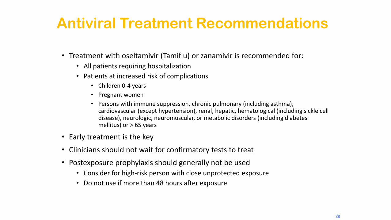
Antiviral Treatment Recommendations
• Treatment with oseltamivir (Tamiflu) or zanamivir is recommended for:• All patients requiring hospitalization
• Patients at increased risk of complications
• Children 0-4 years
• Pregnant women
• Persons with immune suppression, chronic pulmonary (including asthma), cardiovascular (except hypertension), renal, hepatic, hematological (including sickle cell disease), neurologic, neuromuscular, or metabolic disorders (including diabetes mellitus) or > 65 years
• Early treatment is the key
• Clinicians should not wait for confirmatory tests to treat
• Postexposure prophylaxis should generally not be used• Consider for high-risk person with close unprotected exposure
• Do not use if more than 48 hours after exposure

Healthy Habits
• When Healthy:• Avoid close contact with those who are sick
• Wash your hands often
• Avoid touching your eyes, nose and mouth to decrease the spread of germs
• When Ill:• Cover your mouth and nose with a tissue (or upper
sleeve) when you sneeze or cough
• Stay home from work or school when you are sick

Key facts
Influenza is an acute viral infection that spreads easily from person to person.
Influenza circulates worldwide and can affect anybody in any age group.
Influenza causes annual epidemics that peak during winter in temperate regions.
Influenza is a serious public health problem that causes severe illnesses and deaths for higher risk populations.
An epidemic can take an economic toll through lost workforce productivity, and strain health services.
Vaccination is the most effective way to prevent infection.

Avian Influenza
• A contagious viral infection and/or disease of many avian species including poultry, wild and exotic birds, ratites, shore birds and migratory waterfowl.
• The highly pathogenic form of the disease is characterized by severe depression, decrease in egg production, high mortality, edema, hemorrhage, and frank necrosis.
• All H5 and H7 infections are reportable to the World Organization for Animal Health (OIE).

Where does AI virus come from?
• All known subtypes of influenza A viruses circulate among wild birds, especially migratory waterfowl (e.g. ducks and geese)which are considered natural reservoirs for influenza A viruses
• Domestic poultry like chickens and turkeysare not natural reservoirs for AI virus and usually develop clinical disease when infected with AI virus


How does AI virus spread?
• Exposure of poultry to migratory waterfowl
• Exposure of commercial poultry to AI-infected backyard, game bird, or hobby flocks
• Contact with AI-infected live bird markets
• Bird to bird contact (through feces)
• Aerosol droplets
• Manure, equipment, vehicles, egg flats, crates, contaminated shoes and clothing
• Wildlife vectors/scavengers

What are the types of Avian Influenza in domestic poultry?
• Low pathogenic avian influenza (LPAI)
• Mild or no clinical signs
• Low to moderate mortality
• However, the low pathogenic H5 and H7 strains are capable of mutating under field conditions into highly pathogenic strains
• Highly pathogenic avian influenza (HPAI)
• Sudden onset
• Severe clinical signs
• High mortality

H1N1/H5N1


WHAT IS SWINE FLU ?
Swine Influenza (swine flu) is a respiratory disease of pigs caused by type A influenza viruses (H1N1 subtype) that causes regular outbreaks in pigs.
People do not normally get swine flu, but human infections can and do happen
Swine flu viruses have been reported to spread from person-to-person, but in the past, this transmission was limited and not sustained beyond three people

The End