influence of the renin-angiotensin system on epidermal
TRANSCRIPT
Kidney International, Vol. 60 (2001), pp. 847–857
HORMONES – CYTOKINES – SIGNALING
Influence of the renin-angiotensin system on epidermalgrowth factor expression in normal andcyclosporine-treated rat kidney
CHUL WOO YANG, HEE JONG AHN, WAN YOUNG KIM, MI JUNG SHIN, SUNG KWON KIM,JOO HYUN PARK, YOUNG OK KIM, YONG SOO KIM, JIN KIM, and BYUNG KEE BANG
Departments of Internal Medicine and Anatomy, The Catholic University of Korea, Seoul, Korea
Influence of the renin-angiotensin system on epidermal growth Chronic cyclosporine (CsA)-induced nephrotoxicity isfactor expression in normal and cyclosporine-treated rat kidney. characterized by progressive renal failure and irrevers-
Background. Epidermal growth factor (EGF) plays an im- ible renal striped interstitial fibrosis, tubular atrophy,portant role in renal tubular regeneration after ischemic injuryand hyalinosis of the afferent arteriole [1]. The precisein kidney. The present study reports the association betweenmechanism of chronic CsA nephrotoxicity is not wellthe renin-angiotensin system (RAS) and EGF, and the effect
of angiotensin II blockade with losartan (LSRT) on EGF ex- understood, but has been attributed to sustained afferentpression in an experimental model of chronic cyclosporine arteriolar vasoconstriction and arteriopathy [2, 3].(CsA) nephrotoxicity in rats.
The lack of an animal model of chronic CsA nephro-Methods. Two separate experiments were performed. In thetoxicity has hampered the study of the mechanisms offirst experiment, rats on the normal-salt diet (NSD; 0.3%) or
low-salt diet (LSD; 0.05%) were treated with or without LSRT renal damage. However, a reproducible animal modelfor four weeks. In the second experiment, rats on the NSD or of chronic CsA nephrotoxicity was established from theLSD were given vehicle (VH group, olive oil, 1 mg/kg per day) observation that sodium depletion exacerbates CsAor CsA (15 mg/kg per day) or CsA (15 mg/kg per day) plus LSRT
nephrotoxicity [4, 5]. In this model, CsA treatment in(100 mg/L per day). Renal function, histopathology, TUNELstaining, plasma renin activity (PRA), and the expression of rats on a low-salt diet (LSD) induced a histologic featurerenin and EGF were studied. similar to that described in patients on long-term CsA
Results. Normal rats on the LSD showed significantly in- therapy [6]. Salt depletion activates the renin-angioten-creased EGF expression (cortex, 2.6-fold; medulla, 1.7-fold)sin system (RAS), which has been implicated in theand significantly decreased EGF expression with the LSRTchanges in renal hemodynamics and function that followtreatment compared with the rats treated with the NSD (cortex,
74.8 vs. 10%; medulla, 22.5 vs. 5%). In contrast, the CsA- CsA administration [7]. Furthermore, angiotensin-con-treated rats on the LSD had a significantly lower EGF expres- verting enzyme inhibitor or angiotensin II (Ang II) re-sion (cortex, 98 vs. 53%; medulla, 94 vs. 14%); however, con-
ceptor antagonist is effective in preventing interstitialcomitant administration of LSRT increased the EGF expres-fibrosis [8] by inhibiting the expression of intrarenalsion (cortex, 91- vs. 3.8-fold; medulla, 19- vs. 2.4-fold) compared
with the rats on the NSD. In the normal and CsA-treated transforming growth factor-�1 (TGF-�1) [9].LSD rats, EGF expression was well correlated with PRA. In Epidermal growth factor (EGF) is a 53-amino acidaddition, EGF expression was well correlated with the intersti-
(6.2 kD) polypeptide that stimulates the proliferationtial fibrosis score (r � 0.664, P � 0.01) or number of TUNEL-and differentiation of epithelial cells as well as beingpositive cells (r � 0.822, P � 0.01) in CsA-treated LSD rats.
Conclusions. These results suggest that angiotensin II block- mitogenic for various other cell types in culture [10, 11].ade with LSRT decreases EGF expression in normal rats on In addition, EGF is thought to be important in the main-the LSD, but it protects EGF expression in CsA-induced neph- tenance of renal tubule integrity and in the tubular regen-rotoxicity. This finding provides a new perspective on the reno-
erative response to injury [12, 13]. Animal studies showprotection of angiotensin II blockade in chronic CsA nephro-toxicity. that EGF accelerates renal tubular regeneration in rat
models of acute renal failure [14], whereas treatmentwith anti-EGF antibody inhibits tubular cell prolifera-Key words: losartan, nephrotoxicity, progressive renal disease, intersti-
tial fibrosis, tubular atrophy, tubular regeneration, ischemic injury. tion during compensatory renal growth [15].Epidermal growth factor expression has been wellReceived for publication November 29, 2000
studied in clinical and experimental transplantation. Jor-and in revised form February 2, 2001Accepted for publication February 13, 2001 gensen et al reported that urinary excretion of EGF in
living human kidney recipients is 40% lower than in donor 2001 by the International Society of Nephrology
847
Yang et al: RAS and EGF848
kidneys, and urinary expression is well correlated with Protocol 1. (1) NSD rats received a 0.4% sodium con-tent diet for four weeks (N � 6). (2) NSD-LSRT ratscompensatory renal growth, suggesting decreased urinary
EGF excretion by ischemic or CsA-induced toxicity [16]. received a 0.4% sodium content diet plus LSRT(100 mg/L) in the drinking water for 28 days (N � 6).Di Paolo et al reported that the renal expression and
the urinary concentration of the EGF signal are ex- (3) LSD rats received a 0.05% sodium content diet forfour weeks (N � 6). (4) LSD-LSRT rats received a 0.05%tremely weak or even absent in patients with acute rejec-
tion, acute tubular necrosis, and CsA nephrotoxicity [17]. sodium content diet and LSRT (100 mg/L) in the drink-ing water for 28 days (N � 6).Stein-Oakley et al reported that chronic renal allograft
rejection shows decreased expression of EGF in a rat Protocol 2. This study was designed to evaluate therole of RAS in regulating EGF expression in CsA-treatedmodel [18]. These findings suggest that tubular damage
by ischemia, nephrotoxic drugs, or rejection decreases rat kidney and to examine the effect of blocking the AT1receptor on EGF expression. The NSD or an LSD wasEGF expression.
The association of EGF expression and RAS is well administered to the each group. (1) The vehicle (VH)group of rats received a daily subcutaneous injection ofknown from studies in vitro and in the developing kidney
[19]. Angiotensin II potentiates the mitogenic action of olive oil, 1 mg/kg for four weeks (N � 6). (2) CsA ratsreceived a daily subcutaneous injection of CsA (15 mg/kg)EGF on proximal tubular cells [20], and the inhibition
of angiotensin II type 1 (AT1) receptors decreases the for four weeks (N � 6). (3) CsA � LSRT rats received adaily subcutaneous injection of CsA (15 mg/kg) and LSRTrenal expression of EGF in neonatal rats [21]. Based on
these observations, we hypothesized that EGF expression (100 mg/L) was added to the drinking water for fourweeks (N � 6).may be influenced by RAS in chronic CsA nephrotox-
icity. To test this hypothesis, two separate experiments This dosage and route of administration for LSRTwere selected because they have been previously shownwere performed. First, we evaluated the difference in
EGF expression of normal rats on a low-salt diet (LSD) to blockade Ang II receptors significantly [22].or a normal-salt diet (NSD), and evaluated the influence
Basic protocolof Ang II blockade on EGF expression. Second, EGFexpression was evaluated in CsA-treated rats with an After one week of the animals on the NSD or LSD,
weight-matched rats were randomly assigned to the dif-LSD or an NSD, and the influence of Ang II blockadeon EGF expression was evaluated in normal and CsA- ferent treatment groups. Daily body weights were re-
corded. After each treatment period, the systolic bloodtreated rat kidneys. Our study clearly demonstrates theinfluences of RAS on intrarenal EGF expression in rats pressure was measured with a plethysmography using a
tail manometer-tachometer system (BP-2000; Visitechwith or without CsA treatment.Systems, Apex, NC, USA), and 24-hour urine sampleswere collected in metabolic cages (Nalge Co., Rochester,
METHODSNY, USA). The following day, animals were anesthe-
Animals tized with ketamine, and a blood sample and tissue speci-mens were obtained.Male Sprague-Dawley rats (Charles River, Wilming-
ton, MA, USA), weighing 225 to 250 g, were housed inFunctional studiesindividual cases in a temperature- and light-controlled
environment. They received the LSD (0.05% sodium; Urinary and serum creatinine was measured by a Co-bas autoanalyzer (Roche Diagnostics, Div. Hoffman-LaTeklad Premier, Madison, WI, USA) or NSD (0.4%
sodium; Teklad Premier). NSD rats were pair fed with Roche Inc., Nutley, NJ, USA). The creatinine clearance(CCr) was calculated using a standard formula. The whole-LSD animals.blood CsA concentrations were measured by monoclonal
Drugs radioimmunoassay (Incstar Co., Stillwater, MN, USA).Plasma renin activity (PRA) was measured by radio-Cyclosporine provided by Sandoz Research (East Han-
over, NJ, USA) was diluted in olive oil to a final con- immunoassay (Angiotensin I-Biotecx radioimmunoassay;Biotecx, Friendswood, TX, USA).centration of 15 mg/mL. Losartan (LSRT), provided by
Cardiovascular Diseases Research, DuPont-Merck Phar-Histologymaceutical (Wilmington, DE, USA) was dissolved in
sterile water to a final concentration of 100 mg/L. These histologic findings were evaluated as previouslydescribed [23]. Tubulointerstitial fibrosis (TIF) was esti-
Experimental groups mated semiquantitatively using a color image analyzer(Mustek Paragon 800 SP, Macintosh PowerPC 7100, NIHTwo separate experiments were conducted. The first ex-
periment (protocol 1) was designed to evaluate the role of Image version 1.5) by counting the percentage of injuredareas per field of cortex and medulla. Scores of 0 to 3�RAS in regulating EGF expression in normal rat kidney.
Yang et al: RAS and EGF 849
Table 1. Changes in cyclosporine A (CsA) whole blood level,were given as follows: score 0 � normal interstitium;systolic blood pressure (SBP), serum creatinine (SCr), and
score 0.5 � �5%; score 1.0 � 5 to 15%; score 1.5 � 16 creatinine clearance (CCr) in the different experimental groupsto 25%; score 2.0 � 26 to 35%; score 2.5 � 36 to 45%;
CsA level SBP SCr CCrand score 3.0 � �45%. ng/mL mm Hg mg/dL mL/min/100 g
NSDTdt-mediated dUTP-biotin nick end-labelingVH 138 �11 0.55�0.02 0.55�0.02
(TUNEL) method CsA 3826 �310 157�21a 0.80 �0.09a 0.39�0.03a
CsA�LSRT 4273�170 139�12b 0.85 �0.11a 0.38�0.12a
Cells undergoing apoptosis were identified by the LSDApopTag in situ apoptosis detection kit (Oncor, Gaith- VH 127 �5 0.57�0.02 0.61�0.08
CsA 3100 �380 128�7 1.02�0.06a 0.31�0.06aersburg, MD, USA). The sections were dewaxed andCsA�LSRT 3260�520 110�4ab 1.05 �0.17a 0.30�0.05a
treated with proteinase K and then incubated with equili-Data are presented as mean values � SEM of six rats. Abbreviations are:bration buffer in a humidified chamber for 10 minutes at NSD, normal salt diet; VH, vehicle; LSRT, losartan.a P � 0.05 vs. VHroom temperature, followed by incubation with working-b P � 0.05 vs. CsAstrength Tdt enzyme solution in a humidified chamber
at 37�C for two hours. The reaction was terminated byincubation in working-strength stop/wash buffer for 30 min-utes at 37�C. After being rinsed with phosphate-bufferedsaline (PBS), the sections were incubated with anti-digoxi- 1:10,000; Bio-Rad] for 30 minutes. Antibody-reactivegenin peroxidase in a humidified chamber for 30 minutes protein was detected using enhanced chemiluminescenceat room temperature. Sections were then incubated with (Amersham Life Science, Little Chalfont, Buckingham-diaminobenzidine and 0.01% H2O2 for five minutes at shire, UK). Densitometric analysis was performed usingroom temperature. After being rinsed with PBS, the sec- Gelexpert software (Nucleotech Corp., San Carlos, CA,tions were counterstained with hematoxylin and exam- USA). Optical densities (mean � SEM) were obtainedined by light microscopy. To quantitate TUNEL-positive after three determinations for each band.cells, 16 fields of outer medulla were randomly selectedin each section and observed under a microscope (magni- Immunohistochemistry of EGF and reninfication �400). The number of apoptotic cells was ex- Sections were dewaxed. Before incubation with pri-pressed per 1000 of the total tubular cells in each section. mary antibody, the sections were blocked with normal
rabbit serum diluted 1:10 in 0.3% bovine serum albuminImmunoblotting for EGF(BSA) for 30 minutes and incubated for two hours at
Kidneys were homogenized in RIPA [10 mmol/L Tris 4�C in mouse antiserum against EGF (Biomedical Tech-Cl, pH 7.6, 150 mmol/L NaCl, 1% (wt/vol) sodium deoxy- nologies Inc., Stoughton, MA, USA) diluted 1:200 in BSA.cholate, 1% (vol/vol) Triton X-100, 0.1% (wt/vol) so- The sections were rinsed in TBS and incubated for 30dium dodecyl sulfate, 1% (vol/vol) aprotinin, 2 mmol/L minutes in peroxidase-conjugated rabbit anti-mouse IgGNa3VO4, and freshly added leupeptin (1 g/mL), pep-
(Jackson ImmunoResearch Laboratories Inc., West Grove,statin (1 g/mL), and 1 mmol/L phenylmethylsulfonyl
PA, USA). Sections were incubated with a mixture offluoride (PMSF)]. Homogenates were centrifuged at
0.05% 3,3-diaminobenzidine and 0.01% H2O2 for five16,000 � g for 15 minutes at 4�C, and protein concentra-minutes at room temperature, washed with Tris-HCl buf-tions were determined using a protein microassay offer, counterstained with hematoxylin, and examined byBradford method (Bio-Rad, Hercules, CA, USA). Ho-light microscopy. The method for renin immunohisto-mogenates were heated at 94�C for five minutes. Proteinschemistry was same as EGF immunohistochemistry. Thewere separated by sodium dodecyl sulfate-polyacryl-number of renin-positive glomeruli was counted per 50amide gel electrophoresis (SDS-PAGE) in 15% poly-glomeruli.acrylamide gels and were electroblotted onto Bio-Blot
nitrocellulose (Costar, Cambridge, MA, USA). Nonspe-Statistical analysiscific binding was blocked by incubating the blots for
Data reported are mean � SEM, and all statisticalone hour in 5% (wt/vol) nonfat milk. Epidermal growthanalyses were calculated with SYSTAT for Macintoshfactor was detected by incubating for one hour with aversion 5.2 (SYSTAT Inc., Chicago, IL, USA). Compari-polyclonal anti-EGF antibody (Santa Cruz Biotechnol-sons between groups were done by analysis of varianceogy, Inc., Santa Cruz, CA, USA) diluted 1:1000. Primary(Kruskal-Wallis test followed by Tukey or Dunnet test)antibody incubation was followed by six washes of Tris-or unpaired t test. Nonparametric (Spearman) correla-buffered saline with 0.005% Tween 20 (TBS-T). The blottion coefficient was calculated. The level of statisticalwas then incubated with secondary antibody [goat–anti-
rabbit IgG-horseradish peroxidase (HRP) conjugate at significance was P � 0.05.
Yang et al: RAS and EGF850
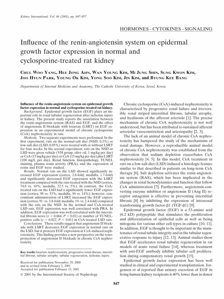
Fig. 1. Effect of angiotensin II blockade withlosartan (LSRT) on cyclosporine A (CsA)-induced chronic nephropathy. (A) Vehicle-treated rat kidney. (B) The sodium-depletedanimal treated with CsA. There is a diffuseinterstitial fibrosis, mononuclear cell infiltrate,and tubular atrophy. (C ) CsA and LSRT-treated rat kidney. Note the decrease in inter-stitial fibrosis and tubular atrophy. Trichromestain, magnification �100.
RESULTS
Blood pressure and functional assessment
Table 1 shows the blood pressure and renal functionin the experimental groups. At four weeks, systolic bloodpressure was increased in the CsA group with an NSD,but there was no significant change in the CsA groupwith an LSD. The addition of LSRT decreased systolicblood pressure compared with the VH group on theNSD and LSD. The CsA group with an NSD and anLSD showed a significant decrease in CCr and a significantincrease in serum creatinine compared with the VHgroup (P � 0.05, respectively). However, there were nosignificant differences in CCr between CsA and CsA �LSRT groups with NSD or LSD.
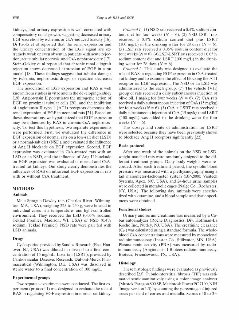
Histologic assessmentFig. 2. Comparison of tubulointerstitial fibrosis (TIF) between the nor-Low salt diet rats treated with CsA had characteristicmal-salt diet (NSD; �) and the low-salt diet (LSD; ) ingesting vehiclemorphologic findings that were usually evident at four(VH) or cyclosporine A (CsA)-treated rats. In the LSD rats, there was
weeks and were similar to the chronic human CsA renal a significant increase in TIF score in CsA-treated rats and a dramaticdecrease of TIF with LSRT treatment in CsA-treated rats. In contrast,lesions (Fig. 1B). Focal TIF, tubular atrophy, and mono-there was no significant difference of TIF in rats with the NSD. *P �nuclear inflammatory cell infiltration were observed. The0.05 vs. NSD; #P � 0.05 vs. VH; ##P � 0.05 vs. CsA.
concurrent administration of LSRT dramatically decreasedTIF (Fig. 1C). When the extent of changes in TIF wasgraded with a 0 to 3� semiquantitative score, there wereno differences in the TIF scores among VH (0.17 � 0.14), with LSRT treatment. Figure 4 shows the comparisonCsA (0.21 � 0.19), and CsA � LSRT groups (0.19 � of TUNEL-positive cells in the CsA-treated rats on the0.18) on a NSD. However, in the LSD group, there was NSD and LSD. In the NSD group, there was no signifi-a significant increase in TIF score in the CsA group cant increase in the number of TUNEL-positive cellscompared with the VH group (2.07 � 0.56 vs. 0.19 �
with CsA treatment compared with the VH group (4.8 �0.13, P � 0.05). The concomitant administration of LSRT2.5 vs. 2.2 � 1.6, P � 0.05), and LSRT treatment diddecreased the TIF score in comparison with the CsAnot affect apoptotic cell death (3.9 � 1.7). In the LSDgroup (1.25 � 0.25; Fig. 2).group, CsA treatment increased the number of TUNEL-
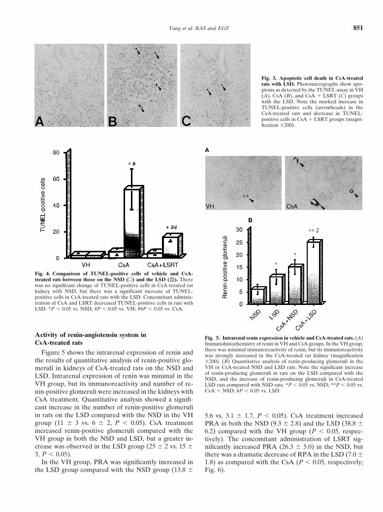
Apoptoic cell death in CsA-treated rats positive cells compared with the VH group (52.1 � 15.5vs. 2.3 � 0.9, P � 0.05), and concomitant administrationFigure 3 shows TUNEL-staining in chronic CsA neph-of LSRT significantly decreased the number of TUNEL-rotoxic rats on the LSD. In the VH group, TUNEL-posi-positive cells (15.6 � 3.0) as compared with the CsAtive cells were rarely observed. The number of TUNEL-
positive cells increased with CsA treatment, but decreased group (P � 0.05).
Yang et al: RAS and EGF 851
Fig. 3. Apoptotic cell death in CsA-treatedrats with LSD. Photomicrographs show apo-ptosis as detected by the TUNEL assay in VH(A), CsA (B), and CsA � LSRT (C) groupswith the LSD. Note the marked increase inTUNEL-positive cells (arrowheads) in theCsA-treated rats and decrease in TUNEL-positive cells in CsA � LSRT groups (magni-fication �200).
Fig. 4. Comparison of TUNEL-positive cells of vehicle and CsA-treated rats between those on the NSD (�) and the LSD ( ). Therewas no significant change of TUNEL-positive cells in CsA-treated ratkidney with NSD, but there was a significant increase of TUNEL-positive cells in CsA-treated rats with the LSD. Concomitant adminis-tration of CsA and LSRT decreased TUNEL-positive cells in rats withLSD. *P � 0.05 vs. NSD; #P � 0.05 vs. VH; ##P � 0.05 vs. CsA.
Activity of renin-angiotensin system in Fig. 5. Intrarenal renin expression in vehicle and CsA-treated rats. (A)CsA-treated rats Immunohistochemistry of renin in VH and CsA groups. In the VH group,
there was minimal immunoreactivity of renin, but its immunoreactivityFigure 5 shows the intrarenal expression of renin and was strongly increased in the CsA-treated rat kidney (magnificationthe results of quantitative analysis of renin-positive glo- �200). (B) Quantitative analysis of renin-producing glomeruli in the
VH or CsA-treated NSD and LSD rats. Note the significant increasemeruli in kidneys of CsA-treated rats on the NSD andof renin-producing glomeruli in rats on the LSD compared with theLSD. Intrarenal expression of renin was minimal in the NSD, and the increase of renin-producing glomeruli in CsA-treated
VH group, but its immunoreactivity and number of re- LSD rats compared with NSD rats. *P � 0.05 vs. NSD; **P � 0.05 vs.CsA � NSD; #P � 0.05 vs. LSD.nin-positive glomeruli were increased in the kidneys with
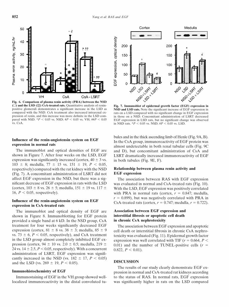
CsA treatment. Quantitative analysis showed a signifi-cant increase in the number of renin-positive glomeruliin rats on the LSD compared with the NSD in the VH 5.6 vs. 3.1 � 1.7, P � 0.05). CsA treatment increasedgroup (11 � 3 vs. 6 � 2, P � 0.05). CsA treatment PRA in both the NSD (9.3 � 2.8) and the LSD (38.8 �increased renin-positive glomeruli compared with the 6.2) compared with the VH group (P � 0.05, respec-VH group in both the NSD and LSD, but a greater in- tively). The concomitant administration of LSRT sig-crease was observed in the LSD group (25 � 2 vs. 15 � nificantly increased PRA (26.3 � 5.0) in the NSD, but3, P � 0.05). there was a dramatic decrease of RPA in the LSD (7.0 �
In the VH group, PRA was significantly increased in 1.8) as compared with the CsA (P � 0.05, respectively;Fig. 6).the LSD group compared with the NSD group (13.8 �
Yang et al: RAS and EGF852
Fig. 6. Comparison of plasma renin activity (PRA) between the NSD(�) and the LSD ( ) CsA-treated rats. Quantitative analysis of renin- Fig. 7. Immunoblot of epidermal growth factor (EGF) expression inpositive glomeruli demonstrates a significant increase in the LSD as NSD and LSD rats. Note the significant increase of EGF expression incompared with the NSD. CsA treatment also increased intrarenal ex- rats on a LSD compared with no significant change in EGF expressionpression of renin, and this increase was more definite in the LSD com- in those on a NSD. Concomitant administration of LSRT decreasedpared with NSD. *P � 0.05 vs. NSD; #P � 0.05 vs. VH; ##P � 0.05 EGF expression in LSD rats, but no significant change was observedvs. CsA. in NSD rats. *P � 0.05 vs. NSD; #P � 0.05 vs. LSD.
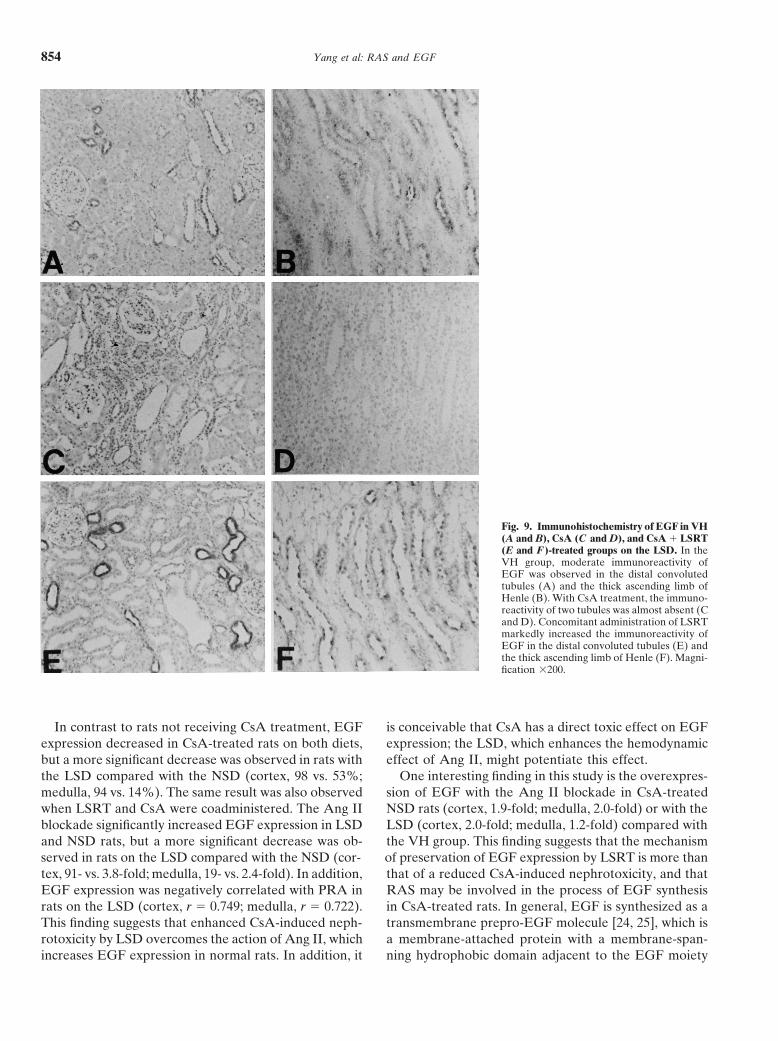
bules and in the thick ascending limb of Henle (Fig. 9A, B).Influence of the renin-angiotensin system on EGF In the CsA group, immunoreactivity of EGF protein wasexpression in normal rats almost undetectable in both renal tubular cells (Fig. 9C
The immunoblot and optical densities of EGF are and D), but concomitant administration of CsA andshown in Figure 7. After four weeks on the LSD, EGF LSRT dramatically increased immunoreactivity of EGFexpression was significantly increased (cortex, 40 � 3 vs. in both tubules (Fig. 9E, F).103 � 8; medulla, 77 � 15 vs. 151 � 19, P � 0.05,
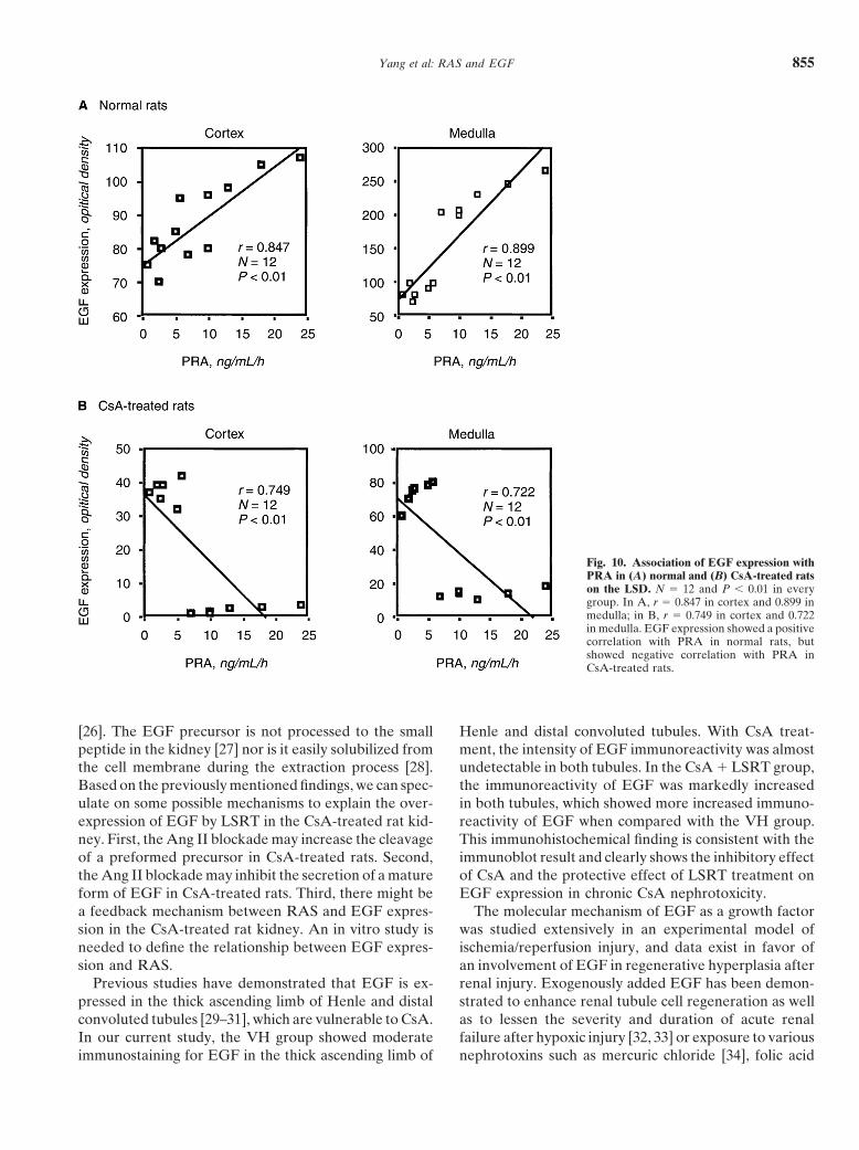
Relationship between plasma renin activity andrespectively) compared with the rat kidney with the NSDEGF expression(Fig. 7). A concomitant administration of LSRT did not
affect EGF expression in the NSD, but there was a sig- The association between RAS with EGF expressionnificant decrease of EGF expression in rats with the LSD was evaluated in normal and CsA-treated rats (Fig. 10).(cortex, 103 � 8 vs. 26 � 5; medulla, 151 � 19 vs. 117 � With the LSD, EGF expression was positively correlated16, P � 0.05, respectively). with PRA in normal rats (cortex, r � 0.847; medulla,
r � 0.899), but was negatively correlated with PRA inInfluence of the renin-angiotensin system on EGF CsA-treated rats (cortex, r � 0.747; medulla, r � 0.722).expression in CsA-treated rats
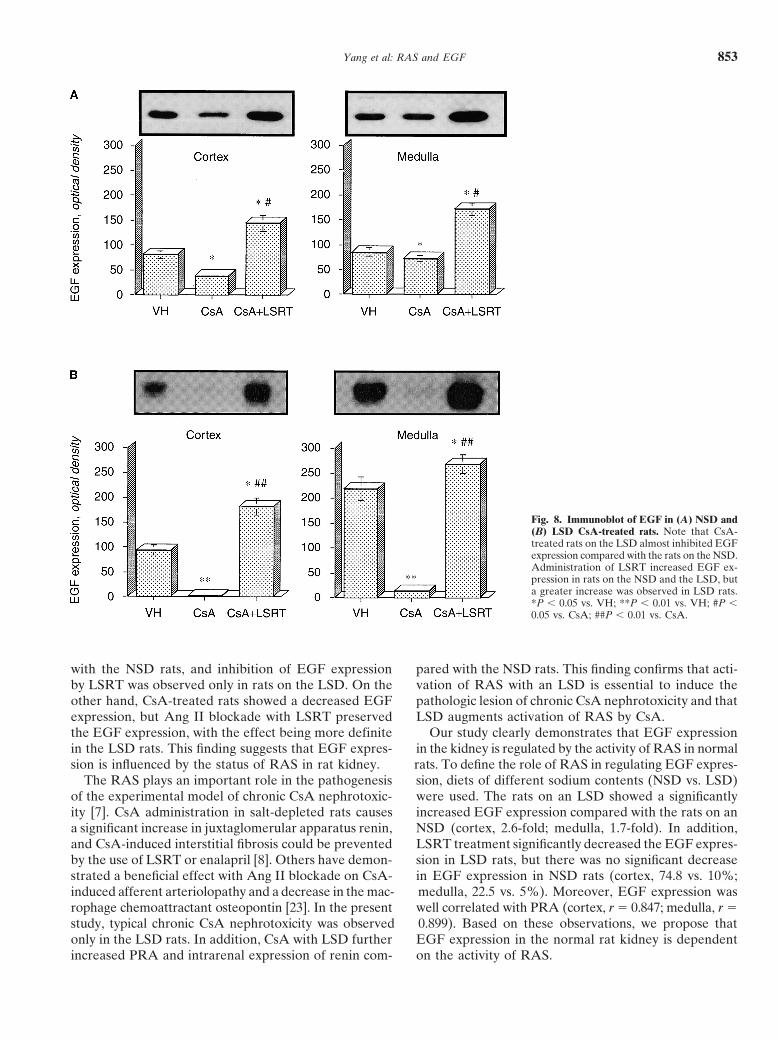
Association between EGF expression andThe immunoblot and optical density of EGF areinterstitial fibrosis or apoptotic cell deathshown in Figure 8. Immunoblotting for EGF proteinin chronic CsA nephrotoxicityprovided a single band at 6 kD. In the NSD group, CsA
treatment for four weeks significantly decreased EGF The association between EGF expression and apoptoticexpression (cortex, 81 � 8 vs. 38 � 3; medulla, 85 � 9 cell death or interstitial fibrosis in chronic CsA nephro-vs. 73 � 6, P � 0.05, respectively), and CsA treatment toxicity was evaluated (Fig. 11). Epidermal growth factorin the LSD group almost completely inhibited EGF ex- expression was well correlated with TIF (r � 0.664, P �pression (cortex, 94 � 10 vs. 2.0 � 0.5; medulla, 219 � 0.01) and the number of TUNEL-positive cells (r �24 vs. 14 � 2.5, P � 0.05, respectively). With a concurrent 0.822, P � 0.01).administration of LSRT, EGF expression was signifi-cantly increased in the NSD (vs. 182 � 17, P � 0.05)
DISCUSSIONand the LSD (vs. 269 � 19, P � 0.05).The results of our study clearly demonstrate EGF ex-
Immunohistochemistry of EGF pression in normal and CsA-treated rat kidneys accordingto the status of RAS. In normal rats, EGF expressionImmunostaining of EGF in the VH group showed well-
localized immunoreactivity in the distal convoluted tu- was significantly higher in rats on the LSD compared
Yang et al: RAS and EGF 853
Fig. 8. Immunoblot of EGF in (A) NSD and(B) LSD CsA-treated rats. Note that CsA-treated rats on the LSD almost inhibited EGFexpression compared with the rats on the NSD.Administration of LSRT increased EGF ex-pression in rats on the NSD and the LSD, buta greater increase was observed in LSD rats.*P � 0.05 vs. VH; **P � 0.01 vs. VH; #P �0.05 vs. CsA; ##P � 0.01 vs. CsA.
with the NSD rats, and inhibition of EGF expression pared with the NSD rats. This finding confirms that acti-vation of RAS with an LSD is essential to induce theby LSRT was observed only in rats on the LSD. On the
other hand, CsA-treated rats showed a decreased EGF pathologic lesion of chronic CsA nephrotoxicity and thatLSD augments activation of RAS by CsA.expression, but Ang II blockade with LSRT preserved
the EGF expression, with the effect being more definite Our study clearly demonstrates that EGF expressionin the kidney is regulated by the activity of RAS in normalin the LSD rats. This finding suggests that EGF expres-
sion is influenced by the status of RAS in rat kidney. rats. To define the role of RAS in regulating EGF expres-sion, diets of different sodium contents (NSD vs. LSD)The RAS plays an important role in the pathogenesis
of the experimental model of chronic CsA nephrotoxic- were used. The rats on an LSD showed a significantlyincreased EGF expression compared with the rats on anity [7]. CsA administration in salt-depleted rats causes
a significant increase in juxtaglomerular apparatus renin, NSD (cortex, 2.6-fold; medulla, 1.7-fold). In addition,LSRT treatment significantly decreased the EGF expres-and CsA-induced interstitial fibrosis could be prevented
by the use of LSRT or enalapril [8]. Others have demon- sion in LSD rats, but there was no significant decreasein EGF expression in NSD rats (cortex, 74.8 vs. 10%;strated a beneficial effect with Ang II blockade on CsA-
induced afferent arteriolopathy and a decrease in the mac- medulla, 22.5 vs. 5%). Moreover, EGF expression waswell correlated with PRA (cortex, r � 0.847; medulla, r �rophage chemoattractant osteopontin [23]. In the present
study, typical chronic CsA nephrotoxicity was observed 0.899). Based on these observations, we propose thatEGF expression in the normal rat kidney is dependentonly in the LSD rats. In addition, CsA with LSD further
increased PRA and intrarenal expression of renin com- on the activity of RAS.
Yang et al: RAS and EGF854
Fig. 9. Immunohistochemistry of EGF in VH(A and B), CsA (C and D), and CsA � LSRT(E and F)-treated groups on the LSD. In theVH group, moderate immunoreactivity ofEGF was observed in the distal convolutedtubules (A) and the thick ascending limb ofHenle (B). With CsA treatment, the immuno-reactivity of two tubules was almost absent (Cand D). Concomitant administration of LSRTmarkedly increased the immunoreactivity ofEGF in the distal convoluted tubules (E) andthe thick ascending limb of Henle (F). Magni-fication �200.
In contrast to rats not receiving CsA treatment, EGF is conceivable that CsA has a direct toxic effect on EGFexpression; the LSD, which enhances the hemodynamicexpression decreased in CsA-treated rats on both diets,
but a more significant decrease was observed in rats with effect of Ang II, might potentiate this effect.One interesting finding in this study is the overexpres-the LSD compared with the NSD (cortex, 98 vs. 53%;
medulla, 94 vs. 14%). The same result was also observed sion of EGF with the Ang II blockade in CsA-treatedNSD rats (cortex, 1.9-fold; medulla, 2.0-fold) or with thewhen LSRT and CsA were coadministered. The Ang II
blockade significantly increased EGF expression in LSD LSD (cortex, 2.0-fold; medulla, 1.2-fold) compared withthe VH group. This finding suggests that the mechanismand NSD rats, but a more significant decrease was ob-
served in rats on the LSD compared with the NSD (cor- of preservation of EGF expression by LSRT is more thanthat of a reduced CsA-induced nephrotoxicity, and thattex, 91- vs. 3.8-fold; medulla, 19- vs. 2.4-fold). In addition,
EGF expression was negatively correlated with PRA in RAS may be involved in the process of EGF synthesisin CsA-treated rats. In general, EGF is synthesized as arats on the LSD (cortex, r � 0.749; medulla, r � 0.722).
This finding suggests that enhanced CsA-induced neph- transmembrane prepro-EGF molecule [24, 25], which isa membrane-attached protein with a membrane-span-rotoxicity by LSD overcomes the action of Ang II, which
increases EGF expression in normal rats. In addition, it ning hydrophobic domain adjacent to the EGF moiety
Yang et al: RAS and EGF 855
Fig. 10. Association of EGF expression withPRA in (A) normal and (B) CsA-treated ratson the LSD. N � 12 and P � 0.01 in everygroup. In A, r � 0.847 in cortex and 0.899 inmedulla; in B, r � 0.749 in cortex and 0.722in medulla. EGF expression showed a positivecorrelation with PRA in normal rats, butshowed negative correlation with PRA inCsA-treated rats.
[26]. The EGF precursor is not processed to the small Henle and distal convoluted tubules. With CsA treat-ment, the intensity of EGF immunoreactivity was almostpeptide in the kidney [27] nor is it easily solubilized from
the cell membrane during the extraction process [28]. undetectable in both tubules. In the CsA � LSRT group,the immunoreactivity of EGF was markedly increasedBased on the previously mentioned findings, we can spec-
ulate on some possible mechanisms to explain the over- in both tubules, which showed more increased immuno-reactivity of EGF when compared with the VH group.expression of EGF by LSRT in the CsA-treated rat kid-
ney. First, the Ang II blockade may increase the cleavage This immunohistochemical finding is consistent with theimmunoblot result and clearly shows the inhibitory effectof a preformed precursor in CsA-treated rats. Second,
the Ang II blockade may inhibit the secretion of a mature of CsA and the protective effect of LSRT treatment onEGF expression in chronic CsA nephrotoxicity.form of EGF in CsA-treated rats. Third, there might be
a feedback mechanism between RAS and EGF expres- The molecular mechanism of EGF as a growth factorwas studied extensively in an experimental model ofsion in the CsA-treated rat kidney. An in vitro study is
needed to define the relationship between EGF expres- ischemia/reperfusion injury, and data exist in favor ofan involvement of EGF in regenerative hyperplasia aftersion and RAS.
Previous studies have demonstrated that EGF is ex- renal injury. Exogenously added EGF has been demon-strated to enhance renal tubule cell regeneration as wellpressed in the thick ascending limb of Henle and distal
convoluted tubules [29–31], which are vulnerable to CsA. as to lessen the severity and duration of acute renalfailure after hypoxic injury [32, 33] or exposure to variousIn our current study, the VH group showed moderate
immunostaining for EGF in the thick ascending limb of nephrotoxins such as mercuric chloride [34], folic acid

Yang et al: RAS and EGF856
fibrosis in CsA-induced nephropathy. Moreover, theprotective effect on EGF expression by LSRT might beassociated with improvements in both parameters. Asexpected, EGF expression was well correlated with inter-stitial fibrosis (r � 0.664) and TUNEL-positive cells (r �0.822). This finding suggests that the loss of endogenousEGF during chronic CsA nephrotoxicity is associatedwith increased apoptotic cell death resulting in interstitialfibrosis. On the other hand, experimental approachesaiming to block EGF receptor signaling seem to amelio-rate the evolution of renal diseases. Terzi et al reporteda reduction of TIF after renal injury in mice with targetedtubular expression of a dominant negative EGF receptor[39]. This finding suggests that inhibition of EGF signal-ing in the kidney may prevent the development of renallesions after renal injury. At present, the fate of EGFreceptor in renal tissues exposed to CsA or LSRT is un-known because our experimental investigations focusedon EGF itself. However, we suggest that intrarenal EGFmay be associated with healing processes, but that it maybe helpful or harmful according to the clinical situation.When the kidney is damaged by ischemic or toxic injury,EGF may act as a growth factor in the regeneration ofrenal tubular cells. In contrast, EGF may have a harmfulaction in the progression of renal disease in an animalmodel of uremia (5/6 nephrectomy model).
Our study clearly demonstrates that EGF expressionis influenced by the status of RAS in normal and CsA-treated rats. EGF expression is decreased by CsA, but isprotected by the Ang II blockade with LSRT. This findingprovides a new perspective to the renoprotection of Ang IIblockade in cases of chronic CsA nephrotoxicity.
ACKNOWLEDGMENTS
Part of this study was presented at the American Society of Nephrol-ogy meeting, Toronto, Canada, in November 2000. The authors thankMs. Yoo Hee Kim and Mr. Je Keun Woo (MSD in Korea) for theirassistance in preparing this manuscript.
Fig. 11. Association of EGF expression with TUNEL-positive cells Reprint requests to Byung Kee Bang, M.D., Department of Internal(A) or interstitial fibrosis score (B) in CsA-treated rat on a LSD. N � Medicine, Kangnam St. Mary’s Hospital, The Catholic University of12 and P � 0.01 in both groups; r � 0.664 in (A) and r � 0.822 in (B). Korea, 505 Banpo-Dong, Seocho-Ku, Seoul, 137-040, Korea.EGF expression was well correlated with TUNEL-positive cells or E-mail: [email protected] fibrosis score.
REFERENCES
1. Myers BD, Ross JC, Newton LD: Cyclosporine-associated chronicnephropathy. N Engl J Med 311:600–705, 1984[35], or gentamicin [36]. In the experimental model of
2. Myers BD: Cyclosporine nephrotoxicity. Kidney Int 30:964–974,chronic CsA nephrotoxicity using salt depletion, intersti-1986
tial fibrosis [37] and apoptotic cell death [38] are closely 3. Myers BD, Sibley R, Newton L, et al: The long-term course ofcyclosporin-associated chronic nephropathy. Kidney Int 33:590–associated with activation of RAS, and the blockade of600, 1988RAS with LSRT or enalapril decreases both parameters
4. Elzinga LW, Rosen S, Bennett WM: Dissociation of glomerular[8, 38]. Based on the previously mentioned findings, our filtration rate from tubulointerstitial fibrosis in experimental
chronic cyclosporin nephropathy: Role of sodium intake. J Aminvestigations were undertaken on the assumption thatSoc Nephrol 4:214–221, 1993EGF possibly acts as a survival factor in maintaining
5. Rosen S, Greenfeld Z, Brezis M: Chronic cyclosporin-inducedtubular cells, and that the loss of EGF might be associ- nephropathy in the rat. Transplantation 49:445–452, 1990
6. Mihatsch MJ, Antovych T, Bohman SO, et al: Cyclosporin Aated with increased apoptotic cell death or interstitial
Yang et al: RAS and EGF 857
nephropathy: Standardization of the evaluation of kidney biopsies. 23. Pichler RH, Franceschini N, Young BA, et al: Pathogenesis ofcyclosporine nephropathy: Roles of angiotensin II and osteopontin.Clin Nephrol 41:23–32, 1994
7. Mason J, Muller-Schweinitzer E, Dupont M, et al: Cyclosporine J Am Soc Nephrol 6:1186–1196, 199524. Gray A, Dull TJ, Ullrich A: Necleotide sequence of epidermaland the renin-angiotensin system. Kidney Int 39(Suppl 32):S28–
S32, 1991 growth factor cDNA predicts a 128,000 molecular weight precur-sor. Nature 303:722–725, 19838. Burdmann EA, Andoh TF, Nast CC, et al: Prevention of experi-
mental cyclosporin-induced interstitial fibrosis by losartan and ena- 25. Scott J, Urdea M, Quiroga M, et al: Structure of a mouse submax-illary messenger RNA encoding epidermal growth factor and sevenlapril. Am J Physiol 269:F491–F499, 1995
9. Shihab FS, Bennett WM, Tanner AM, Andoh TF: Angiotensin II related proteins. Science 221:236–240, 198326. Doolittle RR, Feng DF, Johnson MS: Computer based character-blockade decreases TGF-beta1 and matrix proteins in cyclosporine
ization of epidermal growth factor precursor. Nature 320:558–560,nephropathy. Kidney Int 52:660–673, 1997198610. Burgess AW: Epidermal growth factor and transforming growth
27. Rall LB, Scott J, Bell GI, et al: Mouse prepro-epidermal growthfactor �. Br Med Bull 45:401–424, 1989factor synthesis by the kidney and other tissues. Nature 313:228–11. Das M, Rengaraju M, Samanta A: Epidermal growth factor, in231, 1985Human Cytokines: Handbook for Basic and Clinical Research,
28. Kashimata M, Hiramatsu M, Minami N, Ninami N: Biochemicaledited by Aggarwal BB, Gutterman JU, Boston, Blackwell Scien-properties of epidermal growth factor in the mouse kidney. Comptific Publications, 1992, pp 365–382Biochem Physiol 86:651–653, 198712. Harris RC: Potential physiologic roles for epidermal growth factor
29. Kasselberg AG, Orth DN, Gray ME, Stahlman MT: Immunocy-in the kidney. Am J Kidney Dis 17:627–630, 1991tochemical localisation of human growth factor/urogastrone in sev-13. Taub M, Wang Y, Szczesny TM, Kleinman HK: Epidermaleral human tissues. J Histochem Cytochem 33:315–322, 1985growth factor or transforming growth factor � is required for kid-
30. Salido EC, Lakshmenan J, Fisher DA, et al: Expression of epider-ney tubulogenesis in matrigel cultures in serum-free medium. Cellmal growth factor in the rat kidney. An immunocytochemical andBiol 87:4002–4006, 1990in situ hypridization study. Histochemistry 96:65–72, 199114. Humes HD, Cieslinski DA, Coumbra TM, et al: Epidermal growth
31. Salido EC, Yen PH, Shapiro LJ, et al: In situ hybridization offactor enhances renal tubular cell regeneration and repair andprepro-epidermal growth factor mRNA in the mouse kidney. Amaccelerates the recovery of renal function in postischemic acuteJ Physiol 256:F632–F638, 1989renal failure. J Clin Invest 84:1757–1761, 1989
32. Humes HD, Cieslinski DA, Coimbra TM, et al: Epidermal growth15. Kanda S, Igawa T, Sakai H, et al: Anti-epidermal growth factorfactor enhances renal tubule cell regeneration and accelerates theantibody inhibits compensatory renal hyperplasia but not hypertro-recovery of renal function in postischemic acute renal failure. J Clinphy after unilateral nephrectomy in mice. Biochem Biophys ResInvest 84:1757–1761, 1989Commun 187:1015–1021, 1992 33. Norman J, Tsau YK, Bacay A, Fine LG: Epidermal growth factor16. Jorgensen PE, Kamper AL, Munck O, et al: Urinary excretion accelerates functional recovery from ischemic acute tubular necro-of epidermal growth factor in living human kidney donors and sis in the rat: Role of epidermal growth factor receptor. Clin Sci
their recipients. Eur J Clin Invest 25:442–446, 1995 Lond 78:445–450, 199017. Di Paolo S, Gesualdo L, Stallone G, et al: Renal expression and 34. Coimbra TM, Cieslinski DA, Humes HD: Epidermal growth fac-
urinary concentration of EGF and IL-6 in acutely dysfunctioning tor accelerates renal repair in mercuric chloride nephrotoxins. Amkidney transplanted patients. Nephrol Dial Transplant 12:2687– J Physiol 259:F438–F443, 19902693, 1997 35. Alberti P, Bardella L, Comelli R: Ribosomal protein S6 kinase
18. Stein-Oakley AN, Tzanidis A, Fuller PJ, et al: Expression and is activated after folic acid injury and epidermal growth factordistribution of epidermal growth factor in acute and chronic renal administration but not after unilateral nephrectomy in the ratallograft rejection. Kidney Int 46:1207–1215, 1994 kidney. Nephron 60:330–335, 1992
19. Yoo KH, Wolstenholme JT, Chelvalier RL: Angiotensin-con- 36. Morin NJ, Laurent G, Nonclereq D, et al: Epidermal growthverting enzyme inhibition decreases growth factor expression in factor (EGF) accelerates renal tissue repair in a rat model ofthe neonatal rat kidney. Pediatr Res 42:588–592, 1997 gentamicin nephrotoxicity. Am J Physiol 51:123–140, 1989
20. Taub M, Wang Y, Szczesny TM, Kleinman HK: Epidermal 37. Shihab FS, Andoh TF, Tanner AM, Bennett WM: Sodium deple-growth factor or transforming growth factor � is required for kid- tion enhances fibrosis and the expression of TGF-�1 and matrixney tubulogenesis in matrigel cultures in serum-free medium. Proc proteins in experimental chronic cyclosporine nephropathy. AmNatl Acad Sci USA 87:4002–4006, 1990 J Kidney Dis 30:71–81, 1997
21. Wolf G, Neilson EG: Angiotensin II induces cellular hypertrophy 38. Thomas SE, Andoh TF, Pichler RH, et al: Accelerated apoptosisin cultured murine proximal tubular cells. Am J Physiol 259:F768– characterizes cyclosporine-associated interstitial fibrosis. KidneyF777, 1990 Int 53:897–908, 1998
22. Wong PC, Price WA, Jr, Chiu AT, et al: Hypotensive action of 39. Terzi F, Burtin M, Hekmati M, et al: Targeted expression of aDuP 753, an angiotensin II antagonist, in spontaneously hyperten- dominant-negative EGF-R in the kidney reduces tubulo-interstitial
lesions after renal injury. J Clin Invest 106:225–234, 2000sive rats. Hypertension 15:459–468, 1990