influence of mir-154 on myocardial apoptosis in rats with ... · naling pathway in myocardial cells...
TRANSCRIPT

818
Abstract. – OBJECTIVE: To explore the in-fluence of micro ribonucleic acid (miR)-154 on myocardial apoptosis in rats with acute myo-cardial infarction (AMI), and to analyze whether Wnt/β-catenin signaling pathway was involved in the regulation.
MATERIALS AND METHODS: The Sprague- Dawley (SD) rat model of AMI was established via ligation of left anterior descending artery. Rats were randomly divided into model group (M group, n=12) and ICG-001 intervention group (I group, n=12). At the same time, sham operation group (S group, n=12) was established. In I group, ICG-001 (5 mg/kg) was intraperitoneally injected every day after operation. Meanwhile, an equal amount of normal saline was injected in rats of S group and M group. 21 d after operation, the car-diac function of rats in each group was detected via echocardiography. After that, the rats were immediately executed. MI area in each group was detected via 2,3,5-triphenyltetrazolium chlo-ride (TTC) staining. Myocardial apoptosis level in each group was detected via terminal deoxy-nucleotidyl transferase-mediated dUTP nick end labeling (TUNEL) staining. Moreover, the chang-es of apoptotic proteins in rat myocardial cells were detected via Western blotting. Moreover, the expression level of miR-154 in myocardial cells of rats was detected via quantitative poly-merase chain reaction (qPCR). Furthermore, the influence of miR-154 on Wnt/β-catenin signaling pathway was detected via Western blotting.
RESULTS: Compared with S group, left ventric-ular ejection fraction (LVEF, %) and left ventricu-lar fractional shortening (LVFS, %) were signifi-cantly decreased in M group (p<0.01). However, left ventricular internal diameter at end-diastole (LVIDd) and left ventricular internal diameter at end-systole (LVIDs) were significantly increased (p<0.01). In I group, LVEF (%) and LVFS (%) were significantly higher than those of M group (p<0.05), whereas LVIDs and LVIDd were signifi-cantly lower (p<0.05). MI area in M group was remarkably larger than that of S group (p<0.01). Meanwhile, MI area in I group was significantly
smaller than that of M group (p<0.01). Compared with S group, the number of apoptotic myocar-dial cells and the protein expression level of cleaved caspase-3 were significantly increased in M group (p<0.01). However, the expression level of B-cell lymphoma-2/Bcl-2 associated X protein (Bcl-2/Bax) was significantly decreased (p<0.01). The number of apoptotic myocardial cells and the protein expression level of cleaved caspase-3 were significantly declined in I group when compared with those of M group (p<0.01). However, the expression level of Bcl-2/Bax was significantly increased in I group (p<0.01). The expression level of miR-154 in myocardial cells of M group and I group was remarkably increased when compared with that of S group (p<0.01). Furthermore, the expression levels of β-catenin and Cyclin D1 in myocardial cells of M group were remarkably higher than those of S group and I group (p<0.01).
CONCLUSIONS: AMI significantly increases the expression level of miR-154. Moreover, miR-154 can activate Wnt/β-catenin signaling path-way, eventually promoting myocardial apoptosis.
Key WordsMiR-154, Myocardial infarction, Wnt/β-catenin sig-
naling pathway, Apoptosis.
Introduction
Acute myocardial infarction (AMI) is an ischemic heart disease, which is manifested as persistent ischemia accompanied by myocardial necrosis or apoptosis. Meanwhile, it is also a common cardiovascular disease seriously threat-ening human health1,2. The basic pathogenesis of AMI is that the rupture and hemorrhage of coronary atherosclerotic plaques and thrombo-sis lead to severe luminal stenosis or occlusion. This may result in insufficient or interrupted
European Review for Medical and Pharmacological Sciences 2019; 23: 818-825
H.-Y. SUN1, X.-L. WANG1, L.-C. MA1, M. YANG1, H.-J. YANG1, H.-W. HUANG1, G.-A. ZHAO2
1Department of Cardiology, The Third Affiliated Hospital of Xinxiang Medical University, Xinxiang, China2Department of Cardiology, The First Affiliated Hospital of Xinxiang Medical University, Weihui, China
Corresponding Author: Guoan Zhao, MD; e-mail: [email protected]
Influence of MiR-154 on myocardial apoptosis in rats with acute myocardial infarction through Wnt/β-catenin signaling pathway

Role of MiR-154 in rats with acute myocardial infarction
819
myocardial blood supply and myocardial cell structural damage or death, ultimately reducing myocardial contraction and relaxation ability3,4. A large amount of researches have proved that the prognosis of AMI patients is closely related to the area of myocardial necrosis and the num-ber of surviving myocardial cells. Moreover, the non-renewability of myocardial cells indicates that reversing myocardial remodeling and reduc-ing the number of apoptotic myocardial cells are key steps to improve the prognosis of AMI5,6. Currently, angiotensin-converting enzyme inhib-itors have been widely applied in the treatment of AMI. However, there is still a lack of specific drugs that can reverse myocardial remodeling in clinic. Furthermore, both morbidity and mortali-ty rates of heart failure after AMI remain high7. Wang et al8 have found that the expression levels of various micro ribonucleic acids (miRNAs) in myocardial cells of AMI patients are significantly altered. Interference of miRNA expression may become a new means and target for the treatment of AMI. Dong et al9 have indicated the abnormal expression of miR-154 in myocardial fibrosis. Inhibition of miR-154 can reduce the phenotypic changes of myocardial fibroblasts and effectively improve myocardial fibrosis. Myocardial apopto-sis is an important indicator for poor prognosis of AMI. However, whether miR-154 can regulate myocardial apoptosis through Wnt/β-catenin sig-naling pathway in myocardial cells has not been elucidated yet. In this experiment, we established a rat model of AMI to study the influence of miR-154 on myocardial apoptosis in AMI. Fur-thermore, we analyzed whether Wnt/β-catenin signaling pathway was involved in the regulation.
Materials and Methods
Animals and GroupingA total of 60 Sprague-Dawley (SD) rats weigh-
ing 220-250 g were purchased from the Labora-tory Animal Center of Guangzhou University of Chinese Medicine (Laboratory Animal Produc-tion License No. SCXK2016-0034, Guangzhou, China). Rats were fed in a specific pathogen-free (SPF) room with humidity of (45±2)% at (23±1)°C under normal day-night rhythm. Meanwhile, all rats had free access to food and water to adapt to the environment for 1 week. The Sprague-Daw-ley (SD) rat model of AMI was established via ligation of left anterior descending artery. Totally, 24 successfully modeled rats were selected and
randomly divided into model group (M group, n=12) and ICG-001 intervention group (I group, n=12), respectively. Normal Sprague-Dawley (SD) rats did not undergo ligation of left anterior descending artery, and the remaining operations were the same as M group. Next, 12 surviving rats were selected in sham operation group (S group). ICG-001 (5 mg/kg) was intraperitoneally injected as an intervention in I group every day after operation. However, an equal amount of normal saline was injected in S group and M group. ICG-001 is a kind of specific inhibitor of Wnt/β-catenin signaling pathway, which can effectively inhibit the production of β-catenin and block Wnt/β-catenin signaling pathway. All experiments were in line with the guidelines for feeding and use of laboratory animals issued by the National Institutes of Health. The animal ex-perimental scheme was reviewed and approved by the Ethics Committee of Xinxiang Medical University Animal Center (Weihui, China).
Establishment of AMI ModelAMI model was established via ligation of
left anterior descending artery. Rats were first anesthetized with 10% chloral hydrate and fixed on an operating table. After tracheal intubation, small animal ventilator was connected to assist respiration. The parameters of ventilator were ad-justed as follows: tidal volume: 5 mL, respiratory rate: 80 beats/min, and respiratory ratio: 5:4. The chest hair was removed and the chest was cut open between the 3rd and 4th intercostal spaces to expose the heart. The left anterior descending artery was ligated at about 2 mm on the lower edge of left auricle using the 6-0 wire. Whitening of cardiac apex indicated the successful operation. After the chest was sutured and closed, the rats were treated with anti-infective therapy using penicillin. After resuscitation, the trachea cannula was removed and the rats were placed into the insulation cage.
Determination of Cardiac FunctionThe cardiac function of rats in each group was
detected using Vevo high-resolution small animal ultrasonic imaging system at 21 d after AMI. The rats were first anesthetized with 2% isoflurane and fixed on a thermostatic operating table. The limbs were connected to electrocardiogram electrodes. After the chest hair was removed, the ultrasonic sound-conducting agent was applied on the chest for ultrasonic detection. Left ventricular internal diameter at end-systole (LVIDs), left ventricular internal diameter at end-diastole (LVIDd), left

H.-Y. Sun, X.-L. Wang, L.-C. Ma, M. Yang, H.-J. Yang, H.-W. Huang, G.-A. Zhao
820
ventricular ejection fraction (LVEF) and left ven-tricular fractional shortening (LVFS) of rats were recorded to evaluate cardiac function.
Determination of MI Area The rats were executed immediately after the
determination of cardiac function. The heart was taken and trimmed. Meanwhile, the left ventricle was collected, weighed and sliced into sections. After incubation with pre-heated 1% TTC solu-tion (Beijing Dingguo Biotechnology Co., Ltd., Beijing, China) at 37°C for 15 min in dark, the sections were fixed with 4% paraformaldehyde for 30 min and photographed. The staining area was measured using Image-Pro Plus image analysis software. MI area was calculated by the follow-ing formula: MI area (%) = (MI area per section/myocardial area in the section * myocardial weight in the section) ∑/left ventricular weight * 100%.
Detection of Myocardial Apoptosis LevelThe rats were executed immediately after the
determination of cardiac function. Ischemic tis-sues of left ventricle were taken and prepared into paraffin sections. The level of myocardial apop-tosis in each group was detected according to the instructions of terminal deoxynucleotidyl transfer-ase-mediated dUTP nick end labeling (TUNEL) apoptosis kit (Wuhan Boster Biological Technolo-gy Co., Ltd., Wuhan, China). The number of apop-totic myocardial cells in each group was observed under a confocal fluorescence microscope (Nikon, Tokyo, Japan). After photography, the number of TUNEL-positive cells (green fluorescence) in myocardial tissues of each group was calculated.
Detection of Protein Expression Levels via Western blotting
The rats were executed immediately after the determination of cardiac function. Ischemic tissues of left ventricle were taken, weighed and lysed using RIPA lysis buffer (100 mg/mL) (Be-yotime Biotechnology, Shanghai, China). After homogenization and centrifugation, the super-natant was collected as total protein sample. The concentration of extracted protein in each group was detected using the bicinchoninic acid (BCA) protein quantification kit (Pierce, Rockford, IL, USA). Subsequently, an equal concentration of protein sample was prepared and inactivated via water bath for 15 min. Extracted protein samples were separated by sodium dodecyl sul-phate-polyacrylamide gel electrophoresis (SDS-PAGE) and transferred onto polyvinylidene
difluoride (PVDF) membranes (IPVH00010, Millipore, Billerica, MA, USA). After sealing with 5% skim milk powder for 2 h, the mem-branes were incubated with primary antibodies of cleaved caspase-3, B-cell lymphoma-2 (Bcl-2), Bcl-2 associated X protein (Bax), β-catenin, Cyclin D1 and glyceraldehyde-3-phosphate de-hydrogenase (GAPDH) (purchased from CST, diluted at 1:1000, Danvers, MA, USA) at 4°C overnight. After washing with Tris-buffered sa-line and Tween 20 (TBST) for 3 times, the mem-branes were incubated again with secondary antibodies (Beyotime Biotechnology, Shanghai, China) at room temperature for 1 h. Then, the membranes were washed again with TBST for 3 times. Immunoreactive bands were developed and analyzed using electrochemiluminescence (ECL) solution on a developing machine (Ther-mo Fisher Scientific, Waltham, MA, USA).
Detection of miR-154 Level in Myocardial Cells via Quantitative Polymerase Chain Reaction (qPCR)
The rats were executed immediately after the determination of cardiac function. Ischemic tis-sues of left ventricle were taken, and total RNA was extracted from hippocampus in each group using TRIzol reagent (Invitrogen, Carlsbad, CA, USA). Optical density (OD) value was then deter-mined. Extracted RNA was reversely transcribed into complementary deoxyribose nucleic acid (cDNA) using reverse transcription kit (TaKaRa, Otsu, Shiga, Japan) and stored in a refrigerator at -20°C. Specific reaction conditions were as follows: 42°C for 15 min and 95°C for 2 min. The qPCR system was prepared, and the reaction conditions were as follows: pre-denaturation at 95°C for 5 min, denaturation at 95°C for 30 s, annealing at 60°C for 30 s, extension at 72°C for 1 min, for a total of 30 cycles, and extension at 72°C for 5 min. The relative expression level of gene was calculated by the 2-ΔΔCt method. The primers were synthesized by Invitrogen (Carls-bad, CA, USA), and the sequences were shown in Table I (GAPDH as an internal reference).
Table I. Primer sequences.
Gene Sequences of PCR primers
MiR-154 Sense ACACTCCAGCTGGGTAG Antisense CTCAACTGGTGTCGTGGAGAPDH Sense TATCGGACGCCTGGTTAC Antisense TTCCCATTCTCAGCCTTG

Role of MiR-154 in rats with acute myocardial infarction
821
Statistical AnalysisExperimental data were expressed as (x̅±s).
GraphPad Prism 6.0 (La Jolla, CA, USA) was used for one-way analysis of variance. Tukey’s test was adopted for pairwise comparison of multiple sam-ples. Least Significant Difference (LSD) t-test was adopted for data comparison between two groups. p<0.05 was considered statistically significant.
Results
Cardiac Function The cardiac function of rats in each group was
detected using high-resolution small animal ultrason-ic imaging system at 21 d after AMI. As shown in Figure 1, LVEF (%) and LVFS (%) were significantly decreased in M group when compared with S group (p<0.01). However, LVIDd and LVIDs were signifi-cantly increased in M group (p<0.01). In I group, LVEF (%) and LVFS (%) were significantly higher than those of M group (p<0.05), whereas LVIDs and LVIDd were remarkably lower (p<0.05).
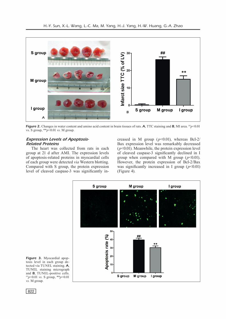
MI AreaThe heart was collected from rats in each
group at 21 d after AMI. The MI area in each group was detected via TTC staining. It was found that the left ventricular MI area of M group was significantly larger than that of S group (p<0.01). However, MI area of in I group was significantly smaller than that of M group after ICG-001 intervention (p<0.01) (Figure 2).
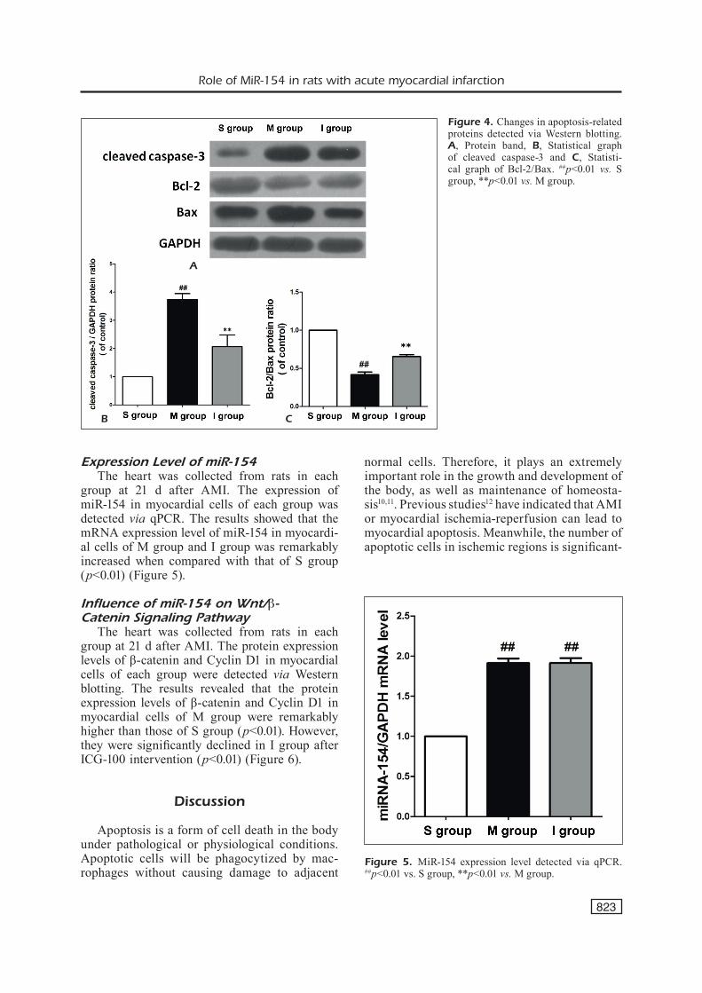
Myocardial Apoptosis Level
The heart was collected from rats in each group at 21 d after AMI. The level of myocar-dial apoptosis in each group was detected via TUNEL staining. As shown in Figure 3, a large number of TUNEL-positive cells were observed in myocardial cells of M group. The number of TUNEL-positive cells in M group was signifi-cantly larger than that of S group (p<0.01). Fur-thermore, the number of TUNEL-positive cells was significantly smaller in I group than M group (p<0.01).
A
C
B
D
Figure 1. Cardiac func-tion. A, LVEF: left ven-tricular ejection fraction, B, LVFS: left ventricular fractional shortening, C, LVIDd: left ventricular in-ternal diameter at end-di-astole and D, LVIDs: left ventricular internal diame-ter at end-systole. ##p<0.01 vs. S group, *p<0.05 vs. M group.
H.-Y. Sun, X.-L. Wang, L.-C. Ma, M. Yang, H.-J. Yang, H.-W. Huang, G.-A. Zhao
822
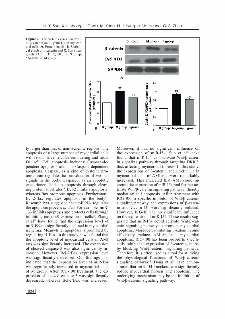
Expression Levels of Apoptosis-Related Proteins
The heart was collected from rats in each group at 21 d after AMI. The expression levels of apoptosis-related proteins in myocardial cells of each group were detected via Western blotting. Compared with S group, the protein expression level of cleaved caspase-3 was significantly in-
creased in M group (p<0.01), whereas Bcl-2/Bax expression level was remarkably decreased (p<0.01). Meanwhile, the protein expression level of cleaved caspase-3 significantly declined in I group when compared with M group (p<0.01). However, the protein expression of Bcl-2/Bax was significantly increased in I group (p<0.01) (Figure 4).
A
B
Figure 2. Changes in water content and amino acid content in brain tissues of rats. A, TTC staining and B, MI area. ##p<0.01 vs. S group, **p<0.01 vs. M group.
A
B
Figure 3. Myocardial apop-tosis level in each group de-tected via TUNEL staining. A, TUNEL staining micrograph and B, TUNEL-positive cells. ##p<0.01 vs. S group, **p<0.01 vs. M group.
Role of MiR-154 in rats with acute myocardial infarction
823
Expression Level of miR-154The heart was collected from rats in each
group at 21 d after AMI. The expression of miR-154 in myocardial cells of each group was detected via qPCR. The results showed that the mRNA expression level of miR-154 in myocardi-al cells of M group and I group was remarkably increased when compared with that of S group (p<0.01) (Figure 5).
Influence of miR-154 on Wnt/β-Catenin Signaling Pathway
The heart was collected from rats in each group at 21 d after AMI. The protein expression levels of β-catenin and Cyclin D1 in myocardial cells of each group were detected via Western blotting. The results revealed that the protein expression levels of β-catenin and Cyclin D1 in myocardial cells of M group were remarkably higher than those of S group (p<0.01). However, they were significantly declined in I group after ICG-100 intervention (p<0.01) (Figure 6).
Discussion
Apoptosis is a form of cell death in the body under pathological or physiological conditions. Apoptotic cells will be phagocytized by mac-rophages without causing damage to adjacent
normal cells. Therefore, it plays an extremely important role in the growth and development of the body, as well as maintenance of homeosta-sis10,11. Previous studies12 have indicated that AMI or myocardial ischemia-reperfusion can lead to myocardial apoptosis. Meanwhile, the number of apoptotic cells in ischemic regions is significant-
A
B C
Figure 4. Changes in apoptosis-related proteins detected via Western blotting. A, Protein band, B, Statistical graph of cleaved caspase-3 and C, Statisti-cal graph of Bcl-2/Bax. ##p<0.01 vs. S group, **p<0.01 vs. M group.
Figure 5. MiR-154 expression level detected via qPCR. ##p<0.01 vs. S group, **p<0.01 vs. M group.
H.-Y. Sun, X.-L. Wang, L.-C. Ma, M. Yang, H.-J. Yang, H.-W. Huang, G.-A. Zhao
824
ly larger than that of non-ischemic regions. The apoptosis of a large number of myocardial cells will result in ventricular remodeling and heart failure13. Cell apoptosis includes: Caspase-de-pendent apoptosis and non-Caspase-dependent apoptosis. Caspase, as a kind of cysteine pro-tease, can regulate the transduction of various signals in the body. Caspase3, as an apoptotic executioner, leads to apoptosis through cleav-ing protein substrates14. Bcl-2 inhibits apoptosis, whereas Bax promotes apoptosis. Furthermore, Bcl-2/Bax regulates apoptosis in the body15. Research has suggested that miRNA regulates the apoptotic process in vivo. For example, miR-133 inhibits apoptosis and protects cells through inhibiting caspase9 expression in cells16. Zhang et al17 have found that the expression level of miR-199a is significantly declined in myocardial ischemia. Meanwhile, apoptosis is promoted by regulating HIF-a. In this study, it was found that the apoptotic level of myocardial cells in AMI rats was significantly increased. The expression of cleaved caspase-3 was also significantly in-creased. However, Bcl-2/Bax expression level was significantly decreased. Our findings also indicated that the expression level of miR-154 was significantly increased in myocardial cells of M group. After ICG-100 treatment, the ex-pression of cleaved caspase-3 was significantly decreased, whereas Bcl-2/Bax was increased.
Moreover, it had no significant influence on the expression of miR-154. Sun et al18 have found that miR-154 can activate Wnt/β-caten-in signaling pathway through targeting DKK2, thus affecting myocardial fibrosis. In this study, the expressions of β-catenin and Cyclin D1 in myocardial cells of AMI rats were remarkably increased. This indicated that AMI could in-crease the expression of miR-154 and further ac-tivate Wnt/β-catenin signaling pathway, thereby mediating cell apoptosis. After treatment with ICG-100, a specific inhibitor of Wnt/β-catenin signaling pathway, the expressions of β-caten-in and Cyclin D1 were significantly reduced. However, ICG-10 had no significant influence on the expression of miR-154. These results sug-gested that miR-154 could activate Wnt/β-cat-enin signaling pathway to promote myocardial apoptosis. Moreover, inhibiting β-catenin could effectively reduce AMI-induced myocardial apoptosis. ICG-100 has been proved to specifi-cally inhibit the expression of β-catenin, there-by blocking Wnt/β-catenin signaling pathway. Therefore, it is often used as a tool for studying the physiological functions of Wnt/β-catenin signaling pathway19. Dong et al9 have demon-strated that miR-154 knockout can significantly reduce myocardial fibrosis and apoptosis. The underlying mechanism may be the inhibition of Wnt/β-catenin signaling pathway.
A
B C
Figure 6. The protein expression levels of β-catenin and Cyclin D1 in myocar-dial cells. A, Protein bands, B, Statisti-cal graph of β-catenin and C, Statistical graph of Cyclin D1. ##p<0.01 vs. S group, **p<0.01 vs. M group.

Role of MiR-154 in rats with acute myocardial infarction
825
Conclusions
We found that AMI significantly increased the expression level of miR-154. Meanwhile, miR-154 could promote myocardial apoptosis through ac-tivating Wnt/β-catenin signaling pathway. In ad-dition, inhibiting β-catenin could significantly re-duce AMI-induced MI and myocardial apoptosis.
Conflict of InterestsThe authors declared no conflict of interest.
References
1) Khoury S, MargoliS g, ravid d, rozenbauM z, Keren g, ShachaM y. Outcomes of early and reversible renal impairment in patients with ST segment elevation myocardial infarction undergoing percu-taneous coronary intervention. Eur Heart J Acute Cardiovasc Care 2018: 1419924616.
2) chen h, lu n, zheng M. A high circulating FGF21 level as a prognostic marker in patients with acute myocardial infarction. Am J Transl Res 2018; 10: 2958-2966.
3) SharMa n, lee J, aponte cS, MarMur Jd, lawSon we, Mann nn, Salifu Mo, youSSef i, Mcfarlane Si. Clinical Characteristics and angiographic findings of acute myocardial infarction associated with marijuana use: consecutive case series. SciFed J Cardiol 2017; 2:
4) robberS l, niJveldt r, beeK aM, teuniSSen p, hol-lander Mr, bieSbroeK pS, everaarS h, van de ven pM, hofMan M, van royen n, van roSSuM ac. The influence of microvascular injury on native T1 and T2* relaxation values after acute myocardial infarction: implications for non-contrast-enhanced infarct assessment. Eur Radiol 2018; 28: 824-832.
5) Kozel M, KocKa v, liSa l, budeSinSKy t, touSeK p. Im-mune-inflammatory response after bioresorbable vascular scaffold implantation in patients with acute myocardial infarction with ST elevation in a long-term perspective. Heart Vessels 2018; 3: 165-178.
6) li J, wang l, wang Q, Xin z, liu y, zhao Q. Di-agnostic value of carotid artery ultrasound and hypersensitive C-reactive protein in Type 2 diabe-tes mellitus patients with acute myocardial infarc-tion in Chinese population. Medicine (Baltimore) 2018; 97: e12334.
7) claeSSen be, Mehran r. How to manage chronic total occlusions in the setting of acute myocardi-al infarction complicated by cardiogenic shock? Catheter Cardiovasc Interv 2018; 92: 464-465.
8) wang y, chang w, zhang y, zhang l, ding h, Qi h, Xue S, yu h, hu l, liu d, zhu w, wang y, li p. Cir-
culating miR-22-5p and miR-122-5p are promising novel biomarkers for diagnosis of acute myocardial infarction. J Cell Physiol 2018; 7: 556-573.
9) dong p, liu wJ, wang zh. MiR-154 promotes myocardial fibrosis through beta-catenin signal-ing pathway. Eur Rev Med Pharmacol Sci 2018; 22: 2052-2060.
10) fogarty ce, diwanJi n, lindblad Jl, tare M, aM-cheSlavSKy a, MaKhiJani K, brucKner K, fan y, berg-Mann a. Extracellular reactive oxygen species drive apoptosis-induced proliferation via drosoph-ila macrophages. Curr Biol 2016; 26: 575-584.
11) bin-Jaliah i, huSSein aM, SaKr hf, eid ea. Effects of low dose of aliskiren on isoproterenol-induced acute myocardial infarction in rats. Physiol Int 2018; 105: 127-144.
12) zhang y, Shen t, liu b, dai d, cai J, zhao c, du l, Jia n, he Q. Cardiac shock wave therapy atten-uates cardiomyocyte apoptosis after acute myo-cardial infarction in rats. Cell Physiol Biochem 2018; 49: 1734-1746.
13) yu by, dong b. LncRNA H19 regulates cardiomyo-cyte apoptosis and acute myocardial infarction by targeting miR-29b. Int J Cardiol 2018; 271: 25.
zhou z, liu z, wang l, luo J, li h. Oxidative stress, apoptosis activation and symbiosis dis-ruption in giant clam Tridacna crocea under high temperature. Fish Shellfish Immunol 2018 Oct 11; 84: 451-457. doi: 10.1016/j.fsi.2018.10.033. [Epub ahead of print]
14) pan S, zhao X, wang X, tian X, wang y, fan r, feng n, zhang S, gu X, Jia M, li J, yang l, wang K, guo h, pei J. Sfrp1 attenuates TAC-induced cardiac dysfunction by inhibiting Wnt signaling pathway- mediated myocardial apoptosis in mice. Lipids Health Dis 2018; 17: 202.
15) yu J, cao X, zheng y, yan l, wang J. Abnormal expression of miR133a in patients with acute myocardial infarction following radical surgery for gastric cancer and the underlying mechanism. Mol Med Rep 2018; 6: 91-112.
16) zhang h, li S, zhou Q, Sun Q, Shen S, zhou y, bei y, li X. Qiliqiangxin attenuates phenylephrine-in-duced cardiac hypertrophy through downregula-tion of MiR-199a-5p. Cell Physiol Biochem 2016; 38: 1743-1751.
17) Sun ly, bie zd, zhang ch, li h, li ld, yang J. MiR-154 directly suppresses DKK2 to activate Wnt signaling pathway and enhance activation of cardiac fibroblasts. Cell Biol Int 2016; 40: 1271-1279.
18) lin zw, zhang w, Jiang Sd, wei wb, li Xf. In-hibition of microRNA-940 suppresses the mi-gration and invasion of human osteosarcoma cells through the secreted frizzled-related protein 1-mediated Wnt/beta-catenin signaling pathway. J Cell Biochem 2018; 15: 22-36.