inflammatory events in asthma: an expanding equation
TRANSCRIPT
Asthma is a chronic inflammatory disorder that can lead toprogressive, potentially irreversible declines in lung function insome patients. Asthmatic inflammation develops when thesequential interaction of inflammatory cells with resident cellsgenerates a cascade of events that contribute to the chronicinflammation and clinical manifestations associated with thedisease, including further inflammation, airway smooth musclespasm (bronchospasm), airway mucus secretion, airway edemaand narrowing, and bronchial epithelial damage. Because ofthe chronic, progressive nature of asthmatic inflammation andthe early age of onset, the ability to evaluate inflammation inchildren would be useful. Several procedures that quantifyinflammatory mediators (in peripheral blood, induced sputum,bronchoalveolar lavage fluid, and bronchial biopsies) haveshown potential usefulness in the evaluation of and the moni-toring of disease severity in children (and, by extension,adults) with asthma. Further research needs to be devoted tothe elucidation of when the inflammatory process starts andhow it changes over time, to the determination of whether theinflammatory process is the same in all patients with wheezing,regardless of the stimulus, to the definition of the relationshipbetween atopy and asthma, and to the establishment of theusefulness of testing for inflammatory markers to help identifyindividual asthmatic phenotypes, to evaluate disease severity,to measure therapeutic response, and/or to predict potentialoutcomes. (J Allergy Clin Immunol 2000;105:S633-6.)
Key words: Adhesion molecule, cytokine, eosinophil, inflamma-tion, leukotriene, macrophage
Until the past decade, asthma was regarded narrowlyas a disorder of airway smooth muscle in which abnor-mal contractility causes the variable airflow obstructionand symptoms characteristic of the disease.1,2 Morerecent investigations have led to our current understand-ing of asthma as a disorder in which acute inflammatoryepisodes superimposed on a chronic inflammatory stateresult in the periodic onset of clinical symptoms knownas asthma exacerbations.2 In addition to underlying theseacute events, chronic inflammation in asthma is nowknown to be capable of stimulating mechanisms of air-way healing and repair that can lead to progressive,potentially irreversible tissue destruction and airwayremodeling and to consequent declines in lung function.3
Although the recognition of the inflammatory compo-nent of asthma has led to improvements in the ability to
manage the disease, many questions remain with respectto the inflammatory process. At this juncture therefore itis useful to review our current understanding of themechanisms, markers, and potential long-term conse-quences of asthmatic inflammation to provide a contextand perspective for the issues that remain to be resolved.
MECHANISMS OF ASTHMATIC INFLAMMA-
TION
Evident across the spectrum of patients with asthma,4
asthmatic inflammation develops as a result of complexinteractions among a wide variety of inflammatory cellsand mediators.
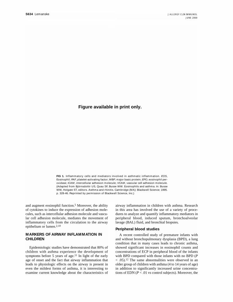
Infiltration of inflammatory cells into the airway walls(predominantly eosinophils and lymphocytes) is a cardi-nal feature of asthma.2 The sequential interaction of thesecells with resident cells (eg, mast cells, macrophages,epithelial cells, endothelial cells) generates a cascade ofevents that contributes to the chronic inflammation andclinical manifestations associated with asthma (Fig 1).5
Mediators, generated by this interaction include hista-mine, platelet-activating factor, derivatives of the arachi-donic acid cascade, such as prostaglandins and the cys-teinyl leukotrienes (LTC4, LTD4, and LTE4), and toxicoxygen radicals.2,5 In turn, these mediators attract otherinflammatory cells, thereby contributing to furtherinflammation, airway smooth muscle spasm (bron-chospasm), airway mucus secretion, and airway micro-vascular leakage that lead to airway edema and narrow-ing.2,5-7 The granular constituents of eosinophil major basicprotein (eosinophil cationic protein [ECP], eosinophil-derived neurotoxin [EDN], and eosinophil peroxidase)induce the epithelial damage seen in the bronchial mucosaof subjects with asthma.5
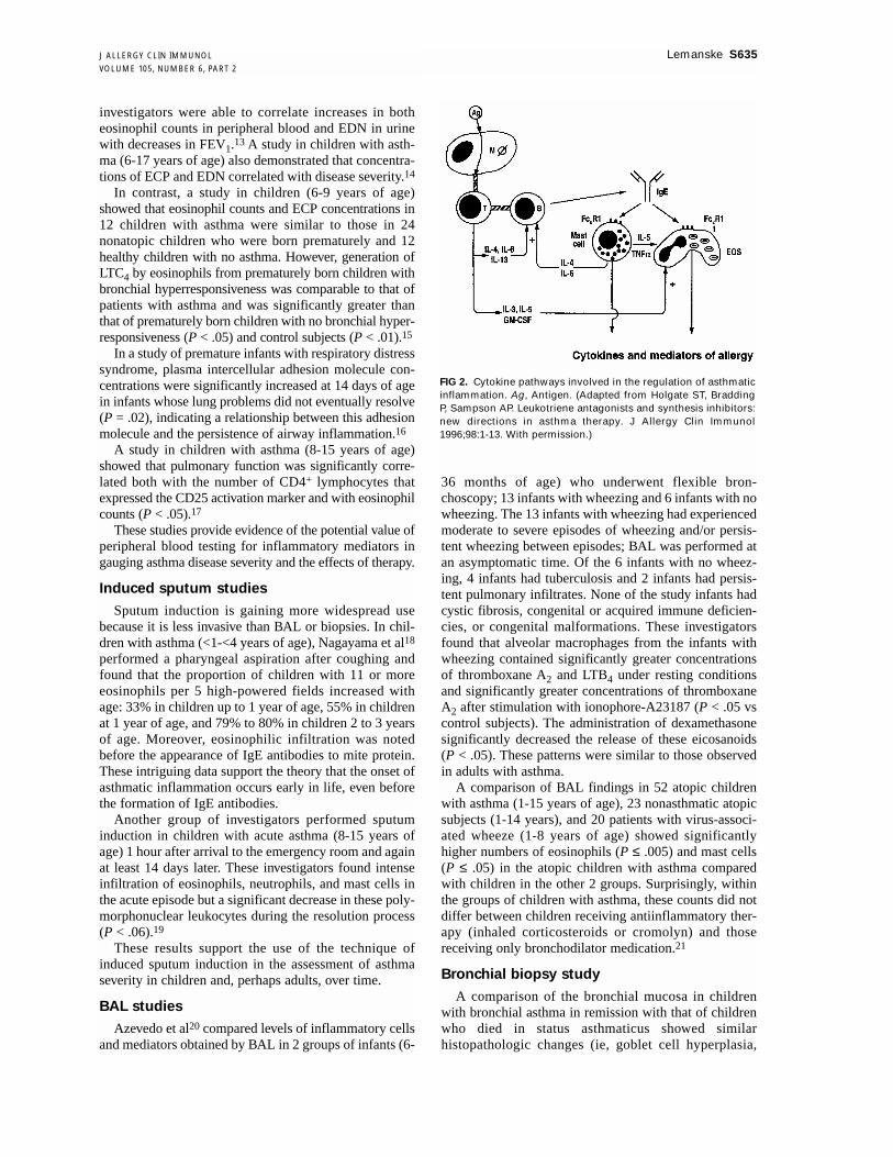
Macrophages, resident cells that are activated inpatients with asthma, are capable of producing a largevariety of cytokines and other mediators involved in theinflammatory response (Fig 2).1,2 These include TNF,IL-1β, IL-2, IL-6, GM-CSF,8 and endothelin.9 Thesecytokines and mediators, in turn, have multiple effects onseveral resident and inflammatory cells. For example, therelease of IL-3, IL-5, and GM-CSF that are generated bylymphocyte activation may prolong eosinophil survival
Inflammatory events in asthma: Anexpanding equation
Robert F. Lemanske, Jr, MD Madison, Wis
S633
From the Departments of Pediatrics and Medicine, University of WisconsinMedical School.
Reprint requests: Robert F. Lemanske, Jr, MD, Professor of Pediatrics andMedicine, University of Wisconsin Hospital, 600 Highland Ave, H4/432,Madison, WI 53792.
Copyright © 2000 by Mosby, Inc.0091-6749/2000 $12.00 + 0 1/0/106155doi:10.1067/mai.2000.106155
Abbreviations usedBAL: Bronchoaveolar lavageBPD: Bronchopulmonary dysplasiaECP: Eosinophil cationic proteinEDN: Eosinophil-derived neurotoxin
S634 Lemanske J ALLERGY CLIN IMMUNOLJUNE 2000
and augment eosinophil function.5 Moreover, the abilityof cytokines to induce the expression of adhesion mole-cules, such as intercellular adhesion molecule and vascu-lar cell adhesion molecule, mediates the movement ofinflammatory cells from the circulation to the airwayepithelium or lumen.2,10
MARKERS OF AIRWAY INFLAMMATION IN
CHILDREN
Epidemiologic studies have demonstrated that 80% ofchildren with asthma experience the development ofsymptoms before 5 years of age.11 In light of the earlyage of onset and the fact that airway inflammation thatleads to physiologic effects on the airway is present ineven the mildest forms of asthma, it is interesting toexamine current knowledge about the characteristics of
airway inflammation in children with asthma. Researchin this area has involved the use of a variety of proce-dures to analyze and quantify inflammatory mediators inperipheral blood, induced sputum, bronchoalveolarlavage (BAL) fluid, and bronchial biopsies.
Peripheral blood studies
A recent controlled study of premature infants withand without bronchopulmonary dysplasia (BPD), a lungcondition that in many cases leads to chronic asthma,showed significant increases in eosinophil counts andconcentrations of ECP in peripheral blood of the infantswith BPD compared with those infants with no BPD (P< .05).12 The same abnormalities were observed in anolder group of children with asthma (4 to 14 years of age)in addition to significantly increased urine concentra-tions of EDN (P < .01 vs control subjects). Moreover, the
FIG 1. Inflammatory cells and mediators involved in asthmatic inflammation. EOS,Eosinophil; PAF, platelet-activating factor; MBP, major basic protein; EPO, eosinophil per-oxidase; ICAM, intercellular adhesion molecule; VCAM, vascular cell adhesion molecule.(Adapted from Björnsdottir US, Quau SF, Busse WW. Eosinophils and asthma. In: BusseWW, Holgate ST, editors. Asthma and rhinitis. Cambridge (MA): Blackwell Science; 1995.p. 328-46. Reprinted by permission of Blackwell Science, Inc.)
Figure available in print only.
J ALLERGY CLIN IMMUNOLVOLUME 105, NUMBER 6, PART 2
investigators were able to correlate increases in botheosinophil counts in peripheral blood and EDN in urinewith decreases in FEV1.13 A study in children with asth-ma (6-17 years of age) also demonstrated that concentra-tions of ECP and EDN correlated with disease severity.14
In contrast, a study in children (6-9 years of age)showed that eosinophil counts and ECP concentrations in12 children with asthma were similar to those in 24nonatopic children who were born prematurely and 12healthy children with no asthma. However, generation ofLTC4 by eosinophils from prematurely born children withbronchial hyperresponsiveness was comparable to that ofpatients with asthma and was significantly greater thanthat of prematurely born children with no bronchial hyper-responsiveness (P < .05) and control subjects (P < .01).15
In a study of premature infants with respiratory distresssyndrome, plasma intercellular adhesion molecule con-centrations were significantly increased at 14 days of agein infants whose lung problems did not eventually resolve(P = .02), indicating a relationship between this adhesionmolecule and the persistence of airway inflammation.16
A study in children with asthma (8-15 years of age)showed that pulmonary function was significantly corre-lated both with the number of CD4+ lymphocytes thatexpressed the CD25 activation marker and with eosinophilcounts (P < .05).17
These studies provide evidence of the potential value ofperipheral blood testing for inflammatory mediators ingauging asthma disease severity and the effects of therapy.
Induced sputum studies
Sputum induction is gaining more widespread usebecause it is less invasive than BAL or biopsies. In chil-dren with asthma (<1-<4 years of age), Nagayama et al18
performed a pharyngeal aspiration after coughing andfound that the proportion of children with 11 or moreeosinophils per 5 high-powered fields increased withage: 33% in children up to 1 year of age, 55% in childrenat 1 year of age, and 79% to 80% in children 2 to 3 yearsof age. Moreover, eosinophilic infiltration was notedbefore the appearance of IgE antibodies to mite protein.These intriguing data support the theory that the onset ofasthmatic inflammation occurs early in life, even beforethe formation of IgE antibodies.
Another group of investigators performed sputuminduction in children with acute asthma (8-15 years ofage) 1 hour after arrival to the emergency room and againat least 14 days later. These investigators found intenseinfiltration of eosinophils, neutrophils, and mast cells inthe acute episode but a significant decrease in these poly-morphonuclear leukocytes during the resolution process(P < .06).19
These results support the use of the technique ofinduced sputum induction in the assessment of asthmaseverity in children and, perhaps adults, over time.
BAL studies
Azevedo et al20 compared levels of inflammatory cellsand mediators obtained by BAL in 2 groups of infants (6-
36 months of age) who underwent flexible bron-choscopy; 13 infants with wheezing and 6 infants with nowheezing. The 13 infants with wheezing had experiencedmoderate to severe episodes of wheezing and/or persis-tent wheezing between episodes; BAL was performed atan asymptomatic time. Of the 6 infants with no wheez-ing, 4 infants had tuberculosis and 2 infants had persis-tent pulmonary infiltrates. None of the study infants hadcystic fibrosis, congenital or acquired immune deficien-cies, or congenital malformations. These investigatorsfound that alveolar macrophages from the infants withwheezing contained significantly greater concentrationsof thromboxane A2 and LTB4 under resting conditionsand significantly greater concentrations of thromboxaneA2 after stimulation with ionophore-A23187 (P < .05 vscontrol subjects). The administration of dexamethasonesignificantly decreased the release of these eicosanoids(P < .05). These patterns were similar to those observedin adults with asthma.
A comparison of BAL findings in 52 atopic childrenwith asthma (1-15 years of age), 23 nonasthmatic atopicsubjects (1-14 years), and 20 patients with virus-associ-ated wheeze (1-8 years of age) showed significantlyhigher numbers of eosinophils (P ≤ .005) and mast cells(P ≤ .05) in the atopic children with asthma comparedwith children in the other 2 groups. Surprisingly, withinthe groups of children with asthma, these counts did notdiffer between children receiving antiinflammatory ther-apy (inhaled corticosteroids or cromolyn) and thosereceiving only bronchodilator medication.21
Bronchial biopsy study
A comparison of the bronchial mucosa in childrenwith bronchial asthma in remission with that of childrenwho died in status asthmaticus showed similarhistopathologic changes (ie, goblet cell hyperplasia,
Lemanske S635
FIG 2. Cytokine pathways involved in the regulation of asthmaticinflammation. Ag, Antigen. (Adapted from Holgate ST, BraddingP, Sampson AP. Leukotriene antagonists and synthesis inhibitors:new directions in asthma therapy. J Allergy Clin Immunol1996;98:1-13. With permission.)

S636 Lemanske J ALLERGY CLIN IMMUNOLJUNE 2000
mucus plugging, increased collagen deposition beneaththe epithelial basement membrane, smooth musclehypertrophy, and local eosinophilia). Notably, the onlyfeatures observed in patients who died in status asthmati-cus that did not occur in the asymptomatic patients weregreater peribronchial eosinophilic infiltration and moreextensive denudation of the airway mucosa, which prob-ably reflected a more acute and active disease process.4
POTENTIAL LONG-TERM CONSEQUENCES
OF ASTHMATIC INFLAMMATION
The chronic inflammation characteristic of asthmastimulates a continuous process of healing and repair inthe airways that can lead to progressive, permanent tissuedestruction. Whereas the bronchial epithelium (whichconstitutes the outermost layer lining the bronchi)appears to regenerate without any trace of the previousinjury, the underlying submucosa regenerates abnormal-ly, resulting in smooth muscle hypertrophy. This defect,in turn, leads to increased airway contractility and isaccompanied by collagen deposition and extracellularmatrix reorganization abnormalities that cause a gradualdecline in pulmonary function.3 The potential for devel-opment of these long-term structural changes in patientswith chronic asthma implies the need for early interven-tion to modify the inflammatory process.2
AIRWAY INFLAMMATION IN ASTHMA:
UNANSWERED QUESTIONS
Although the recognition of the presence of inflam-mation even in mild-to-moderate asthma has improvedtherapeutic approaches to asthma management, manyquestions remain. Specifically, we need to know whenthe inflammatory process starts and how it changes overtime, both in response to therapeutic interventions and asa result of the natural history of asthma. Further studiesalso are needed to determine whether the inflammatoryprocess is the same in all patients who wheeze, regard-less of the stimulus (eg, allergen, exercise, virus). Therelationship between atopy and asthma needs to be clari-fied; as yet, it is not known why some atopic individualsbecome asthmatic and others do not and why some asth-matic individuals are atopic and others are not. Finally,the usefulness of testing for inflammatory markersremains to be established, in terms of its ability to helpidentify individual asthmatic phenotypes, evaluate dis-ease severity, measure therapeutic response, and/or pre-dict potential outcomes. Clarification of these issues willimprove and advance our efforts to evaluate and effec-tively treat patients with asthma.
REFERENCES
1. Holgate ST, Bradding P, Sampson AP. Leukotriene antagonists and syn-thesis inhibitors: new directions in asthma therapy. J Allergy ClinImmunol 1996;98:1-13.
2. Barnes PJ. Frontiers in medicine: new aspects of asthma. J Intern Med1992;231:453-61.
3. Bousquet J, Chanez P, Lacoste JY, White R, Vic P, Godard P, et al. Asth-ma: a disease remodeling the airways. Allergy 1992;47:3-11.
4. Cutz E, Levison H, Cooper DM. Ultrastructure of airways in childrenwith asthma. Histopathology 1978;2:407-21.
5. Björnsdottir US, Quan SF, Busse WW. Eosinophils and asthma. In: BusseWW, Holgate ST, editors. Asthma and rhinitis. Cambridge (MA): Black-well Science; 1995. p. 328-46.
6. Wiggs BR, Bosken C, Paré PD, James A, Hogg JC. A model of airwaynarrowing in asthma and in chronic obstructive pulmonary disease. AmRev Respir Dis 1992;145:1251-8.
7. Barnes PJ, Chung KF, Page CP. Platelet-activating factor as a mediator ofallergic disease. J Allergy Clin Immunol 1988;81:919-34.
8. Broide DH, Lotz M, Cuomo AJ, Coburn DA, Federman EC, WassermanSI. Cytokines in symptomatic asthma airways. J Allergy Clin Immunol1992;89:958-67.
9. Vittori E, Marini M, Fasoli A, de Franchis R, Mattoli S. Increased expres-sion of endothelin in bronchial epithelial cells of asthmatic patients andeffect of corticosteroids. Am Rev Respir Dis 1992;146:1320-5.
10. Leff AR, Hamann KJ, Wegner CD. Inflammation and cell-cell interac-tions in airway hyperresponsiveness. Am J Physiol 1991;260:L189-206.
11. Siegel SC, Rachelefsky GS. Asthma in infants and children: part I. JAllergy Clin Immunol 1985;76:1-15.
12. Yamamoto C, Kojima T, Hattori K, Nogi S, Imamura H, Tsubura A, et al.Eosinophilia in premature infants: correlation with chronic lung disease.Acta Paediatr 1996;85:1232-5.
13. Hoekstra MO, Hovenga H, Gerritsen J, Kauffman HF. Eosinophils andeosinophil-derived proteins in children with moderate asthma. Eur RespirJ 1996;9:2231-5.
14. Zimmerman B, Lanner A, Enander I, Zimmerman LS, Peterson CGB,Ahlstedt S. Total blood eosinophils, serum eosinophil cationic proteinand eosinophil protein X in childhood asthma: relation to disease statusand therapy. Clin Exp Allergy 1993;23:564-70.
15. Schauer U, Alefsen S, Jäger R, Riedel F, Rieger CHL. Blood eosinophils,leukotriene C4 generation, and bronchial hyperreactivity in formerlypreterm infants. Arch Dis Child 1994;71:506-10.
16. Little S, Dean T, Bevin S, Hall M, Ashton M, Church M, et al. Role ofelevated plasma soluble ICAM-1 and bronchial lavage fluid IL-8 levelsas markers of chronic lung disease in premature infants. Thorax1995;50:1073-9.
17. Doi S, Murayama N, Inoue T, Takamatsu I, Kameda M, Omoto Y, et al.CD4 T-lymphocyte activation is associated with peak expiratory flowvariability in childhood asthma. J Allergy Clin Immunol 1996;97:955-62.
18. Nagayama Y, Odazima Y, Nakayama S, Toba T, Funabashi S. Eosinophilsand basophilic cells in sputum and nasal smears taken from infants andyoung children during acute asthma. Pediatr Allergy Immunol1995;6:204-8.
19. Twaddell SH, Gibson PG, Carty K, Woollay KL, Henry RL. Assessmentof airway inflammation in children with acute asthma using induced spu-tum. Eur Resp J 1996;9:2104-8.
20. Azevedo I, de Blic J, Scheinmann P, Vargaftig BB, Bachelet M. Enhancedarachidonic acid metabolism in alveolar macrophages from wheezyinfants. Am J Respir Crit Care Med 1995;152:1208-14.
21. Stevenson EC, Turner G, Heaney LG, Schock BC, Taylor R, Gallagher T,et al. Bronchoalveolar lavage findings suggest two different forms ofchildhood asthma. Clin Exp Allergy 1997;27:1027-35.