inflammatory diseases of the skin - duke … · - inflammation of the subcutaneous fat\r- tend to...
TRANSCRIPT

INFLAMMATORY DISEASES OF THE SKIN
M. Angelica Selim, M.D. Dermatopathology Unit Pathology Department
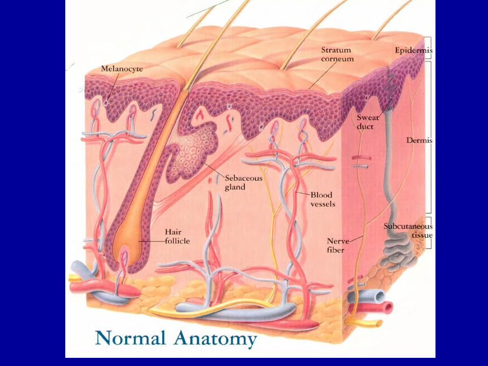
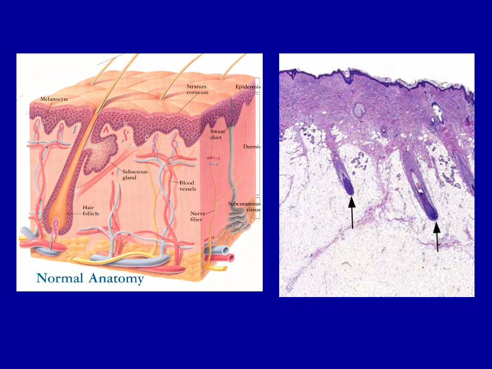
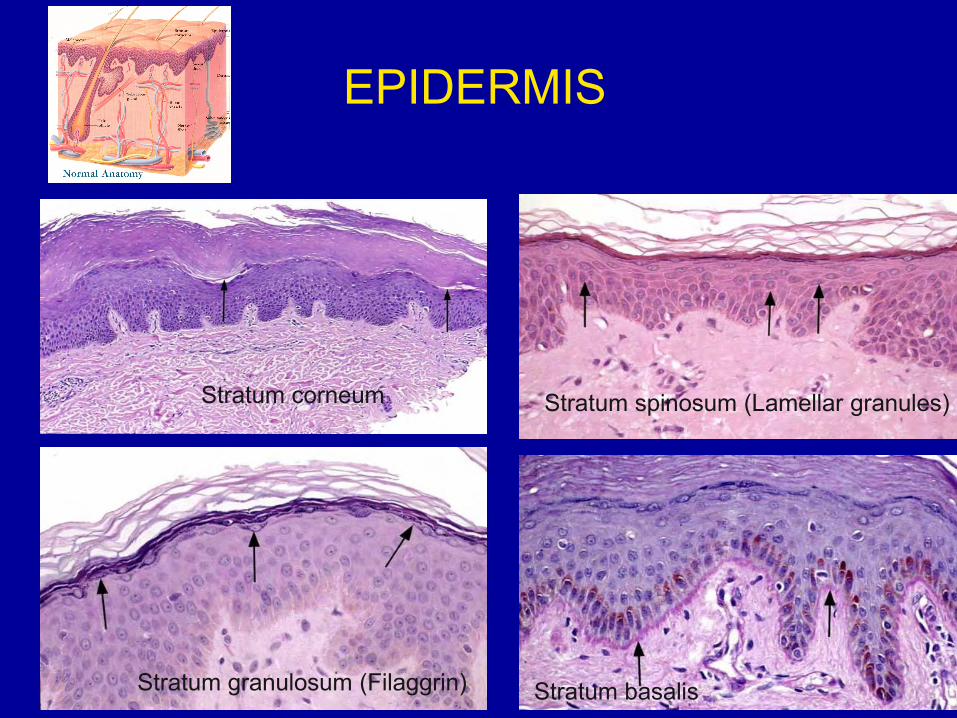
EPIDERMIS
Stratum basalis
Stratum spinosum (Lamellar granules)
Stratum granulosum (Filaggrin)
Stratum corneum
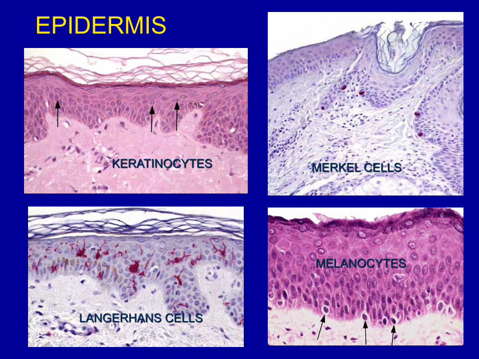
EPIDERMIS
KERATINOCYTES
LANGERHANS CELLS
MERKEL CELLS
MELANOCYTES

DERMIS
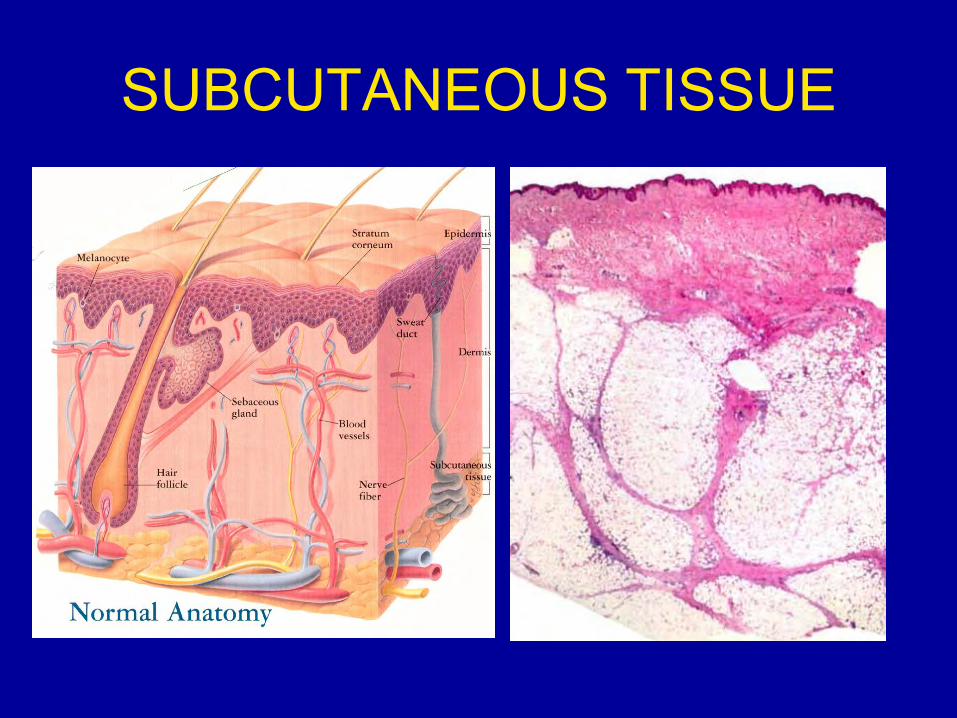
SUBCUTANEOUS TISSUE

ADNEXAL STRUCTURES

FUNCTIONS
• External organ protection: – Impermeable – Melanin
• Temperature control • Vitamin D
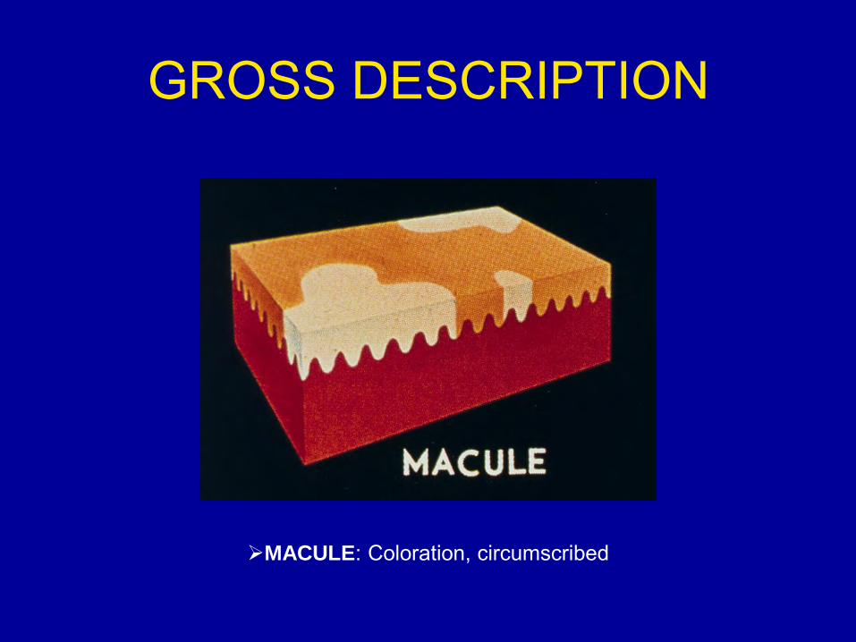
GROSS DESCRIPTION
MACULE: Coloration, circumscribed
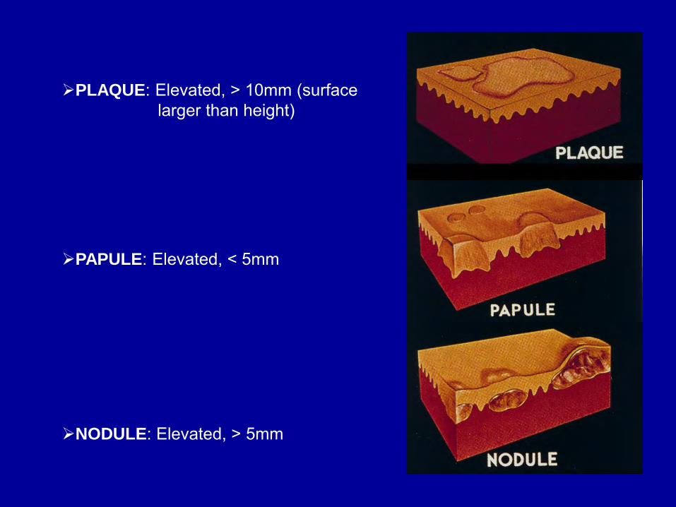
PLAQUE: Elevated, > 10mm (surface larger than height)
PAPULE: Elevated, < 5mm
NODULE: Elevated, > 5mm
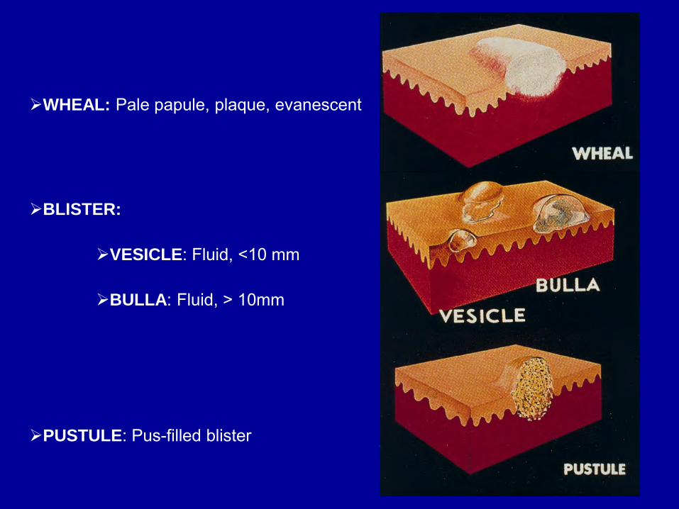
WHEAL: Pale papule, plaque, evanescent
BLISTER:
VESICLE: Fluid, <10 mm
BULLA: Fluid, > 10mm
PUSTULE: Pus-filled blister

CRUST: Serous, purulent exudates
SCALE: Dry, plate-like excrescence
LICHENIFICATION: Thickened, rough
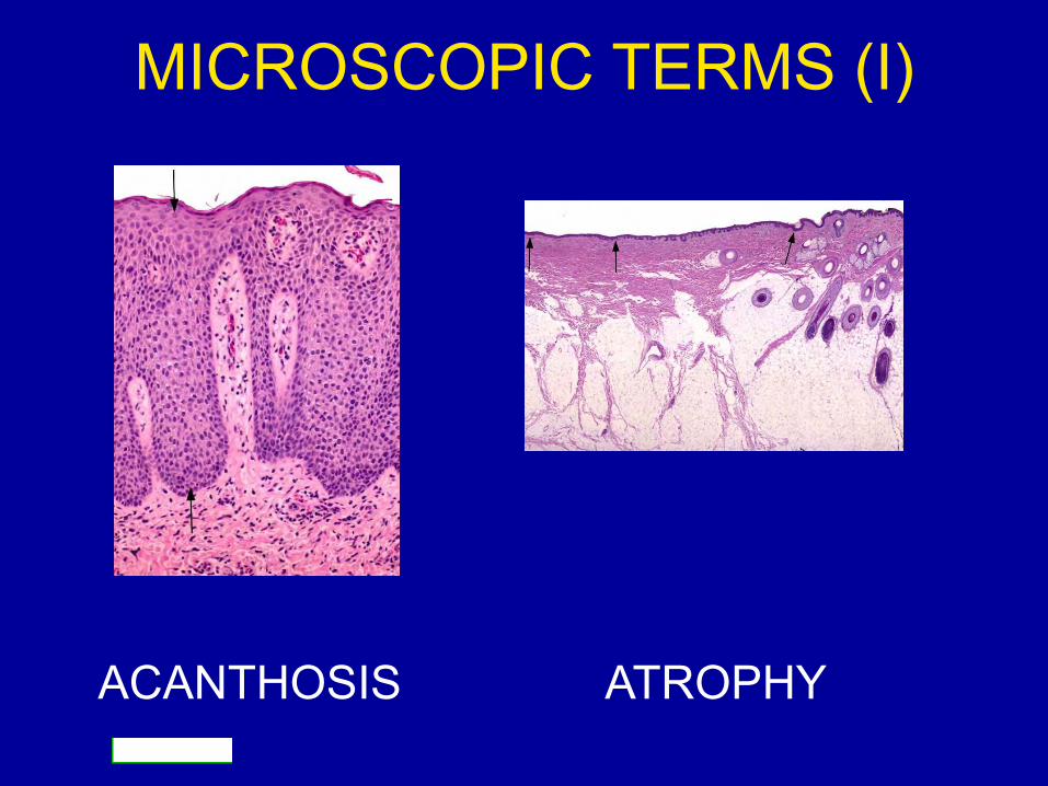
MICROSCOPIC TERMS (I)
ACANTHOSIS ATROPHY

MICROSCOPIC TERMS (II)
HYPERKERATOSIS
HYPERGRANULOSIS ULCER
Orthokeratosis: normal keratin Parakeratosis: nuclei Stratum corneum

MICROSCOPIC TERMS (III)
PAPILLOMATOSIS PSORIASIFORM
ACANTHOLYSIS SPONGIOSIS

TYPES OF BIOPSY: • Shave • Punch • Ellipse • Major excision • ALWAYS CAREFUL !!!!

INDICATIONS FOR BIOPSY • Unknown diagnosis:
– Inflammatory disease – Neoplastic
• Systemic disease: – Vasculitis – Amyloidosis

TECHNIQUES • Hematoxylin and eosin • Histochemistry • Immunohistochemistry • Electron microscopy

DERMATITIS (PATTERNS)
• Location • Superficial/deep • Cellularity
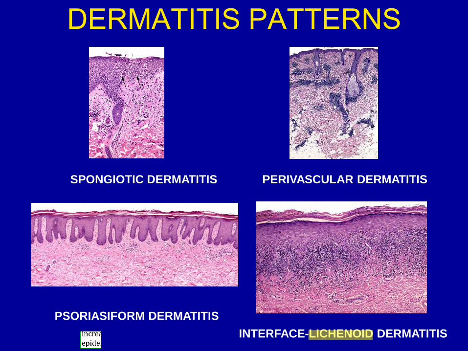
DERMATITIS PATTERNS
SPONGIOTIC DERMATITIS PERIVASCULAR DERMATITIS
PSORIASIFORM DERMATITIS
INTERFACE-LICHENOID DERMATITIS

DERMATITIS PATTERNS:
PANNICULITIS VASCULITIS
ALLERGIC CONTACT DERMATITIS
• Morbidity • Leading occupational disease • Mostly irritant mechanisms • Type IV immune-reaction:
– Sensitization – Elicitation
• Langerhans cells

ALLERGIC CONTACT DERMATITIS: MORPHOLOGY
• ACUTE: – Erythematous macules – Papules and vesicles
• CHRONIC: – Erythema – Scale – Lichenification
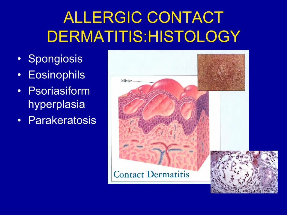
ALLERGIC CONTACT DERMATITIS:HISTOLOGY
• Spongiosis • Eosinophils • Psoriasiform
hyperplasia • Parakeratosis

PSORIASIS
• 1-2 %population in USA • Scalp, acral, extensor surfaces
(elbows/knees) • Nails (pits) • Arthritis
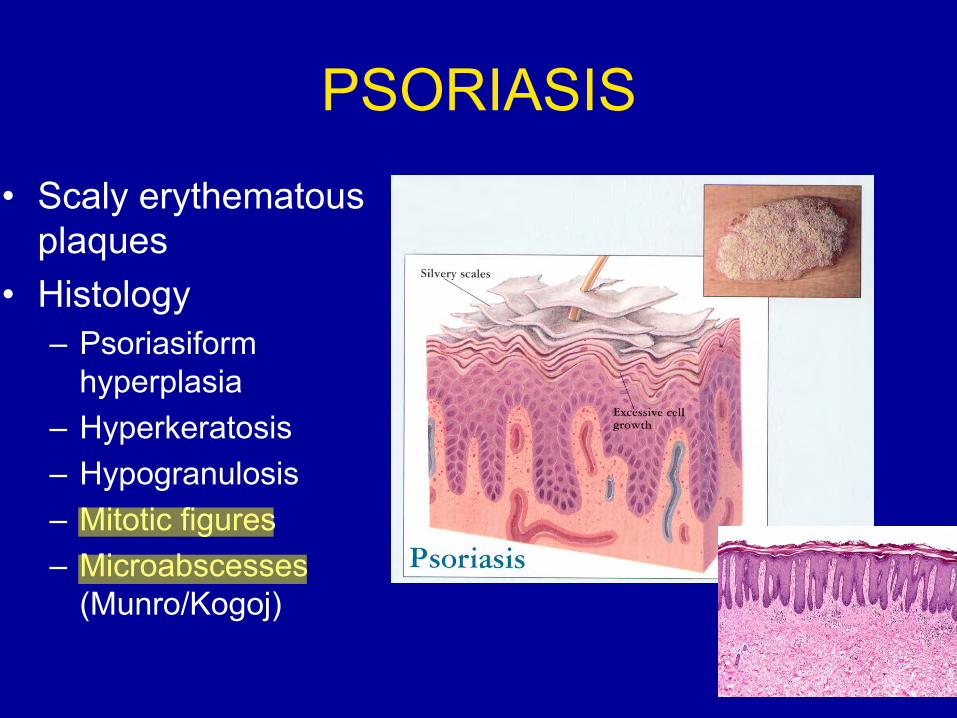
PSORIASIS
• Scaly erythematous plaques
• Histology – Psoriasiform
hyperplasia – Hyperkeratosis – Hypogranulosis – Mitotic figures – Microabscesses
(Munro/Kogoj)

PSORIASIS
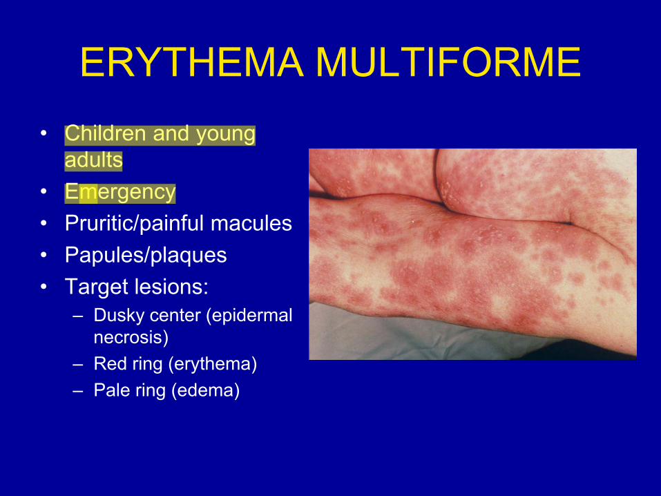
ERYTHEMA MULTIFORME • Children and young
adults • Emergency • Pruritic/painful macules • Papules/plaques • Target lesions:
– Dusky center (epidermal necrosis)
– Red ring (erythema) – Pale ring (edema)
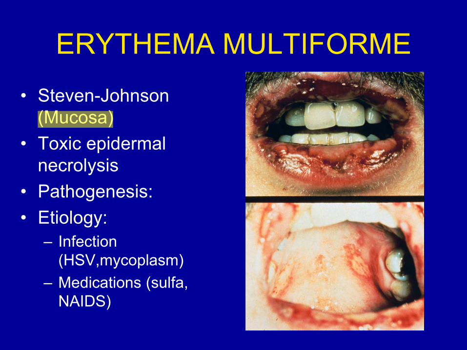
ERYTHEMA MULTIFORME
• Steven-Johnson (Mucosa)
• Toxic epidermal necrolysis
• Pathogenesis: • Etiology:
– Infection (HSV,mycoplasm)
– Medications (sulfa, NAIDS)
ERYTHEMA MULTIFORME

DRUG REACTIONS:
• 2 % of inpatients • 3/1000 Rx • Within 1 week • Amoxicillin, bactrim, ampicillin • Penicillin, barbiturates, benzodiazepines,
thiazides

DRUG REACTIONS PATHOGENESIS
• Immune: – I: IgE (penicillin) – II: cytotoxic – III: immune-complex (vasculitis) – IV: cell mediated (vitamin K)
• Non-immune: – Activation (mast cell degranulation) – Overdose – Side effects (alopecia/ChemoRx) – Photosensitivity (tetracycline) – Others

DRUG REACTIONS MORPHOLOGY
• Lichenoid
• Superficial and deep perivascular

DRUG REACTIONS MORPHOLOGY
VASCULITIS

LUPUS ERYTHEMATOSUS • Multiple organs • Cutaneous or systemic • Diagnosis:
– Clinical – Histologic – Biochemical
• Pathogenesis: – HLA – Medications (hydralazine, procainamine, D-
penicillamine) – Hormonal – Autoimmunity
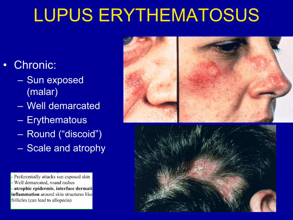
LUPUS ERYTHEMATOSUS
• Chronic: – Sun exposed
(malar) – Well demarcated – Erythematous – Round (“discoid”) – Scale and atrophy

LUPUS ERYTHEMATOSUS
• Subacute:
– Erythematous – Symmetrical – Trunk and arms – Systemic
involvement
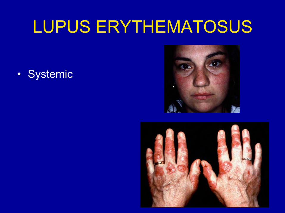
LUPUS ERYTHEMATOSUS
• Systemic
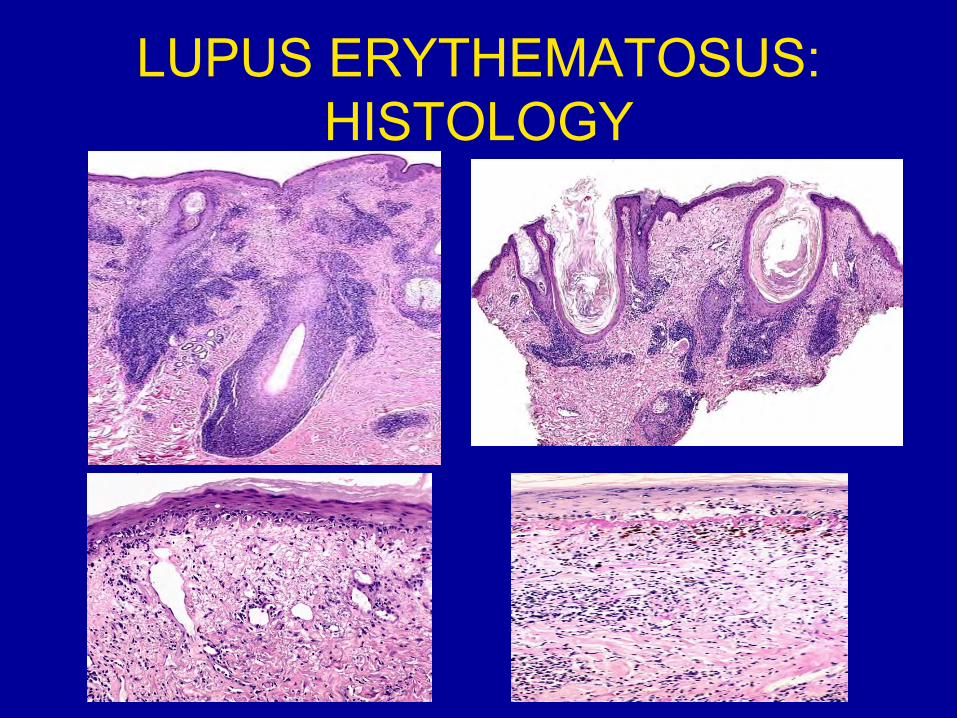
LUPUS ERYTHEMATOSUS: HISTOLOGY
ACNE • Disorder of the pilosebaceous unit • Face, neck, back • Onset:
– Puberty – Neonatal
• Etiology: – Propionibacterium acnes (acids) – Occlusion – Stress – Hormones
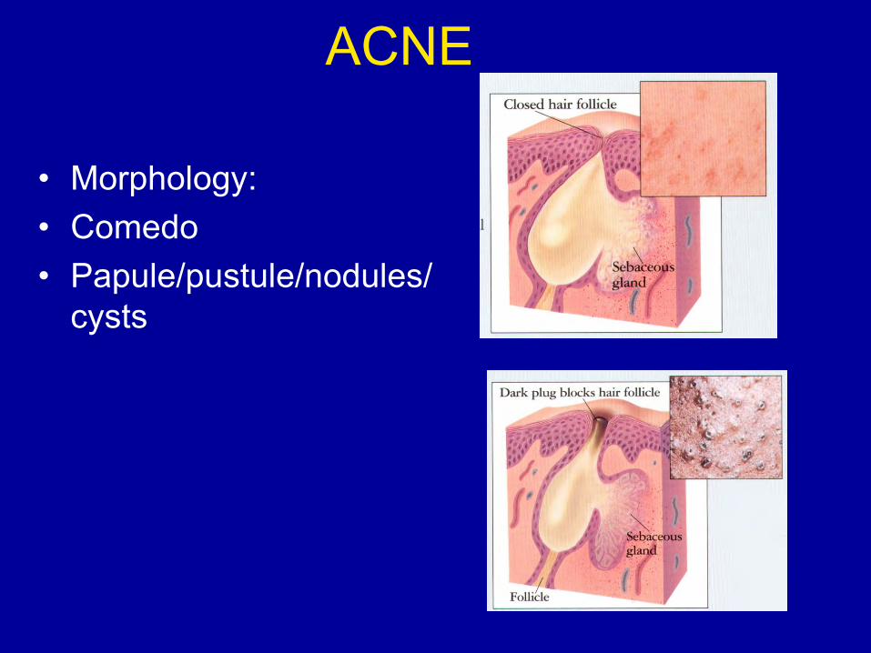
ACNE
• Morphology: • Comedo • Papule/pustule/nodules/
cysts

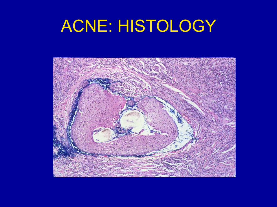
ACNE: HISTOLOGY

ERYTHEMA NODOSUM
• Panniculitis • Bilateral painful/tender • Erythematous/violaceous
nodules • Lower legs • Arthralgias

ERYTHEMA NODOSUM
• Association: – Bacterial (TB, leprosy) – Fungal (histoplasma) – Viral – Medications (contraceptives, sulfas) – IBD – Sarcoidosis – Hodgkin disease

ERYTHEMA NODOSUM

Descriptio
‐ A
‐ Er
‐ V
‐ Is
‐ A
‐ D
on of lesion:
rm
rythematous
esicles‐ bliste
s the patient i
re there syste
iagnosis: acu
o Note l
macules‐ Som
ering of the sk
n pain? Unco
emic findings
te contact de
inear pattern
me areas are
kin
omfortable bu
? No, but he
ermatitis
n‐ means that
flat with redn
ut not in pain
might be cam
he probably
ness
mping recentl
touched som
y around a lo
mething.
ot of trees.

Descriptio
‐ D
‐ Pa
‐ D
‐ D
on of lesion:
iffuse rednes
o The ra
atient is com
oes the patie
iagnosis‐ eryt
o Need t
ss and areas o
sh is in both
plaining of pa
ent have bloo
thema nodos
to find what i
of elevation (n
legs.
ain. Can’t sit b
dy diarrhea?
sum.
is wrong with
nodules) acro
because of pa
Not to Matt’
h the patient!
oss the lower
ain.
s knowledge.
Probably som
leg
.
mething systeemic going onn.
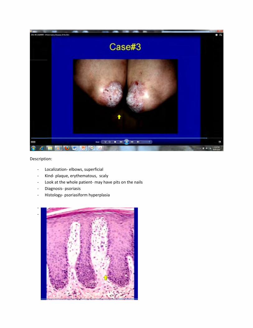
Descriptio
‐ Lo
‐ K
‐ Lo
‐ D
‐ H
‐
‐
on:
ocalization‐ e
ind‐ plaque, e
ook at the wh
iagnosis‐ pso
istology‐ pso
lbows, super
erythematous
hole patient‐
oriasis
riasiform hyp
ficial
s, scaly
may have pit
perplasia
s on the nailss

Describing Lesions:
1. Macule:
a. Change in skin color
b. No elevation or depression
c. Nonpalpable
2. Elevated lesions:
a. Papule = elevated lesion under 5 mm in diameter
b. Nodule = elevated lesion over 5 mm in diameter
c. Plaque = less elevated but surface greater than 1 cm in diameter
3. Wheal = pale (white color) papule or plaque that comes and goes
4. Blisters:
a. Vesicle = fluid filled and under 10 mm
b. Bulla = fluid filled and greater than 10 mm
5. Pustule = blister filled with pus
6. Crust = serous, purulent exudate oozing out of a lesion
7. Scale = dry, plate-like scales coming off
8. Lichenification = thickened, rough
Microanatomy:
1. Acanthosis = thickening of epidermis
2. Atrophy = thinning of epidermis
3. Hyperkeratosis = increased stratum corneum
4. Hypergranulosis = increased stratum granulosum layer (topmost layer before keratin)
5. Papillomatosis = hyperplasia of dermal papillae cause wrinkling:
6. Acantholysis = loss of intercellular connections:
7. Psoriasiform = too much epidermis but pushes down:

8. Spongiosis = epidermis begins to absorb fluid:
Biopsy:
1. Reasons to biopsy:
a. Unknown diagnosis (inflammatory disease, neoplasm)
b. Systemic disease (vasculitis, amyloidosis skin biopsy easier than bronchus)
2. Types of biopsy:
a. Shave (epidermis and some dermis)
b. Punch (gets all layers but small area)
c. Ellipse (cuts off the whole lesion)
d. Major excision (goes all the way to muscle)
Diseases:
1. Dermatitis:
a. Need to know location, superficial vs. deep, and cellularity
b. Types [see slide 25]:
i. Spongiotic = eczema
ii. Perivascular = inflammation around vessels
iii. Psoriasiform = psoriasis
iv. Interface-lichenoid = inflammation between epidermis/dermis
v. Panniculitis = inflammation of dermis (mainly lobules vs. septa)
c. Allergic contact dermatitis:
i. Type IV hypersensitivity reaction via Langerhans cells
ii. Acute see macules (flat) and some papules (small) and vesicles (fluid)
iii. Chronic see more scaling and lichenification
iv. Over time get psoriasiform hyperplasia and parakeratosis
2. Psoriasis:
a. Affects 1-2% of population
b. Main areas are scalp, nails, and extensor surfaces (elbows/knees)
c. Get scaly erythematous plaques
d. Histology: psoriasiform hyperplasia, hyperkeratinosis, hypogranulosis

3. Erythema multiforme:
a. Medical emergency, typically affects children/young adults
b. Get multiform papules and plaques
c. Lesions have red ring/pale ring with dusky centers
d. Causes:
i. Infection (HSV, mycoplasm)
ii. Medications (sulfa, NSAIDs)
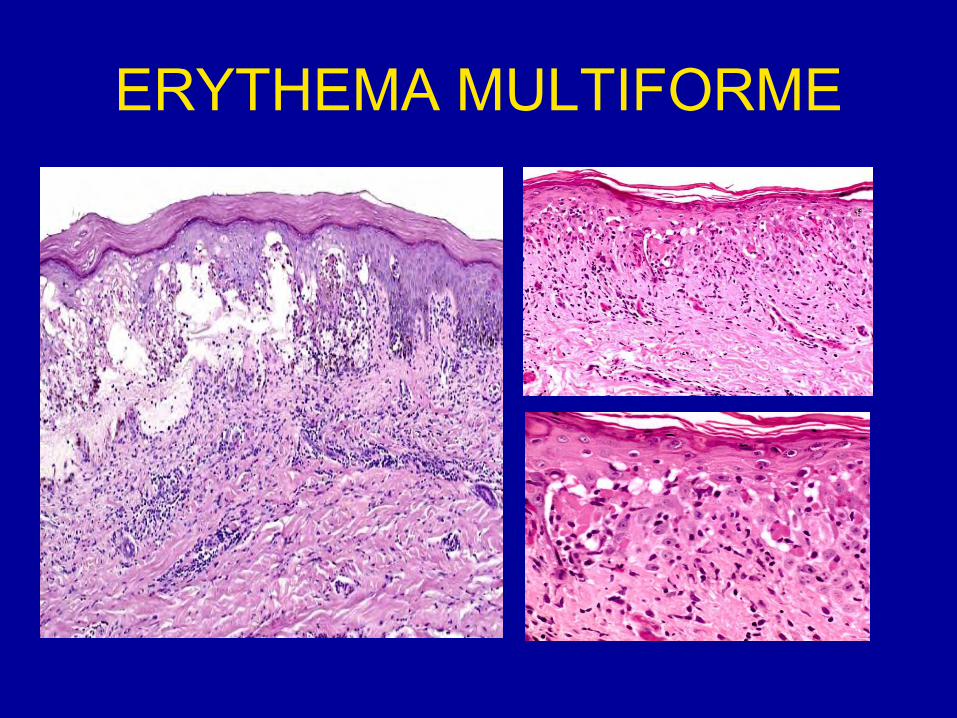
e. Mainly due to immune complex and lymphocytes invading everywhere
4. Lupus erythematosus:
a. Can be cutaneous or systemic
b. Preferentially attacks sun exposed skin
c. Well demarcated, round rashes
d. Histology: atrophic epidermis, interface dermatitis, inflammation around skin
structures like hair follicles
5. Acne:
a. Causes include propioinibacteria, occlusion, stress, hormones
b. Comedo = dilated hair follicle (black head)
c. Pustule is when closed hair follicle fills with neutrophils turns into nodule
6. Erythema nodosum:
a. Inflammation of the subcutaneous fat
b. Tend to see it in the lower legs (both sides)
c. Erythema nodosum is an indication something more systemic is wrong (sarcoid,
Hodgkin, viral, bacteria, etc.)
7. Drug reactions:
a. Happen to 2% of inpatients, within one week of giving a variety of drugs
b. Pathogenesis includes hypersensitivity types I-IV
c. Also includes non-immune causes (overdose, photosensitivity, etc.)
d. Typically get vasculitis, lichenoid