inflammation - faculty of pharmacy and biotechnology - the...
TRANSCRIPT

Inflammation I
Dr. Nabila Hamdi
MD, PhD

http://library.med.utah.edu/WebPath/EXAM/MULTGEN/examidx.htm
2

ILOs • Distinguish between acute and chronic inflammation with respect to causes, nature
of the inflammatory response, and tissue changes.
• Describe the sequence of vascular changes in acute inflammation and their purpose
• Know the mechanisms of increased vascular permeability
• Define the terms edema, transudate, and exudate
• Describe the steps involved in extravasation of leukocytes from the blood to the tissues. Know the steps at which selectins and integrins act.
• Describe the meaning and utility of chemotaxis. Understand the role that chemokines play in inflammation
• Describe the steps involved in phagocytosis and the role of opsonins
• Understand the mechanism of degradation in the phagolysosomes.
• Know the major effects of the chemical mediators of inflammation
• Distinguish between the different patterns of inflammation. Define an abscess.
• Know the cells involved in, and causes of chronic inflammation
• Understand the mechanism of granulomatous inflammation
• Describe the systemic manifestations of inflammation
3

Outline I. Overview Of Inflammation
II. Acute Inflammation 1. Definition
2. Cardinal signs
3. Vascular changes
4. Cellular events
5. Chemical mediators
6. Outcomes
III. Chronic Inflammation 1. Definition
2. Cells & mediators
3. Granulomatous Inflammation
IV. Morphological Patterns of Acute and Chronic Inflammation
V. Systemic Effects of Inflammation
4

Overview
Inflammation is a protective response intended to eliminate the initial cause of cell injury as well as the necrotic cells and tissues resulting from the original
insult.
5

Overview Players of Inflammation
The goal of the inflammatory reaction is to bring cells and molecules of host defense (normally circulating in the blood) to the site of infection or tissue damage. These include blood leukocytes and plasma proteins, cells of vascular walls, and cells and extracellular
matrix (ECM) of the surrounding connective tissue. 6
Tight junctions
Gaps

Acute Inflammation
Definition:
Acute inflammation is a rapid response to injury or microbes and other foreign substances that is designed to deliver leukocytes and plasma proteins to sites of injury.
Stimuli:
• Infections (bacterial, viral, fungal, parasitic)
• Trauma (blunt and penetrating)
• Physical and chemical agents (thermal injury, e.g., burns or frostbite; irradiation or some environmental chemicals)
• Tissue necrosis (from any cause)
• Foreign bodies (splinters, dirt, sutures)
• Immune reactions (also called hypersensitivity reactions)
7

Acute Inflammation
Heat (calor)
redness (rubor)
Swelling (tumor)
Pain (dolor)
loss of function (functio laesa)
Cardinal Signs of Inflammation
8
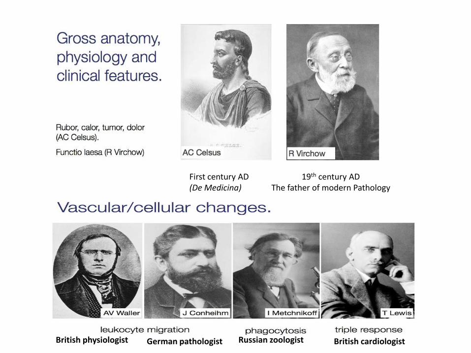
First century AD (De Medicina)
19th century AD The father of modern Pathology
British physiologist German pathologist Russian zoologist British cardiologist
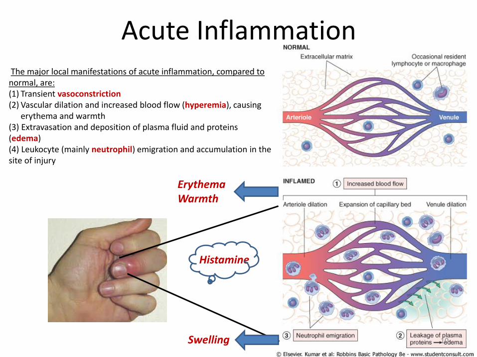
Acute Inflammation The major local manifestations of acute inflammation, compared to normal, are: (1) Transient vasoconstriction (2) Vascular dilation and increased blood flow (hyperemia), causing
erythema and warmth (3) Extravasation and deposition of plasma fluid and proteins (edema) (4) Leukocyte (mainly neutrophil) emigration and accumulation in the site of injury
Erythema Warmth
Swelling
Histamine
10

1. Vascular Changes • Hyperemia • Exudation • Start of emigration
Acute Inflammation
2. Cellular Events • Margination • Rolling • Adhesion • Transmigration • Chemotaxis • Activation • Phagocytosis & killing
3. Chemical Mediators
11

Vascular Changes
12
The mean colloid osmotic pressure is equal to the mean capillary pressure
Fluid leaks out because of increased hydrostatic pressure or decreased osmotic pressure
Formed in inflammation because vascular permeability increases as a result of increased
inter-endothelial spaces
Fluid accumulation in extravascular spaces is called edema; the fluid may be a transudate or exudate. Whereas exudates are typical of inflammation, transudates accumulate in various non-inflammatory conditions.

13
lung sounds - crackles (1).mp4
This chest X-ray shows an area of lung inflammation indicating the presence of pneumonia.
Congestive heart failure with diffuse perihilair Pulmonary edema

Vascular Changes
• After transient vasoconstriction (lasting only for seconds), arteriolar vasodilation occurs, resulting in locally increased blood flow (hyperemia) and engorgement of the down-stream capillary beds. This vascular expansion is the cause of the redness (erythema) and warmth characteristically seen in acute inflammation. The increased volume of blood flow leads to a rise in intravascular hydrostatic pressure, resulting in movement of fluid from capillaries into the tissues (transudate). This fluid is essentially an ultrafiltrate of blood plasma and contains little protein.
• As the microvasculature becomes more permeable, protein-rich fluid moves into the extravascular tissues (exudate). This causes the red blood cells to become more concentrated, thereby increasing blood viscosity and slowing the circulation.
14
Cellular Events
15
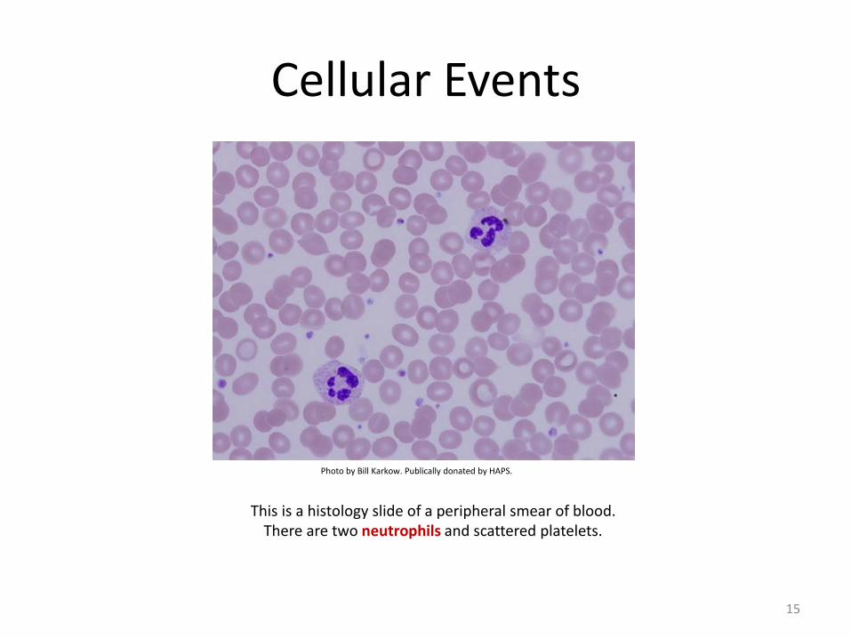
This is a histology slide of a peripheral smear of blood. There are two neutrophils and scattered platelets.
Photo by Bill Karkow. Publically donated by HAPS.

Cellular Events Leukocyte Recruitment
1.Margination
5. Chemotaxis
The leukocytes first roll, then become activated and adhere to endothelium, then transmigrate across the endothelium, pierce the basement membrane, and migrate toward chemoattractants emanating from the source of injury. Different molecules play predominant roles in different steps of this process - selectins in rolling; chemokines in activating the neutrophils to increase avidity of integrins, integrins for stable adhesion. After extravasating from the blood, leukocytes migrate toward sites of infection or injury along a chemical gradient by a process called chemotaxis.
Stasis
4. Diapedesis/ Transmigration
-Bacteria -Cytokines -Complement -Arachidonic acid metabolism
7. Killing 6. Phagocytosis
2. 3. 4.
16

Cellular Events Leukocyte Recruitment
• Margination: The process of leukocyte accumulation at the periphery
of vessels. Leukocytes are pushed out of the central axial column and thus have a better opportunity to interact with lining endothelial cells, especially as stasis sets in.
• Rolling: leukocytes tumble on the endothelial surface, transiently
sticking along the way. This weak and transient adhesion involved in rolling is mediated by the selectin family of adhesion molecules.
• Adhesion: firm adhesion of leukocytes to endothelial surfaces. This
adhesion is mediated by integrins expressed on leukocyte cell surfaces interacting with their ligands on endothelial cells
17

Cellular Events Leukocyte Recruitment
• Transmigration: After being arrested on the endothelial surface, leukocytes migrate through the vessel wall primarily by squeezing between cells at intercellular junctions (diapedesis)
• Chemotaxis: After extravasation from the blood, leukocytes migrate toward sites of infection or injury along a chemical gradient. Both exogenous and endogenous substances can be chemotactic for leukocytes, including:
(1) bacterial products
(2) cytokines
(3) components of the complement system, particularly C5a
(4)products of the lipoxygenase pathway of arachidonic acid (AA) metabolism, particularly leukotriene B4 (LTB4).
18

Cellular Events Leukocyte Recruitment
In most forms of acute inflammation, neutrophils predominate in the inflammatory infiltrate during the first 6 to 24 hours.
Why? Neutrophils are more numerous in the blood, they respond more rapidly to chemokines, and they may attach more firmly to the adhesion molecules that are rapidly induced on endothelial cells (selectins). In addition, after entering tissues, neutrophils are short-lived, they die by apoptosis and disappear within 24 to 48 hours.
Leukocyte Response in Acute Inflammation: Extravasation Medicine, By Ernest
19

Cellular Events Leukocyte Activation
Different classes of cell surface receptors of leukocytes recognize different stimuli. The receptors initiate responses that mediate the functions of the leukocytes.
Only some receptors are depicted. IFN-γ, interferon γ; LPS, lipopolysaccharide.
See next slide
20

Cellular Events Leukocyte Activation
Phagocytosis of a particle (e.g., a bacterium) involves (1) attachment and binding of the particle to receptors on the leukocyte surface, (2) engulfment and fusion of the phagocytic vacuole with granules (lysosomes), and (3) destruction of the ingested particle. iNOS, Inducible nitric oxide synthase; NO, nitric oxide; ROS, reactive oxygen species.
21
oxidative burst

Cellular Events Leukocyte Activation
In leukocytes (mainly neutrophils and macrophages), the phagocyte oxidase (NADPH oxidase) in the phagosome membrane generates superoxide, which can be converted to other free radicals. Myeloperoxidase (MPO) in phagosomes also generates hypochlorite (hydroxy-halide radical) from ROS
22
Oxidative burst
Leukocyte-Induced Tissue Injury??

Leukocyte Activation Phagocytosis
• (1) Recognition and attachment: Leukocytes bind and ingest most microorganisms and dead cells via specific surface receptors, which recognize either components of the microbes and dead cells, or host proteins, called opsonins, that coat microbes and target them for phagocytosis (a process called opsonization)
• (2) Engulfment: Pseudopods are extended around the object, eventually forming a phagocytic vacuole. The membrane of the vacuole then fuses with the membrane of a lysosomal granule, resulting in discharge of the granule's contents into the phagolysosome.
• (3) killing and degradation of the ingested material: The key steps in this reaction are the production of microbicidal substances within lysosomes and fusion of the lysosomes with phagosomes. The most important microbicidal substances are reactive oxygen species (ROS) and lysosomal enzymes (myeloperoxidase). Phagocytosis stimulates an oxidative burst characterized by a sudden increase in oxygen consumption, glycogen catabolism (glycogenolysis), increased glucose oxidation, and production of ROS.
23

Defects in Leukocyte Function
Leukocyte Adhesion Deficiency (LAD) Defect in integrins/selectins
Defects in microbicidal activity A genetic deficiency in one of the several components of the phagocyte oxidase responsible for generating ROS. In these patients, engulfment of bacteria does not result in activation of oxygen-dependent killing mechanisms.
Defects in phagolysosome formation Impairment of the fusion of lysosomes with phagosomes
24

Summary of Vascular & Cellular Events
• Vasodilation is induced by chemical mediators such as histamine, and is the cause of erythema and stasis of blood flow
• Increased vascular permeability allows plasma proteins and leukocytes to enter sites of infection or tissue damage; fluid leak through blood vessels results in edema
• Leukocytes are recruited from the blood into the extravascular tissue where infectious pathogens or damaged tissues may be located and are activated to perform their functions
• Leukocyte recruitment is a multi-step process consisting of loose attachment to and rolling on endothelium (mediated by selectins); firm attachment to endothelium (mediated by integrins); and migration through inter-endothelial spaces.
• Neutrophils predominate in the early inflammatory infiltrate and are later replaced by macrophages.
• Leukocytes can eliminate microbes and dead cells by phagocytosis, followed by their destruction in phagolysosomes. Destruction is caused by free radicals (ROS, NO) generated in activated leukocytes and lysosomal enzymes.
• Enzymes and ROS may be released into the extracellular environment, which may lead to damage of normal tissues.
25

1. What are the cardinal signs of inflammation?
2. Which signs of inflammation are associated with histamine release?
3. What are the vascular changes that occur during acute inflammation?
4. What is the sequence of events in the cellular events of acute inflammation?
5. What is the definition of chemotaxis?
6. What is the mechanism of destruction of a bacteria by the neutrophils?
26

27

References
ROBBINS Basic Pathology 8th Edition Basic Pathology 7th Edition, by Kumar, Cotran and Robbins Inflammation. Vascular and Cellular Processes. http://nfs.unipv.it/nfs/minf/dispense/patgen/lectures/files/autophagy.html