infectious disease cytopathology
TRANSCRIPT

Infectious Disease
Cytopathology
MycologyHeather Ruff
October 26, 2019

Fungi
YeastHyphae

Categorizing Fungal Hyphae
Hyphae
Pigmented hyphae
Dematiaceous molds
Non-septate hyaline hyphae
Zygomycetes
Septate hyaline hyphae
Aspergillus and others
Pseudohypaewith Yeast (Candida)

Assessing Fungal Hyphae
● True hyphae or pseudohyphae● Pigmented or non-pigmented (hyaline)?● Septate or non-septate?
● Branching -○ Acute angle, right angle, variable?○ Dichotomous (equal) vs. non-dichotomous (variable)?
● Arrangement - uniform/parallel vs. disorganized● Other structures
○ Fruiting Bodies (conidia, phialides, vesicle)○ Pseudohyphae○ Yeast

Hyphal Forms
Hyphae Pseudohyphae True Hyphae with Constricted septations

Cervical Pap ThinPrep 29 Female
What is this?

• Oval budding yeast (3-6 µm)• Pseudohyphae• True hyphae (less common)• Blastoconidia (rare)
Candida

• Exophiala, Phialophora, Cladophialophora, Bipolaris, and others
• Pigmented - Usually visible (brown) unstained• If not visible, may be demonstrated with Fontana-Masson
• Narrow (2-12 µm diameter, usually 2-6 µm)• Septate (constricted septations common)• Branching - Irregular• Moniliform (beaded) appearance• May have yeast forms and secondary
structures (e.g. chlamydoconidia)
Dematiaceous (Pigmented) Fungi
Picture from https://www.sciencedirect.com/topics/agricultural-and-biological-sciences/phaeohyphomycosis

Zygomycetes• Mucor spp., Rhizomucor sp., Rhisopuz spp.,
and others broad non-pigmented hyphae
• Non-pigmented• Non-septate (really Pauci-septate)• Broad (5-25 µm diameter, avaerage 12 µm)
• Branching - Irregularly-spaced, non-dichotomous, variable angles ( often 90°)
• Twisted, folded, ribbon-like morphology

Hyaline Fungi
• Aspergillus, Fusarium, Paecilomyces, Acremonium, Scedosporium, Pseudallescheria, and many, many others)
• Non-pigmented• Septate• Narrow (2-12 µm diameter, usually 2-8 µm)
• …

Aspergillus
● Organized● Branching –
○Parallel○Dichotomous (equal)○Acute angle (45°)
● May have fruiting bodies
● Disorganized● Branching –
○Variable orientation○Non-dichotomous (unequal)○Variable angles (45° & 90°)
● May see fruiting bodies
Other Hyaline Fungi

Esophageal Brush 56 F w/ SLE on prednisone.
Diagnosis?

BAL 54 M s/p lung transplant.Diagnosis?

BAL 54 M s/p lung transplant.Diagnosis?

Categorizing Yeast
Yeast
Small
Histoplasma Cryptococcus Pneumocystis Candida
Large
Blastomyces Cryptococcus Coccidioidies

Assessing Yeast
● Size - Small, large, variable● Shape - Round, oval, irregular● Other morphology
● Wall - Thin, thick, refractile● Capsule, pseudocapsule, none● Division – Budding (Broad-based, narrow-based), Fission● Intracellular, extracellular, both● Internal structures
● Stains:● Mucin (Mucicarmine)● Pigment (Fontana-Masson)
Penicillium marneffei (E) - Courtesy of Vicki Schnadig at UTMBParacoccidiioides (*) - Courtesy of Diane Dziedzic at microworld.org *
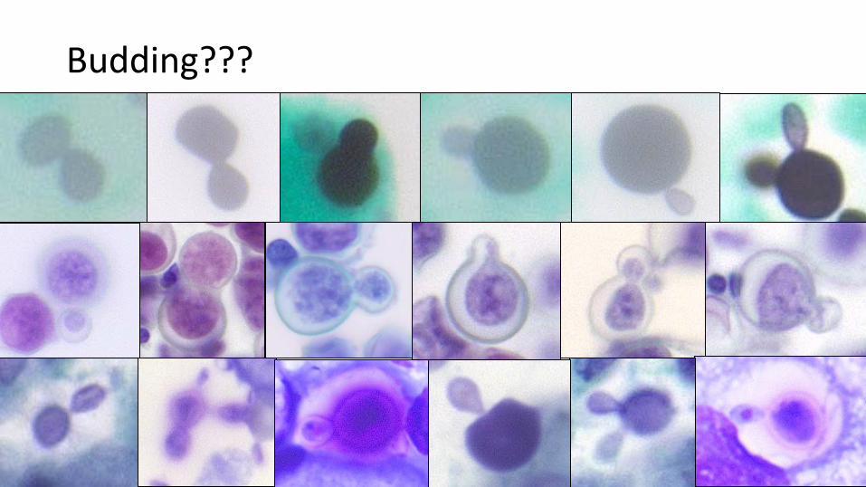
Budding???

Blastomyces sp.
• Round• Large (range 3-40 µm diameter,
usually 8-15 µm)• Thick-walled, refractile
(double-contour)• Broad-based budding (4-5 µm)• No capsule

• Variable shapes (Round, oval, collapsed crescents)
• Variable sizes (range 2-20 µm diameter, usually 4-10)
• Thin-walled, Narrow-based budding• Encapsulated and unencapsulated forms
• Capsule is clear on Romanowsky, Pap• Capsule stains pink on Mucicarmine
• Contains melanin-like pigment• Stains brown with Fontana-Masson• Especially good for unencapsulated small
forms
Cryptococcus sp.
Courtesy of V. Schnadig

Histoplasma sp.
• Ovoid• Small (2-4 µm diameter)
• H. capsulatum v. suboisii, 8-15 µm, West Africa
• Thin-walled• Narrow-based budding• Pseudocapsule• Predominantly intra-cellular• Crescent-shaped chromatin stains dark
violet (purple cap)
Courtesy of V. Schnadig
from: https://academic.oup.com/labmed/article/48/3/249/3098290

Pneumocystis jiroveci
• Round to ovoid cysts • Collapsed crescent, cup appearance
• Small (3-7 µm diameter)• Thin membrane• No capsule, pseudocapsule• No budding!• In “foamy exudate” (collection of cysts)• Romanowsky, Pap - clear cyst with dark dots
(nuclei, intra- and extra-cystic bodies)• GMS - Dot, comma, or parenthesis-shaped
(thickening in membrane)

BAL 73 M Hx rectal ca, carcinoid, immunosuppressed. Diagnosis?

BAL 55 M w/ HIV and diffuse pneumonitis. Diagnosis?
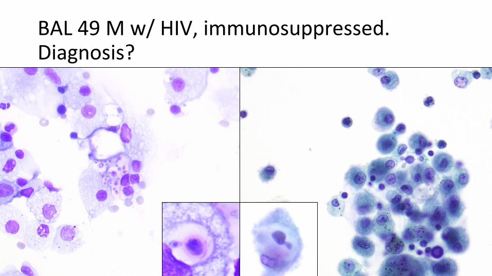
BAL 49 M w/ HIV, immunosuppressed.Diagnosis?

BAL 58 M w/ AML, neutropenia.Diagnosis?

Additional Reference:
(from Diagnostic Pathology of Infectious Disease 2nd Ed.
Kradin)

Differentiating Hyphal Fungal Organisms
Zygomycetes
Non-pigmentedBroadNon/Pauci-septateIrregular, uneven, 90°xTwisted, folded, ribbon-like
Aspergillus
Non-pigmentedNarrowSeptateRegular, even, 45°xParallel, radiating branches+/- fruiting bodies
Other Hyalohyphomycetes
Non-pigmentedNarrowSeptateIrregular, uneven xHaphazard arrangement x+/- fruiting bodies
Phaeohyphomycetes
PigmentedNarrowSeptate (constricted)Irregular, uneven xHaphazard arrangemet x+/- Yeast-like forms and chlamydoconidia

• Round, large thick-walled spherules (5-100 µm, usually 10-100 µm)
• Round small endospores (2-5 µm) • Ruptured, fragmented spherules• Filamentous forms (septate hyphae,
barrel-shaped arthroconidia rarely seen
Coccidioides sp.
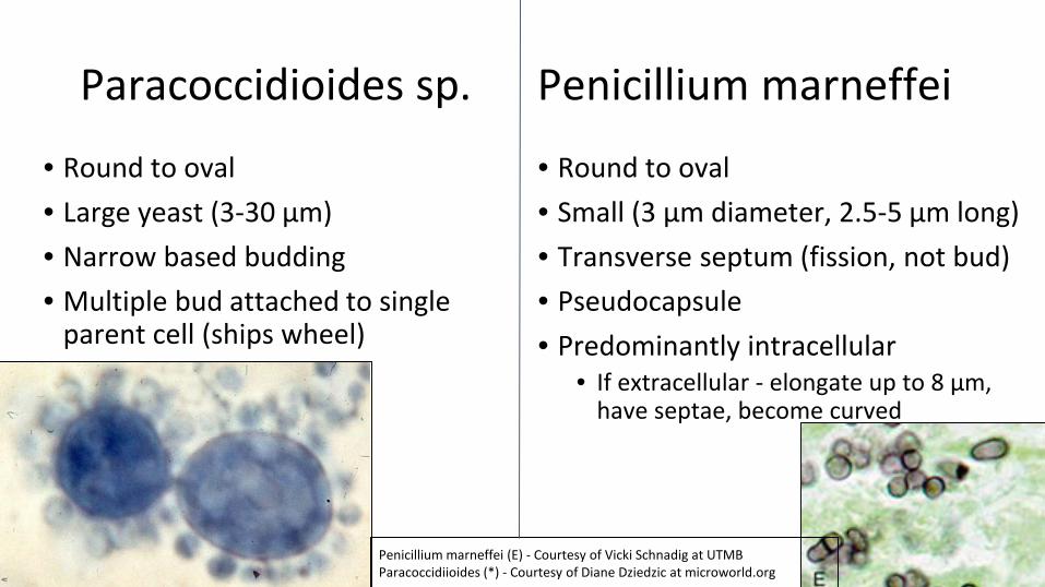
Paracoccidioides sp.
• Round to oval • Large yeast (3-30 µm)• Narrow based budding• Multiple bud attached to single
parent cell (ships wheel)
• Round to oval • Small (3 µm diameter, 2.5-5 µm long)• Transverse septum (fission, not bud)• Pseudocapsule• Predominantly intracellular
• If extracellular - elongate up to 8 µm, have septae, become curved
Penicillium marneffei
Penicillium marneffei (E) - Courtesy of Vicki Schnadig at UTMBParacoccidiioides (*) - Courtesy of Diane Dziedzic at microworld.org

Differentiating Yeast - Large Forms
Blastomyces● All large
● Round● Refractile,
Double contour● Broad budding
● No capsule
Cryptococcus● Variable sizes
● Variable shapes● Thin walled
● Narrow budding
● Capsule● Melanin
pigment
Coccicdioides● Large spherules,
Small endospores
● Round● Thick walled
● No budding
● No capsule
Candida● Small yeast,
Large conidia● Oval● Thin walled
● Buddings
● No capsule

Differentiating Yeast - Small Forms
Pneumoncystis● All small
● Round, cups● No budding● No capsule
● “Foamy exudate”
● GMS → . , “
Cryptococcus● Variable sizes
● Variable shapes● Narrow budding● Capsule
(mucicarmine)● Pigment
(Fontana Masson)
Histoplasma● All small
● Oval● Narrow budding● Pseudocapsule
● Purple cap on Romanowsky
● Mostly Intracellular
Candida● Small yeast,
Large conidia● Oval● Budding● No capsule
● Pseudohyphae, rare true hyphae

Answers To Cases

Esophageal Brush 56 F w/ SLE on prednisone.
Diagnosis:
Fungal organisms consistent with Candida

BAL 54 M s/p lung transplant.Diagnosis:
Fungal hyphae presentHyaline (non-pigmented) and pigmented hyphae
Culture grew Aspergillus fumigatus

BAL 54 M s/p lung transplant.Diagnosis:
Fungal hyphae presentNon-pigmented (hyaline) hyphae
Culture grew Aspergillus fumigatus

BAL 73 M Hx rectal ca, carcinoid, immunosuppressed. Diagnosis:
C/W Blastomyces

BAL 55M w/ HIV and diffuse pneumonitis.Diagnosis:
Pneumocystis jiroveci

BAL 49 M w/ HIV, immunosuppressed.Diagnosis:
Cryptococcus sp.

BAL 58 M w/ AML, neutropenia.Diagnosis:
Pneumocystis & CMV
Don’t forget to look for the second organism!