infection assays in arabidopsis reveal candidate …kamounlab.dreamhosters.com/pdfs/mpp_2016.pdf ·...
TRANSCRIPT
Infection assays in Arabidopsis reveal candidate effectors from the poplar
rust fungus that promote susceptibility to bacteria and oomycete
pathogens
Hugo Germain1, 2Ψ
, David L. Joly3, Caroline Mireault
1, Mélodie B. Plourde
1, Claire
Letanneur1, Donald Stewart
2, Marie-Josée Morency
2, Benjamin Petre
4,5, Sébastien
Duplessis5, Armand Séguin
2.
(1) Department of Chemistry, Biochemistry and Physics, Université du Québec à Trois-
Rivières, 3351 boul. des Forges, Trois-Rivières, QC, G9A 5H7, Canada
(2) Natural Resources Canada, Canadian Forest Service, Laurentian Forestry Centre,
1055 du P.E.P.S., P.O. Box 10380, Stn. Sainte-Foy, Québec, QC, G1V 4C7, Canada
(3) Département de biologie, Université de Moncton, 18 avenue Antonine-Maillet,
Moncton, NB, E1A 3E9, Canada
(4) The Sainsbury Laboratory, Norwich Research Park, Norwich, NR4 7UH, United
Kingdom
(5) INRA, UMR 1136 Interactions Arbres/Microorganismes, INRA/Université de
Lorraine, Centre INRA Nancy Lorraine, 54280 Champenoux, France
DLJ: [email protected]
MBP : [email protected]
CL : [email protected]
MJM: [email protected]
This article has been accepted for publication and undergone full peer review but has not beenthrough the copyediting, typesetting, pagination and proofreading process which may lead todifferences between this version and the Version of Record. Please cite this article as an‘Accepted Article’, doi: 10.1111/mpp.12514
This article is protected by copyright. All rights reserved.

2
Ψ Author for correspondence: Hugo Germain
Tel: 819-376-5011
Email: [email protected]
Word number: 5382
This article is protected by copyright. All rights reserved.

3
Abstract
Fungi of the Pucciniales order cause rust diseases, which altogether affect thousands of
plant species worldwide and pose major threat to several crops. How rust effectors -
virulence proteins delivered into infected tissues to modulate host functions - contribute
to pathogen virulence remains poorly understood. Melampsora larici-populina is a
devastating and widespread rust pathogen of poplars and its genome encodes 1,184
identified small secreted proteins that could potentially act as effectors. Here, following
specific criteria we selected 16 candidate effector proteins and characterized their
virulence activities and subcellular localizations in the leaf cells of Arabidopsis thaliana.
Infection assays using bacterial (Pseudomonas syringae) and oomycete
(Hyaloperonospora arabidopsidis) pathogens revealed subsets of candidate effectors that
enhanced or decreased pathogen leaf colonization. Confocal imaging of GFP-tagged
candidate effectors constitutively expressed in stable transgenic plants revealed that some
protein fusions specifically accumulate in nuclei, chloroplasts, plasmodesmata and
punctate cytosolic structures. Altogether, our analysis suggests that rust fungal candidate
effectors target distinct cellular components in host cells to promote parasitic growth.
Keywords: Fungus, parasite, obligate biotroph, confocal microscopy, virulence
assays.
This article is protected by copyright. All rights reserved.

4
Introduction
Plant-associated organisms secrete effectors that alter the host’s cellular structure
and functions to promote colonization (Hogenhout et al., 2009). The plethora of plant
processes modulated by effectors indicates that effectors target most, if not all, cell
structures (Giraldo & Valent, 2013, Petre et al., 2016, Petre et al., 2015b), and that the
study of effectors is useful to deepen our understanding of pathogenic processes (Win et
al., 2012). Specifically, effectors are used to probe into plant functions that govern plant
resistance (i.e. immune response-related pathways) or plant susceptibility (i.e.
susceptibility genes). Identifying and manipulating such functions is key to developing
resistant crops (Dangl et al., 2013). Using effectors as probes of the plant proteome to
pinpoint the precise protein targeted by an effector may identify susceptibility genes in
the host. Altering these genes, which are critical for compatibility, could provide durable
resistance.
Obligate biotrophic pathogens have to evade and/or suppress host recognition for
a lengthy period of time to complete their life cycle whilst their host remains alive. This
leads to an intricate battle in which the pathogen rewires the host’s cells to meet its needs
and to thwart host defenses. Suppression and evasion of innate immunity is undoubtedly
an important aspect of obligate biotrophy; it is, however, only a starting point in the
establishment of a successful infection. Indeed, it has recently emerged that effectors also
target cellular structures and processes that are not directly related to the plant immune
system, thus suggesting that pathogens do not only suppress immune responses but also
achieve a deep manipulation of their hosts (reviewed in (Chaudhari et al., 2014, Giraldo
& Valent, 2013, Win et al., 2012)).
Rust fungi have a complex life cycle, they cannot be cultured in vitro and are not
easily amenable to transformation, three traits hindering functional investigations.
Moreover, these fungi do not infect model plants such as Arabidopsis thaliana (Lawrence
et al., 2010) and consequently very little is known about the molecular basis of their
pathogenicity (Petre et al., 2014). Melampsora larici-populina is the causative agent of
poplar leaf rust, which has had devastating consequences on poplar plantations
worldwide (Duplessis et al., 2009, Feau et al., 2007, Hacquard et al., 2011, Duplessis et
al., 2011b). In order to assess the full weaponry at the pathogen’s disposal, thorough
This article is protected by copyright. All rights reserved.

5
genomic information is required. The combination of transcriptome and genome analyses
has provided a first glimpse into the putative effector arsenal of M. larici-populina
(Duplessis et al., 2011a, Hacquard et al., 2010, Hacquard et al., 2012, Joly et al., 2010).
Hacquard et al. (2012) identified and annotated 1,184 small secreted proteins that they
highlighted as candidate secreted effector proteins (CSEPs). Most of these putative
effectors show no sequence similarity to proteins from species outside of Pucciniales and
are found in multigene families. A subset of this CSEP repertoire is expressed in
haustoria (Hacquard et al., 2012, Hacquard et al., 2010, Joly et al., 2010) and is thus
probably enriched in proteins which are effectively delivered inside poplar cells during
the infection.
Most of the progress made in effector biology during the last decade relied on
heterologous systems enabling the study of effectors in plant cells (Fabro et al., 2011,
Sohn et al., 2007, Rafiqi et al., 2012). Agrobacterium mediated heterologous expression
or Pseudomonas-mediated delivery of tagged effectors in leaf cells are methods of choice
for studying effectors in planta (Torto et al., 2003, Vleeshouwers et al., 2008).
Subsequently, confocal imaging of green fluorescent proteins (GFP) fusions is widely
used to determine the cellular compartments targeted by effectors (Caillaud et al., 2012,
Schornack et al., 2010, Takemoto et al., 2012, Gaouar et al., 2016). More recently,
Nicotiana benthamiana was used to identify cellular compartments targeted by M. larici-
populina candidate effectors, as well as putative plant-interacting proteins (Petre et al.,
2015b). Effector delivery systems can be coupled with pathogen growth assays to identify
effectors possessing virulence activities (Dou et al., 2008, Fabro et al., 2011). However,
the use of stable Arabidopsis lines has several advantages over transient assays in
N. benthamiana: it offers the possibility to use well characterized genetic tools such as
knock-out lines of putative interactors to search for phenotype similarities; it also enables
the assessment of protein localization in guard cells, which are not transformed in
transient assays; and it renders possible the generation of stable single insertion
homozygous lines which can further be used for crosses, transcriptomic or metabolomic
studies.
In this study, we investigated a set of 16 CSEPs of the poplar rust fungus
M. larici-populina by measuring their ability to promote bacterial and oomycete pathogen
This article is protected by copyright. All rights reserved.

6
growth in a heterologous system. These CSEPs were stably transformed in Arabidopsis
thaliana and these lines were used for confocal imaging to assess candidate effectors’
subcellular localization in planta. Here we demonstrate that CSEPs target various
subcellular structures and promote pathogen colonization of leaves.
Results
Selection of 16 M. larici-populina CSEPs
We selected 16 CSEPs from a set of 1,184 SSPs previously described (Hacquard
et al. 2010, Hacquard et al. 2012) (Table 1). All selected CSEPs were less than 300 amino
acids long, had no sequence similarity with proteins from species outside the Pucciniales
order, showed induction of transcript expression during poplar leaf infection, and
reflected the diversity of CSEPs families in M. larici-populina. Among the most
informative sources of expression data are a haustoria-specific cDNA library and a
transcriptome analysis of laser-microdissected rust-infected poplar leaves (Hacquard et
al., 2010, Joly et al., 2010). Several of the selected CSEPs (MLP72983, MLP106078,
MLP123218, MLP106078, MLP102036, MLP123531, MLP124305, MLP124518,
MLP123531, MLP124305, MLP124466, MLP102036) displayed a very high expression
ratio in microdissected palisade mesophyll enriched in haustoria and infection hyphae
when compared with microdissected uredinia, mostly composed of spores and
sporogenous hyphae (Hacquard et al., 2010). MLP124497, MLP124499 and MLP124518
[Mlp0032_0018] are different members of the same CSEP family, which was previously
designated CPGH1 (Hacquard et al., 2012). This family of very small proteins (<100
amino acids) presents signatures of positive selection and is over-represented in the
haustorial library (Hacquard et al., 2012, Joly et al., 2010). MLP37347 was selected for
its homology to AvrL567, a previously reported effector of the flax rust fungus
Melampsora lini (Dodds et al., 2004). MLP123227 (SSP15), shows a bimodal expression
profile in planta and immunolocalization data indicates it has an intriguing localization
pattern in planta, both in haustoria and sporogenous hyphae (Hacquard et. 2012). The last
two CSEPs (MLP123437 and MLP124111) belong to different classes of SSPs conserved
in Puccinia spp. (classes VI and III; Hacquard et al., 2012) and they display a conserved
This article is protected by copyright. All rights reserved.

7
exon/intron structure and a characteristic spacing of Cys residues (including the presence
of Y/FxC motifs) (Hacquard et al., 2012).
Three CSEPs promote the growth of P. syringae in planta independently of PTI
suppression
To test whether the 16 CSEPs can promote pathogen growth, we performed
infection assays using the P. syringae pv. tomato DC3000/A. thaliana pathosystem (Sohn
et al., 2007). In this system, the sequences coding for the mature form of the CSEP, i.e.
without the predicted signal peptide, are fused to the N-terminus of AvrRPM1 to drive
their translocation inside host cells via the type three secretion apparatus of P. syringae.
Five out of sixteen CSEPs significantly increased bacterial growth in planta
(MLP124499, MLP102036, MLP124266, MLP124497, MLP106078) (Figure 1A).
Conversely, two candidate effectors decreased bacterial growth (MLP123227,
MLP37347), while the other nine did not affect bacterial growth when compared to the
control. Standard growth assays confirmed that the CSEPs did not affect the rate of
bacterial cell division (data not shown), thus suggesting that alteration of in planta
bacterial growth is caused by the effect of the CSEP on plant cells. We conclude that five
CSEPs promote the growth of P. syringae when delivered by the bacteria, suggesting that
they have a virulence activity in leaf cells.
To complement the effector delivery assays, we generated stable A. thaliana
transgenic lines constitutively expressing CSEP-GFP fusions. Transgenic lines were
generated for 14 CSEPs, while lines MLP124111 and MLP123437 could not be retrieved.
Then we performed infection assays using wild-type P. syringae pv tomato DC3000 on
the aforementioned transgenic lines. Five of the fourteen CSEPs promoted the bacterial
growth (MLP123218, MLP124466, MLP124497, MLP124499 and MLP106078), while
two (MLP37347 and MLP123227) showed less growth compared to the control plants
(Figure 1B). The combined results from these two experiments indicate that three CSEPs
(MLP124499, MLP124497, MLP106078) promoted bacterial growth while two
(MLP37347 and MLP123227) reduced bacterial growth in both assays.
In order to understand how these CSEPs (MLP123218, MLP124466,
MLP124266, MLP124497 and MLP106078) contribute to bacterial growth increase, we
This article is protected by copyright. All rights reserved.
8
selected the transgenic lines that displayed increased bacterial growth and infected them
with the type III secretion-deficient bacterial strain P. syringae pv. DC3000 hrcC-.
Although this strain is unable to produce a functional T3SS and inject effectors in host
cells, it still carries pathogen associate molecular patterns that will initiate PTI. Therefore,
we monitored bacterial growth at day 0 and day 3 in transgenic lines, Col-0 and a
transgenic line expressing GFP without any effector (control). The results shown in
Figure 1C demonstrate that P. syringae pv. DC3000 hrcC- growth is not affected by the
effectors when compared to the control. Taken together these results suggest that PTI is
not suppressed by the effectors that contribute to increased bacterial growth.
Eleven CSEPs promote the growth of Hyaloperonospora arabidopsidis in planta
Since M. larici-populina is an obligate biotrophic filamentous pathogen, we
sought to test whether the CSEPs would affect the growth of a pathogen with a similar
lifestyle. Since no rust infect A. thaliana, we used the oomycete Hyaloperonospora
arabidopsidis to challenge the transgenic plants constitutively expressing the candidate
effectors. In total, 11 out of 14 transgenic lines expressing CSEPs supported more
H. arabidopsidis sporulation than control plants (mean ratio of two, 15,000 to 25,000 vs
10,000 spores/g of leaf tissue) (Figure 2). However, no CSEP-GFP line promoted the
growth of H. arabidopsidis at a comparable level to the infection control line eds1, which
is hypersensitive to H. arabidopsidis. We conclude that the majority (> 75%) of the tested
CSEPs promote H. arabidopsidis growth on A. thaliana.
Only two CSEPs (MLP124497, MLP124499) promote pathogen growth in the three
infection assays
In order to evaluate the consistency of the virulence activities detected for the
CSEPs, we compared the results from the three sets of infection assays. This analysis
revealed that all CSEPs promoted pathogen growth in at least one experiment, three did
so in the two experiments involving P. syringae, while only two (MLP124497,
MLP124499) promoted pathogen growth in all three experiments (Figures 1, 2, 3).
Conversely, while two candidate effectors consistently reduced the growth of P. syringae
in both in planta experiments, they did not affect H. arabidopsidis growth. Some
This article is protected by copyright. All rights reserved.

9
candidate effectors did not alter bacterial growth (MLP124256, MLP124518,
MLP124266, MLP123531, MLP124305, MLP72983), but did significantly promote
H. arabidopsidis growth in planta (Figures 1, 2 and 3). Interestingly, the CSEP that
promoted the highest bacterial growth (MLP106078) did not significantly enhance
H. arabidopsidis growth. Based on these results we conclude that MLP124497 and
MLP124499 promote the growth of unrelated pathogens in A. thaliana, while other
CSEPs exhibit a pathogen-specific growth-promoting effect.
The transgenic line 124499 shows delayed senescence
We assessed the phenotypes of the transgenic lines to see if they displayed any
growth or morphological alteration that could be caused by the expression of the fungal
genes. To this end, the plants were grown at 20º C as well as at 16º C and senescence was
monitored as they aged. Supplementary figures 1 and 2 show representative plants from
each transgenic line when grown at 20 ºC (Supplementary figure 1) and 16 ºC
(Supplementary figure 2). Most lines did not show any striking phenotype, except
MLP124499 plants, whose leaves remained green much longer than all other transgenic
and Col-0 plants. To ensure that this was not a positional effect in the flat, we started a
new flat with alternating rows of Col-0 and MLP124499 plants (Supplementary figure 3;
tagged as H1) and we again observed that the rows of MLP124499 remained green while
the Col-0 plant had completely dried. We concluded that, except for the transgenic line
MLP124499, the phenotype of the CSEPs expressing plants was unaffected.
Three candidate effectors specifically accumulate in chloroplasts, cytosolic bodies,
and at plasmodesmata
To gain further insight into the growth-promoting effect of CSEPs, we used
confocal microscopy to determine the subcellular localization of the CSEP-GFP fusions
in leaf epidermal cells (i.e. guard and pavement cells) obtained from the A. thaliana
stable transgenic lines. Three candidate effectors specifically accumulated in chloroplasts
(MLP72983) and cytosolic puncta (MLP37347, MLP124305) (Figure 4A). The
fluorescence signal of MLP72983 was distributed homogenously within the chloroplasts,
suggesting that it accumulates in the stroma. The fluorescence signal of MLP37347 was
This article is protected by copyright. All rights reserved.

10
restricted to small, bright, and circular cytosolic structures of approximately 1 µm in
diameter and of unknown nature, whereas the fluorescence signal of MLP124305
accumulated in small cytosolic dots that were excluded from nuclei (Figure 4A). In order
to investigate the nature of these cytosolic foci we performed co-localisation experiments
of MLP37347 and MLP124305 with PDCB1-mCherry, a plasmodesmata marker, and
RBP47b-CFP, a stress granule marker. MLP37347-GFP and PDCB1-mCherry
fluorescent signals overlapped in punctate structures, indicating that this CSEP
accumulates at the plasmodesmata (Figure 4B). MLP124305 neither localized to the
plasmodesmata nor the stress granules (not shown). To find the true localization of
MLP124305-GFP we crossed the transgenic line overexpressing MLP124305-GFP with
DCP1-CFP (a processing body marker) but did not observe co-localization, therefore we
do not know what the structure targeted by this effector is. The eleven remaining CSEPs
showed a non-informative distribution in the cytosol and in the nucleus, similar to the
free GFP control (Figure 4A). Anti-GFP western blots revealed a single band signal at the
expected size for all but one CSEP-GFP, suggesting that the fusions were neither cleaved
nor modified in leaf cells (Supplementary figure 4). MLP124305 yielded multiple bands
of lower molecular weight, suggesting protein degradation (data not shown). We
conclude that out of the sixteen CSEPs, three target specific cellular structures in leaf
cells: some undefined cytosolic puncta, the chloroplast and plasmodesmata.
Discussion
In this study, we used A. thaliana as a heterologous system to perform the
functional analysis of a subset of 16 candidate secreted effector proteins (CSEPs) from
the poplar leaf rust fungus Melampsora larici-populina. Notably, we identified several
CSEPs that promote bacterial or oomycete pathogen growth in planta, as well as some
that accumulate in distinct plant cell compartments. These proteins were flagged as
putative virulence factors that needed to be investigated further.
Functional studies of rust fungi are challenging due to the obligate biotrophic
status of these pathogens, and high throughput ‘effectoromics’ approaches are needed
(Petre et al., 2014, Petre et al., 2015a, Petre et al., 2015b). Such procedures are not fully
available yet in most rust pathosystems. We sought to partially fulfill this need by using
This article is protected by copyright. All rights reserved.

11
heterologous systems, a strategy increasingly used in rust effector biology (Petre et al.,
2015a, Petre et al., 2016, Petre et al., 2015b). To this end, we created a collection of
A. thaliana transgenic lines to investigate the subcellular localization and virulence
activities of rust CSEPs, complementing the efforts accomplished thus far using
N. benthamiana as a heterologous system. These lines represent a valuable resource for
the community. For instance, they could be used in proteomic approaches to find
interaction partners, confirming co-immunoprecipitation results from N. benthamiana
and/or to study the impact of effectors on the plant transcriptome and metabolome.
Ideally, the confirmation that a small secreted protein is a bona fide effector
requires the demonstration that it has the capacity to interfere with a host component to
ultimately favor pathogenesis. Hence we used pathogen growth as a proxy to measure the
effect of these CSEPs on host cells. Three out of the sixteen candidate effectors tested
(18%) robustly enhanced bacterial growth in planta. In contrast, when using a similar
experimental set-up, Fabro et al. (2011) showed that 70% of Hpa candidate effectors set
caused an increase in bacterial growth. There are two possibilities that may explain the
difference of number between this study and our results. First, our assays were performed
in a non-host plant, in which M. larici-populina CSEPs might not be functional; second,
Fabro et al. (2011) used RxLR candidate effectors that are all likely to be host-
translocated, while our portfolio may have included apoplastic candidate effectors or
even non-effectors that are predicted secreted proteins and remain in the apoplastic space
of the native poplar-poplar rust pathosystem.
Bacterial infection assays revealed dissimilarities depending on whether CSEPs
were bacteria delivered or constitutively expressed in planta. For example, MLP123218
and MLP124466 had a positive effect on pathogen growth when expressed in planta, but
none when they were bacterially delivered. Conversely, MLP102036 and MLP124266
had a positive effect on pathogen growth when they were bacterially delivered, but none
when they were constitutively expressed. Two factors may explain the differences. First,
bacterially produced CSEPs may be mistargeted or misfolded due to their passage
through the T3SS to reach the plant cell. Second, GFP tags may generate steric
interference causing a mistargeting or misfolding of the CSEPs, especially if they are
small.
This article is protected by copyright. All rights reserved.
12
The demonstrated function of many effectors is to suppress PTI (Gohre &
Robatzek, 2008, Zhang et al., 2007). To study this possibility, we used the P. syringae pv
tomato DC3000 hrcC- strain, which is deficient in the production of the type III secretion
system and which triggers PTI but not ETI. Our infection results with bacteria and
oomycete suggested that the increased pathogen growth observed with MLP106078,
MLP124497 and MLP124499 were not caused by PTI suppression. Remarkably, it
suggests that the CSEPs assayed favor pathogen infection without suppressing plant
immunity. Other aspects of the plant immunity, such as ROS production and signaling
pathways, are currently being investigated as part of a more detailed analysis of these
CSEPs. Since obligate biotrophs are extremely sophisticated parasites that often establish
long-standing associations with host tissues, we speculate that many of their effectors not
only suppress immune responses like many hemi-biotrophs do, but deeply modulate host
functions.
Our results show that infection assays using different pathogens having various
lifestyles, i.e. bacteria and oomycete, can yield strikingly different results. Hpa and
M. larici-populina are both obligate biotrophic filamentous pathogens of dicot plants and
they share some similarities in their propagation mode in leaf tissues. Therefore, one
would expect that Hpa pathogenicity would be more likely to be affected by rust effectors
than P. syringae bacterial pathogenicity. Indeed, our results showed that 11 CSEPs
promoted the growth of Hpa, which is more than twice the number of CSEPs promoting
the growth of P. syringae. It is possible that M. larici-populina CSEPs target mechanisms
that specifically assist in leaf infection by biotrophic pathogens.
We could identify rust CSEPs affecting pathogen growth in A. thaliana, a non- host plant.
Since many effectors exert their activities by associating with plant proteins, this
observation suggests that some CSEPs may have the ability to associate with A. thaliana
proteins despite the fact that they do not under natural conditions. In line with this
hypothesis, Petre et al. (2015, 2016) identified rust CSEPs that associate with specific
N. benthamiana proteins which have conserved homologs in the natural host plants
poplar. Since our present report includes candidate effectors that were also investigated
by Petre et al. (2015), we assessed whether some of the putative targets identified by co-
This article is protected by copyright. All rights reserved.

13
immunoprecipitation/mass spectrometry could explain the pathogen assays results that we
observed. Only two effectors common between the two studies had specific interactors,
MLP37347 and MLP124111. MLP124111 did not show any effect on pathogen growth
when the effector was delivered by P. syringae and could unfortunately not be assayed
with the effector constitutively expressed in planta as it did not produce a viable
transgenic line. MLP37347 on the other hand displayed an interesting effect: it decreased
bacterial growth (in both systems) but increased oomycete growth. In the co-
immunoprecipitation assay, MLP37347 had a single specific interactor, which is the
glutamate decarboxylase 1 (GAD1). It should be noted that MLP37347 is the effector
localized to the plasmodesmata. Evaluation of the potential role of the glutamate
decarboxylase 1 as a putative virulence target of MLP37347 will require further
investigation.
Two CSEPs (MLP37347, MLP123227) had a negative effect on bacterial growth
but a positive effect on H. arabidopsidis growth. One option for that decreased bacterial
growth is their possible recognition by A. thaliana’s immune receptors and the triggering
of the defense responses. M. larici-populina is non-host on Arabidopsis nor is any rust
fungus, and for this reason it seems surprising that A. thaliana would recognize these
candidate effectors. We rather hypothesize that Arabidopsis does not directly recognize
those CSEPs; but that instead they may target proteins guarded by a R-protein in
A. thaliana (Dangl & Jones, 2001, Van der Biezen & Jones, 1998). Thus, the
identification of the target of these CSEPs could provide information with regards to
decoy or bait, and ultimately, by homology, allow the identification of important
components of the plant immune system (Khan et al., 2016). We also have to keep in
mind that the expression of a given effector in P. syringae may affect the secretion of
other of its endogenous effectors and may thus decrease its virulence.
Confocal microscopy assays revealed that some candidate effectors accumulated
in chloroplasts, plasmodesmata and cytosolic foci in leaf cells, while most displayed non-
informative nucleocytosolic distribution. It is important to consider that these
localizations were obtained using expression driven by the 35S promoter, so it could be
different from what would be observed when the CSEPs are delivered by the pathogen.
Petre et al. (2015a) studied the localization and interaction partners of some M. larici-
This article is protected by copyright. All rights reserved.
14
populina CSEPs. In both studies, the subcellular localization of MLP102036,
MLP123227, MLP124266, MLP124497, MLP124499 and MLP37347 was identical.
Interestingly, several rust candidate effectors accumulate in chloroplasts (Petre et al.,
2015a, 2015b, 2016). Despite intensive cell biology screens, no other effectors of
filamentous pathogens were reported to target chloroplasts. Whether chloroplasts are
important organelles for rust fungi to manipulate remains to be investigated. The similar
localization of the common CSEPs in both studies strengthens the use of heterologous
systems to infer CSEPs localization, putative function and interaction partner studies.
We could not recover stable transgenic lines expressing MLP124111 and
MLP123437, which suggests that their accumulation in cells could be toxic to plants.
MLP123437 was not investigated by Petre et al. 2015, however using N. benthamiana
Mlp124111 localized in chloroplasts as well as in large, bright, and irregular cytosolic
structures that were probably aggregates. It is possible that these aggregates, as they may
be toxic to the cell, were the reason why a stable transgenic line for MLP124111 could
not be recovered, although no sign of necrosis was reported by Petre et al. (2015).
Alternatively, chloroplastic localization could trigger the chlorosis we observed in the
unique transgenic T1 line obtained which never reached maturity. Our results
demonstrate that advances in rust fungi biology, as well as in the biology of other
obligate biotrophs that cannot be easily and/or routinely transformed, rely on the
combination of parallel approaches (e.g. in vitro and heterologous assays). Our approach
is much valuable due to the large pool of genetic knowledge and resources that are
available for Arabidopsis compared to Nicotiana.
This article is protected by copyright. All rights reserved.

15
Material and methods
Plasmid constructs
DNA sequences encoding all mature small secreted proteins were ordered from
GenScript in a lyophilized form (the stop codon was removed to enable GFP fusion). All
constructs were transferred by BP recombination to the Gateway pDONR Zeo vector
(Invitrogen, Carlsbad, CA, USA). From pDONR™/Zeo, contructs were either sent to
pB7FWG2 (Karimi et al., 2002) to generate stable C-terminally GFP-tagged transgenic
Arabidopsis plants or pVSP-PsSPdes (courtesy of Guus Bakkeren’s laboratory) for
Pseudomonas infection assays. pVSP-PsSPdes harbors the AvrRPM1 secretion signal
and a C-terminal HA tag (Rentel et al., 2008).
Transformation of A. thaliana, pathogen assay and western blotting
Five week old soil-grown Arabidopsis thaliana plants were transformed using the floral
dip method with minor modification (Clough & Bent, 1998). Modifications included
substituting Silwet L-77 by OFX-0309 (Norac Concept Inc, Guelph, ON, Canada)
(Mireault et al., 2014) and dipping the plants a second time one week later to increase the
number of transformants. T1 plants were selected using ammonium glufosinate (Basta) at
60 mg/L. T2 plants grown in Petri dishes (with Basta at 25 mg/L), and lines displaying a
3:1 ratio were selected to obtained single insertion lines. T3 plants were also grown in
Petri dishes, and lines showing 100% germination were selected as homozygotes. An
average of twelve transgenic lines for each construct were selected and screened for GFP
expression by Western blot (Supplementary Figure 1), and the strongest expressing line
was kept for each construct. This primary screen was necessary to ensure that the GFP
was not being cleaved from the small secreted proteins in which case we would have
observed fluorescence signal from a free GFP in confocal microscopy. It should also be
noted that two constructs did not yield any transgenic lines despite many transformation
attempts; these are SSPs MLP124111 and MLP123437. In fact, one line was recovered
from MLP124111 but it showed important chlorosis, stunted morphology, it did not grow
higher than 2 cm in seven weeks, it never produced any viable seeds and died. Additional
attempts to produce more transgenic lines overexpressing MLP124111 failed. We
conclude that constitutive in planta expression of MLP124111 and perhaps MLP123437
is most likely toxic to the plant. Infections were carried out on one line per construct.
This article is protected by copyright. All rights reserved.
16
Western blots and infection assays were performed as previously described (Germain et
al., 2010, Germain et al., 2007). Briefly, bacterial infections were performed with four
week-old Arabidopsis plants grown at 22ºC in a 16/8h light/dark cycle. Ps pv. tomato
diluted at OD600 0.001 were leaf-inoculated on the abaxial side of the leaf. A minimum of
36 leaves were infected for each effector or transgenic line. Leaf punches were taken
from 12 leaves at day 0 and 24 were taken at day 3 to assess bacterial quantity. Hpa
infection were performed as described in Dong et al. 2016 with no modification to the
protocol (Dong et al., 2016). For Western blots, two leaf punches (0.384 cm2) were taken
from 3-weeks-old T3 plants and stored at -80ºC. Leaf punches were disrupted using a
TissueLyser (Qiagen, Toronto, ON, Canada) and a 5 mm stainless steel bead agitated at
26 cycles/sec for 10 sec. Laemli sample buffer (100µL) was added and the tube was
heated at 95ºC for 5 min and spun at 13,000 g for 1 min. Supernatant (10µL) was loaded
on a 10% acrylamide gel. Western blot was carried out as previously described (Germain
et al., 2007); the mouse anti-GFP antibody used to detect the recombinant protein was
purchased from Cedarlane (Burlington, ON, Canada) and used at 1:5000.
Confocal imaging
T3 seedlings 6-10 days’ old collected from Petri dishes containing ½ MS media +
ammonium glufosinate (25 mg/L) were placed in a water drop under a cover slip and
imaged immediately. Before each imaging session, wild-type plants were visualized to
adjust the settings in order to exclude the autofluorescence of the chloroplasts. Images
were captured with a Carl Zeiss LSM700 or Nikon A1 confocal microscope. 40X oil
PlanApo immersion objectives were used. Excitation laser (488 nm) was used and
photons were collected between 510 and 520 nm. Image analysis was performed with
ImageJ (http://imagej.nih.gov/ij/).
Acknowledgments
We are thankful to Carl Zeiss Corporation and Nikon Instruments for giving us the
opportunities to freely use the Zeiss 410 and Nikon A1 confocal microscope respectively.
We also thank Isabelle Lamarre and Franck Stefani (Laurentian Forestry Centre) for
editorial work. HG’s and DLJ’ post-doctoral fellowships were supported by the Natural
This article is protected by copyright. All rights reserved.
17
Sciences and Engineering Research Council of Canada (NSERC). Funding for the project
was also provided by NSERC Discovery Grants to HG and AS. The project in HG’s
laboratory was also partially funded by an institutional research chair held by HG. CM
received an undergraduate student award from NSERC. BP was supported by an INRA
Contrat Jeune Scientifique, by the European Union, (as part of the Marie-Curie FP7
COFUND People Programme, through an AgreenSkills’ fellowship (under grant
agreement no. 267196), and by the Laboratory of Excellence ARBRE, through a mobility
grant (12RW53). SD is supported by the French National Research Agency through the
Laboratory of Excellence ARBRE (ANR-12-LABXARBRE-01) and the Young Scientist
Grant POPRUST (ANR-2010-JCJC-1709-01). We are grateful to Jean-François Laliberté
(INRS Armand-Frappier, Qc Canada) for providing the plasmodesmata marker pDCB1-
mCherry. We also thank professor Dan Robert for providing the RBP47CFP construct.
This article is protected by copyright. All rights reserved.

18
Figure legends
Figure 1. Bacterial growth in planta differs when effectors are delivered by bacteria
or are constitutively expressed
A) Growth of Pst DC3000 without (EV) or with candidate effectors (all other lanes) was
measured on the day of infection and 3 days after infection of 4-week-old plants by leaf
infiltration. A bacterial suspension of OD600 = 0.001 was used as inoculum. Statistical
significance was evaluated using Student’s t-test (p<0.05); asterisks indicate statistically
significant differences between the effector and empty vector. The experiment was
repeated at least three times (a representative experiment is shown).
B) Growth of Pst DC3000 wild-type infecting plants expressing an effector, Col-0 or
Col-0 expressing GFP was measured on the day of infection and 3 days after infection of
4-week-old plants by leaf infiltration. A bacterial suspension of OD600 = 0.001 was used
as inoculum. Statistical significance was evaluated using Student’s t-test (p<0.05);
asterisk indicates statistically significant difference between plants carrying effector and
Col-0 expressing GFP. Five replicates were included for each genotype. To facilitate the
comparison with Figure 1A, each transgenic line is presented in the same order. The
experiment was repeated at least three times (a representative experiment is shown).
C) Growth of Pst DC3000 hrcC- strain infecting plants expressing an effector, Col-0 or
Col-0 expressing GFP was measured on the day of infection and 3 days after infection of
4-week-old plants by leaf infiltration. A bacterial suspension of OD600 = 0.001 was used
as inoculum. Statistical significance was evaluated using Student’s t-test (p<0.05);
asterisk indicates statistically significant difference between plants carrying effector and
Col-0 expressing GFP. Five replicates were included for each genotype. The experiment
was repeated at least three times (a representative experiment is shown). Note that no
transgenic lines were recovered for 123437 and 124111.
This article is protected by copyright. All rights reserved.

19
Figure 2. Most candidates effector affect the growth of H.arabidopsidis Noco2 when
constitutively expressed in planta.
Two week-old soil-grown plants were inoculated with H. arabidopsidis Noco2 at a
concentration of 20,000 conidiospores per ml and the number of conidiospores was
quantified 7 days after inoculation. Bars represent the mean of four replicates. Statistical
significance was established using Student’s t-test (p<0.05), and statistically significant
differences are represented by an asterisk between plants carrying effector and Col-0
expressing GFP. Each transgenic line is ordered as in Figure 1 for easier comparison. The
experiment was repeated five times (representative experiment is shown).
Figure 3. Venn diagram showing the candidate effectors that induced increased
virulence (in black) or decreased virulence (in red) .
Note that no transgenic lines were recovered for 123437 and 124111.
Figure 4. Subcellular localization of the different candidate effectors.
A) Confocal images of leaf epidermal cells of 7 day old transgenic plantlets
expressing candidate effectors fused to GFP (SSP-GFP) in Col-0 genetic
background. The top three effectors are the ones displaying informative
localization, while the bottom three rows display the effectors showing
nucleocytoplasmic localization.
B) Colocalization between PDCB1-mCherry , a plasmodesmata marker and 37347-
GFP to infer its localization. Left panel red channel, center panel green channel,
right panel overlay with DIC.
Supplementary Figure 1. Pictures of plant after 4 weeks of growth at 20ºC.
Supplementary Figure 2. Pictures of plant after 4 weeks of growth at 16ºC.
Supplementary Figure 3. Pictures of 124499 and Col-0 plants after 6 weeks of growth at
20ºC to vizualize senescence.
Supplementary Figure 4. Western blot showing the molecular weight of the different
candidate effectors tagged with the GFP protein
This article is protected by copyright. All rights reserved.

20
Immunoblot analysis performed using an anti-GFP antibody. Molecular weight was
calculated using the BioRad Low molecular weight protein standard. Only the transgenic
line that was selected for infection and confocal imaging is shown.
Supplementary Table 1.
Full DNA and peptide sequences of the candidate effectors. --- indicates where the
candidate effector is predicted to be cleaved. The C-terminal section was used as the
mature protein.
This article is protected by copyright. All rights reserved.

21
Table 1.
ProteinIDa
CPG or Class
(number of
members)b
Protein (signal
peptide)
lengthc
Cysteine
residuesd
Evidence of
expressione
Signature
of positive
selectionf
Homology
to M. lini
HESPsg
Homology
in Pgtg
Effect on
pathogen
growthh
Sub-cellular
localisation
37347 - 152 (23) 2 OA - AvrL567 - -, -, + Plasmodesmata
72983 CPG332-CPG333
(13)
220 (26) 8
S, I, H, OA,
A3 - - Yes
N, N, + Chloroplast
102036 CPG2528 (5) 107 (25) 0 H, OA, A3 - - - +, N, N Nucleocytoplasmic
106078 - 137 (21) 10 I, OA, A3 - - - +, +, N Nucleocytoplasmic
123218 CPG543 (7) 209 (19) 6 I, OA, A3 - - Yes N, +, + Nucleocytoplasmic
123227
(SSP15) CPG1059 (2)
124 (24) 3 I, OA - - -
-, -, + Nucleocytoplasmic
123437 Class VI (6) 180 (20) 11 I, H, OA - HESP-C49 Yes N Not available
123531 CPG4557 (2) 102 (21) 8 I, OA, A3 - - - N, N, + Nucleocytoplasmic
124111 Class III (8) 135 (21) 10 H - - Yes N Not available
124256 CPG5464 (13) 89 (25) 6 I Yes AvrP4 - N, N, + Nucleocytoplasmic
124266 CPG5464 (13) 92 (25) 7 I, H Yes AvrP4 - +, N, + Nucleocytoplasmic
124305 CPG5184 (2) 147 (25) 12 OA, A3 - - - N, N, + Cytoplasmic foci
124466 - 76 (24) 0 H, OA, A3 - - - N, +, N Nucleocytoplasmic
124497 CPGH1 (33) 77 (21) 4 I, H, OA Yes - - +, +, + Nucleocytoplasmic
124499 CPGH1 (33) 72 (21) 3 I, H Yes - - +. +, + Nucleocytoplasmic
124518 CPGH1 (33) 76 (21) 3 I, H, OA, A3 Yes - - N, N, + Nucleocytoplasmic
a Best predicted gene model in the M. larici-populina isolate 98AG31 JGI genome sequence.
b Families, clusters of paralogous genes (CPGs) and classes of small-secreted proteins as described in Duplessis et al., 2011 and Hacquard et al.,
2012. Number between brackets indicate the number of members in gene families.
This article is protected by copyright. All rights reserved.

22
c Numbers of amino acids are indicated.
d Number of cysteine residues in the mature form of the protein (i.e. without signal peptide)
eEvidence of expression among S (Expressed Sequence Tags from resting spores, (Duplessis et al., 2011a), I (Roche 454 data from infected
leaves, (Hacquard et al., 2012), H (Expressed Sequence Tags from isolated haustoria, (Joly et al., 2010), OA (oligoarray data from infected
leaves, (Duplessis et al., 2011a), A3 (most highly upregulated transcripts from oligoarray data in the palisade mesophyll versus sporogenous
microdissected structures, (Hacquard et al., 2010).
f As described in Hacquard et al., (2012).
g Homology searches were carried out against UNIPROT and Puccinia Group Database using blastp (E value <1e-6). Pgt = Puccinia graminis f.
sp. tritici.
hFirst position indicate effect on bacterial growth when the effector is delivered by Pst, second position denote effect on bacterial growth when the
effector is expressed in planta, third position denote effect on Hpa growth when the effector is expressed in planta. + =positive effect, N= no
effect, - = negative effect.
This article is protected by copyright. All rights reserved.

23
Bibliography
Caillaud, M. C., Piquerez, S. J., Fabro, G., Steinbrenner, J., Ishaque, N., Beynon, J., et al. (2012)
Subcellular localization of the Hpa RxLR effector repertoire identifies a tonoplast-
associated protein HaRxL17 that confers enhanced plant susceptibility. The Plant journal
: for cell and molecular biology, 69, 252-265.
Chaudhari, P., Ahmed, B., Joly, D. L. and Germain, H. (2014) Effector biology during biotrophic
invasion of plant cells. Virulence, 5, 1-7.
Clough, S. J. and Bent, A. F. (1998) Floral dip: a simplified method for Agrobacterium-mediated
transformation of Arabidopsis thaliana. The Plant Journal, 16, 735-743.
Dangl, J. L. and Jones, J. D. G. (2001) Plant pathogens and integrated defence responses to
infection. Nature, 411, 826-833.
Dodds, P. N., Lawrence, G. J., Catanzariti, A. M., Ayliffe, M. A. and Ellis, J. G. (2004) The
Melampsora lini AvrL567 avirulence genes are expressed in haustoria and their products
are recognized inside plant cells. The Plant cell, 16, 755-768.
Dong, O. X., Meteignier, L. V., Plourde, M. B., Ahmed, B., Wang, M., Jensen, C., et al. (2016)
Arabidopsis TAF15b Localizes to RNA Processing Bodies and Contributes to snc1-
Mediated Autoimmunity. Molecular plant-microbe interactions : MPMI,
MPMI11150246R.
Dou, D., Kale, S. D., Wang, X., Chen, Y., Wang, Q., Wang, X., et al. (2008) Conserved C-terminal
motifs required for avirulence and suppression of cell death by Phytophthora sojae
effector Avr1b. The Plant cell, 20, 1118-1133.
Duplessis, S., Cuomo, C. A., Lin, Y. C., Aerts, A., Tisserant, E., Veneault-Fourrey, C., et al. (2011a)
Obligate biotrophy features unraveled by the genomic analysis of rust fungi.
Proceedings of the National Academy of Sciences of the United States of America, 108,
9166-9171.
Duplessis, S., Joly, D. L. and Dodds, P. N. (2011b) Rust Effectors. In: Effectors in Plant–Microbe
Interactions. Wiley-Blackwell, pp. 155-193.
Duplessis, S., Major, I., Martin, F. and Séguin, A. (2009) Poplar and Pathogen Interactions:
Insights from Populus Genome-Wide Analyses of Resistance and Defense Gene Families
and Gene Expression Profiling. Critical Reviews in Plant Sciences, 28, 309-334.
Fabro, G., Steinbrenner, J., Coates, M., Ishaque, N., Baxter, L., Studholme, D. J., et al. (2011)
Multiple candidate effectors from the oomycete pathogen Hyaloperonospora
arabidopsidis suppress host plant immunity. PLoS Pathogen, 7, e1002348.
Feau, N., Joly, D. L. and Hamelin, R. C. (2007) Poplar leaf rusts: model pathogens for a model
treeThis minireview is one of a selection of papers published in the Special Issue on
Poplar Research in Canada. Canadian Journal of Botany, 85, 1127-1135.
Gaouar, O., Morency, M.-J., Letanneur, C., Séguin, A. and Germain, H. (2016) The 124202
candidate effector of Melampsora larici-populina interacts with membranes in Nicotiana
and Arabidopsis. Canadian Journal of Plant Pathology, null-null.
Germain, H., Houde, J., Gray-Mitsumune, M., Sawasaki, T., Endo, Y., Rivoal, J., et al. (2007)
Characterization of ScORK28, a transmembrane functional protein receptor kinase
predominantly expressed in ovaries from the wild potato species Solanum chacoense.
FEBS Letters, 581, 5137-5142.
Germain, H., Qu, N., Cheng, Y. T., Lee, E., Huang, Y., Dong, O. X., et al. (2010) MOS11: a new
component in the mRNA export pathway. PLoS Genetics, 6, e1001250.
This article is protected by copyright. All rights reserved.
24
Giraldo, M. C. and Valent, B. (2013) Filamentous plant pathogen effectors in action. Nature
reviews. Microbiology, 11, 800-814.
Gohre, V. and Robatzek, S. (2008) Breaking the barriers: microbial effector molecules subvert
plant immunity. Annual review of phytopathology, 46, 189-215.
Hacquard, S., Delaruelle, C., Legue, V., Tisserant, E., Kohler, A., Frey, P., et al. (2010) Laser
capture microdissection of uredinia formed by Melampsora larici-populina revealed a
transcriptional switch between biotrophy and sporulation. Molecular plant-microbe
interactions, 23, 1275-1286.
Hacquard, S., Joly, D. L., Lin, Y. C., Tisserant, E., Feau, N., Delaruelle, C., et al. (2012) A
comprehensive analysis of genes encoding small secreted proteins identifies candidate
effectors in Melampsora larici-populina (poplar leaf rust). Molecular plant-microbe
interactions, 25, 279-293.
Hacquard, S., Petre, B., Frey, P., Hecker, A., Rouhier, N. and Duplessis, S. (2011) The poplar-
poplar rust interaction: insights from genomics and transcriptomics. Journal of
pathogens, 2011, 716041.
Hogenhout, S. A., Van der Hoorn, R. A., Terauchi, R. and Kamoun, S. (2009) Emerging concepts in
effector biology of plant-associated organisms. Molecular plant-microbe interactions :
MPMI, 22, 115-122.
Joly, D. L., Feau, N., Tanguay, P. and Hamelin, R. C. (2010) Comparative analysis of secreted
protein evolution using expressed sequence tags from four poplar leaf rusts
(Melampsora spp.). BMC genomics, 11, 422.
Karimi, M., Inze, D. and Depicker, A. (2002) GATEWAY vectors for Agrobacterium-mediated plant
transformation. Trends in plant science, 7, 193-195.
Khan, M., Subramaniam, R. and Desveaux, D. (2016) Of guards, decoys, baits and traps:
pathogen perception in plants by type III effector sensors. Current opinion in
microbiology, 29, 49-55.
Lawrence, G. J., Dodds, P. N. and Ellis, J. G. (2010) Transformation of the flax rust fungus,
Melampsora lini: selection via silencing of an avirulence gene. The Plant journal : for cell
and molecular biology, 61, 364-369.
Mireault, C., Paris, L.-E. and Germain, H. (2014) Enhancement of the Arabidopsis floral dip
method with XIAMETER OFX-0309 as alternative to Silwet L-77 surfactant. Botany, 92, 1-
3.
Petre, B., Joly, D. L. and Duplessis, S. (2014) Effector proteins of rust fungi. Frontiers in plant
science, 55, 416.
Petre, B., Lorrain, C., Saunders, D. G. O., Win, J., Sklenar, J., Duplessis, S., et al. (2015a) Rust
fungal effectors mimic host transit peptides to translocate into chloroplasts. Cellular
Microbiology, n/a-n/a.
Petre, B., Saunders, D. G., Sklenar, J., Lorrain, C., Krasileva, K. V., Win, J., et al. (2016)
Heterologous Expression Screens in Nicotiana benthamiana Identify a Candidate
Effector of the Wheat Yellow Rust Pathogen that Associates with Processing Bodies. PloS
one, 11, e0149035.
Petre, B., Saunders, D. G., Sklenar, J., Lorrain, C., Win, J., Duplessis, S., et al. (2015b) Candidate
Effector Proteins of the Rust Pathogen Melampsora larici-populina Target Diverse Plant
Cell Compartments. Molecular plant-microbe interactions : MPMI, 28, 689-700.
Rafiqi, M., Ellis, J. G., Ludowici, V. A., Hardham, A. R. and Dodds, P. N. (2012) Challenges and
progress towards understanding the role of effectors in plant-fungal interactions.
Current opinion in plant biology, 15, 477-482.
This article is protected by copyright. All rights reserved.
25
Rentel, M. C., Leonelli, L., Dahlbeck, D., Zhao, B. and Staskawicz, B. J. (2008) Recognition of the
Hyaloperonospora parasitica effector ATR13 triggers resistance against oomycete,
bacterial, and viral pathogens. Proceedings of the National Academy of Sciences of the
United States of America, 105, 1091-1096.
Schornack, S., van Damme, M., Bozkurt, T. O., Cano, L. M., Smoker, M., Thines, M., et al. (2010)
Ancient class of translocated oomycete effectors targets the host nucleus. Proceedings
of the National Academy of Sciences of the United States of America, 107, 17421-17426.
Sohn, K. H., Lei, R., Nemri, A. and Jones, J. D. (2007) The downy mildew effector proteins ATR1
and ATR13 promote disease susceptibility in Arabidopsis thaliana. The Plant cell, 19,
4077-4090.
Takemoto, D., Rafiqi, M., Hurley, U., Lawrence, G. J., Bernoux, M., Hardham, A. R., et al. (2012)
N-terminal motifs in some plant disease resistance proteins function in membrane
attachment and contribute to disease resistance. Molecular plant-microbe interactions,
25, 379-392.
Torto, T. A., Li, S., Styer, A., Huitema, E., Testa, A., Gow, N. A., et al. (2003) EST mining and
functional expression assays identify extracellular effector proteins from the plant
pathogen Phytophthora. Genome research, 13, 1675-1685.
Van der Biezen, E. A. and Jones, J. D. G. (1998) Plant disease-resistance proteins and the gene-
for-gene concept. Trends in Biochemical Sciences, 23, 454-456.
Vleeshouwers, V. G., Rietman, H., Krenek, P., Champouret, N., Young, C., Oh, S. K., et al. (2008)
Effector genomics accelerates discovery and functional profiling of potato disease
resistance and Phytophthora infestans avirulence genes. PloS one, 3, e2875.
Win, J., Chaparro-Garcia, A., Belhaj, K., Saunders, D. G., Yoshida, K., Dong, S., et al. (2012)
Effector biology of plant-associated organisms: concepts and perspectives. Cold Spring
Harbor symposia on quantitative biology, 77, 235-247.
Zhang, J., Shao, F., Li, Y., Cui, H., Chen, L., Li, H., et al. (2007) A Pseudomonas syringae effector
inactivates MAPKs to suppress PAMP-induced immunity in plants. Cell host & microbe,
11, 175-185.
This article is protected by copyright. All rights reserved.

Figure 1. Bacterial growth in planta differs when effectors are delivered by bacteria or constitutively expressed
A) Growth of Pst DC3000 without (EV) or with candidate effectors (all other lanes) was measured on the day
of infection and 3 days after infection of 4-week-old plants by leaf infiltration. A bacterial suspension of OD600 = 0.001 was used as inoculum. Statistical significance was evaluated using Student’s t-test
(p<0.05); asterisks indicate statistically significant differences between the effector and empty vector. The experiment was repeated at least three times (a representative experiment is shown).
B) Growth of Pst DC3000 wild-type infecting plants expressing an effector, Col-0 or Col-0 expressing GFP was measured on the day of infection and 3 days after infection of 4-week-old plants by leaf infiltration. A bacterial suspension of OD600 = 0.001 was used as inoculum. Statistical significance was evaluated using Student’s t-test (p<0.05); asterisk indicates statistically significant difference between plants carrying effector and Col-0 expressing GFP. Five replicates were included for each genotype. To facilitate the comparison with Figure 1A, each transgenic line is presented in the same order as in figure 1A. The
experiment was repeated at least three times (a representative experiment is shown).
C) Growth of Pst DC3000 hrcC- strain infecting plants expressing an effector, Col-0 or Col-0 expressing GFP was measured on the day of infection and 3 days after infection of 4-week-old plants by leaf infiltration. A
Page 26 of 29
This article is protected by copyright. All rights reserved.

bacterial suspension of OD600 = 0.001 was used as inoculum. Statistical significance was evaluated using Student’s t-test (p<0.05); asterisk indicates statistically significant difference between plants carrying
effector and Col-0 expressing GFP. Five replicates were included for each genotype. The experiment was repeated at least three times (a representative experiment is shown). Note that no transgenic lines were
recovered for 123437 and 124111.
159x180mm (300 x 300 DPI)
Page 27 of 29
This article is protected by copyright. All rights reserved.
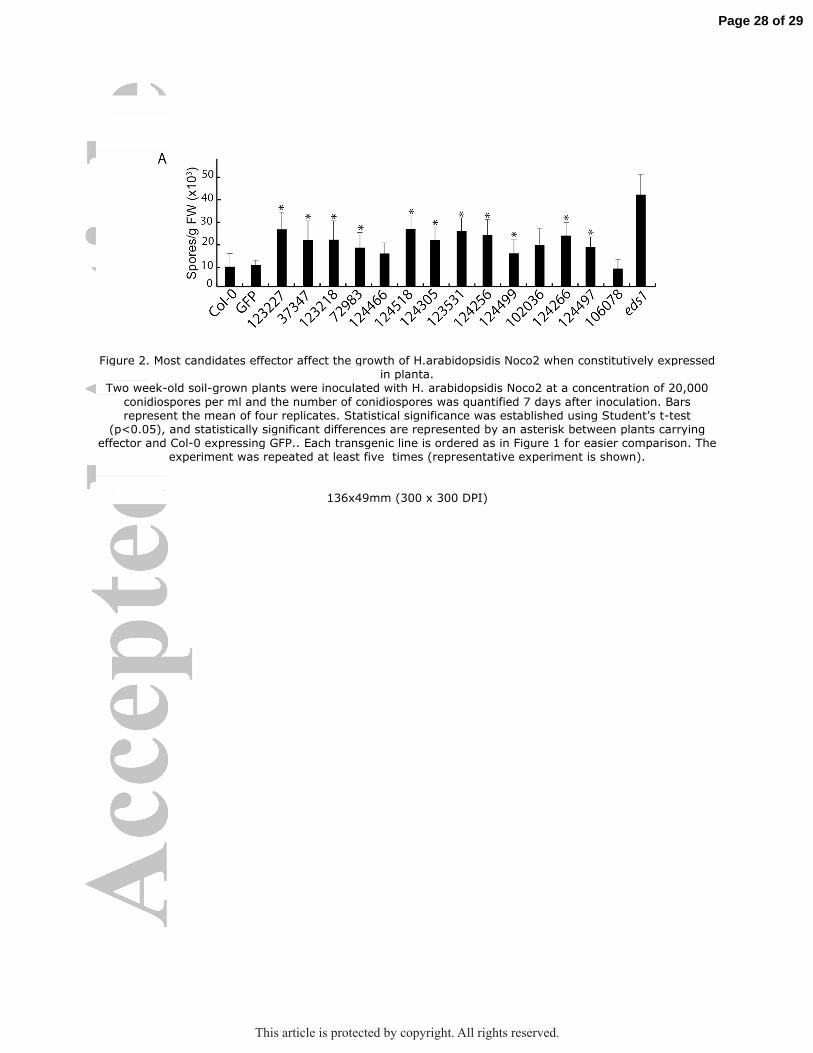
Figure 2. Most candidates effector affect the growth of H.arabidopsidis Noco2 when constitutively expressed in planta.
Two week-old soil-grown plants were inoculated with H. arabidopsidis Noco2 at a concentration of 20,000
conidiospores per ml and the number of conidiospores was quantified 7 days after inoculation. Bars represent the mean of four replicates. Statistical significance was established using Student’s t-test
(p<0.05), and statistically significant differences are represented by an asterisk between plants carrying effector and Col-0 expressing GFP.. Each transgenic line is ordered as in Figure 1 for easier comparison. The
experiment was repeated at least five times (representative experiment is shown).
136x49mm (300 x 300 DPI)
Page 28 of 29
This article is protected by copyright. All rights reserved.

Figure 3. Venn diagram showing the candidate effectors that induced increased virulence (in black) or decreased virulence (in red) .
Note that no transgenic lines were recovered for 123437 and 124111.
48x69mm (300 x 300 DPI)
Page 29 of 29
This article is protected by copyright. All rights reserved.

Figure 4. Subcellular localization of the different candidate effectors. A) Confocal images of leaf epidermal cells of 7 day old transgenic plantlets expressing candidate effectors
fused to GFP (SSP-GFP) in Col-0 genetic background. The top three effectors are the ones displaying informative localization, while the bottom three rows display the effectors showing nucleocytoplasmic
localization. B) Colocalization between PDCB1-mCherry , a plasmodesmata marker and 37347-GFP to infer its localization. Left panel red channel, center panel green channel, right panel overlay with DIC.
212x193mm (300 x 300 DPI)
Page 30 of 29
This article is protected by copyright. All rights reserved.