infants and children: acute management of community ... · guideline summary gl2018_007 issue date:...
TRANSCRIPT

Infants and Children: Acute Management of Community Acquired Pneumonia
Summary This Guideline provides evidence based guidance for clinicians in the acute assessmentand management of community acquired pneumonia in infants and children.
Document type Guideline
Document number GL2018_007
Publication date 16 March 2018
Author branch Agency for Clinical Innovation
Branch contact (02) 9424 5944
Review date 16 March 2023
Policy manual Patient Matters Manual for Public Health Organisations
File number H17/47033
Status Active
Functional group Clinical/Patient Services - Baby and Child, Medical Treatment, Nursing and Midwifery
Applies to Affiliated Health Organisations, Board Governed Statutory Health Corporations,Community Health Centres, Local Health Districts, Public Health Units, Public Hospitals,Specialty Network Governed Statutory Health Corporations
Distributed to Divisions of General Practice, Government Medical Officers, Ministry of Health, PrivateHospitals and Day Procedure Centres, Public Health System, Tertiary Education Institutes
Audience Emergency Departments, Medical, Clinicians, Nursing
Guideline
Secretary, NSW Health

GUIDELINE SUMMARY
GL2018_007 Issue date: March-2018 Page 1 of 2
INFANTS AND CHILDREN: ACUTE MANAGEMENT OF COMMUNITY ACQUIRED PNEUMONIA
PURPOSE
This Clinical Practice Guideline provides evidence based direction to clinicians in the acute management of community acquired pneumonia. It is aimed at achieving the best paediatric clinical care in the assessment and management of acute community acquired pneumonia and appropriate escalation responses across New South Wales.
KEY PRINCIPLES
This Guideline applies to all facilities where paediatric patients are managed. It requires Chief Executives of all Local Health Districts and specialty health networks to determine where local adaptations are required or whether it can be adopted in its current format in all hospitals and facilities required to manage children with community acquired pneumonia.
The Clinical Practice Guideline reflects what is currently regarded as a safe and appropriate approach to the acute management of community acquired pneumonia in infants and children. However, as in any clinical situation there may be factors which cannot be covered by a single set of guidelines. This document should be used as a guide, rather than as a complete authoritative statement of procedures to be followed in respect of each individual presentation. It does not replace the need for the application of clinical judgement to each individual presentation.
USE OF THE GUIDELINE
Chief Executives must ensure:
This Guideline is adopted or local protocols are developed based on the Infants and Children: Acute Management of Community Acquired Pneumonia, March 2018 Clinical Practice Guideline.
Local protocols are in place in all hospitals and facilities likely to be required to assess or manage paediatric patients with community acquired pneumonia.
Ensure that all staff treating paediatric patients are educated in the use of the locally developed paediatric guidelines.
Directors of Clinical Governance are required to inform relevant clinical staff treating paediatric patients of this new guideline.

GUIDELINE SUMMARY
GL2018_007 Issue date: March-2018 Page 2 of 2
REVISION HISTORY
Version Approved by Amendment notes
March 2018 (GL2018_007)
Deputy Secretary, Strategy and Resources
Guideline amendments include: -revision of alternatives for penicillin allergy -additional alternatives and improved clarity on dosage and duration of recommended medications.
March 2015 (GL2015_005)
Deputy Secretary, Population and Public Health
New Guideline
ATTACHMENT
1. Infants and Children: Acute Management of Community Acquired Pneumonia: Guideline

Infants and Children: Acute Management of Community Acquired Pneumonia
GUIDELINE
Issue date: March-2018
GL2018_007

Infants and Children: Acute Management of Community Acquired Pneumonia
GUIDELINE
GL2018_007 Issue date: March-2018 Contents page
CONTENTS
1 INTRODUCTION ...................................................................................................................... 3
2 OVERVIEW ............................................................................................................................... 3
3 SCOPE OF THE GUIDELINE .................................................................................................. 3
4 ALGORITHM ............................................................................................................................ 5
5 CLINICAL FEATURES ............................................................................................................. 6
History ............................................................................................................................... 6
Examination ...................................................................................................................... 6
Viral vs. bacterial pneumonia ........................................................................................... 7
Atypical pneumonia .......................................................................................................... 7
Alternative diagnoses and missed diagnosis ................................................................... 7
Summary ........................................................................................................................... 8
6 ASSESSMENT OF SEVERITY ................................................................................................ 8
Clinical features of mild pneumonia ................................................................................. 8
Clinical features of severe pneumonia ............................................................................. 9
Indications for admission to hospital ................................................................................ 9
7 DIAGNOSTIC TESTS ............................................................................................................. 10
Chest radiography .......................................................................................................... 10
Other tests....................................................................................................................... 11
Pulse oximetry ................................................................................................................ 11
Laboratory evaluation ..................................................................................................... 11
Complete blood count ..................................................................................................... 12
Acute phase reactants .................................................................................................... 12
Full blood count ............................................................................................................... 12
Urea and electrolytes ...................................................................................................... 12
Microbiological investigations ......................................................................................... 12
Blood culture ................................................................................................................... 12
Sputum Gram stain and culture ...................................................................................... 13
Nasopharyngeal bacterial culture ................................................................................... 13
Nasopharyngeal aspirates .............................................................................................. 13
Pleural fluid ..................................................................................................................... 13
Ultrasound ....................................................................................................................... 13
Other investigations ........................................................................................................ 14
Serum .............................................................................................................................. 14
8 MANAGEMENT ...................................................................................................................... 14
Outpatient management ................................................................................................. 15
Inpatient management .................................................................................................... 15
9 SPECIFIC MANAGEMENT AND ANTIBIOTIC AND DOSE RECOMMENDATIONS ......... 16
Term neonates: ≤ 1 month of age .................................................................................. 16
Infants: 1 to 3 months ..................................................................................................... 17

Infants and Children: Acute Management of Community Acquired Pneumonia
GUIDELINE
GL2018_007 Issue date: March-2018 Contents page
Infants and children: 4 months to 16 years .................................................................... 18
10 SPECIFIC PATHOGENS ....................................................................................................... 19
Staphylococcus aureus................................................................................................... 19
Pertussis ......................................................................................................................... 20
11 PENICILLIN ALLERGY .......................................................................................................... 21
12 DISCHARGE CRITERIA ........................................................................................................ 21
Follow up ......................................................................................................................... 22
Follow up chest x-ray ...................................................................................................... 22
13 COMPLICATIONS .................................................................................................................. 22
Pleural empyema ............................................................................................................ 22
Lung abscess .................................................................................................................. 24
14 APPENDIXES ......................................................................................................................... 25
Appendix 1: References.......................................................................................................... 25
Appendix 2: Incidence and mortality ....................................................................................... 28
Appendix 3: Prevention ........................................................................................................... 30
Appendix 4: Parent information .............................................................................................. 31
Appendix 5: Expert Working Group ........................................................................................ 32
Appendix 6: Glossary .............................................................................................................. 33

Infants and Children: Acute Management of Community Acquired Pneumonia
GUIDELINE
GL2018_007 Issue date: March-2018 Page 3 of 34
1 INTRODUCTION
This guideline presents the current best evidence for acute management of community acquired pneumonia in infants and children. Its purpose is to inform practice for NSW health care providers. This guideline is primarily targeted to clinicians caring for infants and children who have community acquired pneumonia.
The document should not be seen as a stringent set of rules to be applied without the clinical input and discretion of the managing professionals. Each patient should be individually evaluated and a decision made as to appropriate management in order to achieve the best clinical outcome.
In 2017, a targeted review of the 2015 guideline was undertaken to revise antibiotic guidance to align with recommendations in the eTherapeutic Guidelines (version15, 2014). A comprehensive review of this guideline is scheduled for 2020.
This guideline was developed by a representative group of NSW clinicians with expertise in acute and community paediatric care.
In the interests of patient care it is critical that contemporaneous, accurate and complete documentation is maintained during the course of patient management from arrival to discharge.
Management of community acquired pneumonia for Aboriginal children requires an awareness and respect of the cultural differences and factors influencing the health of Aboriginal and Torres Strait Islander people. Refer to your local Aboriginal Liaison Officer or for further information see Respecting the Difference the NSW Health Aboriginal Cultural Training Framework and/or NSW Health’s Communicating Positively – A Guide to Appropriate Aboriginal Terminology.
2 OVERVIEW
Community Acquired Pneumonia (CAP) is a relatively common presentation to emergency departments and correct management of CAP improves patient outcomes. Cases of severe pneumonia due to strains of community Methicillin-resistant Staphylococcus aureus (MRSA) are becoming more frequent in NSW.1,2 There has been a significant increase in the incidence of pleural effusion and empyema. At the same time, the risk of development of resistant organisms increases with the use of broad spectrum antibiotics and should be considered when prescribing these drugs.
3 SCOPE OF THE GUIDELINE
Inclusions:
All children less than 16 years of age
Parental anxiety should not be discounted:
it is often of significance even if the child does not appear especially unwell.

Infants and Children: Acute Management of Community Acquired Pneumonia
GUIDELINE
GL2018_007 Issue date: March-2018 Page 4 of 34
Community acquired pneumonia.
Exclusions:
Sepsis (Refer to the Paediatric Sepsis Pathway)
Immunocompromised patients (where e.g. Pneumocystis jiroveci (previously Pneumocystis carinii) pneumonia (PCP) should be considered)
Cystic fibrosis
Herpes simplex virus (HSV) pneumonitis
Hospital acquired pneumonia
Congenital heart or lung conditions
Tuberculosis
Patients with recent overseas travel and “tropical” pneumonias
Premature babies who have NOT yet reached “Term” as per their corrected gestational age
Aspiration of foreign body and/or gastric contents
Non-cystic fibrosis bronchiectasis.
Infants and Children: Acute Management of Community Acquired Pneumonia
GUIDELINE
GL2018_007 Issue date: March-2018 Page 5 of 34
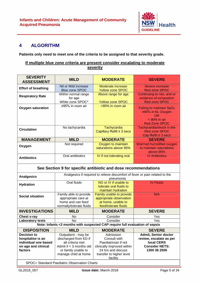
4 ALGORITHM
Patients only need to meet one of the criteria to be assigned to that severity grade.
If multiple blue zone criteria are present consider escalating to moderate severity
SEVERITY
ASSESSMENT MILD MODERATE SEVERE
Effort of breathing Nil or Mild increase Blue zone SPOC
Moderate increase Yellow zone SPOC
Severe increase Red zone SPOC
Respiratory Rate Within normal range
for age White zone SPOC*
Above range for age
Yellow zone SPOC
Continuing to rise, and or evidence of exhaustion
Red zone SPOC
Oxygen saturation ≥95% in room air <95% in room air
Failing to maintain SpO2 ≥95% in 6L Oxygen
OR < 90% in air
Red Zone SPOC
Circulation No tachycardia Tachycardia
Capillary Refill ≥ 3 secs Tachycardia/shock in the
Red zone SPOC Cap Refill ≥ 3 secs
MANAGEMENT MILD MODERATE SEVERE
Oxygen Not required Oxygen to maintain
saturations above 95% Warmed humidified oxygen
to maintain saturations above 95%
Antibiotics
Oral antibiotics IV if not tolerating oral IV Antibiotics
See Section 9 for specific antibiotic and dose recommendations
Analgesics Analgesics if required to relieve discomfort of fever or pain related to the
pneumonia
Hydration Oral fluids NG or IV if unable to
tolerate oral fluids to maintain hydration
IV Fluids
Social situation Family able to provide
appropriate care at home and can feed
normally/tolerate fluids
Family unable to provide appropriate observation
at home, unable to feed/tolerate fluids
N/A
INVESTIGATIONS MILD MODERATE SEVERE
Chest x-ray No Consider Yes
Laboratory tests No Consider Yes
Note: Infants <3 months with suspected CAP require full evaluation of sepsis
DISPOSITION MILD MODERATE SEVERE Decision to hospitalise is an individual one based on age and clinical factors
Outpatient - may be discharged from ED if
all criteria met Admit if < 3 months old
or family unable to manage child at home
Admission Consult with
Paediatrician if not clinically improved within
24 hrs and discuss transfer to higher level
facility
Admit, Senior doctor review, escalate as per
local CERS Consider NETS:
1300 36 2500
SPOC= Standard Paediatric Observation Charts
Infants and Children: Acute Management of Community Acquired Pneumonia
GUIDELINE
GL2018_007 Issue date: March-2018 Page 6 of 34
5 CLINICAL FEATURES
Pneumonia can manifest in a variety of ways dependent on the age of the child, the severity and the underlying micro-organism. This section refers to the clinical features as identified on history taking and on examination. Additional information on atypical pneumonia follows and important points related to alternative diagnoses are discussed.
History
Commonly, children with pneumonia present with fever, cough, and may have increased respiratory rate (RR) and increased work of breathing.
The NSW Health Standard Paediatric Observations Charts ‘Yellow Zone’ criteria define tachypnoea as:
RR greater than 65 in infants less than 3 months
RR greater than 55 in infants aged 3-12 months
RR greater than 50 in children aged 1-4 years
RR greater than 35 in children aged 5-11 years
RR greater than 30 in children older than 12 years.
Tachypnoea is the most sensitive sign for predicting children who have pneumonia evident on chest x-ray.3 Absence of tachypnoea makes pneumonia very unlikely.4
In children over the age of one month, cough is a common feature. An older child may describe pleuritic pain.5
Fever is common but not always present at the time of assessment. Fever alone occurring without cough or respiratory distress may still be pneumonia.6
Reduced oral intake and vomiting often occur as well as occasionally loose motions or diarrhoea.
Some patient groups with underlying disease are at a greater risk for developing pneumonia. Such diseases include cystic fibrosis, bronchiectasis, immunodeficiency and conditions associated with recurrent aspiration. A careful history is needed in those with recurrent pulmonary infections without known underlying disease.
Determination of the child’s immunisation status, recent travel and antibiotic use is needed. These factors may provide clues to the organism as well as guide the choice of antibiotic.
Examination
Vital signs are abnormal in proportion with disease severity; this includes increased respiratory rate, increased heart rate or low oxygen saturation. Fever is often present. The child may also be pale, sweaty and dry mucous membranes may be present. The blood pressure is normal unless the pneumonia is very severe and/or the child is profoundly dehydrated. Tachycardia, poor capillary refill and lethargy is evident in children who are shocked.
Infants and Children: Acute Management of Community Acquired Pneumonia
GUIDELINE
GL2018_007 Issue date: March-2018 Page 7 of 34
On inspection, increased work of breathing is present with the use of accessory muscles, as evidenced by tracheal tug, subcostal and intercostal recessions. Infants may also demonstrate nasal flare, head bobbing, grunting and episodes of apnoea.
Focal crackles on auscultation are not always present.5 Even when focal crackles are present, they are not sensitive nor specific for pneumonia.7 Less commonly bronchial breathing, a pleural rub or wheeze may be present. Reduced air entry and dullness to percussion may occur in the presence of pleural effusion and empyema.
Late and serious signs include severe respiratory distress, cyanosis, hypotension, and altered level of consciousness.
Viral vs. bacterial pneumonia
Viral lower respiratory tract infection is more often associated with mild symptoms and tends to occur in infants and younger children. The onset of symptoms for viral pneumonia can be more gradual. Bilateral signs on auscultation are evident. There may be rhinorrhoea, sore throat, arthralgia or a rash. Fever may be low-grade or absent.5 Viral lower respiratory tract infections are often preceded by rhinorrhoea and congestion. There is no reliable way of distinguishing the causative organism based on clinical features.
In viral pneumonia temperatures are generally lower than in bacterial pneumonia. Studies have shown a lower probability of having chest pain and rigors in viral pneumonias.
Bacterial pneumonia, especially in older children, typically begins with a rigor followed by high fever, cough and chest pain. Physical findings depend on the stage of pneumonia. Early findings include scattered focal crackles and rhonchi heard over the affected field. With progression of illness, signs of effusion and consolidation may become evident.
Atypical pneumonia
Although certain features on history and examination may be associated with specific micro-organisms, there is no reliable way of distinguishing the causative organism based on clinical features.8 In atypical pneumonia, wheeze is more often seen than in typical bacterial pneumonia.
Mycoplasma pneumoniae is more common in school-aged children than toddlers. Patients with mycoplasma may describe a malaise, sore throat, dry cough, headache, rash, myalgia and arthralgia associated with low-grade fever.5
Chlamydia pneumoniae usually presents with mild symptoms in adolescents and younger children.
Alternative diagnoses and missed diagnosis
There are a few other conditions that should be considered in children with this presentation. Bronchiolitis in babies manifests with rhinorrhoea, fever and tachypnoea. Bilateral crackles and/or wheeze may be evident. Children with upper respiratory tract infections have normal saturations and a clear chest on auscultation. Babies with cardiac failure often have a known history of congenital heart disease and may have bilateral chest signs without fever. Urinary tract infection and bacteraemia should be considered in

Infants and Children: Acute Management of Community Acquired Pneumonia
GUIDELINE
GL2018_007 Issue date: March-2018 Page 8 of 34
children with fever who have minimal respiratory symptoms or signs. Tachypnoea alone may be a sign of underlying metabolic acidosis e.g. diabetic ketoacidosis.
Occasionally, lower lobe pneumonia may present with abdominal pain and fever. In these patients, the increased respiratory rate and low saturations may aid the diagnosis; however these signs can be absent or minimal. Children with pneumonia may also present with fever alone.
Summary
The clinical presentation of children with pneumonia varies widely based on the child’s age, disease severity and the causative organism. Pneumonia is common and a high index of suspicion and careful attention to the findings on history and examination will identify the majority of children with this illness.
6 ASSESSMENT OF SEVERITY
The objective of the initial clinical assessment is to decide if the child’s history and physical examination findings are suggestive of CAP. The severity of the condition can range from mild to life threatening. Children with mild to moderate respiratory symptoms can often be managed safely at home providing the family is able to provide appropriate care, observations and supervision.
An assessment of pneumonia severity is necessary to determine the need for laboratory and imaging studies and the appropriate treatment setting. The severity of pneumonia is assessed by the child's overall clinical appearance and behaviour, including an assessment of his or her degree of alertness and willingness to eat or drink.9 Only one criterion needs to be met for the child to be assigned to a particular severity grade – vomiting excluded. If multiple blue zone criteria are present, consider escalating to moderate severity.
Temperature (°C): A temperature of greater than 38.5°C with features of tachypnoea, increased work of breathing, tachycardia and poor feeding can be indicators of moderate to severe disease. Neonates and young infants may not have the characteristic signs of serious infection (temperature can be high or low).10
Clinical features of mild pneumonia
These children:
may have abnormal findings on auscultation
often have temperature less than 38.5 degrees celsius
have mild or absent respiratory distress
may have an increased respiratory rate but will not display signs of increased effort of breathing
have oxygen saturations greater than or equal to 95% in room air
have no cyanosis
may have a normal heart rate when afebrile
are mentally alert

Infants and Children: Acute Management of Community Acquired Pneumonia
GUIDELINE
GL2018_007 Issue date: March-2018 Page 9 of 34
are able to feed normally.
Clinical features of severe pneumonia
These children:
will often have temperatures greater than 38.5 degrees celsius and
have moderate to severe respiratory distress
have tachypnoea and moderate/severe increased work of breathing
have tachycardia
have an inability to feed or maintain hydration
may have grunting, nasal flaring or apnoea
may have cyanosis
may have altered mental status with hypoxaemia.
Indications for admission to hospital
The decision to hospitalise a child with pneumonia must be an individual one based on age and clinical factors. Hospitalisation should be considered for all infants less than three months of age and for a child of any age whose family cannot provide appropriate care and assure compliance with the therapeutic plan.
The age appropriate Standard Paediatric Observation Chart (SPOC)11 should be used as part of the assessment to aid the clinician in determining how unwell the infant, child or adolescent is and the severity of their illness. Escalation should be according to local CERS policy.
Additional indications for hospitalisation include: 9, 12
Hypoxaemia (oxygen saturation consistently less than 95% in room air)
Dehydration, or inability to maintain hydration orally; inability to feed in an infant
Moderate to severe respiratory distress:
o Respiratory rate greater than 55 breaths/minute in infants less than 12 months or
o Respiratory rate greater than 50 breaths/minute in older children
o Difficulty breathing, apnoea, or grunting
Signs of toxicity (drowsy, lethargic or irritable, pale, mottled and/or tachycardic) 11
Underlying conditions that may predispose to a more serious course of pneumonia such as, cardiopulmonary disease, chronic lung disease, prematurity, history of malignancy
Presence of complications (e.g. effusion/empyema)
Failure of outpatient therapy (worsening or no response in 24 to 72 hours).

Infants and Children: Acute Management of Community Acquired Pneumonia
GUIDELINE
GL2018_007 Issue date: March-2018 Page 10 of 34
7 DIAGNOSTIC TESTS
The diagnosis of pneumonia requires historical or physical examination evidence of an acute infectious process with signs or symptoms of respiratory distress or radiologic evidence of an acute pulmonary infiltrate. The diagnostic approach depends to some extent upon the setting (inpatient or outpatient), the severity of illness and the age of the patient.
In the appropriate clinical setting the diagnosis can be made without radiographs.
In children with severe CAP, the diagnosis should be confirmed by chest x-ray and a full investigative process undertaken.
In general, aetiologic diagnosis should be sought in children who require admission to hospital and those who fail to respond to initial treatment.13
Chest radiography
A chest x-ray should not be considered a routine test in children with mild CAP.
Children that are well enough to be discharged from the ED with clear clinical signs of pneumonia do not need a chest x-ray to confirm the diagnosis.
However most children that require hospital admission will have moderate to severe disease and will require a chest x-ray.
It is recommended that a chest x-ray be obtained when:14,9
the pneumonia is classified as moderate to severe
clinical findings are unclear
exclusion of alternate explanation for respiratory distress (foreign body, heart failure)
a complication such as pleural effusion is suspected
the pneumonia is prolonged or unresponsive to antimicrobials
in a rural setting where access to after-hours diagnostics is limited, or decisions regarding escalation of care need to be made early.
There are a number of important points to consider when deciding whether to obtain a chest x-ray in the context of CAP:
Radiologic findings are poor indicators of aetiologic diagnosis
The finding of segmental consolidation is reasonably specific for bacterial pneumonia but lacks sensitivity. Pulmonary consolidation in young children sometimes appears to be spherical (round pneumonia). They tend to be greater than 3cm, solitary, located posteriorly and mostly caused by Streptococcus pneumoniae. They usually respond to appropriate antimicrobial therapy.
However if the lesion fails to resolve, a referral to a paediatric respiratory service should be made for consideration of an alternate diagnosis. Alternate diagnosis may include any of the following:
Infants and Children: Acute Management of Community Acquired Pneumonia
GUIDELINE
GL2018_007 Issue date: March-2018 Page 11 of 34
° Congenital malformations of the lungs and airways (e.g. pulmonary sequestration, bronchogenic cyst, congenital pulmonary airway malformation [CPAM])
° Thoracic tumors (e.g. bronchial carcinoid, bronchogenic carcinomas, pleuropulmonary blastoma, metastatic Wilms tumor)
° Mediastinal masses (neurogenic tumor, lymphoma, enlarged lymph nodes with tuberculosis)
° Predisposing conditions (e.g. foreign body inhalation, aspiration, immune deficiencies)
° The findings of pneumatoceles or large effusions are supportive of bacterial etiology. 14, 9
Chest x-ray changes may lag behind clinical findings and ultrasound.
Chest x-ray views
The recommended view depends upon the age of the child. In children older than four years the front posterior upright chest view is usually obtained to minimise the cardiac shadow. In younger children the position does not affect the cardiothoracic shadow, and the anterioposterior supine view is preferred.
Lateral chest x-ray should not be routinely performed.
Other tests
Ultrasound is simple, radiation-free, and is as good as chest x-ray in identifying pleuropulmonary alterations in children with suspected pneumonia.15
Pulse oximetry
Pulse oximetry should be performed in every child that presents with CAP (refer to local practice guidelines when undertaking continuous pulse oximetry). Hypoxaemia is well established as a risk factor for poor outcome in children with systemic disease especially respiratory disease. In a prospective study from Zambia the risk of death from pneumonia was significantly increased when hypoxaemia was present.8 Continuous pulse oximetry in respiratory disease is important to monitor. A good trace and trend is required continuously to accurately assess pulse oximetry, hence the need to monitor continuous pulse oximetry and not “one off” spot checks.
Laboratory evaluation
The laboratory evaluation of the child with CAP depends on the clinical scenario, age, severity of illness, presence of potential complications, underlying comorbidities, and requirement for admission. As a general rule children who are managed as outpatients do not require any investigations unless significant comorbidities. Young infants (i.e. less than three months) in whom pneumonia is suspected, particularly those who are febrile and have signs of toxicity will require further investigation to exclude other causes of infection, refer to the Paediatric Sepsis Pathway.

Infants and Children: Acute Management of Community Acquired Pneumonia
GUIDELINE
GL2018_007 Issue date: March-2018 Page 12 of 34
Complete blood count
Table 2: When to perform complete blood count
MILD MODERATE SEVERE
It is not necessary, unless significant co-morbidities i.e. (immunodeficiency)
In children with moderate disease it may be considered as it may provide useful information in conjunction with the clinical presentation to allow a decision to be made regarding requirement for admission to hospital. White blood cell counts greater than 15,000 per microlitre are suggestive of bacterial disease; eosinophilia may be present in children infected with Chlamydia trachomatis.
Should be undertaken in all children admitted with severe CAP.
Acute phase reactants
Erythrocyte sedimentation rate (ESR), C-reactive protein (CRP) and serum procalcitonin do not distinguish between bacterial and viral causes of CAP and should only be considered in moderate to severe disease.16
In patients with moderate to severe disease, acute-phase reactants may be used in conjunction with clinical findings to assess response to therapy.
Full blood count
What to look for:
Neutrophilia – may occur in either bacterial or in some acute viral infections
Leucopaenia – may occur with a viral infection or overwhelming sepsis or Q fever (Coxiella burnetti)
Lymphocytosis – Bordetella pertussis associated.
Urea and electrolytes
In moderate to severe disease only, urea and electrolyte testing may be helpful in assessing the degree of dehydration and whether hyponatraemia is present.
Microbiological investigations
For children admitted to hospital with CAP it is important to attempt a microbiological diagnosis. It is clear from a number of studies that between 10 to 30% of infections will have a mixed viral and bacterial etiology.17
Blood culture
Blood cultures should be obtained if the child requires admission to hospital. Blood cultures are positive in 10 to 20% of children with pneumonia. The yield increases to 30 to 40% in patients with a parapneumonic effusion or empyema. Pneumococcal

Infants and Children: Acute Management of Community Acquired Pneumonia
GUIDELINE
GL2018_007 Issue date: March-2018 Page 13 of 34
pneumonia is seldom a bacteraemic illness. Streptococcus pneumoniae is cultured in the blood in less than 5%.18
Blood cultures should be obtained in children with moderate to severe CAP. Repeat blood cultures in Staphylococcus aureus bacteraemia should be performed every 24 hrs until they are “no growth” to document efficacy of therapy. This should occur regardless of clinical status and antimicrobial therapy should continue for the full duration.
Sputum Gram stain and culture
The Gram stain of a good sputum specimen (presence of leucocytes, absence of squamous epithelial cells) has reasonable sensitivity and specificity for presumptive detection of Streptococcus pneumoniae. However, good specimens are often difficult to obtain in children.18
Nasopharyngeal bacterial culture
This is uninformative and should not be routinely undertaken. Bacterial growth in the nasoparynx does not indicate infection in the lower airways.
Nasopharyngeal aspirates
Nasopharyngeal secretions should be considered for viral detection using PCR and/or immunofluorescence on all children less than 18 months admitted with CAP.
There is substantial evidence that the risk of serious bacterial infection is low in children with laboratory confirmed viral infection. However, diffuse lower respiratory tract inflammation induced by viral respiratory tract infections predispose to bacterial super infection. Viral and bacterial co-infections were detected in 23% of children with pneumonia evaluated at a tertiary care children’s hospital.16
Antibacterial therapy is not necessary for children with a positive viral result in the absence of clinical laboratory or radiographic findings suggestive of bacterial co infection.
Sensitivity and specificity for PCR testing is over 90%.
Concordance between nasopharyngeal aspirates and nose throat swabs (taken for viral studies) have been found to be 89%.7
Pleural fluid
If the required expertise is available, pleural fluid may be aspirated for diagnostic purposes when there is evidence of an effusion present. However, in children it would be rare to perform this procedure due to its traumatic nature. Ultrasound guided thoracocentesis is the accepted clinical standard in children as it reduces the risk for iatrogenic pneumothorax.19 A specimen should be sent for biology and virology. Culture positive rates are in the order of 17–20%.
Ultrasound
Ultrasound is indicated as a first line investigation to confirm and quantify pleural fluid and empyema if suspected clinically or radiologically. Lung abscesses located within
Infants and Children: Acute Management of Community Acquired Pneumonia
GUIDELINE
GL2018_007 Issue date: March-2018 Page 14 of 34
consolidated lung can also be visualised. Ultrasound is more sensitive than chest x-ray for the detection of community-acquired pneumonia in children.15
Other investigations
Urine should not be taken for pneumococcal antigenuria as the specificity is too poor to be a useful test in diagnosis of CAP. False positivity occurs due to nasopharyngeal pneumococcal colonisation. 27
Legionellosis is rare in childhood and so urinary antigen testing for that is rarely indicated.
Serum
Paired serology remains the mainstay for diagnosing Mycoplasma pneumoniae and Chlamydia pneumoniae. Acute and convalescent serology should be undertaken if the patient is admitted with severe pneumonia or the clinical presentation is supportive of an infection with Mycoplasma or Chlamydia. During primary infection the immunoglobulin M (IgM) antibody appears 2-3 weeks after illness onset. The immunoglobulin G (IgG) antibody may not reach a diagnostically high (fourfold rise) titre until 6-8 weeks after illness onset.
8 MANAGEMENT
Consideration of the patient’s clinical condition using the “Severity score” and “Disposition Criteria” (see algorithm page 3) will assist in determining whether the child can be managed as an outpatient or if the child requires inpatient management.
Please note that this management guideline EXCLUDES the following conditions:
Sepsis, please refer to the Paediatric Sepsis Pathway
Immunocompromised patients (where Pneumocystis jiroveci should be considered)
Cystic fibrosis
Aspiration pneumonia
HSV pneumonitis
Hospital acquired pneumonia
Congenital heart or lung conditions
Tuberculosis
Patients with recent overseas travel and “tropical” pneumonias
Premature babies who are not ‘term’ as per their corrected gestational age.
Non-cystic fibrosis bronchiectasis.
IMPORTANT NOTE:
Aboriginal and Torres Strait Islander children and Pacific Islander children have a higher incidence of Staphylococcal pneumonia caused by both methicillin sensitive and methicillin resistant Staphylococcal aureus. When treating these patients this should be taken into consideration.

Infants and Children: Acute Management of Community Acquired Pneumonia
GUIDELINE
GL2018_007 Issue date: March-2018 Page 15 of 34
Consider Staphylococcus aureus pneumonia in any child with a severe pneumonia not
responding to antibiotic treatment. Please refer to the Specific Pathogens regarding
antibiotic cover for Staphylococcus aureus pneumonia.
Outpatient management
Analgesia can be given to relieve discomfort from fever or pain related to the pneumonia
Follow-up should be arranged to evaluate the patient for any deterioration.
Inpatient management
Analgesia can be given to relieve discomfort from fever or pain related to the pneumonia
Oxygen should be provided to patients to keep their saturations equal to or above 95%
When oxygen therapy required, consider warm, humidified oxygen (if available)
Patients exclusively on intravenous fluids require daily monitoring of their electrolytes to monitor for syndrome of inappropriate antidiuretic hormone (SIADH) 20
Chest physiotherapy has not been shown to be beneficial and is not recommended 20
In any progressively unwell child, consideration should be given to transfer the patient to a higher care facility. The advice/use of NETS 1300 36 2500 should be considered.

Infants and Children: Acute Management of Community Acquired Pneumonia
GUIDELINE
GL2018_007 Issue date: March-2018 Page 16 of 34
9 SPECIFIC MANAGEMENT AND ANTIBIOTIC AND DOSE RECOMMENDATIONS
(based on eTherapeutic Guidelines (version 15, 2014).
Term neonates: ≤ 1 month of age
ALL NEONATES REQUIRE ADMISSION FOR INPATIENT MANAGEMENT - MILD, MODERATE OR SEVERE
Up to 7 days of age 21 BENZYLPENICILLIN (Penicillin G) 50 mg/kg IV 12-hourly (max 300 mg/dose)
Plus
GENTAMICIN 4 mg/kg IV once daily (max 24 mg/dose)
8 to 28 days of age 21 BENZYLPENICILLIN (Penicillin G) 50 mg/kg IV 6-hourly (max 300 mg/dose)
Plus
GENTAMICIN 4 mg/kg IV once daily (max 24 mg/dose)
NOTE: Herpes simplex virus pneumonitis may present between days 3 and 7 and requires expert advice regarding management.21 Consider:
ACICLOVIR 20 mg/kg IV 8-hourly if risk factors for HSV pneumonitis
Consider adding the following treatment for Chlamydia trachomatis*: AZITHROMYCIN 20 mg/kg orally daily for 3 days 22 (max 120 mg/dose) or CLARITHROMYCIN 7.5 mg/kg orally 12-hourly for 7 days Consider adding the following for Bordetella pertussis* : AZITHROMYCIN 10 mg/kg orally daily for 5 days (max 50 mg/dose) or CLARITHROMYCIN 7.5 mg/kg orally 12-hourly for 7 days (max 37.5 mg/dose)
Beware of atypical presentations e.g. apnoea.
*Azithromycin is the only drug recommended for Bordatella pertussis treatment or prophylaxis or treatment of Chlamydia trachomatis in children <1 month of age, it is associated with infantile hypertrophic pyloric stenosis, especially in babies <2 weeks old, though the benefit of using azithromycin outweighs this risk.
Infants and Children: Acute Management of Community Acquired Pneumonia
GUIDELINE
GL2018_007 Issue date: March-2018 Page 17 of 34
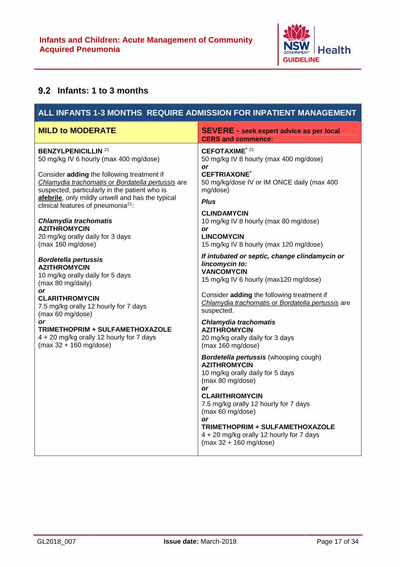
Infants: 1 to 3 months
ALL INFANTS 1-3 MONTHS REQUIRE ADMISSION FOR INPATIENT MANAGEMENT
MILD to MODERATE SEVERE - seek expert advice as per local
CERS and commence:
BENZYLPENICILLIN 21 50 mg/kg IV 6 hourly (max 400 mg/dose) Consider adding the following treatment if Chlamydia trachomatis or Bordatella pertussis are suspected, particularly in the patient who is afebrile, only mildly unwell and has the typical clinical features of pneumonia21: Chlamydia trachomatis AZITHROMYCIN 20 mg/kg orally daily for 3 days (max 160 mg/dose) Bordetella pertussis AZITHROMYCIN 10 mg/kg orally daily for 5 days (max 80 mg/daily) or CLARITHROMYCIN 7.5 mg/kg orally 12 hourly for 7 days (max 60 mg/dose) or TRIMETHOPRIM + SULFAMETHOXAZOLE 4 + 20 mg/kg orally 12 hourly for 7 days (max 32 + 160 mg/dose)
CEFOTAXIME* 21 50 mg/kg IV 8 hourly (max 400 mg/dose) or CEFTRIAXONE* 50 mg/kg/dose IV or IM ONCE daily (max 400 mg/dose)
Plus
CLINDAMYCIN 10 mg/kg IV 8 hourly (max 80 mg/dose) or LINCOMYCIN 15 mg/kg IV 8 hourly (max 120 mg/dose)
If intubated or septic, change clindamycin or lincomycin to: VANCOMYCIN 15 mg/kg IV 6 hourly (max120 mg/dose) Consider adding the following treatment if Chlamydia trachomatis or Bordatella pertussis are suspected.
Chlamydia trachomatis AZITHROMYCIN 20 mg/kg orally daily for 3 days (max 160 mg/dose)
Bordetella pertussis (whooping cough) AZITHROMYCIN 10 mg/kg orally daily for 5 days (max 80 mg/dose) or CLARITHROMYCIN 7.5 mg/kg orally 12 hourly for 7 days (max 60 mg/dose) or TRIMETHOPRIM + SULFAMETHOXAZOLE 4 + 20 mg/kg orally 12 hourly for 7 days (max 32 + 160 mg/dose)
Infants and Children: Acute Management of Community Acquired Pneumonia
GUIDELINE
GL2018_007 Issue date: March-2018 Page 18 of 34
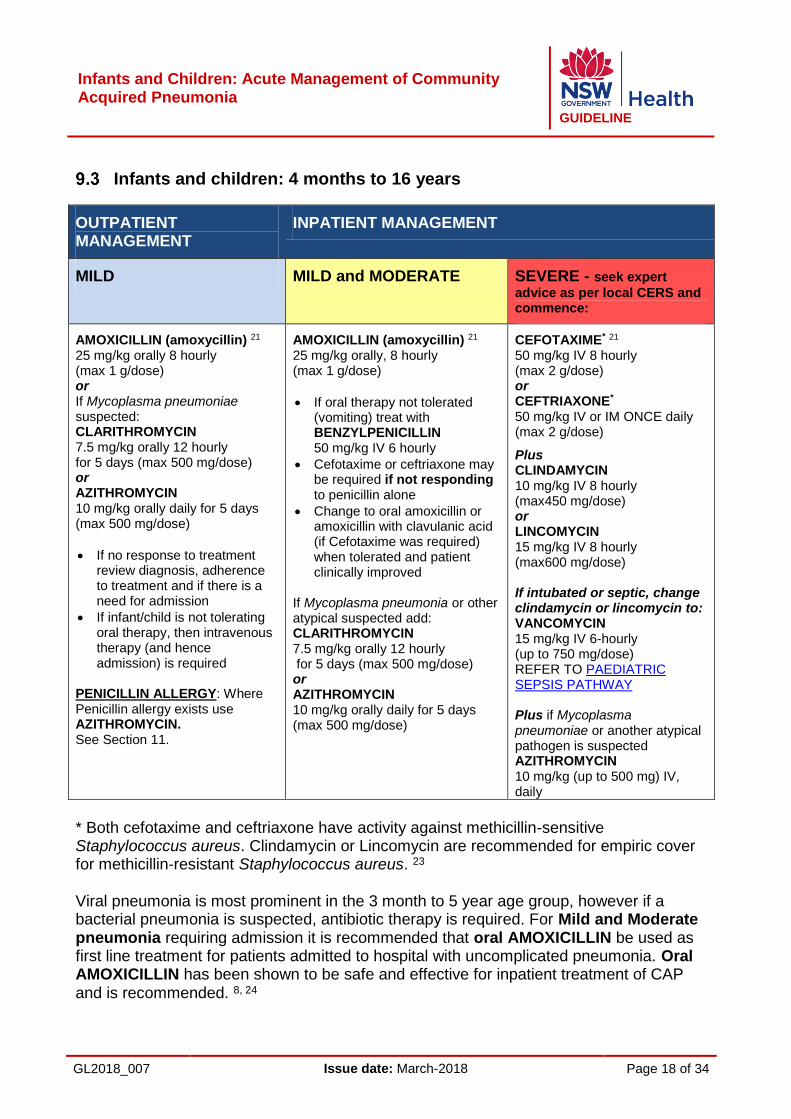
Infants and children: 4 months to 16 years
OUTPATIENT MANAGEMENT
INPATIENT MANAGEMENT
MILD MILD and MODERATE SEVERE - seek expert
advice as per local CERS and commence:
AMOXICILLIN (amoxycillin) 21 25 mg/kg orally 8 hourly (max 1 g/dose) or If Mycoplasma pneumoniae suspected: CLARITHROMYCIN 7.5 mg/kg orally 12 hourly for 5 days (max 500 mg/dose) or AZITHROMYCIN 10 mg/kg orally daily for 5 days (max 500 mg/dose)
If no response to treatment review diagnosis, adherence to treatment and if there is a need for admission
If infant/child is not tolerating oral therapy, then intravenous therapy (and hence admission) is required
PENICILLIN ALLERGY: Where Penicillin allergy exists use AZITHROMYCIN. See Section 11.
AMOXICILLIN (amoxycillin) 21 25 mg/kg orally, 8 hourly (max 1 g/dose)
If oral therapy not tolerated (vomiting) treat with BENZYLPENICILLIN 50 mg/kg IV 6 hourly
Cefotaxime or ceftriaxone may be required if not responding to penicillin alone
Change to oral amoxicillin or amoxicillin with clavulanic acid (if Cefotaxime was required) when tolerated and patient clinically improved
If Mycoplasma pneumonia or other atypical suspected add: CLARITHROMYCIN 7.5 mg/kg orally 12 hourly for 5 days (max 500 mg/dose) or AZITHROMYCIN 10 mg/kg orally daily for 5 days (max 500 mg/dose)
CEFOTAXIME* 21 50 mg/kg IV 8 hourly (max 2 g/dose) or CEFTRIAXONE* 50 mg/kg IV or IM ONCE daily (max 2 g/dose)
Plus CLINDAMYCIN 10 mg/kg IV 8 hourly (max450 mg/dose) or LINCOMYCIN
15 mg/kg IV 8 hourly (max600 mg/dose) If intubated or septic, change clindamycin or lincomycin to: VANCOMYCIN 15 mg/kg IV 6-hourly (up to 750 mg/dose) REFER TO PAEDIATRIC SEPSIS PATHWAY Plus if Mycoplasma pneumoniae or another atypical pathogen is suspected AZITHROMYCIN 10 mg/kg (up to 500 mg) IV, daily
* Both cefotaxime and ceftriaxone have activity against methicillin-sensitive Staphylococcus aureus. Clindamycin or Lincomycin are recommended for empiric cover for methicillin-resistant Staphylococcus aureus. 23
Viral pneumonia is most prominent in the 3 month to 5 year age group, however if a bacterial pneumonia is suspected, antibiotic therapy is required. For Mild and Moderate pneumonia requiring admission it is recommended that oral AMOXICILLIN be used as first line treatment for patients admitted to hospital with uncomplicated pneumonia. Oral AMOXICILLIN has been shown to be safe and effective for inpatient treatment of CAP and is recommended. 8, 24
Infants and Children: Acute Management of Community Acquired Pneumonia
GUIDELINE
GL2018_007 Issue date: March-2018 Page 19 of 34
10 SPECIFIC PATHOGENS
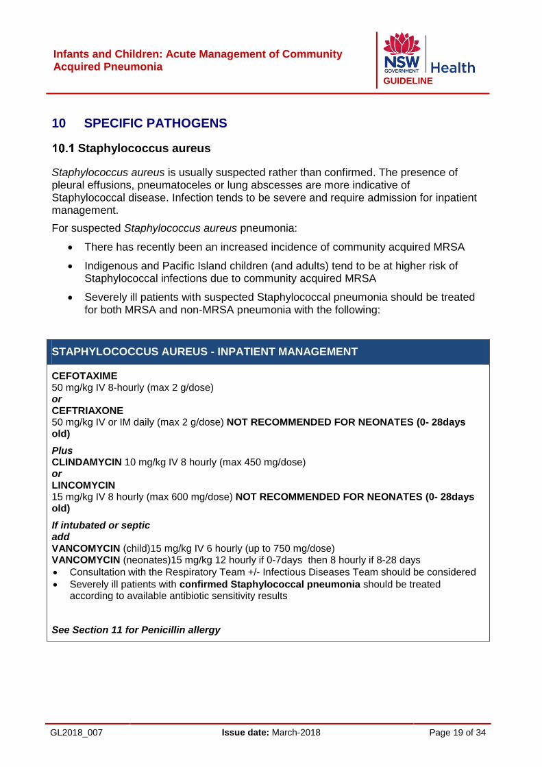
Staphylococcus aureus
Staphylococcus aureus is usually suspected rather than confirmed. The presence of pleural effusions, pneumatoceles or lung abscesses are more indicative of Staphylococcal disease. Infection tends to be severe and require admission for inpatient management.
For suspected Staphylococcus aureus pneumonia:
There has recently been an increased incidence of community acquired MRSA
Indigenous and Pacific Island children (and adults) tend to be at higher risk of Staphylococcal infections due to community acquired MRSA
Severely ill patients with suspected Staphylococcal pneumonia should be treated for both MRSA and non-MRSA pneumonia with the following:
STAPHYLOCOCCUS AUREUS - INPATIENT MANAGEMENT
CEFOTAXIME 50 mg/kg IV 8-hourly (max 2 g/dose) or CEFTRIAXONE 50 mg/kg IV or IM daily (max 2 g/dose) NOT RECOMMENDED FOR NEONATES (0- 28days old)
Plus CLINDAMYCIN 10 mg/kg IV 8 hourly (max 450 mg/dose) or LINCOMYCIN 15 mg/kg IV 8 hourly (max 600 mg/dose) NOT RECOMMENDED FOR NEONATES (0- 28days old)
If intubated or septic add VANCOMYCIN (child)15 mg/kg IV 6 hourly (up to 750 mg/dose) VANCOMYCIN (neonates)15 mg/kg 12 hourly if 0-7days then 8 hourly if 8-28 days
Consultation with the Respiratory Team +/- Infectious Diseases Team should be considered
Severely ill patients with confirmed Staphylococcal pneumonia should be treated according to available antibiotic sensitivity results
See Section 11 for Penicillin allergy

Infants and Children: Acute Management of Community Acquired Pneumonia
GUIDELINE
GL2018_007 Issue date: March-2018 Page 20 of 34
Pertussis
Admit all infants under 6 months with suspected pertussis and children with cyanosis or apnoea. Frequent observations and monitoring with pulse oximetry is essential. Intensive care may be needed for children with episodes of cyanosis or apnoea. Pertussis, suspected or confirmed at any age should be treated with the following 23:
PERTUSSIS
OUTPATIENT MANAGEMENT - only infants and children greater than 6 months of age
PERTUSSIS
INPATIENT MANAGEMENT - all infants under 6 months of age require admission
AZITHROMYCIN 10 mg/kg orally for the first dose only (max 500 mg/dose) then 5 mg/kg daily for next 4 days (max 250 mg/dose) or CLARITHROMYCIN 7.5mg/kg orally 12 hourly for 7 days (max 500 mg/dose) or TRIMETHOPRIM + SULFAMETHOXAZOLE 4 + 20 mg/kg orally 12 hourly for 7 days (max 160 + 800 mg/dose)
Under 1 month: AZITHROMYCIN 10 mg/kg orally or IV daily (max 50 mg/dose) for 5 days or CLARITHROMYCIN 7.5mg/kg orally 12 hourly for 7 days (max 37.5 mg/dose) Age 1 – 6 months: AZITHROMYCIN 10 mg/kg orally for the first dose only (max 80 mg/dose) followed by 5 mg/kg oral daily for next 4 days (max 40 mg/dose) or CLARITHROMYCIN 7.5 mg/kg orally 12 hourly for 7 days (max 60 mg/dose) or TRIMETHOPRIM + SULFAMETHOXAZOLE 4 + 20 mg/kg orally 12-hourly for 7 days (max 32 + 160 mg/dose)
Infants and Children: Acute Management of Community Acquired Pneumonia
GUIDELINE
GL2018_007 Issue date: March-2018 Page 21 of 34
11 PENICILLIN ALLERGY
1. Child known to tolerate cephalosporins:
MILD to MODERATE
3 months to 2 years CEFUROXIME 10 mg/kg/dose oral 12 hourly (max 125 mg/dose)
2 years to 12 years CEFUROXIME 15 mg/kg/dose oral 12 hourly (max 250 mg/dose)
Greater than 12 years CEFUROXIME 250mg/dose oral 12 hourly (max 500 mg/dose)
If parenteral therapy is required:
Greater than 1 month CEFOTAXIME 50 mg/kg IV 8 hourly (max 2 g/dose) or CEFTRIAXONE 50 mg/kg IV daily (max 2 g/dose)
2. There is a 7% chance of cross-reaction of cephalosporin with penicillin. Non-
beta-lactam alternatives are Erythromycin, Clarithromycin or if greater than 8 years of age consider Doxycycline:
MILD to MODERATE
Greater than 28 days ERYTHROMYCIN 10 mg/kg/dose oral 6 hourly (max 500 mg/dose) or
All ages CLARITHROMYCIN 7.5 mg/kg/dose oral12 hourly (max 500 mg/dose) or
8 years or greater DOXYCYCLINE 2 mg/kg/dose oral 12 hourly (max 100 mg/dose)
Streptococcus Pneumoniae or Staphylococcus aureus may be resistant to these above agents.
SEVERE
0 - 6 days VANCOMYCIN 15 mg/kg/dose IV 12 hourly (max 90 mg/dose) plus CIPROFLOXACIN 10 mg/kg/dose IV 12 hourly (max 60 mg/dose)
7 to 28 days VANCOMYCIN 15 mg/kg/dose IV 8 hourly (max 90 mg/dose) plus CIPROFLOXACIN 10 mg/kg/dose IV 12 hourly (max 60 mg/dose)
Greater than 28 days VANCOMYCIN 15 mg/kg/dose IV 6 hourly (max 750 mg/dose) plus CIPROFLOXACIN 10 mg/kg/dose IV 12 hourly (max 400 mg/dose)
12 DISCHARGE CRITERIA
Patients are eligible for discharge when there is overall clinical improvement, good oral intake, resolution of symptoms with consistent pulse oximetry measurements equal to or greater than 95% for at least 12 hours.
Infants and Children: Acute Management of Community Acquired Pneumonia
GUIDELINE
GL2018_007 Issue date: March-2018 Page 22 of 34
Patients are not appropriate for discharge if they have substantially increased work of breathing or sustained tachypnoea or tachycardia. Parents should be able to administer and children able to comply with taking oral antibiotics prior to discharge.
If a patient has had a chest tube, the chest tube should have been removed for at least 24 hours prior to discharge.
Follow up
All patients with pneumonia require follow up examination with a medical officer at 4-6 weeks.
In patients with recurrent pneumonia or atelectasis consider:
Aspiration
Foreign body
Congenital malformation
Cystic fibrosis
Immunosuppression.
Follow up chest x-ray
A follow up chest x-ray is not required in those who are previously healthy and recovering well with no ongoing symptoms. In patients with uncomplicated pneumonia repeat chest radiographs are unwarranted.
However in patients with pleural effusions, pneumatoceles, or pulmonary abscess a repeat chest radiograph should be done to ensure resolution. In round pneumonia a follow up chest radiograph should be done to ensure tumour masses are not missed. A follow up chest x-ray is also warranted where there are continuing symptoms.
A chest x-ray should be obtained in children who fail to demonstrate clinical improvement within 48 – 72 hours after initiation of antibiotic therapy or any child that has progressive symptoms or clinical deterioration.
Repeat chest x-ray 4 – 6 weeks after diagnosis should only be obtained in patients with recurrent pneumonia, round pneumonia or persisting symptoms (such as shortness of breath, cough, fever or chest pain).
However it must be remembered that radiologic abnormalities lag behind clinical resolution. Follow up chest x-rays obtained 3 - 6 weeks after initial imaging revealed residual abnormalities in 10 – 30% of children with radiographically confirmed CAP.20
13 COMPLICATIONS
Pleural empyema
Pleural empyema is defined as accumulation of purulent material consisting of leukocytes, fibrin, and pathogens between the visceral and parietal pleura. The

Infants and Children: Acute Management of Community Acquired Pneumonia
GUIDELINE
GL2018_007 Issue date: March-2018 Page 23 of 34
prevalence of empyema complicates 0.7% of childhood pneumonias with an incidence of 0.7-3.3 per 100,000 in Australia.25
The clinical features may resemble those of uncomplicated pneumonia and in addition include pleuritic (stabbing, worsening with deep inspiration) chest pain which may occur due to inflammation of the parietal pleura. Radiation into ipsilateral shoulder (para-diaphragmatic pleural empyema), and non-pleuritic pain (dull, aching) signifies direct involvement of parietal pleura (e.g. abscess). On examination, dullness on percussion, decreased/absent breath sounds, decreased chest movements, scoliosis, and splinting may be present.
The aetiology is usually a bacterial infection, most commonly Streptococcus pneumoniae and less commonly Staphylococcus aureus (MRSA/MSSA).
Diagnosis is suspected clinically and by chest x-ray and confirmed with chest ultrasound. The collection of specimens (blood culture and pleural fluid) for microbiological analyses is recommended as it may aid in guiding antibiotic treatment even though in the majority of cases the cultures are negative. Empyema commonly requires further interventions in addition to antibiotic treatment and as such all children with empyema should be managed in a hospital with appropriate expertise and under the care of a respiratory paediatrician.
The treatment consists of effective pain relief (non-steroidal anti-inflammatory drugs (NSAIDs); consider patient controlled analgesia (PCA)/opioids post-surgery as indicated), maintaining appropriate oxygenation (SaO2 equal to or greater than 95%), and antibiotics. Video-assisted thoracoscopic surgery (VATS) or insertion of percutaneous small bore drainage with instillation of fibrinolytics (e.g. 6 doses of urokinase over 3 days) is often required.
Repeated ultrasounds may be required in the case of clinical deterioration as fluid may accumulate rapidly. A prolonged course of oral antibiotics may be required (1-6 weeks) and follow-up until clinically improved should be arranged. Chest x-ray may remain abnormal for up to 6 months after treatment. Usually children fully recover without long term sequelae, however complications include:
Bronchopleural fistula
Cutaneous fistula
Bacteraemia
Peri/endocarditis
Pneumothorax
Necrotising pneumonia
Pneumatoceles
Persistent lobar collapse (Query of Foreign body)
Lung abscess. Other Complications
Metastatic infection (i.e. in bones)
Sepsis
Haemolytic Uremic Syndrome (HUS).

Infants and Children: Acute Management of Community Acquired Pneumonia
GUIDELINE
GL2018_007 Issue date: March-2018 Page 24 of 34
Lung abscess
A lung abscess is a cavity in the lung filled with purulent material caused by an infection that arises from aspiration, impaired mucociliary clearance, or haematogenous spread/septic emboli. Clinical symptoms and signs are often indistinguishable from pneumonia, although persistent fever and cough despite appropriate antibiotic treatment, chest pain, haemoptysis, dullness on percussion, and localized reduced air entry may raise suspicion. Diagnosis is made by chest x-ray and supported by chest ultrasound or contrast-enhanced chest computer tomography. Lung abscess may require further radiological or surgical intervention in addition to a prolonged course of antibiotic treatment, which is not only guided by clinical severity but also pre-existing risk factors (e.g. cover for anaerobes in children at risk of recurrent aspirations, gram-negative bacilli in children with cystic fibrosis, MRSA in children colonised with MRSA, recurrent skin abscesses, etc.) 2, 26.
A respiratory paediatrician (via NETS where necessary) should always be consulted for children presenting with a lung abscess, as they commonly require management in a hospital with appropriate expertise under the care of a respiratory paediatrician.

Infants and Children: Acute Management of Community Acquired Pneumonia
GUIDELINE
GL2018_007 Issue date: March-2018 Page 25 of 34
14 APPENDIXES
Appendix 1: References
1. NSW Admitted Patient Data Collection (SAPHaRI). Centre for Epidemiology and Evidence, NSW Ministry of Health, 2012.
2. NSW Emergency Department Data Collection (SAPHaRI). Centre for Epidemiology and Evidence, NSW Ministry of Health, 2012.
3. Margolis, P., Gadomski, A. The rational clinical examination. Does this infant have pneumonia? JAMA, 1998; 279 (4): 308.
4. http://emedicine.medscape.com/article/967822-clinical “Paediatric Pneumonia Clinical Presentation” NJ Bennett. [Accessed 23/11/11]
5. Bachur, R., Perry, H., Harper, M.B. Occult pneumonias: empiric chest radiographs in febrile children with leukocytosis. Ann Emerg Med, 1999; 33 (2): 166.
6. Mahabee-Gittens, E.M., Grupp-Phelan, J., Brody, A.S., Donnelly, L.F., Bracey, S.E., Duma, E.M. Identifying children with pneumonia in the emergency department. Clin Pediatr (Phila), Jun 2005; 44 (5): 427-35.
7. British Thoracic Society, Guidelines for Community Acquired Pneumonia in Children, 2011.
8. Community Acquired Pneumonia Guideline Team, Cincinnati Children's Hospital Medical Center. Evidence-based care guidelines for medical management of community acquired pneumonia in children 60 days to 17 years of age – Guideline 2014. www.cincinnatichildrens.org/svc/alpha/h/health-policy/ev-based/pneumonia.htm [Accessed September 2014].
9. Bradley, J.S., Byington, C.L., Shah, S.S. The management of community-acquired pneumonia in infants and children older than 3 months of age: Clinical practice guidelines by the Pediatric Infectious Diseases Society and the Infectious Diseases Society of America. Clin Infect Dis, 2011; 53:e25.
10. NSW Health. Standard Paediatric Observation Charts. 2011 11. NSW Ministry of Health, PD2010_063 Infants and children: Acute Management
of Fever, second edition, 2010. 12. Harris, M., Clark, J., Coote, N. British Thoracic Society guidelines for the
management of community acquired pneumonia in children: update 2011. Thorax, 2011; 66:ii1.
13. Up to date clinical features and diagnosis of community acquired pneumonia in children May 2011 http://www.uptodate.com
14. Swingler, G.H. Randomised controlled trial of clinical outcome after chest radiograph. Lancet, 1998; 351:404.
15. Caiulo, V. A., Gargani, L., Caiulo, S., Fisicaro, A., Moramarco, F., Latini, G., Picano, E. and Mele, G. Lung ultrasound characteristics of community-acquired pneumonia in hospitalized children. Pediatric. Pulmonology (2012),. doi: 10.1002/ppul.22585
16. Bradley J.S., Byington C.L., Shah S.S. Management of Community-Acquired Pneumonia in Infants and Children Older Than 3 Months of Age: Clinical Practice Guidelines by the Pediatric Infectious Diseases Society of America. Clinical Infectious Diseases 2011;53(7)e25-276

Infants and Children: Acute Management of Community Acquired Pneumonia
GUIDELINE
GL2018_007 Issue date: March-2018 Page 26 of 34
17. Juven, T. Etiology of community- acquired pneumonia in 254 hospitalised children. Pediatr Infect Dis J, 2000; 19: 217-223.
18. Korppi, M. Pneumococcal serology in childrens respiratory infections. Eur J Clin Microbiol, 2008: 25: 559-560.
19. Barnes TW, Morgenthaler TI, Olson EJ, Sonographically guided thoracentesis and rate of pneumothorax. Journal of Clinical Ultrasound 2005;33: 442–6.
20. Francois P, Desrumaux A, Cans C, Prevalence and risk factors of suppurative complications in children with pneumonia. Acta Paediatric 2010;99:861–6.
21. eTG complete [Internet]. Melbourne: Therapeutic Guidelines Limited; 2015 Nov 22. Azythromycin(systemic): Pediatric drug information Lexicomp, Up to Date
https://www.uptodate.com.acs.hcn.com.au/contents/azithromycin-systemic-pediatric-drug-information?source=search_result&search=azithromycin&selectedTitle=2~148
23. Kaplan SL, Hulten KG, Mason EO (2014) Staphylococcus aureus infections. In Feigin and Cherry’s Textbook of Pediatric Infectious Diseases. Eds Cherry JD, Harrison GJ, Kaplan SL, Steinbach WJ , Hotez PJ pp 1113- 1131 Elsevier Philadelphia.
24. Sydney Children’s Hospital Empiric Antibiotic Guideline 2015. 25. Strachan, Cornelius, Gilbert et al., Paediatric Empyema Thoracis, Pediatric
Respiratory Medicine, 2011 second edition, MOSBY Elsevier, Taussig and Landau.
26. Thomas R, Ferguson J, Coombs GW, Gibson PG. Community-acquired methicillin-resistant staphylococcus aureus pneumonia: a clinical audit. Respirology. 2011; 16:926-3.
27. Garcia-Suarez MD, Cron LE, Suarez-Alvarez B et al (2009) Diagnostic Detection of Streptococcus pneumoniae PpmA in urine. Clin Microbiol Infect. 15(5) 443-53.
Further resources: 1. Commonwealth of Australia, Aboriginal and Torres Strait Islander Health
Performance Framework, 2011. 2. Pediatric Respiratory Medicine, second edition, MOSBY Elsevier, Taussig and
Landau, 2008. 3. James Cherry & Gail J. Demmler-Harrison & Sheldon L. Kaplan & William J.
Steinbach & Peter Hotez, Paediatric Community Acquired Pneumonia in Feigin and Cherry’s Textbook of Paediatric Infectious Diseases 7th edition Saunders, 2014.
4. Finnish guidelines for the treatment of community-acquired pneumonia and pertussis in children. Tapiainen T. Aittoniemi J. Immonen J. et al. Acta Paediatrica. vol 105, no 1, January 2016, pp 39-43.
5. Review of guidelines for evidence-based management for childhood community-acquired pneumonia in under-5 years from developed and developing countries. Nascimento-Carvalho CM; Madhi SA; O'Brien KL. Pediatric Infectious Disease Journal. 32(11):1281-2, 2013 Nov.
6. Community-acquired pneumonia in children. Cardinale F; Cappiello AR; Mastrototaro MF; Pignatelli M; Esposito S. Early Human Development. 89 Suppl 3:S49-52, 2013 Oct.
7. The Management of Community-Acquired pneumonia in Infants and Children Older than 3 Months of Age: Clinical Practice Guidelines by the Pediatric

Infants and Children: Acute Management of Community Acquired Pneumonia
GUIDELINE
GL2018_007 Issue date: March-2018 Page 27 of 34
Infectious Diseases Society of America. Bradley JS, Byington CL, Shah SS et al Clinical Infectious Diseases 2011;53(7)e25-276
Infants and Children: Acute Management of Community Acquired Pneumonia
GUIDELINE
GL2018_007 Issue date: March-2018 Page 28 of 34
Appendix 2: Incidence and mortality
Pneumonia in children worldwide is associated with significant morbidity and mortality. Most paediatric deaths from pneumonia occur in developing countries, with only low mortality reported in developed countries such as Australia. Over the past decade, a trend of increasing complications of pneumonia (e.g. empyema) and the emergence in the community of resistant strains of staphylococcal pneumonias are reported.1,2,3 There are significant challenges in determining precise rates of viral and bacterial CAP as well as identifying pathogens. It is likely that new pathogens will be identified over the next decade. In many cases, there is a mixture of pathogens, including both viral and bacterial pathogens with European and Asian studies identifying 8 - 40% of mixed infections, while a pathogen is not detected in up to 60% of cases.4
Age
The variety of pathogens, the developing immune system and age-related exposures means that different pathogens are associated with different age groups. Vaccination also plays a role in the incidence of pneumonia in children. See Table 1 (Appendix 4)
Infants under 1 year have the highest rate of hospitalisations for pneumonia in developed countries such as Australia, with ill and preterm/low birth weight babies at increased risk.5 In NSW from 2007 to 2011 22,018 children under 17 years of age were hospitalised with a diagnosis of pneumonia, with almost 34% of these children aged between two and four years of age, and a further 32% aged under two years of age.6 A 4% increase in presentations to NSW Emergency Departments (ED) for pneumonia in children aged under 17 years has been reported for the five year period 2007-11. Over 20,000 presentations to 82 NSW ED’s occurred in this time period, with the total number of cases likely to be much higher.7
Pneumonia in the neonatal period is described as either early or late onset. Early onset (within 72 hrs of birth) is typically associated with maternal bacterial pathogens and is acquired in three ways: from intrauterine aspiration of infected amniotic fluid; from transmission across the placenta or from aspiration of infected amniotic fluid either during or after birth.
Late onset pneumonia in the neonate occurs after the first week of life. It is associated with nosocomial infection in hospitalised neonates, and thus is not community-acquired. However, following discharge of the neonate it may then be associated with colonisation from infected individuals in the community.
Causative organisms in the pre-school aged child are usually viruses, most commonly respiratory syncytial virus (RSV), with streptococcus pneumonia the most common bacterial agent. In children aged five years and older, the most common pathogens are the bacteria Mycoplasma pneumoniae and Chlamydia pneumoniae.
Pathogens in pleural effusions, empyema and lung abscess
Pleural effusions, empyema, lung abscess and other complications of pneumonia have been reported to be increasing around the world4, with one Australian study identifying a majority of empyema cases caused by non-vaccine related strains of

Infants and Children: Acute Management of Community Acquired Pneumonia
GUIDELINE
GL2018_007 Issue date: March-2018 Page 29 of 34
Streptococcus pneumonia.2 Staphylococcal empyemas accounted for 9% of reported cases, with over one third MRSA positive.2 Community acquired MRSA pneumonia appears to be increasing in Australia and is implicated in more severe cases seen in NSW in recent years, including those patients who have died.3 Necrotising pneumonia is associated with significant morbidity and mortality.3,4 Causative organisms responsible for necrotising pneumonia are Panton-Valentine leukocidin (PVL) positive strains of MRSA and pneumococcal strains. PVL-positive MRSA variants are usually associated with community-acquired infections that generally affect previously healthy young children and young adults.
Risk groups
Aboriginal and Torres Strait Islander children appear to be at increased risk. Community acquired MRSA pneumonia was first reported in the 1980s amongst indigenous communities in Western Australia.3 In almost 7% of paediatric pneumonia hospitalisations in NSW, children were recorded as being from Aboriginal or Torres Strait Islander background, though the actual figure is likely to be higher.6 Other at-risk groups include children with pre-existing morbidity such as underlying cardiac and respiratory conditions and immunocompromised children.

Infants and Children: Acute Management of Community Acquired Pneumonia
GUIDELINE
GL2018_007 Issue date: March-2018 Page 30 of 34
Appendix 3: Prevention
A major factor in the prevention of CAP is the general improvement in public health. Ensuring adequate nutrition, preventing low birth weight and improved hand washing have had good effects in the developed world. However much still needs to be done to improve housing standards, crowding and smoking inside the house especially in the Indigenous community. In addition to this, the uptake of routine vaccinations needs ongoing emphasis by all health professionals.
Vaccination has had a major impact on pneumonia and child survival worldwide.
Haemophilus Influenzae type B vaccination has reduced radiologically confirmed pneumonia by 20-30% in the developing world.
Bordetella pertussis continues to be seen and NSW continues to experience frequent epidemics. Infants less than six months have the highest morbidity and mortality. While improved uptake of the primary pertussis vaccination helps prevent cases, another important factor is the increasing pool of susceptible older children and adults. Therefore a booster vaccination is given at four years and in high school (years 7 or 10). A booster vaccination is also recommended for all parents planning a pregnancy, all household members, carers and grandparents of infants under 12 months, and all adults working with young children, especially health care and child care workers.
Note that acellular pertussis vaccines available in Australia that contain three pertussis antigens are 85% effective in preventing pertussis and between 71% and 78% effective in preventing mild disease however immunity wanes over time. This means that even fully immunised children can still develop whooping cough (although they often have less severe illness).
Streptococcus pneumoniae. The introduction of the pneumococcal conjugate vaccine (PCV) has been the biggest recent change in pneumonia prevention. These vaccines are immunogenic in children from two months of age and have around 97% efficacy against invasive pneumococcal disease. Recently Prevenar 13 has replaced Prevenar 7 on the National Immunisation Program. Prevenar 13 provides protection against six additional serotypes, including serotype 19A, which is the dominant serotype responsible for invasive pneumococcal disease (IPD) in children under three years in recent years in Australia.
Influenza is a common cause of CAP and children are important in the spread of influenza in the community. Annual seasonal influenza vaccination is recommended for any person aged six months and over who wishes to reduce the likelihood of becoming ill with influenza, but is only available for free for all adults aged 65 and over, all Aboriginal and Torres Strait Islander peoples aged 15 years and over, all pregnant women and children aged six months and over with medical conditions predisposing them to severe influenza.
Respiratory syncytial virus (RSV) prophylaxis with monoclonal RSV immunoglobulin can decrease the risk of severe pneumonia in high risk infants. Recent analysis has suggested that Palivizumab may be cost effective in selected high risk infants.

Infants and Children: Acute Management of Community Acquired Pneumonia
GUIDELINE
GL2018_007 Issue date: March-2018 Page 31 of 34
Appendix 4: Parent information
Parents and carers should be provided with verbal and written information about pneumonia on discharge. A range of factsheets on health and safety topics have been developed by Sydney Children's Hospitals Network and Kaleidoscope Hunter Children’s Health Network. Access to the Pneumonia fact sheet can be found at:
http://www.schn.health.nsw.gov.au/parents-and-carers/fact-sheets/pneumonia

Infants and Children: Acute Management of Community Acquired Pneumonia
GUIDELINE
GL2018_007 Issue date: March-2018 Page 32 of 34
Appendix 5: Expert Working Group
Dr Mark Lee (Chair) Director Emergency, John Hunter Children’s Hospital, Paediatrician, HNELHD
Helen Stevens Paediatric Clinical Nurse Consultant,
Children’s Healthcare Network, HNELHD Dr Maureen Van Rossumdu Chattel Paediatrician,
Manning Rural Referral Hospital (Taree) Professor Joerg Mattes Respiratory & Sleep Medicine Paediatrician John Hunter Children’s Hospital, Chair, Paediatrics and Child Health School of Medicine and Public Health,
The University of Newcastle Melinda Simpson-Collins Paediatric Clinical Nurse Consultant,
Children’s Healthcare Network, NSLHD Tina Kendrick Paediatric Clinical Nurse Consultant, NETS Dr Miguel Taliana Emergency Physician (FACEM),
Maitland Hospital Dr Mandy Fletcher Fellow Paediatric Emergency Medicine,
Sydney Children’s Hospital’s Network, Randwick Sarah Patterson Project Officer, (Secretariat until February 2014) Paediatric Clinical Practice Guidelines
Co-ordinator, Clinical Excellence Commission
Professor Alison Kesson Department Head Infectious Diseases and Microbiology and Chair, Diagnostic Division,
Sydney Children’s Hospital’s Network, Westmead
Michelle Jenkins Pharmacist John Hunter Children’s Hospital Jane Cichero Paediatric Guideline Coordinator (Secretariat from February 2014) NSW Kids and Families Acknowledgement given to the following expert for specific input: Dr Jim Newcombe, Infectious Diseases Fellow, Sydney Children’s Hospital’s Network, Randwick & Royal Prince Alfred Hospital

Infants and Children: Acute Management of Community Acquired Pneumonia
GUIDELINE
GL2018_007 Issue date: March-2018 Page 33 of 34
Appendix 6: Glossary
BGL Blood glucose level ED Emergency department CERS Clinical Emergency Response System CNS Central nervous system CSF Cerebrospinal fluid CRP C-reactive protein CT Computed tomography DI Diabetes insipidus DIC Disseminated intravascular coagulation Diff Differential ESR Erythrocyte sedimentation rate FBC Full blood count FDPs Fibrin degradation products GCS Glasgow Coma Scale Hib Haemophilus influenzae type b HSV Herpes Simplex Virus HUS Haemolytic Uremic Syndrome IgG Immunoglobulin G IgM Immunoglobulin M IM Intramuscular IV Intravenous LFTs Liver function tests LP Lumbar puncture MRI Magnetic resonance imaging MRSA Methicillin-resistant Staphylococcus aureus MSSA Methicillin-sensitive Staphylococcus aureus MTB Mycobacterium tuberculosis M/C/S Microscopy, culture and sensitivity Na+ Serum sodium NETS NSW Newborn and paediatric Emergency Transport Service NHMRC National Health and Medical Research Council NSAIDS Non-steroidal anti-inflammatory drugs PCA Patient controlled analgesia PCP Pneumocystis carinii pneumonia PCR Polymerase chain reaction PCV Pneumococcal conjugate vaccine PPE Personal protective equipment PHU Public Health Unit PMN Polymorphonuclear PVL Panton-Valentine leukocidin RBC Red blood cell RSV Respiratory syncytial virus SIADH Syndrome of inappropriate antidiuretic hormone VATS Video-assisted thoracoscopic surgery WBC White blood cell

Infants and Children: Acute Management of Community Acquired Pneumonia
GUIDELINE
GL2018_007 Issue date: March-2018 Page 34 of 34
WCC White cell count