induction of maturogenesis by partial pulpotomy: 1 year follow-up
TRANSCRIPT

Hindawi Publishing CorporationCase Reports in DentistryVolume 2013, Article ID 975834, 5 pageshttp://dx.doi.org/10.1155/2013/975834
Case ReportInduction Of Maturogenesis by Partial Pulpotomy:1 Year Follow-Up
A. Bacaksiz and A. Alaçam
Department of Pediatric Dentistry, Gazi University, 06510 Ankara, Turkey
Correspondence should be addressed to A. Bacaksiz; [email protected]
Received 11 July 2013; Accepted 7 October 2013
Academic Editors: K.-Y. Fu and E. Nicolas
Copyright © 2013 A. Bacaksiz and A. Alacam. This is an open access article distributed under the Creative Commons AttributionLicense, which permits unrestricted use, distribution, and reproduction in any medium, provided the original work is properlycited.
In cariously exposed immature permanent teeth, the treatment choice is controversial in pediatric dentistry. Radical root canaltreatment usually appears to be the solution for these teeth. Even partial pulpotomy is a vital treatment for traumatically exposedimmature permanent teeth; extending the borders of indication towards cariously exposed immature permanent teeth withreversible pulpitis may abolish the necessity of pulpectomy. This article describes the partial pulpotomy of a cariously affectedimmature permanent teeth and the follow-up for 1 year. A healthy 11-year-old male patient was referred to Gazi UniversityFaculty of Dentistry Department of Pediatric Dentistry. The patient had reversible pulpitis symptoms on teeth numbered 45. Atradiographic examination, immature apex and deep caries lesion were observed and partial pulpotomy was performed by usingcalcium hydroxide to maintain vitality of the pulp and allow continued development of root dentin expecting the root will attainfull maturity. Clinical and radiographic follow-up demonstrated a vital pulp besides not only closure of the apex (apexogenesis), butalso physiologic root development (maturogenesis) after 1 year. Partial pulpotomy is an optional treatment for cariously exposedimmature permanent teeth for preserving vitality and physiological root development.
1. Introduction
The treatment of cariously exposed permanent teeth shouldmaintain an arrestment of carious process and vital pulp freeof inflammation [1].The aim of vital pulp therapy is to protectthe vitality and function of the coronal or remaining radicularpulp tissue [2]. If cariously exposed teeth not treated, pulpalnecrosis or periradicular lesions may occur and patients maycomplain about pain, swelling, or any discomfort [3]. Thetreatment of option can be decided due to the maturationof the teeth. In deciduous teeth the aim is maintaining thefunction thereby, removing the infected coronal pulp ormummifying the radicular pulp tissue are the options. Inpermanent teeth, the border extends and usually following apulpectomy, obturation of the root canal system is performed.
Different factors such as traumaor caries can affect imma-ture permanent teeth’s root development and maturation.If the damage occurs including the pulp of the immaturepermanent teeth, the treatment becomes a challenge. It hasbeen stated that pulpectomy is the best treatment choice
to prevent and/or heal apical periodontitis [4]. It has beenproved that, root canal treatment on vital teeth showssucceeding results [5, 6]. Nevertheless, the survival rate ofendodontically treated teeth is not as successful as vitalteeth, especially in molars (hazard ratio, 7 : 1) [7]. Becauseof that, preserving a vital pulp should be the main goal oftreatment options. In cariously exposed immature permanentteeth, besides pulpectomy, vital pulp therapy is an alternativeprocedure because of the high healing capacity of pulp tissuein young patients [8–10]. By this procedure, while treatingthe reversible pulpal symptoms, the tissues remain vitaland physiologic dentin deposition and root developmentcontinues. Thus, further interventional approaches such asroot canal treatment may become unnecessary [11].
The overall success of vital pulp therapy mainly dependson which technique is performed, the inflammatory statusof the teeth, the type of the agent which is used, the successcriteria, and the period of follow-up [12].
As Cvek described in 1978 [13], partial pulpotomy isa form of vital pulp therapy that consists of the surgical

2 Case Reports in Dentistry
amputation of 2 to 3mm of damaged and inflamed coronalpulp tissue. After removal of the damaged tissue, a dressingagent is placed to stimulate healing and maintain vitalityof the remaining pulp [14]. It has showed successful resultsin the treatment of complicated crown fractures (95%) [15]and in cariously exposed immature posterior, asymptomaticpermanent teeth (91–93%) [16, 17].
Calcium hydroxide, has been the material of choiceto stimulate dentin formation after a carious exposure ofimmature permanent teeth for many years [11]. Cvek hasreported that partial pulpotomy and pulp capping by usingcalcium hydroxide in immature permanent teeth had asuccess rate of 96% [13]. With calcium hydroxide application,a dentine bridge will consist above the healthy pulp tissue. Inthe absence of a dentin bridge, the remaining pulp becomesunattached which is followed by degeneration, atrophy andshrinkage from the dentine. Therefore, a dentin bridge for-mation provides a physical barrier from the external factorsto protect the pulp.
Not only calcium hydroxide but also mineral trioxideaggregate (MTA) has been suggested as a suitable materialto be used in vital pulp therapy [18]. It has good physicalcharacteristics and is biocompatible [19]. It also provides agood seal [20, 21] and has great marginal adaptation [22].Moreover, it was shown in vitro that MTA did not induceapoptosis of pulp cells but instead induced proliferation ofthese cells [23].
The term “maturogenesis” has been used for the con-tinued physiologic root development which is not restrictedto the apical segment [24]. In this manner, the continueddeposition of the root dentin provides a resistant structureagainst fracture. A case report is presented in which thevitality of the immature teethmaintained after performing anCa(OH)
2partial pulpotomy allowing the physiological root
development.
2. Case Report
An 11-year-old Turkish boy consulted to the Gazi UniversityFaculty of Dentistry Department of Pediatric dentistry clinic(Ankara, Turkey) with sensitivity to cold and sweets. Nospontaneous pain was reported by the patient. The medicalhistory was noncontributory. In the clinical examinationgross occlusodistal caries was observed on his mandibularright second premolar (number 45) without signs of extraoralor intraoral swelling or sinus tract formation (Figure 1). Thelower second premolar tested negative to percussion andpalpation tests and themobility was within the normal limits.Pulp vitality tests using electric pulp test showed a normalpositive response without lingering sensation. Additionally,the adjacent teeth responded positive and within the normallimits to the electric pulp test. Radiographic examinationrevealed a close relation between caries and the pulp hornsand undeveloped root with a wide-open apex and no evi-dence of periradicular pathology (Figure 2).
As a result of clinical and radiographic assessments,the pulpal status of the lower right second premolar wasdetermined as vital with reversible pulpitis due to caries. The
Figure 1
Figure 2
treatment plan included the removal of the carious lesion andclinical evaluation of the pulp exposure. Vital pulp therapyincluding partial pulpotomy with calcium hydroxide wasplanned and an informed consentwas obtained frompatient’sfather.
After local anesthesia, the tooth was cleaned with pumiceand isolated with a rubber dam. Caries removal was per-formed with a sterile high-speed 801-016ML diamond roundbur with copious irrigation which was continued with steelround bur and exposure of pulp horns with moderate bleed-ing was observed. The amputation of the 2-3mm damagedpulp was executed. The cavity was rinsed with saline anda sterile cotton pellet moistened with saline was used toapply moderate pressure to the exposed pulp for 5min. Afterthe homeostasis was achieved, calcium hydroxide was gentlyplaced on the exposed pulp. A glass ionomer cement (KavitanPro, Spofa Dental, Czech Republic) was used as base materialand the tooth was restored with amalgam (Figure 3).
The patient was scheduled for 1 month follow-up in orderto assess root developmentvitality and examine any signs orsymptoms.The parents were informed to call the dental clinicif the patient reported any pain or discomfort.
At the 1-month follow-up, the clinical examinationshowed an intact restoration and absence of any abnormalsigns or symptoms. The patient reported no pain or discom-fort. The tooth tested positive to the electrical pulp test andradiographic examination showed continued development ofroot and maturation of the root (Figure 4).
Case Reports in Dentistry 3
Figure 3
Figure 4
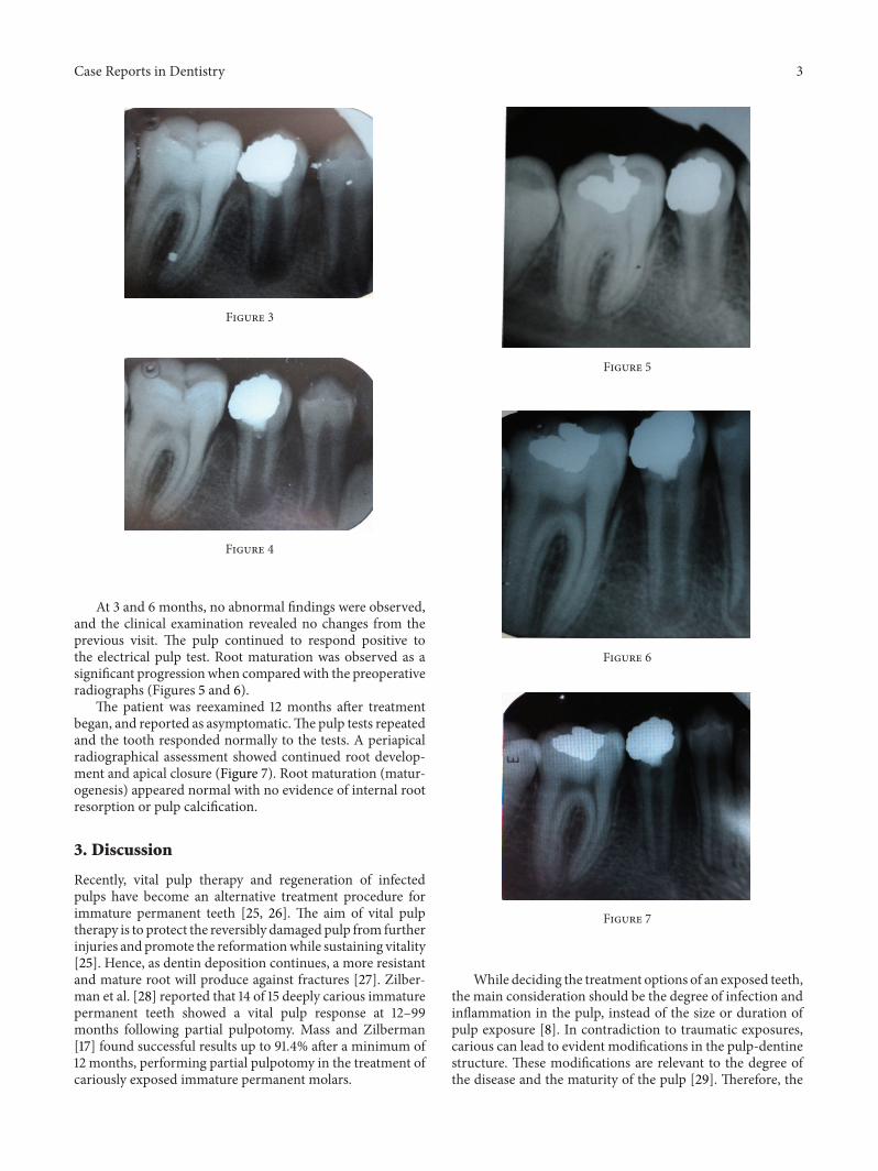
At 3 and 6 months, no abnormal findings were observed,and the clinical examination revealed no changes from theprevious visit. The pulp continued to respond positive tothe electrical pulp test. Root maturation was observed as asignificant progressionwhen comparedwith the preoperativeradiographs (Figures 5 and 6).
The patient was reexamined 12 months after treatmentbegan, and reported as asymptomatic.The pulp tests repeatedand the tooth responded normally to the tests. A periapicalradiographical assessment showed continued root develop-ment and apical closure (Figure 7). Root maturation (matur-ogenesis) appeared normal with no evidence of internal rootresorption or pulp calcification.
3. Discussion
Recently, vital pulp therapy and regeneration of infectedpulps have become an alternative treatment procedure forimmature permanent teeth [25, 26]. The aim of vital pulptherapy is to protect the reversibly damaged pulp from furtherinjuries and promote the reformationwhile sustaining vitality[25]. Hence, as dentin deposition continues, a more resistantand mature root will produce against fractures [27]. Zilber-man et al. [28] reported that 14 of 15 deeply carious immaturepermanent teeth showed a vital pulp response at 12–99months following partial pulpotomy. Mass and Zilberman[17] found successful results up to 91.4% after a minimum of12 months, performing partial pulpotomy in the treatment ofcariously exposed immature permanent molars.
Figure 5
Figure 6
Figure 7
While deciding the treatment options of an exposed teeth,the main consideration should be the degree of infection andinflammation in the pulp, instead of the size or duration ofpulp exposure [8]. In contradiction to traumatic exposures,carious can lead to evident modifications in the pulp-dentinestructure. These modifications are relevant to the degree ofthe disease and the maturity of the pulp [29]. Therefore, the

4 Case Reports in Dentistry
important concern should be the selection of the case andtreatment planning to acquire better results [30].
Although many new agents were introduced to themarket, Ca(OH)
2is still a well-accepted material to stimulate
dentin formation after a carious exposure of immature per-manent teeth [11]. In the literature,many studies reported thatpartial pulpotomy of cariously exposed immature permanentteeth with calcium hydroxide have sustained a success rateof 93 [16, 17, 28]. It is still the most accessible material anddentists can perform easily in clinics. Caliskan [2] reportedtwenty-six permanent vital molars with carious pulp expo-sures and radiographic periapical involvement were treatedusing a full coronal pulpotomy, with calcium hydroxideplaced directly on the radicular pulp tissue once bleedinghad stopped. Of these, twenty-four teeth showed resolutionof radiographic periapical involvement and dentine bridgeformation after 16–72 months. The presence of radiographicperiapical change accompanying reversible pulpitis can be asa sequel to reversible pulpal inflammation, rather than as adirect response to an infected pulp.
Clinical and radiographical evaluations at 1, 3, 6, and 12months showed pulpal vitality maintenance. Current vitalitytests are still based on neurological inducement, and this isstill a contradictory in immature teeth [12]. The radiograph-ical assessment of periapical pathology in immature teethis also questionable due to the normal radiolucency of thedeveloping root.
In spite of the fact that histological success cannot be eval-uated, clinical success can be assessed by the absence of anyclinical or radiographic signs of failure and the substantiationof the development of the root dentin and apex closure.Thus,the patient should be recalled and reexamined periodicallyagainst any failure even if absence of visible symptoms.
The optimal choice of treatment is, to maintain vitality ofan exposed pulp rather than replacing it with a root fillingmaterial. However, the technique, the type of the agent whichis used, the inflammatory status of the teeth is variable and achallenge to the dentists.
In the light of the literature and this case, for cariouslyexposed immature permanent teeth, partial pulpotomy is anoptional treatment for preserving vitality and physiologicalroot development. In this manner, as well as a vital pulp, notonly closure of the apex (apexogenesis) but also physiologicroot development (maturogenesis) occurs.
References
[1] J. Ward, “Vital pulp therapy in cariously exposed permanentteeth and its limitations,” Australian endodontic journal : thejournal of the Australian Society of Endodontology Inc, vol. 28,no. 1, pp. 29–37, 2002.
[2] M. K. Caliskan, “Pulpotomy of carious vital teeth with periapi-cal involvement,” International endodontic journal, vol. 28, no.3, pp. 172–176, 1995.
[3] I. V. Nosrat and C. A. Nosrat, “Reparative hard tissue for-mation following calcium hydroxide application after partialpulpotomy in cariously exposed pulps of permanent teeth,”International Endodontic Journal, vol. 31, no. 3, pp. 221–226,1998.
[4] A. Gesi, M. Hakeberg, J. Warfvinge, and G. Bergenholtz,“Incidence of periapical lesions and clinical symptoms afterpulpectomy - A clinical and radiographic evaluation of 1-versus 2-session treatment,” Oral Surgery, Oral Medicine, OralPathology, Oral Radiology and Endodontology, vol. 101, no. 3, pp.379–388, 2006.
[5] K. Kojima, K. Inamoto, K. Nagamatsu et al., “Success rate ofendodontic treatment of teeth with vital and nonvital pulps.a meta-analysis,” Oral Surgery, Oral Medicine, Oral Pathology,Oral Radiology, and Endodontics, vol. 97, no. 1, pp. 95–99, 2004.
[6] Y.-L. Ng, V. Mann, S. Rahbaran, J. Lewsey, and K. Gulabivala,“Outcome of primary root canal treatment: Systematic review ofthe literature - Part 2. Influence of clinical factors,” InternationalEndodontic Journal, vol. 41, no. 1, pp. 6–31, 2008.
[7] D. J. Caplan, J. Cai, G. Yin, and B. A. White, “Root canal filledversus non-root canal filled teeth: A retrospective comparisonof survival times,” Journal of Public Health Dentistry, vol. 65, no.2, pp. 90–96, 2005.
[8] C. D. Fong and M. J. Davis, “Partial pulpotomy for immaturepermanent teeth, its present and fixture,” Pediatric Dentistry,vol. 24, no. 1, pp. 29–32, 2002.
[9] P. Aguilar and P. Linsuwanont, “Vital pulp therapy in vitalpermanent teeth with cariously exposed pulp: A systematicreview,” Journal of Endodontics, vol. 37, no. 5, pp. 581–587, 2011.
[10] E. Mass, U. Zilberman, and A. B. Fuks, “Partial pulpotomy:another treatment option for cariously exposed permanentmolars,” ASDC journal of dentistry for children, vol. 62, no. 5,pp. 342–345, 1995.
[11] R. Weisleder and C. R. Benitez, “Maturogenesis: Is it a newconcept?” Journal of Endodontics, vol. 29, no. 11, pp. 776–778,2003.
[12] P. J. Waterhouse, J. H. Nunn, J. M. Whitworth, and J. V.Soames, “Primary molar pulp therapy - Histological evaluationof failure,” International Journal of Paediatric Dentistry, vol. 10,no. 4, pp. 313–321, 2000.
[13] M. Cvek, “A clinical report on partial pulpotomy and cappingwith calciumhydroxide in permanent incisorswith complicatedcrown fracture,” Journal of Endodontics, vol. 4, no. 8, pp. 232–237, 1978.
[14] J. H. Camp, E. J. Barrett, and F. Pulver, “Pediatric endodontics:endodontic treatment for the primary and young permanentdentition.,” in Pathway of the Pulp, S. B. R. Cohen, Ed., pp. 823–833, Mosby, St. Loius, Mo, USA, 8th edition, 2002.
[15] A. B. Fuks, E. Bimstein, and A. Bruchim, “Radiographic andhistologic evaluation of the effect of two concentrations offormocresol on pulpotomized primary and young permanentteeth inmonkeys,”Pediatric dentistry, vol. 5, no. 1, pp. 9–13, 1983.
[16] I. Mejare andM. Cvek, “Partial pulpotomy in young permanentteeth with deep carious lesions,” Endodontics & dental trauma-tology, vol. 9, no. 6, pp. 238–242, 1993.
[17] E. Mass and U. Zilberman, “Clinical and radiographic evalu-ation of partial pulpotomy in carious exposure of permanentmolars,” Pediatric dentistry, vol. 15, no. 4, pp. 257–259, 1993.
[18] M. Torabinejad and N. Chivian, “Clinical applications of min-eral trioxide aggregate,” Journal of Endodontics, vol. 25, no. 3, pp.197–205, 1999.
[19] J. Camilleri, F. E. Montesin, S. Papaioannou, F. McDonald, andT. R. Pitt Ford, “Biocompatibility of two commercial forms ofmineral trioxide aggregate,” International Endodontic Journal,vol. 37, no. 10, pp. 699–704, 2004.

Case Reports in Dentistry 5
[20] K. M. Barrieshi-Nusair and H. M. Hammad, “Intracoronalsealing comparison of mineral trioxide aggregate and glassionomer,” Quintessence International, vol. 36, no. 7-8, pp. 539–545, 2005.
[21] M. Tselnik, J. Craig Baumgartner, and J. Gordon Marshall,“Bacterial leakage with mineral trioxide aggregate or a resin-modified glass ionomer used as a coronal barrier,” Journal ofEndodontics, vol. 30, no. 11, pp. 782–784, 2004.
[22] M. Torabinejad, P. W. Smith, J. D. Kettering, and T. R. Pitt Ford,“Comparative investigation of marginal adaptation of mineraltrioxide aggregate and other commonly used root-end fillingmaterials,” Journal of Endodontics, vol. 21, no. 6, pp. 295–299,1995.
[23] M. Fridland and R. Rosado, “MTA solubility: A long termstudy,” Journal of Endodontics, vol. 31, no. 5, pp. 376–379, 2005.
[24] D. R. Anthony and E. S. Senia, “The use of calcium hydroxide asa temporary paste fill,” Texas dental journal, vol. 99, no. 10, pp.6–10, 1981.
[25] D. Tziafas, A. J. Smith, and H. Lesot, “Designing new treatmentstrategies in vital pulp therapy,” Journal of Dentistry, vol. 28, no.2, pp. 77–92, 2000.
[26] F. Banchs and M. Trope, “Revascularization of immaturepermanent teeth with apical periodontitis: New treatmentprotocol?” Journal of Endodontics, vol. 30, no. 4, pp. 196–200,2004.
[27] R. T. Webber, “Apexogenesis versus apexification,” Dental Clin-ics of North America, vol. 28, no. 4, pp. 669–697, 1984.
[28] U. Zilberman, E. Mass, and H. Sarnat, “Partial pulpotomy incarious permanent molars,” American journal of dentistry, vol.2, no. 4, pp. 147–150, 1989.
[29] D. Ricketts, “Management of the deep carious lesion and thevital pulp dentine complex,” British Dental Journal, vol. 191, no.11, pp. 606–610, 2001.
[30] R. Patel andN. Cohenca, “Maturogenesis of a cariously exposedimmature permanent tooth usingMTA for direct pulp capping:A case report,”Dental Traumatology, vol. 22, no. 6, pp. 328–333,2006.

Submit your manuscripts athttp://www.hindawi.com
Hindawi Publishing Corporationhttp://www.hindawi.com Volume 2014
Oral OncologyJournal of
DentistryInternational Journal of
Hindawi Publishing Corporationhttp://www.hindawi.com Volume 2014
Hindawi Publishing Corporationhttp://www.hindawi.com Volume 2014
International Journal of
Biomaterials
Hindawi Publishing Corporationhttp://www.hindawi.com Volume 2014
BioMed Research International
Hindawi Publishing Corporationhttp://www.hindawi.com Volume 2014
Case Reports in Dentistry
Hindawi Publishing Corporationhttp://www.hindawi.com Volume 2014
Oral ImplantsJournal of
Hindawi Publishing Corporationhttp://www.hindawi.com Volume 2014
Anesthesiology Research and Practice
Hindawi Publishing Corporationhttp://www.hindawi.com Volume 2014
Radiology Research and Practice
Environmental and Public Health
Journal of
Hindawi Publishing Corporationhttp://www.hindawi.com Volume 2014
The Scientific World JournalHindawi Publishing Corporation http://www.hindawi.com Volume 2014
Hindawi Publishing Corporationhttp://www.hindawi.com Volume 2014
Dental SurgeryJournal of
Drug DeliveryJournal of
Hindawi Publishing Corporationhttp://www.hindawi.com Volume 2014
Hindawi Publishing Corporationhttp://www.hindawi.com Volume 2014
Oral DiseasesJournal of
Hindawi Publishing Corporationhttp://www.hindawi.com Volume 2014
Computational and Mathematical Methods in Medicine
ScientificaHindawi Publishing Corporationhttp://www.hindawi.com Volume 2014
PainResearch and TreatmentHindawi Publishing Corporationhttp://www.hindawi.com Volume 2014
Preventive MedicineAdvances in
Hindawi Publishing Corporationhttp://www.hindawi.com Volume 2014
EndocrinologyInternational Journal of
Hindawi Publishing Corporationhttp://www.hindawi.com Volume 2014
Hindawi Publishing Corporationhttp://www.hindawi.com Volume 2014
OrthopedicsAdvances in