indiana university seminar 10.15.11 disclosures …mri/seminars/slides/10.15.11.rowley_iu_ct...
TRANSCRIPT
Radiation Dosimetry and Dose Reduction Techniques in CT
Howard A. Rowley, M.D. University of Wisconsin, Madison
Indiana University Seminar 10.15.11
Learning Objectives
Review radiation dose terminology and risks
Outline strategies to reduce CT dose
Become familiar with iterative reconstruction
Many thanks to: L. Gentry, F. Ranallo, and M. Lev
Disclosures – Howard Rowley
• Honoraria / consulting / agreements
GE Healthcare research, MR patents
Bracco contrast
Bayer contrast
Guerbet contrast
Trial consulting: Lundbeck, HL Gore, Eli Lilly, ImagePace
• Off-label use of contrast
Perfusion
• NIH 5R01 EB007021-04 (CT reconstruction)
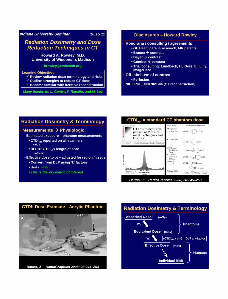
Radiation Dosimetry & Terminology
• Measurements Physiologic
–Estimated exposure – phantom measurements
CTDIvol reported on all scanners • mGy
DLP = CTDIvol x length of scan • mGy-cm
–Effective dose to pt – adjusted for region / tissue
Convert from DLP using ‘k’ factors
Units: mSv
This is the key metric of interest
Bauhs, J RadioGraphics 2008; 28:245–253
CTDIvol = standard CT phantom dose
Bauhs, J RadioGraphics 2008; 28:245–253
CTDI: Dose Estimate - Acrylic Phantom Radiation Dosimetry & Terminology
Absorbed Dose
Equivalent Dose
Effective Dose
Individual Risk
Phantoms
Humans
(mGy)
(mSv)
(mSv)
WR
WT (CTDIvol x cm) = DLP x k-factor

McCollough et al Radiology 2011; 259:311–316
Converting CTDI to Effective Dose
CTDIvol x cm = DLP
DLP x k-factor = E (mSv) = Pt Dose
Christner et al AJR 2010; 194:881–889
Radiation Risk:
Tissue Weighting Factors
Christner et al AJR 2010; 194:881–889
Organ-Specific Tissue Risk Estimation
Tissue Weighting Factors (WT)
Brain = 0.01
Christner et al AJR 2010; 194:881–889
Effective Dose Calculation by Region
DLP x k-factor = E (mSv)
Christner et al AJR 2010; 194:881–889
Effective Dose Calculation by Region
DLP x k-factor = E (mSv)
Quick Method to Estimate Dose
from DLP:
Head CT: DLP / 100
4
Body CT: DLP / 100
1.5
Radiation Risks in Perspective
• Routine Exposure mSv / year
–Environment 3-30
–Medical (population avg) 3
–Worker (allowable) 50
• Medical Exams mSv / exam
–Chest X-ray 0.1
–Head CT 1-2
–Coronary CT 9
–Barium enema 15
–Cardiac nuclear scan 41
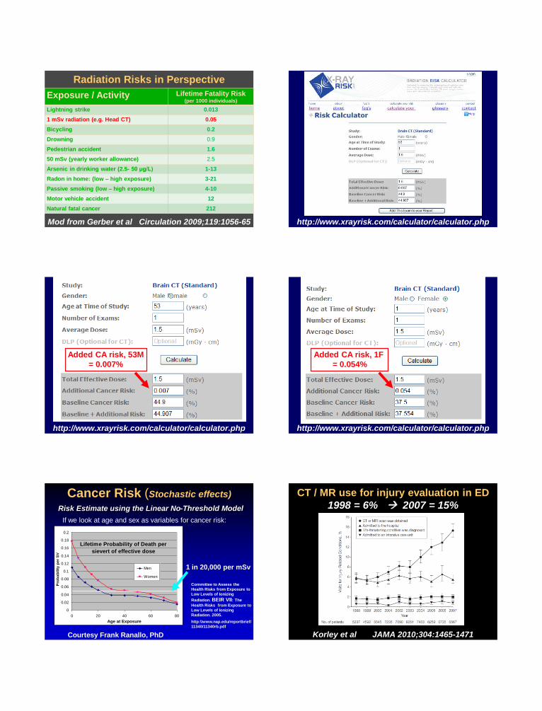
Mod from Gerber et al Circulation 2009;119:1056-65
Radiation Risks in Perspective
Exposure / Activity Lifetime Fatality Risk (per 1000 individuals)
Lightning strike 0.013
1 mSv radiation (e.g. Head CT) 0.05
Bicycling 0.2
Drowning 0.9
Pedestrian accident 1.6
50 mSv (yearly worker allowance) 2.5
Arsenic in drinking water (2.5- 50 μg/L) 1-13
Radon in home: (low – high exposure) 3-21
Passive smoking (low – high exposure) 4-10
Motor vehicle accident 12
Natural fatal cancer 212
http://www.xrayrisk.com/calculator/calculator.php
http://www.xrayrisk.com/calculator/calculator.php
Added CA risk, 53M
= 0.007%
http://www.xrayrisk.com/calculator/calculator.php
Added CA risk, 1F
= 0.054%
Courtesy Frank Ranallo, PhD
Cancer Risk (Stochastic effects) Risk Estimate using the Linear No-Threshold Model
1 in 20,000 per mSv
If we look at age and sex as variables for cancer risk:
Lifetime Probability of Death per
sievert of effective dose
0
0.02
0.04
0.06
0.08
0.1
0.12
0.14
0.16
0.18
0.2
0 20 40 60 80
Age at Exposure
Pro
ba
bilit
y p
er
SV
Men
Women
Committee to Assess the
Health Risks from Exposure to
Low Levels of Ionizing
Radiation. BEIR VII: The
Health Risks from Exposure to
Low Levels of Ionizing
Radiation. 2005.
http:\\www.nap.edu/reportbrief/
11340/11340rb.pdf
Korley et al JAMA 2010;304:1465-1471
CT / MR use for injury evaluation in ED
1998 = 6% 2007 = 15%
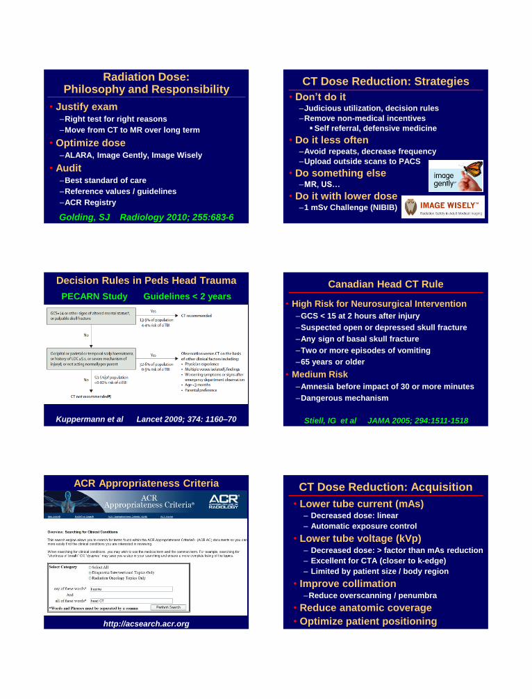
Radiation Dose: Philosophy and Responsibility
• Justify exam –Right test for right reasons
–Move from CT to MR over long term
• Optimize dose –ALARA, Image Gently, Image Wisely
• Audit –Best standard of care
–Reference values / guidelines
–ACR Registry
Golding, SJ Radiology 2010; 255:683-6
CT Dose Reduction: Strategies
• Don’t do it –Judicious utilization, decision rules
–Remove non-medical incentives
Self referral, defensive medicine
• Do it less often –Avoid repeats, decrease frequency
–Upload outside scans to PACS
• Do something else –MR, US…
• Do it with lower dose –1 mSv Challenge (NIBIB)
Kuppermann et al Lancet 2009; 374: 1160–70
Decision Rules in Peds Head Trauma
PECARN Study Guidelines < 2 years
Canadian Head CT Rule
• High Risk for Neurosurgical Intervention
–GCS < 15 at 2 hours after injury
–Suspected open or depressed skull fracture
–Any sign of basal skull fracture
–Two or more episodes of vomiting
–65 years or older
• Medium Risk
–Amnesia before impact of 30 or more minutes
–Dangerous mechanism
Stiell, IG et al JAMA 2005; 294:1511-1518
http://acsearch.acr.org
ACR Appropriateness Criteria CT Dose Reduction: Acquisition
• Lower tube current (mAs) – Decreased dose: linear
– Automatic exposure control
• Lower tube voltage (kVp) – Decreased dose: > factor than mAs reduction
– Excellent for CTA (closer to k-edge)
– Limited by patient size / body region
• Improve collimation –Reduce overscanning / penumbra
• Reduce anatomic coverage
• Optimize patient positioning
McCollough et al RadioGraphics 2006; 26:503–512
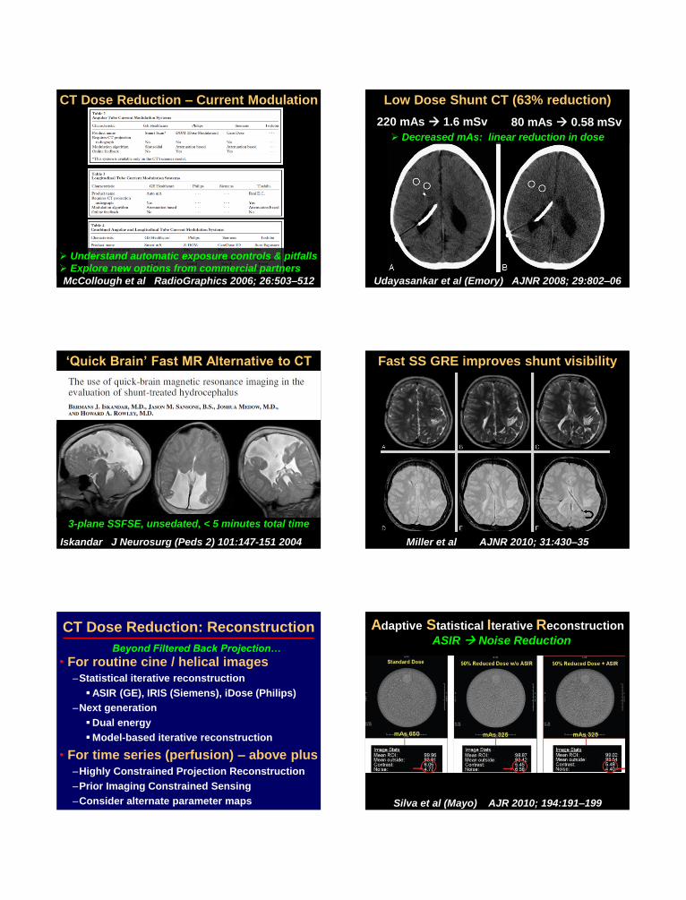
CT Dose Reduction – Current Modulation
Understand automatic exposure controls & pitfalls
Explore new options from commercial partners
Udayasankar et al (Emory) AJNR 2008; 29:802–06
Low Dose Shunt CT (63% reduction)
220 mAs 1.6 mSv 80 mAs 0.58 mSv
Decreased mAs: linear reduction in dose
Iskandar J Neurosurg (Peds 2) 101:147-151 2004
‘Quick Brain’ Fast MR Alternative to CT
3-plane SSFSE, unsedated, < 5 minutes total time
Miller et al AJNR 2010; 31:430–35
Fast SS GRE improves shunt visibility
CT Dose Reduction: Reconstruction
• For routine cine / helical images
–Statistical iterative reconstruction
ASIR (GE), IRIS (Siemens), iDose (Philips)
–Next generation
Dual energy
Model-based iterative reconstruction
• For time series (perfusion) – above plus
–Highly Constrained Projection Reconstruction
–Prior Imaging Constrained Sensing
–Consider alternate parameter maps
Beyond Filtered Back Projection…
Silva et al (Mayo) AJR 2010; 194:191–199
Adaptive Statistical Iterative Reconstruction
ASIR Noise Reduction

Adaptive Statistical Iterative Reconstruction
User choices at console mA reduction dose reduction
Noise index
% Dose reduction
mA Range: min & max
Screenshot courtesy of Frank Ranallo, PhD
Adaptive Statistical Iterative Reconstruction
Old exam – No ASIR
mA kV Pitch Rot’n
Time
Noise
Index
ASIR
%
CTDI
vol
DLP E
mSv
New 274 140 0.53 0.60 11.4 30% 42 1438 4.4
Old 201 140 0.63 1.0 11.4 0% 57 2054 6.4
New – 30% ASIR / Dose ↓
DLP*0.0031 (for head & neck) = E (mSv)
Silva et al (Mayo) AJR 2010; 194:191–199
Adaptive Statistical Iterative Reconstruction
Variable ‘blending’ of FBP with % ASIR
0.6 mSv Temporal Bone using iDose
Courtesy of Terry McGinn, Philips, and MetroHealth
Low Dose CT Reconstruction Methods
Courtesy of Saad Sirohey, PhD, and GE HealthCare
CTP Concerns: Radiation Dose
Imanishi et al Eur Radiol (2005) 15:41–46
Day 37 after 1st CTP: four CTA/CTP and two DSA exams in 2 weeks
120 kV, 100 mAs, and 50 rotations
http://www.fda.gov/MedicalDevices/Safety/AlertsandNotices/ucm185898.htm
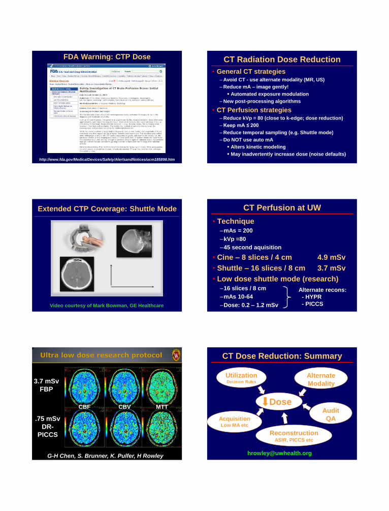
FDA Warning: CTP Dose CT Radiation Dose Reduction
• General CT strategies –Avoid CT - use alternate modality (MR, US)
–Reduce mA – image gently!
Automated exposure modulation
–New post-processing algorithms
• CT Perfusion strategies –Reduce kVp = 80 (close to k-edge; dose reduction)
–Keep mA ≤ 200
–Reduce temporal sampling (e.g. Shuttle mode)
–Do NOT use auto mA
Alters kinetic modeling
May inadvertently increase dose (noise defaults)
Extended CTP Coverage: Shuttle Mode
Video courtesy of Mark Bowman, GE Healthcare
CT Perfusion at UW
• Technique
–mAs = 200
–kVp =80
–45 second aquisition
• Cine – 8 slices / 4 cm 4.9 mSv
• Shuttle – 16 slices / 8 cm 3.7 mSv
• Low dose shuttle mode (research)
–16 slices / 8 cm
–mAs 10-64
–Dose: 0.2 – 1.2 mSv
Alternate recons:
- HYPR
- PICCS
.75 mSv
DR-
PICCS
3.7 mSv
FBP
CBF CBV MTT
G-H Chen, S. Brunner, K. Pulfer, H Rowley
CT Dose Reduction: Summary
Dose
Utilization Decision Rules
Alternate
Modality
Acquisition Low MA etc
Reconstruction ASIR, PICCS etc
Audit
QA