indian journal of - · pdf fileindian journal of 2014 : vol. 2 guest ... lucknow amit vora,...
TRANSCRIPT
EDITORS | Dr. Yash Lokhandwala � Dr. Rabin Chakraborty
INDIAN JOURNAL OF
2014 : Vol. 2
GUEST EDITOR | Dr. Sanjay Bindra � Dr. Parag Barwad


C o n t e n t s
Editorial ....................................................................................................... 2
Message from Past President..................................................................... 3
Evolution of Defibrillator Therapy ............................................................ 5
Evaluation and Management of Ventricular Bigeminy ........................... 7
Quantification of Myocardial Infarction by Cardiac Magnetic Resonance Imaging, Electrocardiography, Echocardiography ............ 12
Spectrum of Arrhythmias in Heart Failure ............................................ 19
T-wave Inversions : A Review .................................................................. 24
ECG Quiz ................................................................................................... 31
ISE Membership Form ............................................................................ 39
executive Committee ofIndIan soCIety
ofeleCtroCardIology
PRESIDENT
ajay naik, Ahmedabad
IMM. PaST PRESIDENTsB gupta, Mumbai
PRESIDENT ELECT Praveen Jain, Jhansi
VICE PRESIDENTSsK dwivedi, Lucknowamit Vora, MumbaiKK Varshney, Aligarh
HON. GENERaL-SECRETaRYashish nabar, Mumbai
TREaSURERKetan K Mehta, Mumbai
JOINT SECRETaRIESsadanand shetty, Mumbaisnehal Kothari, Mumbai
MEMBERSUlhas Pandurangi, ChennaiUday Jadhav, Navi Mumbaiaditya Kapoor, Lucknowramesh dargad, MumbaiaB Patil, Mumbains neki, AmritsarH Prabhakar, Mangaloreajit Bhagwat, Aurangabad
JOURNaL EDITORSyash lokhandwala, Mumbairabin Chakraborty, Kolkata
CO-OPTED MEMBERSPrashant JagtapOrganising SecretaryIseCon 2015 (nagpur)
SECRETaRIaTS. B. GUPTAPaST PRESIDENT
Indian Society of Electrocardiology102, Rail Mitra, Plot # 125, Sector I, Charkop, Kandivali (W), Mumbai 400067. Mobile : 0 98213 64565 • e-mail : [email protected] • www.iseindia.org

Dear Friends,
as heart failure is one of the prime killers all over the world and the prevalence is going to rise in view of higher longevity, we would be dealing with more patients of arrhythmia in heart failure in our daily practice. Understanding the complex interplay of causative factors, mediators and manifestation of these arrhythmia and its effect on the management of such patients is of utmost importance. Two arrhythmias, atrial fibrillation (af) and Ventricular arrhythmias [Premature ventricular complexes (PVC), non-sustained ventricular tachycardia (nsVt), and sustained Vt] are of prime importance and thus have been discussed in details in the current issue. as mentioned earlier the complex interplay between the mechanism of its causation and its effect on heart failure leads to a vicious cycle.
as myocardial infarction (MI) is leading cause of mortality and morbidity worldwide, apart from acute management for the patient it is important to analyze the effect of MI on the heart and to prognosticate the patient about the future. thus it is important to quantify the amount to myocardium affected in the previous MI. there are various investigational modalities available for the quantification such as ECG, Echocardiography, Cardiac Magnetic resonance imaging (CMrI), single photon electron computed tomography (sPeCt) and other radionucleotide imaging. as eCg and echocardiography are the most easily and widely available modalities we should have a thorough understanding of the various techniques of MI quantification. However, an important aspect of future prognostication such as viability assessment cannot be determined only on the basis of eCg. It require further investigation, at least stress echocardiography to demonstrate viability in the myocardium. Cardiac MrI is an excellent investigational modality which demonstrate accurately the amount to infarcted myocardium and viable myocardium. But its availability and lack to expertize to interpretation is a big hindrance for its routine use.
t wave inversion is the most common eCg abnormality. Inverted t waves are broadly classified into primary and secondary T wave changes. The spectrum varies from normal variants in children and middle-aged women to findings highly suggestive of acute ischemia. so one must be careful to avoid over-diagnosis in patients with benign t wave inversions and at the same time not miss the diagnosis in patients with malignant t wave inversions. the current review gives a detailed insight into the various caused of t wave inversion and its clinical implication in real world practice.
Ventricular bigeminy has always been a cause of concern when diagnosed on eCg. It is not a disease, but it can be a marker of possible underlying heart conditions that may increase the risk of cardiac death. In patients with structurally normal heart it is rarely of any clinical significance and the prognosis is generally excellent. Suppression of ventricular bigeminy with ant-iarrhythmic medication is not indicated routinely, unless the patient is symptomatic or at risk of tachycardia-induced cardiomyopathy owing to the very high frequency of premature ventricular contractions. the mechanism and imp0-lications have been discussed.
there is a short summary about the development of ICd and the recently launched “non-vascular” subcutaneous ICd. the eCg Quiz features as always. looking forward to mid-term IseCon 2014 in Vishakapatnam!
Parag Barwad Yash Lokhandwala faculty, dept of Cardiology, arrhythmia associates PgI, Chandigarh
Editorial

From Past President’s Desk
Dear Members,
It is our great pleasure in bringing out the 2nd issue of Indian Journal of electrocardiology of the year 2014 on the eve of Mid-term Conference of Indian society of electrocardiology at Vizag.
IseCon 2014 – the annual Conference of Indian society of electrocardiology was organized by dr. s.K. dwivedi, dr. aditya Kapoor and the team at lucknow on 8th and 9th March 2014. It was a great scientific feast! Our heartiest congratulations to dr. s.K. dwivedi, dr. aditya Kapoor and their team.
Mid-term Conference of Indian society of electrocardiology is being organized on 11th and 12th october 2014 with the great efforts of dr. C. narasimhan and dr. g.s.r. Murthy and their team at Vizag and I am sure it will be an academic bonanza.
IseCon 2015 is being organized at nagpur by dr. Uday Mahorkar, dr. Prashant Jagtap on 7th and 8th february 2015. for more details, kindly log on www.iseindia.org
Ise has initiated Pacemaker/Crt/ICd survey. the results will be published soon in one of the leading journals.
My sincere thanks to dr. yash lokhandwala, dr. sanjay Bindra, dr. Parag Barwad and the editorial team for bringing out the Ise Journal – 2014, 2nd Volume.
long live Indian society of electrocardiology
Dr. S.B. GuptaPast President Indian Society of Electrocardiology


5
Evolution of Defibrillator TherapyVivek Reddy*, Sanjay Bindra**
*research assistant, **regional Medical Center, san Jose Ca
Development of the implantable cardioverter defibrillator (ICd) was pioneered by Michel Mirowski in the late 1960s after the death of a close friend and mentor from ventricular tachyarrhythmias. His frustration with the limitations of available treatments for high risk individuals led to the idea of an implantable device that would continuously monitor the cardiac rhythm and deliver defibrillating shocks when ventricular tachyarrhythmias occurred. during the 1970s, experimental models were built and refined by Mirowski and Morton Mower, leading to the first implantation of a defibrillator in a patient, with two previous cardiac arrests in 1980s.1 since the mid 1980s experience with these devices expanded along with evidence of effectiveness of defibrillators in terminating malignant ventricular arrhythmias. By 1991, when two expert panels published independent recommendations, indications had broadened to include ventricular fibrillation or ventricular tachycardia not amenable to drug treatment.2 Because of further refinements in device technology, loss of faith in the universal effectiveness of drug treatment, and accumulating evidence from randomized clinical trials, ICds have become the treatment of choice for patients at high risk for life-threatening arrhythmia.3
numerous large-scale clinical trials have demonstrated the benefit of implantable cardioverter defibrillators (ICDs) in primary and secondary prevention of sudden cardiac death (sCd), especially in post MI and heart failure with left ventricular dysfunction.4,5
Current ICd indications have expanded to include prophylactic implantation in individuals who have a high risk of sCd (primary prevention).
ICd therapy has evolved from requiring a thoracotomy with epicardial patches, to introduction of transvenous ICds (tV-ICds) and now the subcutaneous ICd (s-ICd).
While the minituarization of the ICd to a transvenous system reduced morbidity and mortality from thoracotomy and epicardial patch electrode placement, tV-ICds may be associated with significant morbidity.6
the risks associated with transvenous ICd systems include procedural risks, inappropriate device therapy and long term complications such as lead failure.7
late infections including endocarditis, vessel occlusions, lead dislodgement, valvular dysfunctions, and intrinsic lead defects with consequent inappropriate/ineffective therapy are noted with endocardial leads. Published reports of lead failure rates of up to 20% per year for 8 year old systems.8
Moreover, lead failure and infection of systems is a complex
clinical condition, potentially leading to need for procedural revision and lead extraction with considerable risk.
therefore, strategies that simplify the ICd implant procedure with the conceptualization of a non tV ICd lead system lead to the creation of the subcutaneous ICd (s-ICd). early studies confirmed that multiple device configurations provide reliable defibrillation with lower energy than expected. Innovation with a dedicated detection algorithm and a custom-built electrode configuration for a reproducible implant of ICD in the subcutaneous space.
gold et al proved that arrhythmia detection using subcutaneous electrodes did not differ in sensitivity and specificity when compared with tV-ICd system.9
Studies have confirmed that the S-ICD system is a safe and effective alternative to conventional ICd therapy in patients at risk of death from Vt/Vf.10
The S-ICD system
The system (Boston Scientific) consists of a subcutaneous pulse generator (place in the axilla along the 6th rib) and a subcutaneous lead placed along the life side of the sternum, provided with a single high-voltage and low-impedance shock coil and 2 sensing electrodes. the s-ICd system can sense from 3 different vectors: proximal ring electrode to generator (primary vector), distal tip electrode to the pulse generator (secondary vector), and a distal tip connected to proximal ring (alternate vector).
the main advantage of the s-ICd system consists of the simplicity of implantation (and explantation, when necessary) and the potential removal of complications associated with the implantation of a transvenous lead. Clinically relevant infections are often confined to the subcutaneous phase.
Indications
s-ICd system is indicated in patients for primary or secondary prophylaxis in all patients without the need for bradycardia pacing, antitachycardia pacing or cardiac resynchronization therapy. the pediatric patient population including congenital heart disease patients (intracardiac shunts and or no access to the right ventricle), the young active patients with hypertrophic cardiomyopathy and channelopathy, and patients on hemodialysis are especially well suited for this novel therapy.
Conclusions
Progress in device therapy has allowed now for defibrillation therapy to be delivered “without touching the heart.” advances

6 Indian Journal of Electrocardiology
in leadless pacing may potentially create a novel system in the future which will allow both for intracardiac pacing and sensing, thus advancing the paradigm further.
References
1. Mirowski M, reid Pr, Mower MM, et al. termination of malignant ventricular arrhythmias with an implanted automatic defibrillator in human beings. N Engl J Med 1980;303:322–24.
2. Lehmann MH, Saksena S. Implantable cardioverter defibrillators in cardiovascular practice: report of the policy conference of the north american society of Pacing and electrophysiology. PACE 1991;14:969–79.
3. gregoratos g, Cheitlin Md, Conill a, et al. aCC/aHa guidelines for implantation of cardiac pacemakers and antiarrhythmia devices: executive summary—a report of the american College of Cardiology/american Heart association task force on Practice guidelines (Committee on Pacemaker Implantation). Circulation 1998;97:1325–35.
4. glikson M, luria d, friedman Pa, et al. are routine arrhythmia inductions necessary in patients with pectoral implantable cardioverter defibrillators? Cardiovasc Electrophysiol 2000;11:127–35.
5. luria d, rasmussen MJ, Hammill sC, friedman Pa. frequency and mode of detection of nonthoracotomy implantable defibrillator lead failure (abst). PACE 1999;22:705.
6. Curtis JP, luebbert JJ, Wang y, et al: association of physician certification and outcomes among patients receiving an implantable cardioverter-defibrillator. JAMA 2009;310:1661-1670.
7. gold Mr, Peters rW, Johnson JW, et al: Complications associated with pectoral implantation of cardioverter defibrillators. PACE 1997;28:208-211.
8. Kleemann t, Becker t, doenges K, et al: annual rate of transvenous defibrillation lead defects in implantable cardioverter-defibrillators over a period of n 10 years. Circulation 2007;115:2474-2480.
9. gold Mr, theuns da, Knight BP, et al: Head-to head comparison of arrhythmia discrimination performance of subcutaneous and transvenous ICd detection algorithms: the start study. J Cardiovasc Electrophysiol 2012;23:359-366.
10. Weiss R, Knight BP, Gold MR, et al: Safety and Efficacy of a Totally Subcutaneous Implantable-Cardioverter Defibrillator. Circulation 2013;128:944-953.

7
Evaluation and Management of Ventricular BigeminyPranil Gangurde, Aziz Kothawala, Pratap Nathanidept of Cardiology, ltMg Hospital, sion, Mumbai
Introduction
Ventricular bigeminy is a commonly encountered entity in clinical practice. these are commonly asymptomatic but can cause upsetting symptoms in some patients. their occurrence in normal hearts is usually of less clinical significance. However, there are occasions where presence of bigeminal rhythm signifies susceptibility towards more sinister arrhythmias, especially in diseased hearts. Ventricular bigeminy is triggered by multiple mechanisms and when it gives rise to malignant ventricular arrhythmias it can be cured by catheter ablation.1 occasionally, premature ventricular contraction especially of rVot origin, may be a possible cause of lV dysfunction and heart failure and focal RF ablation produces significant clinical benefits in these patients.2
Epidemiology
Sir James Mackenzie was the first to describe extrasystoles in 1890 when he noticed that chambers of the heart could beat outside of their correct order.3 an estimated prevalence of ventricular premature beats (PVC) is 1-4% in the general population.4 In a healthy population, PVCs have been detected in 1% of subjects on standard 12-lead eCg and between 40-75% of subjects with 24 to 48 hour Holter monitoring.1 their prevalence is generally age-dependent, ranging from 1% in children5 to 69% in subjects upto 75 years.6 PVC-induced cardiomyopathy may develop in a time-dependent fashion.7 In fact, niwano et al8 demonstrated gradually progressive worsening of lV function in patients with ventricular bigeminy with frequent PVCs (>1000 beats/day) as measured by the lV ejection fraction (lVef) and lV end-diastolic dimension over a follow-up period of 4 to 8 years.
Definition and Pathophysiology
Bigeminy is described as cardiac irregularity which refers to a continuous alteration of short and long cardiac cycles, corresponding to the phenomenon of pulsus bigeminus diagnosed at the bedside by palpation of radial pulse. different mechanisms such as a re-entry mechanism and parasystolic mechanism involving disturbance of impulse formation or impulse conduction or both may be involved.9 the most common variety is intermittent bigeminy due to ventricular premature systoles. In patients with atrial fibrillation (AF) in particular a beat terminating a short cycle subsequent to a long one tends to exhibit a bizarre contour and Qrs prolongation, caused by aberrant ventricular conduction. It is of considerable practical importance to distinguish between ectopic ventricular premature systoles and aberrant ventricular conduction of early supraventricular impulses. Such a distinction may sometimes be difficult since under both circumstances the resulting bizarre beat tends to be “coupled”
to a beat terminating a long cycle. Whereas the occurrence of an ectopic premature systole in the wake of a long ventricular pause can be ascribed to partial recovery of conductivity in a re-entry path, aberrant ventricular conduction occurring under such circumstances is explained by impairment of conductivity in ordinary ventricular conduction pathways caused by prolongation of the normal ventricular refractory phase concomitant with the lengthening of the cardiac cycle. Usually it is the right sided bundle-branch system which is affected by this mechanism.
the following three criteria in most instances should permit the correct interpretation of early bizarre beats during irregular ventricular beating.
1. Beats of ectopic origin tend to have a fixed coupling while the short r-r interval of aberrant ventricular conduction tends to vary in a wider range.
2. aberrant ventricular complexes almost invariably show a pattern of right bundle-branch system block, with Qrs prolonged in its terminal portion in contrast to ectopic beats which show a variety of bizarreness, with Qrs wid-ened throughout.
3. Unlike premature beats of ectopic origin, aberrant ven-tricular beats do not give rise to a “compensatory” pause, and it is the absence of such a pause which prevents con-tinuation of aberrant conduction in the form of bigeminy. aberrant ventricular conduction may, however, continue in the form of consecutive rapid beats with a similar bi-zarre contour and prolonged Qrs duration and thus imi-tate ventricular paroxysmal tachyeardia.10
Structurally normal heart
Kennedy et al and engstrom et al found no correlation between ventricular bigeminy and mortality or cardiac events in men without Cad.11,12 However, in the framingham Heart study men with frequent PVCs had a two-fold increase in the five-year mortality risk and cardiac event rate (after correction of other cardiovascular risk factors) compared to those without.14 Despite the conflicting data, in general, PVCs in healthy individuals without any structural heart disease do not usually pose any risk of sCd.
Structural heart disease
the incidence, frequency and complexity of ventricular arrhythmias are greater in patients with ventricular bigeminy with known or suspected heart disease,12,13 so exclusion of structural and coronary heart diseases is important for management.

8 Indian Journal of Electrocardiology
Post myocardial infarction
the incidence of spontaneous ventricular arrhythmias in the post-infarction period can be as high as 85% for premature ventricular complexes.14 Ventricular bigeminy itself was an independent risk factor for total cardiac death and sudden cardiac death in the first six months following the infarction.15 left ventricular ejection fraction (lVef) below 0.3 was found to be a better predictor of early mortality (less than 6 months) and the presence of ventricular arrhythmias was a better predictor of late mortality (after 6 months).16 therefore, it is reasonable to conclude that the presence of ventricular bigeminy after acute myocardial infarction is associated with an increased total mortality in some patients, particularly those with impaired left ventricular function.
Cardiomyopathy
It has been estimated that 70-95% of patients with cardiomyopathy and congestive heart failure have frequent premature ventricular contractions (which include ventricular bigeminy) and these are associated with higher mortality.17 frequent ventricular contractions in ventricular bigeminy itself can give rise to cardiomyopathy by different mechanisms as shown in above diagram (figure 1).
Exercise-induced ventricular bigeminy
Ventricular bigeminy may occur after exercise in a patients with structurally normal heart18. Ventricular bigeminy predicted cardiovascular mortality to the same degree as a positive ischaemic response, and its predictive value was independent of the presence or absence of ischaemic changes on the exercise eCg.19 the association between exercise-induced ventricular bigeminy and cardiovascular mortality is plausible, but the extent of its clinical implication in asymptomatic individuals with structurally normal hearts is as yet uncertain.
Pregnancy
Premature ventricular complexes may increase in frequency during pregnancy in otherwise healthy women. though most of these are benign & require just reassurance as treatment, sometimes antiarrhythmic drugs may be required for treatment. Most antiarrhythmic drugs are safe except for amiodarone, which has been associated with significant fetal abnormalities.20
Hypertrophic cardiomyopathy (HCM)
Ventricular bigeminy with frequent PVCs, are indicative of an
Alterations inintracellular calciumand membrane ionic
currentsAlterations in
heart ratedynamics
Tachycardia-induced
cardiomyopathy
PVC-inducedcardiomyopathy
Myocardial andperipheral vascular
autonomicdysregulation
Ventriculardyssynchrony
Increasedoxygen
consumption
Hemodynamicimpairment
Figure 1 : Mechanisms of ventricular bigeminy leading to cardiomyopathy
9
increased risk of sustained ventricular arrhythmia. It however has a low positive and relatively high negative predictive value for sudden death despite its frequent occurrence in HCM patients. In a 24-hour Holter monitoring study, 88% of patients with HCM had premature ventricular contractions, in which 12% had frequent premature ventricular complexes.21 generally treatment is warranted only in symptomatic patients.
Congenital heart disease
a meta-analysis of 39 studies, including 4,627 patients with corrected congenital heart disease, showed that the combination of ventricular dysfunction and complex PVCs correlates with late sCd22. However, in the absence of ventricular dysfunction or symptoms, isolated PVCs have minimal prognostic significance in these individuals. antiarrhythmic therapy is not indicated for asymptomatic patients with isolated PVCs.
Athletes
athletes without structural heart disease who have premature ventricular complexes (ventricular bigeminy) at rest which get suppressed during exercise testing, can participate in all competitive sports. should the premature ventricular complexes increase in frequency during exercise or exercise testing to the extent that they produce symptoms of impaired consciousness, significant fatigue, or dyspnea, the athlete can participate in class Ia competitive sports such as golf, billiards, bowling, cricket, curling and riflery only. Athletes with structural heart disease who are in high-risk groups and have premature ventricular complexes (with or without treatment) can participate in class Ia competitive sports only.23
Approach to patient with ventricular bigeminy
a detailed history helps to identify high-risk patients requiring further risk stratification. According to symptoms and severity of underlying heart disease, in addition to the clinical presentation the prognosis and management are individualized. Physical examination is often unrevealing in such patients who present with ventricular bigeminy. the main focus of clinical examination is toward evaluating underlying structural heart disease.
a standard 12-lead resting eCg with a rhythm strip helps to localize the origin of ventricular bigeminy and to identify the related important electrical abnormalities such as long or short Qt syndrome, Brugada syndrome, arrhythmogenic right ventricular dysplasia, ischemic heart disease and electrolyte disturbances. In addition, Qrs duration and repolarization abnormalities are both independent predictors of sCd. In some studies, st segment depression or t-wave abnormalities are associated with up to four times increased risk of cardiovascular death and sCd.24
Blood sampling is important to diagnose electrolytes disturbances such as hypokalaemia and hypomagnesaemia. Holter ambulatory monitoring is useful if the symptoms are suggestive and 12-lead eCg is unrevealing. While a 24- to
48-hour continuous Holter recording is appropriate whenever the arrhythmia is known or suspected to occur at least once a day. echocardiography should be performed in patients with suspected structural heart disease and in the subset of patients at high risk for development of serious ventricular arrhythmias or sCd, such as acute myocardial infarction survivors. exercise stress test may be considered in the investigation of ventricular bigeminy in middle-aged or older patients without other evidence of Cad25. exercise-induced ventricular ectopic increases mortality at 12 months by three-fold relative to patients with PVCs only at rest26. However, exercise-induced PVCs in apparently normal individuals should not be used to dictate therapy unless associated with documented ischaemia or sustained Vt. eP testing is probably useful in the evaluation of patients with remote myocardial infarction and symptoms suggestive of ventricular tachyarrhythmia, such as palpitations, presyncope and syncope.
Treatment
Most patients are asymptomatic, and reassurance is the only therapy required if there is no evidence of structural heart disease. for symptomatic patients, avoidance of precipitating factors such as smoking, excessive alcohol and caffeine intake may be helpful. of greater importance is the exclusion of underlying structural heart disease or other conditions that may precipitate bigeminy, such as electrolyte imbalance or drug toxicity. after correctable factors have been addressed, medications such as beta-blockers can be used in the setting of a hyperadrenergic state or myocardial ischaemia if there is a need to suppress the PVCs. lignocaine may be used during the peri-infarct period. exercise-induced PVCs should be treated if there is documented ischaemia or sustained Vt. suppression of asymptomatic PVCs is no longer considered a therapeutic aim for prevention of death in post-infarction or cardiomyopathy patients. If an antiarrhythmic is indicated, beta-blockers are the first-line for suppression of symptomatic PVCs. they have been conclusively demonstrated to reduce mortality in post-infarction and heart failure patients, and thus should be part of the standard therapy. the role of amiodarone as a second-line antiarrhythmic in this setting is supported by findings in the Basel Antiarrhythmic Study of Infarct survival, which suggested that amiodarone (i.e. 200 mg/day) in patients with persisting asymptomatic complex arrhythmias after myocardial infarction decreases mortality in the first year after myocardial infarction27. a meta-analysis involving 6,500 post-myocardial infarction and heart failure patients with a median frequency of PVCs at 18 per hour demonstrated that amiodarone results in overall reduction of 13% in total mortality.28 radiofrequency ablation is now a well-recognised, non-pharmacological technique for the elimination of frequent symptomatic ventricular ectopic beats when pharmacological treatment has failed or is not preferred. the 2006 aCC/aHa/esC guidelines gave this a Class IIa indication for patients who are otherwise at low risk for sCd and who have frequent symptomatic, predominantly monomorphic PVCs that are drug resistant, or for those who are drug intolerant or do not

10 Indian Journal of Electrocardiology
wish to have long-term drug therapy. In addition, ablation of asymptomatic PVCs may also be considered when the PVCs are very frequent, so as to avoid or treat tachycardia-induced cardiomyopathy (Class IIb).25
Conclusion
Ventricular bigeminy rhythm is not a disease, but it can be a marker of possible underlying heart conditions that may increase the risk of cardiac death. However, it is important to know that bigeminal rhythm is common in people with no structural heart disease in which, the prognosis is generally excellent. suppression of ventricular bigeminy with antiarrhythmic medication is not indicated routinely, unless the patient is symptomatic or at risk of tachycardia-induced cardiomyopathy owing to the very high frequency of premature ventricular complexes. Where pharmacological therapy has failed, there is now the option of radiofrequency ablation for treatment of symptomatic patients. the eCg is a simple yet useful tool to improve risk assessment, especially in those with known cardiovascular disease.
Case 1
54 year old woman with a history of sudden onset fast regular palpitations with syncope since 20 years. she was diagnosed as a case of arrythmogenic right ventricular cardiomyopathy (arVC) with left ventricular involvement for which she
underwent eP study. on her routine followup now, her eCg (figure 2) showed bigeminal rhythm with heart rate of 68 per minute, left axis deviation, ventricular premature beats originating from right ventricular outflow tract. She was evaluated with cardiac MrI which showed severe global hypokinesia and biventricular dysfunction. she also underwent ICd implantation.
Case 2
12 year old boy, 2nd by birth order diagnosed as case of congenital cyanotic heart disease (corrected - transposition of great arteries) presented with history of multiple episodes of syncope. there was intermittent complete heart block (figure 3) with juctional escape and multiple ventricular premature complexes. electrocardiogram looks more like bigeminy but there is no sinus beat so it cannot be a ventricular bigeminy. Patient was advised surgical repair and permanent pacemaker implantation.
Case 3
34 year old woman with systemic hypertension since last 2 years had come for regular check-up. she was absolutely asymptomatic. Her eCg (figure 4) shows ventricular bigeminy, the premature ventricular complexes (PVC) having a left bundle branch block morphology and inferior axis, suggesting that they originate from the right ventricular outflow tract (RVOT). Her echocardiogram was normal.
References
1. tg andré ng treating patients with ventricular ectopic beats. Heart 2006;92:1707-1712.
2. takemoto M, yoshimura H, ohba y, Matsumoto y, yamamoto U, Mohri M, et al. radiofrequency catheter ablation of premature ventricular complexes from right ventricular outflow tract improves left ventricular dilatation and clinical status in patients without structural heart disease. J Am Coll Cardiol 2005;45:1259-65
3. royal College of general Practitioners. available at: www.rcgp.org.uk /about_us/history_heritage__ archives/archives/personal_papers/sir_james_mackenzie_collection/mackenzie_biography.aspx.
4. Kennedy Hl, Whitlock Ja, sprague MK, Kennedy lJ, Buckingham
Figure 2 : eCg of case no.1
Figure 3 : eCg of case no.2
Figure 4 : eCg of case no.3

11
ta, goldberg rJ. long-term follow-up of asymptomatic healthy subjects with frequent and complex ventricular ectopy. N Engl J Med 1985;312:193–197.
5. southall dP, Johnston f, shinebourne ea, Johnston Pg. 24-hour electrocardiographic study of heart rate and rhythm patterns in population of healthy children. Br Heart J 1981;45:281–291.
6. Camm aJ, evans Ke, Ward de, Martin a. the rhythm of the heart in elderly subjects. Am Heart J 1980;99:598–603.
7. yarlagadda rK, Iwai s, stein KM, Markowitz sM, shah BK, Cheung JW, tan V, lerman BB, Mittal s. reversal of cardiomyopathy in patients with repetitive monomorphic ventricular ectopy originating from the right ventricular outflow tract. Circulation 2005;112:1092–1097.
8. niwano s, Wakisaka y, niwano H, fukaya H, Kurokawa s, Kiryu M, Hatakeyama Y, Izumi T. Prognostic significance of frequent premature ventricular contractions originating from the ventricular outflow tract in patients with normal left ventricular function. Heart 2009;95:1230–1237
9. langendorf r, Pick a, Winternitz M: Mechanisms of intermittent ventricular bigeminy. I. appearance of ectopic beats dependent upon length of the ventricular cycle, the “rule of Bigeminy. Circulation 1955;11:422
10. Gouaux J. L, Ashman R.: Auricular fibrillation with aberration simulating ventricular paroxysmal tachycardia. Am Heart J 1947;34:366.
11. Kennedy Hl, Whitlock Ja, sprague MK, et al. long-term follow-up of asymptomatic healthy subjects with frequent and complex ventricular ectopy. N Engl J Med 1985;312:193-7.
12. engström g, Hedblad B, Janzon l, Juul-Möller s. Ventricular arrhythmias during 24-h ambulatory eCg recording: incidence, risk factors and prognosis in men with and without a history of cardiovascular disease. J Intern Med 1999;246:363-72.
13. Bikkina M, larson Mg, levy d. Prognostic implications of asymptomatic ventricular arrhythmias: the framingham Heart study. Ann Intern Med 1992;117:990-6.
14. santini M, ricci CP. Controversies in the Prevention of sudden death. J Clin Basic Cardiol 2001;4:275-8.
15. Kostis JB, Byington r, friedman lM, goldstein s, furberg C. Prognostic significance of ventricular ectopic activity in survivors of acute myocardial infarction. J Am Coll Cardiol 1987;10:231-42.
16. Bigger Jt Jr, fleiss Jl, Kleiger r, Miller JP, rolnitzky lM. the relationships among ventricular arrhythmias, left ventricular dysfunction, and mortality in the 2 years after myocardial infarction. Circulation 1984;69:250-8.
17. Podrid PJ, fogel rI, fuchs tt. Ventricular arrhythmia in congestive heart failure. Am J Cardiol 1992;69:82g-95g; discussion 95 g-96 g.
18. raZZaK Ma. Bigeminy on exertion. Circulation 1963;28:32-4.
19. Jouven X, Zureik M, desnos M, Courbon d, ducimetière P. long-term outcome in asymptomatic men with exercise-induced premature ventricular depolarizations. N Engl J Med 2000; 343:826-33.
20. omar ar, lee lC, seow sC, teo sg, Poh KK. Managing ventricular ectopics: are ventricular ectopic beats just an annoyance? Singapore Med J 2011;52:707.
21. adabag as, Casey sa, Kuskowski Ma, Zenovich ag, Maron BJ. Spectrum and prognostic significance of arrhythmias on ambulatory Holter electrocardiogram in hypertrophic cardiomyopathy. J Am Coll Cardiol 2005;45:697-704.
22. garson a Jr. Ventricular arrhythmias after repair of congenital heart disease: who needs treatment? Cardiol Young 1991;1:177-81
23. Zipes dP, ackerman MJ, estes na 3rd, et al. task force 7: arrhythmias. J Am Coll Cardiol 2005;45:1354-63
24. Kors Ja, de Bruyne MC, Hoes aW, van Herpen g, et al. t axis as an indicator of risk of cardiac events in elderly people. Lancet 1998;352:601-5.
25. european Heart rhythm association, Heart rhythm society, Zipes dP, et al. aCC/aHa/esC 2006 guidelines for management of patients with ventricular arrhythmias and the prevention of sudden cardiac death: a report of the american College of Cardiology/american Heart association task force and the european society of Cardiology Committee for Practice guidelines (Writing Committee to develop guidelines for Management of Patients With Ventricular arrhythmias and the Prevention of sudden Cardiac death). J Am Coll Cardiol 2006;48:e247-346.
26. Podrid PJ, graboys tB. exercise stress testing in the management of cardiac rhythm disorders. Med Clin North Am 1984;68:1139-52
27. Burkart F, Pfisterer M, Kiowski W, Follath F, Burckhardt D. Effect of antiarrhythmic therapy on mortality in survivors of myocardial infarction with asymptomatic complex ventricular arrhythmias: Basel antiarrhythmic study of Infarct survival (BasIs). J Am Coll Cardiol 1990;16:1711-8.
28. effect of prophylactic amiodarone on mortality after acute myocardial infarction and in congestive heart failure: meta-analysis of individual data from 6500 patients in randomised trials. amiodarone trials Meta-analysis Investigators. Lancet 1997;350:1417-24.

12 Indian Journal of Electrocardiology
Quantification of Myocardial Infarction by Cardiac Magnetic Resonance Imaging, Electrocardiography, EchocardiographyAshar Khan, Amot Kinare, Alpa Bharati*, Ajay Mahajan*departements of Cardiology and radiology, ltMg Hospital, sion, Mumbai
Introduction
While developed nations have been able to reduce the mortality rates with Cad, it has increased by about 300% in India and the trend is expected to worsen over the coming years.1 Patients with acute myocardial infarction in India are more likely to be younger, have considerable delay between onset of symptoms to presentation in hospital and are more likely to be fatal as compared with their western counterparts.2 among the many demographic, clinical and instrumental variables that influence the short- and long-term prognosis of acute MI, the extent of injury and left ventricular function has the maximum clinical relevance.3,4 Various studies have confirmed that the size of the infarct and the extent of LV damage are the main determinant of survival in patients of aMI.5-7
geltman et al5 in their study found larger myocardial necrosis in anterior than in non-anterior MI patients, suggesting that MI size but not its location was an independent predictor of post-infarction prognosis. subsequently studies quantifying infarct size by post-contrast CMrI reiterated the fact that the size of the infarct irrespective of its location is an independent determinant of lV remodelling and dysfunction.6,7 Mauri et al8 used the sum of number of leads with st elevations as a marker of infarct size. They classified Myocardial Infarction into 4 groups as small (a), modest (B), large (C) and extensive (d); group a (small Infarct) having st segment elevations in 2-3 leads as compared with ≥ 8 leads for Group D (Extensive Infarct). at 30 days, according to the extent of myocardial injury, the relative risk (RR) of death was significantly higher for groups B, C and d, compared with group a. even at 10 years, the survival rate was found to be related to the extent of myocardial injury, as evaluated by st-segment elevation on the admission eCg
the extent of injury can be measured by different modalities
including eCg, echocardiography, single Photon emission Computed tomography scan (sPeCt scan) and CMrI.
Cardiac Magnetic Resonance Imaging (CMRI) for Infarct Quantification
over the past decade, CMrI has developed into a clinical tool with excellent spatial and temporal resolution, unrestricted tomographic fields and no exposure to radiation. It has revolutionized the assessment of cardiac pathologies and dysfunction in a living heart and provides insight to the ongoing disease process. It is a new age, cutting edge technology with a huge potential to benefit the physician and the patients.9 CMr with late gadolinium enhancement (lge) offers currently the most precise and accurate non invasive method to quantify infarct size and morphology of Myocardial Infarction.10
the basic principle involved in the quantifying infarct size by late gadolinium enhancement (lge) by CMr is inversion-recovery imaging after 10-15 minutes delay after intravenous administration of contrast. the mechanism of lge in infarcted myocardium is most likely due to the inability of the gadolinium chelates to cross the intact cell membranes of the normal myocardium. normal myocardium has myocytes which are densely packed and about 70-80% of tissue volume is intracellular. as a result of this compact arrangement of intact myocardium, the distribution volume of gadolinium is small and tissue concentration is low. However in patients with aMI, there is membrane rupture allowing gadolinium to diffuse inside the cell resulting in increased gadolinium concentration and consequent signal enhancement. as a result of this, the normal myocardium appears nulled or black while the infarcted myocardium appears bright or enhanced.11
the total myocardial mass and volume are calculated on cine images as per standard CMr protocols described.12 the size of the infarct is quantified by manually delineating the region of interest around the infarcted tissue. a number of other
Abstract
With the ever increasing burden of coronary artery disease, newer tools for risk stratification and prognostication are needed. Cardiac magnetic resonance imaging (CMrI) has been established as the most accurate modality to diagnose and quantify infarct/scar size in post myocardial infarction (MI) patients. However its restricted availability and cost tend to preclude its routine clinical use. twelve lead electrocardiography (eCg) and echocardiography are both time tested, widely available, inexpensive tools to evaluate patients with acute myocardial infarction. Many scoring systems had been developed to assess infarct size in MI with eCg to help prognosticate patients. the selvester score was widely validated in normal as well as patients with MI in the 1980s. the new scoring system upgraded for eCg confounders in 2009 has led to renewed interest in eCg to correctly identify and quantify infarct size/scar in ischemic patients. determining wall motion scoring index by echocardiography is semi-quantitiative method of determining systolic dysfunction in patients with MI. tissue doppler imaging (tdI) and speckle tracking are novel modalities to detect infarct size by echocardiography.

13
Figure 1 : A CMR protocol is summarized in this figure taking into account the pathophysiological substrates underlying different MRI findings. SSFP indicates steady-state free precession; IR GRE, inversion recovery gradient echo; LV, left ventricle; rV, right ventricle; aar, area of myocardium at risk.
techniques have also been described to quantify and delineate the size of the infarcts and scars. these include the 2 sd, 3 sd, 4 sd, 5 sd, 6 sd and full width at half maximum (fWHM) techniques.11 automated easy to use commercial softwares and computer algorithms are available which give the exact size of the infarcted myocardium.
studies have validated the diagnostic utility of CMrI for regional wall motion abnormality.10 Infarct size or Infarct transmurality by lge co-relates with markers of infarct size such as creatine kinase, time to treatment and incomplete st-segment elevation.13 ricciardi14 et al found CMr highly sensitive in detecting infarcted tissue down to a few grams in target myocardial region of distal embolization during percutaneous coronary interventions in patients with coronary artery disease. Various clinical trials and research work now conclusively prove that CMrI is more accurate than nuclear studies, especially in detection of subendocardial infarction.15-17
ECG Quantification of Infarct Size
the understanding and application of eCg emerged rapidly from the second half of the 19th century when augustus Wallers, first recorded the human ECG to present times where it is the most commonly used bioelectric signal and cardiac diagnostic test.18 during the 1970s and 1980s many attempts were to develop the utility of ECG for risk stratification. these included use of both single eCg abnormalities as well as complex multivariate scoring systems. the scoring systems included the Minnesota Code,19 Cardiac Injury Index score.20,21 aldrich score22,23 and the selvester scoring system.24-26 The scientific basis for these scoring system were developed on the crux of earlier studies which showed degree of eCg changes to correlate with the extent of myocardial damage.27-28 Wilson et al27 in 1944 provided the experimental and clinical base for the use of precordial eCg and that there existed a close relationship between potential variation of precordial electrodes and the potential variation of the underlying ventricular surface. the summation of st segment elevations (∑ST) was used as an index of extent of myocardial
Technique & Sequences Pathophysiologicalpoint of View
MRI analysis
Rest Cine Imaging: multisliceSSFP cine short axisview
T2-weighted spin. echoimages: short axis view
Rest First Pass Perfusion
Early gadolinium enhancement
Lage gadoliniumenhancement (LGE)
Myocardial blood flow
Microvascular function
Myocardial necrosis;persistentmicrovascular damage
Hyperenhancement:myocardium at risk;
Hypoenhancement core:Myocardial hemorrhage
Age of infarction:
Area of myocardium At Risk (AAR %):
Presence, area of myocardial hemorrhage (%)
Positive first pass in chronic infarction (seethickness)
Thrombus detection
Microvascular damage (presence and area)
Infarct size: presence, transmurality: size (gr, %)
Myocardium salvaged: area at risk minus LGE
Persistent microvasclar damage: presence, n*segments: area (%)
Microvascular obstruction (presence, segmentlocation; size)
Contractile function;morfologicalassessment
Global evaluation: LV & RV volumes, mass,stroke volumes, ejection fraction;
Regional evaluation: wall thickness, wallmotion score index
(Slice thickness 6-10mm, gap0-4 mm;spatial resolution: 1-2 mm x 1-2 mm inplane, temporal resolution: <45 ms)
(Slice thickness 8-10 mm)
(3 short axis)
(2 min post-contrast T1-weightedIR GRE)
10-15 min post-contrast T1-weighted IR GRESlice thickness 5-8 mm, gap 0-4 mm; spatialresolution; 1.4-1.8x1.4-18. mm2

14 Indian Journal of Electrocardiology
Figure 2 : Manual delination of the infarcted area for quantification of infarct size
injury in studies and was found to have a direct association with the Killip class as well as the degree of elevation of cardiac enzymes.28,29
The Selvester QRS score was first developed in 1972 as a method for estimating the total percentage of the infarcted lV by using a composite scoring system.24-26 the original scoring system was derived based computer simulations that suggested an orderly and predictable sequence of changes in the Qrs complex associated with infarcts of various locations and sizes. through pilot studies in patients with localized wall motion abnormalities seen by ventriculography, selvester et al refined the results of these computer simulations to produce both qualitative and quantitative criteria for determining infarct size.
the original score consisted of 57 criteria from 10 of 12 eCg leads summing up to 32 points each point is physiologically equivalent to the necrosis of 3% of the left ventricle, thus providing the estimation of the total injured area by the aMI. The Selvester score was modified by Wagner et al30 in 1982 leading to composite score of 37 criteria with 29 points (table 1).
Each criterion in the modified score exhibited at least 95% specificity and the total 29-point scoring system achieved 98% specificity when a score of more than 2 points was required for identification of infarction. The specificity of the Selvester method has been established in normal subjects, and its ability to detect and estimate the anatomically determined sizes of prior infarctions has been documented.31,32
Because prognosis in aMI is determined in large part by the degree of ventricular dysfunction and the extent of myocardial ischemia, the relation of the Qrs score to survival may be explained largely by its relation to left ventricular function.33
Palmeri et al.34 demonstrated a good correlation (I = 0.88) between the Qrs score and left ventricular ejection fraction by radionuclide imaging after aMI. In a recent study of patients undergoing Primary angioplasty in MI, selvester Qrs score was found to be better than the level of cardiac enzymes in predicting the extent of myocardial infarction and the left ventricular ejection fraction.35 similarly infarct size as estimated by Qrs scoring at the time of discharge is an independent and prognostically relevant metric in
contemporary steMI patients undergoing primary PCI.36 In a recent study comparing infarct size by eCg parameters and CMr, it was found that selvester score increased stepwise in relation to global lV infarct size.37
The Selvester ECG scoring system though very specific, could not be applied to all patients with aMI. Patients with pre-existing conduction abnormalities such as bundle branch blocks and pacemakers are not candidates for scoring. these limitations made Qrs scoring impractical for ICd/Crt patients’ evaluation because greater than 50% of potential ICd patients and nearly all Crt patients have eCg confounders.38 similarly in patient with past history of aMI, it may not be completely reflective of infarct size.
to overcome the limitations of the scoring system and applicability in patients with eCg confounders, strauss
Table 1 : Modified Selvester Scoring
Lead Maximum Lead Points
Criteria Points
I 2 Q≥30 ms R/Q ≤1
11
II 2 Q≥40 ms Q≥30 ms
21
aVl 2 Q≥30 msR/Q ≤1
11
avf 5 Q≥50 msQ≥40 msQ≥30 msR/Q ≤1R/Q ≤2
32121
V1 4 any Qr>50 msr>40 msr/s > 1
1211
V2 4 Any Q or R≤20 msR≥60 msR≥50 msR/S≥1.5
1211
V3 1 Any Q or R ≤ 20 msV4 3 Q>20 ms
r/Q or r/s<0.5r/Q or r/s<1
121
V5 3 Q>30 msr/Q or r/s <1r/Q or r/s <2
121
V6 3 Q>30 msr/Q or r/s<1r/Q or r/s<3
121

15
et al38,39 further modified the QRS Score in 2009 for use in the presence of bundle branch blocks and left ventricular hypertrophy. In their study, the modified QRS score for eCg confounders was shown to be able to identify and quantify myocardial scar in comparison to contrast-enhanced magnetic resonance imaging in patients with ischemic cardiomyopathy. They found that QRS scores (modified for each ECG confounder) correctly identified and quantified scar in ischemic patients when compared with the reference standard of cardiac magnetic resonance using late-gadolinium enhancement. Higher Qrs-estimated scar size is associated with increased arrhythmogenesis. another study found that QRS-scoring is unique in that it directly identifies and quantifies the myocardial substrate (infarct/scar) that precipitates and supports re-entrant ventricular arrhythmias.40
Echocardiography in quantification of Myocardial Infarc-tion
echocardiography remains the most commonly used initial imaging modalities in cases of aMI. In 1989, the american society of echocardiography recommended the Wall Motion
scoring Index (WMsI) as a semiquantitive method to assess regional systolic function in ischemic heart disease.41 for the purpose of calculation the WMSI, the left ventricle is artificially divided into different segments. the currently recommended segmentation is a 17-segment model as described in the figure 3.
on the basis of the wall motion analysis, the WMsI is calculated as
(Sum of Wall Motion Score/ Number of segments visual-ised)
a normal left ventricle has a WMsI of 1 since each of the 17 segments receives a wall motion score of 1. the larger the infarct the higher the WMsI since the wall motion abnormalities would be more severe.
In a study comparing estimation of myocardial infarct size by WMsI and technetium99m sestimibi scan,42 it was found that myocardial perfusion defect and wall motion abnormalities correlated fairly well in patients with acute myocardial infarction during the acute phase. Patients with a WMsI more than 1.7 were found to have a perfusion defect of more than 20%. the correlation was found to be better for anterior than inferior or lateral infarcts of smaller size. analysis of regional systolic function requires good endocardial border definition. In patients with suboptimal acoustic windows, contrast administration improves image quality through improvement of endocardial border definition and enhances detection of regional systolic function abnormalities.43
although assessment of regional systolic function is commonly based on visual analysis of myocardial thickening, more recent techniques like tissue doppler Imaging and two-dimensional speckle tracking echocardiography allow quantitative evaluation of regional systolic function.44 studies have demonstrated that assessment of regional and global strain by these novel methods correlate with size and transmural extent of myocardial infarction as determined by contrast-enhanced MrI. the global strain parameter is a valuable predictor of the total extent of myocardial infarction and may therefore be an important clinical tool for risk stratification in the acute phase of myocardial infarction.45,46
Case 1
A 42 year old man, non-hypertensive, presented with com-plaints of rest angina and diaphoresis of 4 hours duration.
He has a total selvester score of 9, which indicates an infarct size of approximately 27% of the left ventricle.
Echocardiograhapic Measurement of WMSI in the same patient
WMSI = 31/17 = 1.8
CMR Quantification of Infarct Size in the same patient
Figure 3 : the left ventricle is divided into 6 walls: anterior, anteroseptal, anterolateral, inferior, inferoseptal and posterolateral. each wall is further divided into a basal, mid and apical segment and the apical cap represents the 17th segment. numerical score is allotted to each segment on the basis of visual assessment of the contractile function of each segment as follows: Normal (≥ 40% thickening in systole) = 1, Hypokinesis (10-40% thickening in systole) = 2, severe hypokinesis (≤10% thickening in systole) = 3, Dyskinesis (paradoxical systolic motion) =4, aneurysm (diastolic deformation) = 5
anterior
anterolateral
anteroseptal
Basal
Apex
Apex
inferoseptal
inferior
inferolateral
HorizontalLong Axis (HLA)
(4 Chamber)
VerticalLong Axis (HLA)
(2 Chamber)
12
3
456
4
anterior
anterior
anterolateral
lateral
anteroseptal
Mid-Cavity
Apical
Short Axis (SA)
inferoseptal
septal
inferior
inferior
inferolateral
7
12
13
14
1516
17
17
9
8
105
11
16 Indian Journal of Electrocardiology
(Create): a prospective analysis of registry data. The Lancet 2008;371:1435-42.
3. Mauri f, franzosi Mg, Maggioni aP, santoro e, santoro l. Clinical value of 12-lead electrocardiography to predict the long-term prognosis of gissi-1 patients. J Am Coll Cardiol 2002;39:1594–1600.
4. Minicucci Mf, farah e, fusco dr, Cogni al, azevedo Ps, okoshi K, Zanati sg, Matsubara BB, Paiva sa, Zornoff la. Infarct size as predictor of systolic functional recovery after myocardial infarction. Arq Bras Cardiol 2014;102:549-56.
5. geltman eM, ehsani aa, Campbell MK, schechtman K, roberts r, Sobel BE. The influence of location and extent of myocardial infarction on long-term ventricular dysrhythmia and mortality. Circulation 1979;60:805-814.
6. orn s, Manhenke C, anand Is, squire I, nagel e, edvardsen t, dickstein K. effect of left ventricular scar size, location, and transmurality on left ventricular remodeling with healed myocardial infarction. Am J Cardiol 2007;99:1109-1114.
7. Masci Pg, ganame J, francone M, desmet W, lorenzoni V, Iacucci I, et al. relationship between location and size of myocardial infarction and their reciprocal influences on post-infarction left ventricular remodelling. Eur Heart J 2011;32:1640-1648.
8. Mauri f, gasparin M, Barbonaglia l, santoro e, franzosi Mg, tognoni G, Rovelli F. Prognostic significance of the extent of myocardial injury in acute myocardial infarction treated by streptokinase (the gIssI trial). The American journal of Cardiology 1989;63:1291-1295
9. Bharati, a H, Merchant sa. Cutting edge 3 tesla Cardiac MrI: How can it Benefit to Patients and Physicians? http: //www.apiindia.org/medicine update 2013/chap17
10. Kim rJ, shahdJ. fundamental concepts in myocardial viability assessment revisited: When knowing how much is ‘alive’ is not enough? Heart 2004;90:137.
11. flett as, Hasleton J, Cook C, Hausenloy d, Quarta g, ariti C, et al. Evaluation of techniques for the quantification of myocardial scar of differing etiology using cardiac magnetic resonance. JACC: Cardiovascular Imaging 2011;4:150-6.
12. Kramer CM, Barkhausen J, flamm sd, Kim rJ, nagel, e. standardized cardiovascular magnetic resonance (CMr) protocols 2013 update. J Cardiovasc Magn Reson 2013;15:91.
13. yang Q, li K, liu X et al: Contrast-enhanced whole-heart coronary magnetic resonance angiography at 3.0 t: a comparative study with X-ray angiography in a single centre. J Am Coll Cardiol 200954;69.
14. ricciardi MJ, Wu e, davidson CJ, et al. Visualization of discrete microinfarction after percutaneous coronary intervention associated with mild creatine kinase-MB elevation. Circulation 2001;103:2780-3.
15. Wagner a, Mahrholdt H, Holly ta, et al. Contrast-enhanced MrI and routine single photon emission computed tomography (sPeCt) perfusion imaging for detection of subendocardial myocardial infarcts: an imaging study. Lancet 2003;361:374-9.
IV1 V4
V5
V6
V2
V3
II
aVL
aVF
score 0 score 1
score 1
score 2
score 2
score 2
score 1
score 0
score 0
score 0
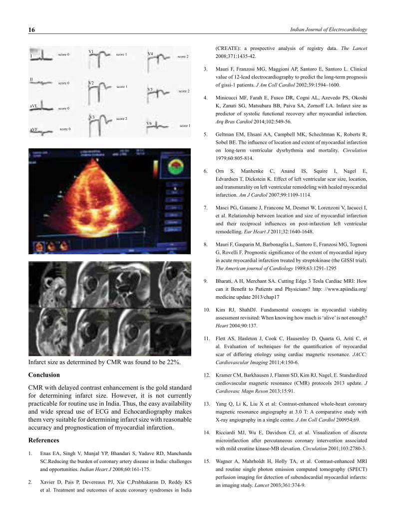
Infarct size as determined by CMr was found to be 22%.
Conclusion
CMr with delayed contrast enhancement is the gold standard for determining infarct size. However, it is not currently practicable for routine use in India. thus, the easy availability and wide spread use of eCg and echocardiography makes them very suitable for determining infarct size with reasonable accuracy and prognostication of myocardial infarction.
References
1. enas ea, singh V, Munjal yP, Bhandari s, yadave rd, Manchanda sC.reducing the burden of coronary artery disease in India: challenges and opportunities. Indian Heart J 2008;60:161-175.
2. Xavier d, Pais P, devereaux PJ, Xie C,Prabhakaran d, reddy Ks et al. treatment and outcomes of acute coronary syndromes in India

17
16. Klein C, nekolla sg, Bengel fM. assessment of Myocardial Viability with Contrast-enhanced Magnetic resonance Imaging. Comparison with Positron emission tomography. Circulation 2002;105:162-7.
17. Kühl HP, Beek aM, van der Weerdt aP, et al. Myocardial viability in chronic ischemic heart disease. Comparison of contrast-enhanced magnetic resonance imaging with 18F-fluorodeoxyglucose positron emission tomography. J Am Coll Cardiol 2003;41:1341-8.
18. Mirvis dM, goldberger al. electrocardiography.(2001) Heart disease: a textbook of cardiovascular medicine. 6th ed. Philadelphia: WB saunders, 100-1.
19. Blackburn H, Keys a, simmons e, rautaharju P. the electrocardiogram in population studies. A classification of system. Circulation 1960;21:1160-75.
20. rautaharju PM, Warren JW, Jain U, Wolf HK, nielsen Cl. Cardiac infarction injury score: an electrocardiographic coding scheme for ischemic heart disease. Circulation 1981;64: 249–56.
21. van domburg rt, Klootwijk P, deckers JW, van Bergen PfMM, Jonker, JJ, simoons Ml. the Cardiac Infarction Injury score as a predictor for long-term mortality in survivors of a myocardial infarction. Eur Heart J 2008;19:1034-41.
22. aldrich Hr, Wagner nB, Boswick J. Use of initial st-segment deviation for prediction of final electrocardiographicsize of acute myocardial infarct. J Am Coll Cardiol 1988;61:749–53.
23. Clemmensen P, grande P, aldrich Hr, Wagner gs. evaluation of formulas for estimating the final size of acute myocardial infarcts from quantitative st-segment elevation on the initial standard 12-lead eCg. J Electrocardiol 1991;24:77–80.
24. selvester r, Wagner J, rubin H. Quantitation of myocardial infarct size and location by electrocardiogram and vectorcardiogram. Quantitation in Cardiology 1972:31.
25. roark sf, Ideker re, Wagner gs, alonso dr, Bishop sP, Bloor CM, selvester rH. evaluation of a Qrs scoring system for estimating myocardial infarct size: III. Correlation with quantitative anatomic findings for inferior infarcts. J Am Coll Cardiol 1983;51:382-9.
26. anderson Wd, Wagner nB, lee Kl, White rd, yuschak J, Behar Vs, Wagner gs . evaluation of a Qrs scoring system for estimating myocardial infarct size. VI: Identification of screening criteria for non-acute myocardial infarcts. J Am Coll Cardiol 1988;61:729-73.
27. Wilson fn, Johnston fd, rosenbaum ff, erlanger H, Kossmann Ce, Hecht H. the precordial electrocardiogram. Am Heart J 1944;27:19-85.
28. Madias Je, Venkataraman K, Hoddm W. Precordial st-segment mapping 1. Clinical studies in the coronary care unit. Circulation 1975;52:799-809.
29. Morris gK, Hampton Jr, Hayes MJ, Mitchell Jra. Predictive value of st segment displacement and other indices after myocardial infarction. Lancet 1974;2:372-4.
30. gs Wagner, CJ freye, s Palmeri et al. the evaluation of a Qrs scoring
system for estimating myocardial infarct size. I. Specificity and observer agreement. Circulation 1982;65:342–347.
31. Hindman nB, schocken dd, Widmann M, et al. evaluation of a Qrs scoring system for estimating myocardial infarct size, V: specificity and method of application of the complete system. Am J Cardiol 1985;55:1485–90.
32. Idekar re, Wagner gs, ruth WK, et al. evaluation of Qrs scoring system for estimating myocardial infarct size. II. Corelation with anatomic findings for anterior infarct. New Eng J Med 1982;306:1423-1424.
33. Bounous eP, Califf rM, Harrell fe, Hinohara t, Mark dB, Idekar RE,et al.Prognostic value of simplified Selvester Score in patients with Coronary artery disease. Am J Cardiol 1988;11:35.
34. Palmeri st, Harrison dg, Cobb fr, Morris Kg, Harrell fe, Ideker re, selvester rH, and Wagner gs. a Qrs scoring system for assessing left Ventricular function after Myocardial Infarction. N Engl J Med 1982;306:4-9.
35. ghayal P, Mathur a, Hashim H. Predictive Value of selvester score in estimating infarct size in patients undergoing primary percutaneous coronary intervention. J Am Coll Cardiol 2014;63(12_s):.doi:10.1016/s0735-1097(14)60153-4.
36. tjandrawidjaja MC, fu y, Westerhout CM, Wagner gs, granger CB, armstrong PW. Usefulness of the Qrs score as a strong prognostic marker in patients discharged after undergoing primary percutaneous coronary intervention for st-segment elevation myocardial infarction. J Am Coll Cardiol 2010;106:630-4.
37. Kochav Jd, okin P, afroz a, gonzalez ar, nguyen t, Wang y, Weinsaft J. Q Wave Duration Stratifies Global Left Ventricular Infarct Burden.An eCg CMr Comparison study. J Am Coll Cardiol 2013;61:e1156.
38. Strauss DG, Selvester RH, Lima JA, et al. ECG quantification of myocardial scar in cardiomyopathy patients with or without conduction defects: correlation with cardiac magnetic resonance and arrhythmogenesis. Circ Arrhythm Electrophysiol 2008;1:327–36.
39. strauss dg, selvester rH. the Qrs complex--a biomarker that “images” the heart: Qrs scores to quantify myocardial scar in the presence of normal and abnormal ventricular conduction. J Electrocardiol 2009;42:85–96.
40. strauss dg, Wu KC. Imaging myocardial scar and arrhythmic risk prediction--a role for the electrocardiogram? J Electrocardiol 2009;42:138, e1–8.
41. schiller nB, shah PM, Crawford M, deMaria a, devereux r, feigenbaum H, et al. recommendations for quantitation of the left ventricle by two-dimensional echocardiography. american society of echocardiography Committee on standards, subcommittee on Quantitation of two-dimensional echocardiograms Journal of the American Society of Echocardiography : Official Publication of the american society of echocardiography 1989;2:358-67.
42. oh JK, gibbons rJ, Christian tf, gersh BJ, Click rl, sitthisook s, et al . Correlation of regional wall motion abnormalities detected by two-dimensional echocardiography with perfusion defect determined by

18 Indian Journal of Electrocardiology
technetium 99m sestamibi imaging in patients treated with reperfusion therapy during acute myocardial infarction. Am Heart J 1996;131:32-7.
43. senior r, Becher H, Monaghan M, agati l, Zamorano J, Vanoverschelde Jl, nihoyannopoulos P. Contrast echocardiography: evidence-based recommendations by european association of echocardiography. Eur J Echocardiogr 2009;10:194-212.
44. Mor-avi V, lang rM, Badano lP,Belohlavek M, Cardim nM, derumeaux g, et al. Current and evolving echocardiographic techniques for the quantitative evaluation of cardiac mechanics: ase/eae consensus statement on methodology and indications endorsed
by the Japanese society of echocardiography. Eur J Echocardiogr 2011;12:167-205.
45. Vartdal t, Brunvand H, Pettersen e, et al. early Prediction of Infarct size by strain doppler echocardiography after Coronary reperfusion. J Am Coll Cardiol 2007;49:1715-1721. doi:10.1016/j.jacc.2006.12.047.
46. gjesdal o, Hopp e, Vartdal t, lunde K, Helle-Valle t, aakhus s, et al. global longitudinal strain measured by two-dimensional speckle tracking echocardiography is closely related to myocardial infarct size in chronic ischaemic heart disease. Clinical Science 2007;113:287-296.

19
Spectrum of Arrhythmias in Heart FailureGoyal P, Barwad P, Sharma YPPost graduate Institute of Medical education and research (PgIMer), Chandigarh
Introduction
In the western world, heart failure (Hf) is third most common cardiovascular disease affecting 2 per cent of the population.1,2 More than 500,000 new cases are diagnosed each year3 and around 30 to 40 per cent of patients die within 1 year after receiving this diagnosis. Hf can be disabling and it can severely reduce a patient’s quality of life. We do not have data regarding the exact incidence and prevalence of heart failure in India but with higher propensity for cardiovascular diseases and ageing population the burden of Hf is likely to be higher with us in comparison to the western population. Coronary artery disease, diabetes, hypertension, valvular heart diseases including rheumatic heart disease and primary muscle diseases are common causes of heart failure in our population.
Cardiac arrhythmias are common accompaniment of all forms of congestive heart failure (CHf). they may be symptomatic or asymptomatic, whether benign or lethally malignant. etiologically, in a patient with mitral valve regurgitation, the predominant arrhythmia may be atrial fibrillation (AF) while on the other hand, in a patient with ischemic cardiomyopathy, cardiac arrhythmia may manifest in the form of ventricular tachycardia (VT) or ventricular fibrillation (VF), potentially leading to sudden cardiac death (sCd). furthermore, both atrial and ventricular arrhythmias are often present in the same patient.
the main arrhythmias in CHf that have drawn considerable attention are Vt/Vf and af. data from many studies indicate that in patients with CHf, the prevalence of premature ventricular beats and/or couplets is approximately 87%, and that of non-sustained Vt could be as high as 45–80%.4-6 euro Heart failure survey of af, showed that up to 45% of patients with CHf also present with af,7 and in hospitalized patients
with CHf new-onset af is an independent predictor of in-hospital mortality (odds ratio: 1.53; 95% CI: 1.1–2.0). data from the framingham Heart study further indicates that CHf itself increases the risk of af 4.5-fold in men and 5.9-fold in women.8 other less common arrhythmias include atrial flutter, atrioventricular nodal reentrant tachycardia (AVNRT), atrioventricular reentrant tachycardia (aVrt), and atrial tachycardia.
Mechanisms of arrhythmias in CHF
In CHf, there occur structural changes in the heart including myocardial stretch, fibrosis and scar formation, and chamber dilatation. furthermore, there is alteration of the cellular ionic current patterns, the receptor distribution and in the internal milieu of gap junctions. these provide adequate substrates for activation of the above said mechanisms and genesis of arrhythmias.
a reduction in pacemaker If current, which is responsible for diastolic depolarization of sinoatrial nodal cells, may lead to bradycardia, and increased expression of If current in non-pacemaker cells may cause enhanced automaticity related ectopic tachycardia.9 early after-depolarizations are promoted by the reduction in repolarizing K+ currents such as Ito, IKs and IKr, and increases in depolarizing currents such as Ina and ICa prolong action potential duration and repolarization. on the other hand, alteration of the na+/Ca+ exchanger promotes delayed after-depolarizations. diseased myocytes, scar tissue and fibrosis leads to regional differences in impulse transmission and therefore trigger re-entry. these anatomical or functional barriers form due to reduced connectivity, due to reduced gap junctions, non-uniform anisotropy, dispersion of refractoriness, and areas of slow conduction may all of which cause anatomical or functional re-entrant arrhythmia such as Vt, with subsequent further wave-breaks or the occurrence of multiple wave-breaks leading to Vf.
recently, the role of ryanodine receptors and abnormal intracellular Ca+2 handling by the sarcoplasmic reticulum has also been attributed to the development of myocardial contractile dysfunction and genesis of ventricular arrhythmias.10
the role of autonomic dysfunction, especially in the genesis of malignant Vt/Vf leading to sCd, cannot be overemphasized. Irrespective of the etiology of heart failure, autonomic dysfunction occurs in the form of an increase in sympathetic outflow to the heart and to the peripheral vasculature, elevated plasma norepinephrine and its spill over, down-regulation of myocardial b-adrenergic receptors, marked depletion of myocardial catecholamine stores and a reduction in cardiac
Figure 1 : Interaction of atrial fibrillation and heart failure
INFLAMMATION
ATRIAL FIBRILLATION HEART FAILURE
STRUCTURAL HEART DISEASES:HYPERTENSION, VALVULAR, ISCHEMICINFILTRATIVE DILATED CARDIOMYOPATHY, RESTRICTIVE PHYSIOLOGY
ANS ACTIVATION
TARGETS FOR ANTI ARRYTHMIC DRUGS
ELECTROPHYIOLOGICAL CHANGES :SLOW CONDUCTION,ABNORMAL REPOLARIZATION,ABNORMAL CALCIUM HANDLING

20 Indian Journal of Electrocardiology
vagal tone.11,12 the renin– angiotensin–aldosterone system also plays an influential role in arrhythmogenesis.13
AF in CHF
af is the most common arrhythmia worldwide and is increasing in prevalence. CHf results in structural remodelling that creates an ideal substrate for af (figure 1). there occur many structural and functional changes in CHf that promote the coexistence of af. Persistent left atrial hypertension from poor left ventricular (lV) chamber compliance and function promotes interstitial fibrosis and decreased gap junction surface area. structural myocyte changes lead to a reduction of repolarizing potassium currents and abnormal intracellular calcium handling. a decline in electrical coupling between neighbouring myocytes slows conduction within the myocardium. Baseline pathophysiological activation of the sympathetic system promotes triggered automaticity. Hf medications cause potassium, calcium and magnesium imbalances which can influence further susceptibility to a proarrhythmic state. finally Hf and af share several risk factors, including coronary artery disease, diabetes mellitus, hypertension, obesity, and obstructive sleep apnea (figure 1).
these contributing factors lead to a high prevalence of af in Hf, affecting 30% of all individuals with Hf, including those with reduced or preserved ejection fraction. furthermore, new onset of af often leads to worsening of nyHa class or precipitation of acute decompensation in CHf. this occurs due to an acute loss of atrial contribution to lV diastolic filling as well as shortening of cardiac cycle due to ensuing tachycardia.
AF an Independent Risk Factor or a Marker of Advanced Disease in heart failure?
The prognostic significance of AF in patients with heart failure is significant because AF is an independent risk factor of adverse outcome. In the framingham Heart study, af was associated with twice the cardiovascular mortality compared
with sinus rhythm.8 In a retrospective analysis of the studies of left Ventricular dysfunction (solVd) trial, which enrolled 6500 patients with lV ejection fraction (lVef) <35%, baseline af was an independent predictor for all-cause mortality, progressive pump failure, and the combined end point of death or hospitalization for heart failure.14 In the solVd registry data, the odds ratio for total mortality among Hf patients with af compared with patients in sinus rhythm was1.81 (P<0.0001). In the Valsartan in acute Myocardial Infarction (ValIant) trial of 14703 patients with acute myocardial infarction complicated by heart failure, af also was associated with greater long-term morbidity and mortality.15 In a retrospective analysis of the Carvedilol or Metoprolol european trial (CoMet), which included 3029 patients with LVEF <35%, baseline AF significantly increased the risk for death and heart failure hospitalization.16 Middlekaufet al17 found that patients with advanced heart failure and af had significantly reduced 1-year survival compared with sinus rhythm patients. Moreover, af seemed to be a stronger predictor of negative outcome in the subset of patients with mild to moderate heart failure. similarly, Corell et al8 found that the presence of af in outpatients with heart failure also was associated with increased morbidity and mortality and that af was a stronger predictor of adverse outcome in patients with better cardiac function (lVef >35%). In the trandolapril Cardiac evaluation (traCe) study, Pedersen et al19 found that long-term mortality was increased in al lsubgroups of patients with af except those with the most advanced disease (lVef <25%). from these trials, it appears that af serves as a negative prognostic marker in patients with systolic heart failure, and the independent effect of af on mortality is inversely related to the severity of heart failure. studies have also found that new-onset af carries a particularly grave prognosis in patients with heart failure. Perry et al20 found that among 944 elderly patients hospitalized with heart failure, the onset of new AF carried a significantly higher risk for death compared with patients with no af or those with chronic af (hazard ratio, 1.41; 95%confidence interval, 1.08 to 1.83). over 80% of patients hospitalized with heart failure and found to have new-onset af died within 4 years of discharge compared with only 61% to 66% in those without af or with persistent af. In a 21-year community-based cohort study of patients with newly diagnosed af, the mortality risk was substantially higher within the first 4 months, with an hazard ratio of 9.62 (95% confidence interval, 8.93 to 10.32) compared with the hazard ratio of 1.66 (95% confidence interval, 1.59 to 1.73) thereafter. In an analysis of CoMet16, new-onset af, but not baseline af, remained an independent predictor of all-cause mortality. development of new af was associated with increased mortality in the framingham Heart study as well. Pozzoli et al21 prospectively studied patients with mild heart failure in sinus rhythm and found that the onset of af was associated with a clinical and hemodynamic deterioration, predisposition to systemic thromboembolism, and overall poorer prognosis.
Figure 2 : Interaction of ventricular tachycardia and heart failure

21
Ventricular Arrhythmias in CHF
Ventricular arrhythmias are frequently seen in patients with lV dysfunction and CHf. Ventricular premature complexes (VPCs) occur in 70% to 95% of heart failure patients, and non-sustained ventricular tachycardia (nsVt) occurs in 20% to 80%. also, 50% to 60% of deaths in patients with CHf are sudden and are attributed to an arrhythmic cause, most often to ventricular tachyarrhythmia. While ventricular arrhythmias are not always symptomatic, their ultimate clinical effect is an increased risk of sudden cardiac death and a higher overall mortality rate. It is therefore of prognostic significance to understand the management of ventricular arrhythmias in patients with left-ventricular (lV) dysfunction and CHf.
sudden cardiac death (sCd) is a sudden, unexpected death caused by loss of heart function. More than fifty percent of deaths attributed to heart disease, and more than 350,000 deaths annually, in the United states are due to sCd.44.45 life-threatening ventricular arrhythmias, including Vt and ventricular fibrillation (VF) are responsible for most sudden deaths.46
Ventricular arrhythmias occur with a much higher prevalence in those with Hf with reduced ejection fraction (Hfref) than in general population. In fact, the primary mode of death in patients with nyHa I, II, or III Hf is sudden death due to ventricular arrhythmia. there are multiple reasons as to why patients with heart failure are predisposed to sudden death. Myocardial fibrosis, loss of cell-cell coupling, and ventricular dilatation contributes to alterations in action potential refractoriness and conduction resulting in slowed electrical conduction and co-existing altered repolarization patterns providing a substrate for re-entry triggered arrhythmias. abnormal triggered activity provides a focal mechanism for arrhythmia. additionally activation of sympathetic nervous system and raas promotes abnormal automaticity. Heart failure drugs promote electrolyte imbalances and ongoing sub-endocardial ischemia in ischemic CHf precipitate Vt/Vf (figure 2).
Conclusion
to summarize, structural remodelling, neurohormonal activation and concomitant drug therapies in patients with Hf create a substrate for arrhythmias which adversely affects causing morbidity and mortality. Beta-blockers remain the ideal treatment for rate control in the patient with af and for both prevention and suppression of Vt. antiarrhythmics have significant toxicities and pro-arrhythmic tendencies, but when used cautiously, can have an important role in the treatment of these patients. furthermore, due to their utility in the acute termination of ventricular arrhythmias in out of hospital settings and prevention of sudden cardiac death ICds have become new pillars of heart failure therapy. In selective cases, radiofrequency ablation has been used successfully in restoring normal rhythm and reducing frequency of recurrence of both atrial and ventricular arrhythmias.
References
1. Jessup M, Brozena s. Heart failure. N Engl J Med 2003;348:2007–18.
2. Kenchaiah s, narula J, Vasan rs. risk factors for heart failure. Med Clin North Am 2004;88:1145–72.
3. Owan TE, Redfield MM. Epidemiology of diastolic heart failure. Prog Cardiovasc Dis 2005;47:320–32.
4. francis gs. development of arrhythmias in the patients with congestive heart failure: pathophysiology, prevalence and prognosis. Am J Cardiol 1986;57:3B–7B.
5. Podrid PJ, fogel rI, fuchs tt. Ventricular arrhythmia in congestive heart failure. Am J Cardiol 1992;69:82g–96g.
6. singh sn, fisher sg, Carson Pe, fletcher rd; department of Veterans Affairs CHF STAT Investigators. Prevalence and significance of nonsustained ventricular tachycardia in patients with premature ventricular contractions and heart failure treated with vasodilator therapy. J Am Col Cardiol 1998;32:942–947.
7. Cleland Jgf, swedberg K, follath f et al.; study group on diagnosis of the Working group on Heart failure of the european society of Cardiology. the euroHeart failure survey programme – a survey on the quality of care among patients with heart failure in europe. Part 1: patient characteristics and diagnosis. Eur Heart J 2003;24:442–463.
8. Benjamin eJ, levy d, Vaziri sM et al. Independent risk factors for atria fibrillation in a population-based cohort: the Framingham Heart Study. JAMA 1994;271:840–844.
9. sartiani l, stillitano f, Cerbai e, Mugelli a. electrophysioilogic changes in heart failure: focus on pacemaker channels. Can J Physiol Pharmacol 2009;87:84–90.
10. yano M. ryanodine receptor as a new therapeutic target of heart failure and lethal arrhythmia. Circ J 2008;72:509–514.
11. Tomaselli GF, Zipes DP. What causes sudden death in heart failure? Circ Res 2004;95:754–763.
12. frenneaux MP. autonomic changes in patients with heart failure and in postmyocardial infarction patients. Heart 2004;90:1248–1255.
13. Makkar KM, sanoski Ca, spinlen sa. role of angiotensin-converting enzyme inhibitors, angiotensin II receptor blockers, and aldosterone antagonist in the prevention of atrial and ventricular arrhythmias. Pharmacotherapy 2009;29:31–48.
14. dries dl, exner dV, gersh BJ, domanski MJ, Waclawiw Ma, Stevenson LW. Atrial fibrillation is associated with an increased risk for mortality and heart failure progression in patients with asymptomatic and symptomatic left ventricular systolic dysfunction: a retrospective analysis of the solVd trials: studies of left Ventricular dysfunction. J Am Coll Cardiol 1998;32:695–703.
15. Velazquez eJ, Pfeffer Ma, McMurray JV, Maggioni aP, rouleau Jl, Van de Werf f, Kober l, White Hd, swedberg K, leimberger Jd, gallo P, sellers Ma,edwards s, Henis M, Califf rM; ValIant Investigators. Valsartan In acute myocardial infarction (ValIant)

22 Indian Journal of Electrocardiology
trial: baseline characteristics in context. Eur J Heart Fail 2003;5:537-44.
16. swedberg K, olsson lg, Charlesworth a, Cleland J, Hanrath P, Komajda M, Metra M, torp-Pedersen C, Poole-Wilson P. Prognostic relevance of atrial fibrillation in patients with chronic heart failure on long-term treatment with beta-blockers: results from CoMet. Eur Heart J 2005;26:1303–1308.
17. Middlekauff Hr, stevenson Wg, stevenson lW. Prognostic significance of atrial fibrillation in advanced heart failure: a study of 390 patients. Circulation 1991;84:40–48.
18. Corell P, gustafsson f, schou M, Markenvard J, nielsen t, Hildebrandt P. Prevalence and prognostic significance of atrial fibrillation in outpatients with heart failure due to left ventricular systolic dysfunction. Eur J Heart Fail 2007;9:258–265.
19. Pedersen od, Bagger H, Køber l, torp-Pedersen C, for the traCe study group. Impact of congestive heart failure and left ventricular systolic function on the prognostic significance of atrial fibrillation and atrial flutter following acute myocardial infarction. Int J Cardiol 2005;100:65–71.
20. Ahmed A, Perry GJ. Incident atrial fibrillation and mortality in older adults with heart failure. Eur J Heart Fail 2005;7:1118–1121.
21. Pozzoli M, Cioffi G, Traversi E, Pinna GD, Cobelli F, Tavazzi L. Predictors of primary atrial fibrillation and concomitant clinical and hemodynamic changes in patients with chronic heart failure: a prospective study in 344 patients with baseline sinus rhythm. J Am Coll Cardiol 1998;32:197–204.
22. Wyse dg, Waldo al, diMarco JP, domanski MJ, rosenberg y, schron eB, Kellen JC, greene Hl, Mickel MC, dalquist Je, Corley sd, for the atrial fibrillation follow-up Investigation of rhythm Management (affIrM) Investigators. a comparison of rate control and rhythm control in patients with atrial fibrillation. N Engl J Med 2002;347:1825–1833.
23. echt ds, liebson Pr, Mitchell lB, Peters rW, obias-Manno d, Barker aH, arensberg d, Baker a, friedman l, greene Hl, Huther Ml, richardson dW, the Cast Investigators. Mortality and morbidity in patients receiving encainide, flecainide, or placebo: the Cardiac arrhythmia suppression trial. N Engl J Med 1991;324:781–788.
24. gottlieb ss, Kukin Ml, Medina n, yushak M, Packer M. Comparative hemodynamic effects of procainamide, tocainide, and encainide in severe chronic heart failure. Circulation 1990;81:860–864.
25. Van gelder IC, Hagens Ve, Bosker Ha, Kingma JH, Kamp o, Kingma t, said sa, darmanata JI, timmermans aJ, tijssen Jg, Crijns HJ, for the rate Control Versus electrical Cardioversion for Persistent atrial fibrillation study group. a comparison of rate control and rhythm control in patients with recurrent persistent atrial fibrillation. N Engl J Med 2002;347:1834–1840.
26. opolski g, torbicki a, Kosior da, szulc M, Wozakowska-Kaplon B, Kolodziej P, achremczyk P, for the Investigators of the Polish How to treat Chronic atrial fibrillation study. rate control vs rhythm control in patients with nonvalvular persistent atrial fibrillation: the results of
the Polish How to treat Chronic atrial fibrillation (Hot Cafe) study. Chest 2004;126:476–486.
27. Carlsson J, Miketic s, Windeler J, Cuneo a, Haun s, Micus s, Walter s, tebbe U, for the staf Investigators. randomized trial of rate-control versus rhythm-control in persistent atrial fibrillation: the Strategies of treatment of atrial fibrillation (staf) study. J Am Coll Cardiol 2003;41:1690–1696.
28. Hohnloser sH, Kuck KH, lilienthal J. rhythm or rate control in atrial fibrillation: Pharmacological Intervention in Atrial Fibrillation (PIAF): a randomised trial. Lancet 2000;356:1789–1794.
29. roy d, talajic M, nattel s, Wyse dg, dorian P, lee Kl, Bourassa Mg, arnold JM, Buxton ae, Camm aJ, Connolly sJ, dubuc M, ducharme a, guerra Pg, Hohnloser sH, lambert J, le Heuzey Jy, o’Hara g, Pedersen od, rouleau Jl, singh Bn, stevenson lW, stevenson Wg, thibault B, Waldo al, for the atrial fibrillation and Congestive Heart failure Investigators. rhythm control versus rate control for atrial fibrillation and heart failure. N Engl J Med 2008;358:2667–2677.
30. ohkusa t, Ueyama t, yamada J, yano M, fujumura y, esato K, Matsuzaki M. alterations in cardiac sarcoplasmic reticulum Ca2_ regulatory proteins in the atrial tissue of patients with chronic atrial fibrillation. J Am Coll Cardiol 1999;34:255–263.
31. Whipple G, Sheffield L, Woodman E, et al. Reversible congestive heart failure due to rapid stimulation of the normal heart. In: Proc New Engl Cardiovasc Soc 1961–1962;20:39–40.
32. Thihalolipavan S, Morin DP. Atrial fibrillation and congestive heart failure. Heart Fail Clin 2014;10:305–18.
33. Joglar Ja, acusta aP, shusterman nH, et al. effect of carvedilol on survival and hemodynamics in patients with atrial fibrillation and left ventricular dysfunction: retrospective analysis of the Us Carvedilol Heart failure trials Program. Am Heart J 2001;142:498–501.
34. yancy CW, Jessup M, Bozkurt B, et al. 2013 aCCf/ aHa guideline for the management of heart failure: a report of the american College of Cardiology foundation/american Heart association task force on practice guidelines. J Am Coll Cardiol 2013;62:e147–239.
35. Pedersen od, Bagger H, Keller n, Marchant B, Køber l, torp-Pedersen C. Efficacy of dofetilide in the treatment of atrial fibrillation-flutter in mpatients with reduced left ventricular function: a Danish Investigationsmof arrhythmia and Mortality on dofetilide (dIaMond) substudy. Circulation 2001;104:292–296.
36. ducharme a, swedberg K, Pfeffer Ma, Cohen-solal a, granger CB, Maggioni aP, Michelson el, McMurray JJ, olsson l, rouleau Jl, young JB, olofsson B, Puu M, yusuf s, for the CHarM Investigators. Prevention of atrial fibrillation in patients with symptomatic chronic heart failure by candesartan in the Candesartan in Heart failure: assessment of reduction in Mortality and Morbidity (CHarM) program. Am Heart J 2006;152:86–92.
37. nasr Ia, Bouzamondo a, Hulot Js, dubourg o, le Heuzey Jy, lechat P. Prevention of atrial fibrillation onset by beta-blocker treatment in heart failure: a meta-analysis. Eur Heart J 2007;28:457–462.
38. tondo C, Mantica M, russo g, et al. Pulmonary vein vestibule ablation
23
for the control of atrial fibrillation in patients with impaired left ventricular function. Pacing Clin Electrophysiol 2006;29:962–70.
39. Pappone C, Vicedomini g, augello g, et al. radiofrequency catheter ablation and antiarrhythmic drug therapy: prospective, randomized, 4-year follow-up trial: the aPaf study. Circ Arrhythm Electrophysiol 2011;4:808–14. 64.
40. Jones dg, Haldar sK, Hussain W, et al. a randomized trial to assess catheter ablation versus rate control in the management of persistent atrial fibrillation in heart failure. J Am Coll Cardiol 2013;61:1894–903.
41. Hsu LF, Jaı¨s P, Sanders P, et al. Catheter ablation for atrial fibrillation in congestive heart failure. N Engl J Med 2004;351:2373–83.
42. Manolis ag, Katsivas ag, lazaris ee, et al. Ventricular performance and quality of life in patients who underwent radiofrequency aV junction ablation and permanent pacemaker implantation due to medically refractory atrial tachyarrhythmias. J Interv Card Electrophysiol 1998;2:71–6.
43. Khan MN, Jaı¨s P, Cummings J, et al. Pulmonary-vein isolation for atrial fibrillation in patients with heart failure. N Engl J Med 2008;359:1778–85.
44. Myerburg rJ, Interian a, Mitrani rM, et al. frequency of sudden cardiac death and profiles of risk. Am J Cardiol 1997;80:10f–9f. 69.
45. Myerburg rJ, Kessler KM, Castellanos a. sudden cardiac death: epidemiology, transient risk, and intervention assessment. Ann Intern Med 1993;119:1187–97.
46. Huikuri HV, Castellanos a, Myerburg rJ. sudden death due to cardiac arrhythmias. N Engl J Med 2001;345:1473–82.
47. Poole Je, Johnson gW, Hellkamp as, et al. Prognostic importance of defibrillator shocks in patients with heart failure. N Engl J Med 2008;359:1009–17.
48. sears sf, todaro Jf, Urizar g, et al. assessing the psychosocial impact of the ICD: a national survey of implantable cardioverter defibrillator health care providers. Pacing Clin Electrophysiol 2000;23:939–45.
49. greene Hl: the CasCade study: randomized antiarrhythmic drug therapy in survivors of cardiac arrest in seattle. Am J Cardiol 1993;72:70f.
50. siebels J, Cappato r, ruppel r, et al: Preliminary results of the Cardiac arrest study Hamburg (CasH). Am J Cardiol 1993;72:109f.
51. The Antiarrhythmics Versus Implantable Defibrillators (AVID) Investigators: a comparison of antiarrhythmic-drug therapy with implantable defibrillators in patients resuscitated from near-fatal ventricular arrhythmias. N Engl J Med 1997;337:1576.
52. Connolly sJ, gent M, roberts rs, et al: Canadian Implantable Defibrillator Study (CIDS): Study design and organization. Am J Cardiol 1993;72:103f.
53. Cappato r: secondary prevention of sudden death: the dutch study, the Antiarrhythmics Versus Implantable Defibrillator Trial, the Cardiac Arrest Study Hamburg, and the Canadian Implantable Defibrillator study. Am J Cardiol 1999;83:68d.
54. Bardy gH, lee Kl, Mark dB, et al: amiodarone or an implantable cardioverter-defibrillator for congestive heart failure. N Engl J Med 2005;352:225.
55. Moss aJ, Hall WJ, Cannom ds, et al: Improved survival with an implanted defibrillator in patients with coronary disease at high risk for ventricular arrhythmia. N Engl J Med 1996;335:1933.
56. Buxton ae, lee Kl, fisher Jd, et al: a randomized study of the prevention of sudden death in patients with coronary artery disease. N Engl J Med 1999;341:1882.
57. Moss aJ, Zareba W, Hall WJ, et al: Prophylactic implantation of a defibrillator in patients with myocardial infarction and reduced ejection fraction. N Engl J Med 2002;346:877.
58. Connolly sJ, dorian P, roberts rs, et al. Comparison of beta-blockers, amiodarone plus betablockers, or sotalol for prevention of shocks from implantable cardioverter defibrillators: the OPTIC Study: a randomized trial. JAMA 2006;295:165–71.
59. singer I, al-Khalidi H, niazi I, et al. azimilide decreases recurrent ventricular tachyarrhythmias in\ patients with implantable cardioverter defibrillators. J Am Coll Cardiol 2004;43:39–43. accessed april 23, 2014.
60. Hohnloser sH, dorian P, roberts r, et al. effect of amiodarone and sotalol on ventricular defibrillation threshold: the Optimal Pharmacological Therapy in Cardioverter Defibrillator Patients (oPtIC) trial. Circulation 2006;114:104–9.
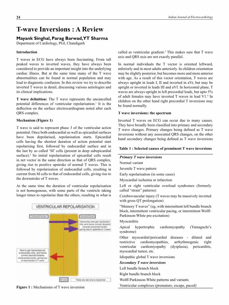
24 Indian Journal of Electrocardiology
T-wave Inversions : A ReviewMayank Singhal, Parag Barwad, YT Sharmadepartment of Cardiology, PgI, Chandigarh
Introduction
t waves in eCg have always been fascinating. from tall peaked waves to inverted waves, they have always been considered to provide an important insight into the underlying cardiac illness. But at the same time many of the t wave abnormalities can be found in normal population and may lead to diagnostic confusion. In this review we try to describe inverted t waves in detail, discussing various aetiologies and its clinical implications.
T wave definition: the t wave represents the uncancelled potential differences of ventricular repolarisation.1 It is the deflection on the surface electrocardiogram noted after each Qrs complex.
Mechanism (Figure 1)
t wave is said to represent phase 3 of the ventricular action potential. once both endocardial as well as epicardial surfaces have been depolarised, repolarisation starts. epicardial cells having the shortest duration of action potential start repolarising first, followed by endocardial surface and in the last by so called ‘M’ cells (present in deep subepicardial surface).2 so initial repolarisation of epicardial cells result in net vector in the same direction as that of Qrs complex, giving rise to positive upstroke of normal t waves. this is followed by repolarisation of endocardial cells, resulting in current from M cells to that of endocardial cells, giving rise to the downstroke of t waves.
at the same time the duration of ventricular repolarisation is not homogenous, with some parts of the ventricle taking longer times to repolarise than the others, resulting in what is
called as ventricular gradient.3 this makes sure that t wave axis and Qrs axis are not exactly parallel.
In normal individuals the t vector is oriented leftward, inferiorly and in most adults anteriorly. In children orientation may be slightly posterior, but becomes more and more anterior with age. as a result of this vector orientation, t waves are always upright in leads I, II and inverted in aVr, but may be upright or inverted in leads III and aVf. In horizontal plane, t waves are always upright in left precordial leads, but upto 5% of adult females may have inverted t waves in lead V1.4 In children on the other hand right precordial t inversions may be found normally.
T wave inversions: the spectrum
Inverted t waves on eCg can occur due to many causes. They have broadly been classified into primary and secondary T wave changes. Primary changes being defined as T wave inversions without any associated Qrs changes, on the other hand secondary changes being defined as T wave inversions
Figure 1 : Mechanisms of t wave inversion
Table 1 : Selected causes of prominent T wave inversions
Primary T wave inversionsnormal variantJuvenile t wave patternearly repolarisation (in some cases)Myocardial ischemia or infarction left or right ventricular overload syndromes (formerly called “strain” patterns)Cerebrovascular injury (t waves may be massively inverted with gross Qt prolongation)“Memory t waves” (eg, with intermittent left bundle branch block, intermittent ventricular pacing, or intermittent Wolff-Parkinson-White pre-excitation)Myocarditisapical hypertrophic cardiomyopathy (yamaguchi’s syndrome)other myocardial/pericardial diseases - dilated and restrictive cardiomyopathies, arrhythmogenic right ventricular cardiomyopathy (dysplasia), pericarditis, myocardial tumor, etc.Idiopathic global t wave inversionsSecondary T wave inversionsleft bundle branch blockright bundle branch blockWolff-Parkinson-White patterns and variantsVentricular complexes (premature, escape, paced)

25
secondary to associated Qrs changes ex. lVH, lBBB etc. The spectrum varies (Table 1) from normal findings as T inversions in right sided precordial leads in children and females to findings highly suggestive of acute ischemia. So while one must be careful to avoid overdiagnosis in patients with benign t wave inversions, at the same time not miss the diagnosis in patients with malignant t wave inversions.
Primary T wave inversions
these abnormalities are independent of changes in Qrs complexes. these may occur either due to abnormal shape and duration of ventricular action potential or abnormal sequence of repolarisation. drugs and electrolyte imbalances may cause reversible t wave inversions due to abnormal shape and duration of ventricular aPs. Whereas majority of other t wave inversions occur due to abnormal sequence of repolarisation.
a. Juvenile pattern (figure 2): as discussed above, t wave inversions is right precordial leads are common in chil-dren, but if the same persist in adulthood, they may rep-resent persistent juvenile pattern. But at the same time it is important to differentiate the same from t wave inver-sions noted in arrhythmogenic right ventricular cardio-myopathy. In a study by Pappadakis et al,5 while studying eCgs of 1710 young athletes, they noted t wave inver-sions in 4% of athletes <16 years of age and undertook their intensive cardiac workup. they concluded that t-wave inversions in V1–V3 are relatively common in athletes <16 years and probably represent the juvenile electrocardiogram pattern. In adolescent athletes, t-wave inversions beyond V2 if ≥16 years, T-wave inversions in the inferior/lateral leads and deep t-wave inversions in any lead are unusual, warranting further investigations for underlying cardiomyopathy. a finnish study6 evalu-ated eCgs of 11000 middle aged subjects and found that t-wave inversions in right precordial leads V1 to V3
were present in 54 (0.5%) of the subjects. In addition, 76 (0.7%) of the subjects had inverted t waves present only in leads other than V1 to V3. right precordial t-wave inversions did not predict increased mortality (not significant for all end points). However, inverted T waves in leads other than V1 to V3 were associated with an in-creased risk of cardiac and arrhythmic death (P<0.001 for both).
b. early repolarisation variant: In the so called ‘early re-polarisation Pattern’ there is at least 1-2 mm of upward concavity st segment elevation with prominent upright t waves in at least 2 contiguous leads. But other pat-terns have also been described. for example - a pattern of st segment coving and symmetric t wave inversion indistinguishable from acute evolving infarction is occa-sionally seen in otherwise healthy athletic young adults.7 eCg changes of erP are extremely common. this is es-pecially true in certain subsets of individuals - including healthy young adults (especially athletic males). a theo-retic basis does exist that might facilitate arrhythmogene-sis in certain predisposed individuals with erP. and erP has been identified with higher frequency in some stud-ies8,9 among some subjects with idiopathic cardiac arrest (despite absence of underlying structural abnormality). a word of caution: a cause-and-effect relationship between erP and cardiac risk has not been established. a large study10 evaluating eCgs of 29000 outpatients concluded that there was no significant association between mortal-ity and any component of ‘early repolarisation Pattern’.
c. Myocardial Ischemia: t-wave inversions are consistent with ischemia if any of the following is true:11
1. the st-segment depression or t-wave inversion is directed in the same direction as the Qrs complex: this is called concordance between the Qrs complex and the st or t abnormality.
2. the t wave has a positive-negative biphasic pattern (figure 3).
3. the t wave is symmetrically inverted and has a pointed configuration, while the ST segment is not deviated or is upwardly bowed (coved) or horizon-tally depressed (figure 4).
4. the magnitude of st-segment depression progresses or regresses on serial tracings, or st-segment de-
Figure 2 : Juvenile pattern
Figure 3 : Ischaemic t wave (biphasic)
26 Indian Journal of Electrocardiology
pression progresses to t-wave abnormality during ischemia-free intervals (dynamic st-segment de-pression).
either the positive-negative biphasic t waves or the deeply inverted (≥ 5 mm) T waves that often follow them, when occurring in the precordial leads V2 and V3, with or without similar changes in V1, V4, and V5, are nearly pathognomonic of very recent severe ischemia or injury in the distribution of the left anterior descending artery and characterize what is known as Wellens syndrome (figure 3).12
Wellens and his colleagues showed that 75% of patients who developed these t-wave abnormalities and who were treated medically without angiographic investigation went on to develop extensive anterior wall myocardial infarction within a mean of 8.5 days.12
In a later investigation of 1,260 patients presenting with unstable angina, 180 patients (14%) had this characteris-tic t-wave pattern.13 all of the latter patients had stenosis of 50% or more in the proximal left anterior descending artery, and 18% had total occlusion of the left anterior descending artery.
there are studies14,15 that have suggested that t wave in-versions in lead aVl in patients with chronic stable angina may predict clinically significant mid LAD lesions. Rec-ognition of this finding and early appropriate referral to a cardiologist may definitely prove to be of utmost benefit.
the terminal t wave inversions in patients with st eleva-tion MI indicate advanced stages of myocardial infarc-tion. In a recent study,16 188 steMI cases undergoing urgent PCI were evaluated and terminal t wave inversion was found to be an independent risk factor for poor inpa-tient prognosis.
on the other hand in a recent study17 in patients with nsteMI, eCgs were analysed and patients were divided into 4 groups: st depression, t wave inversions, transient st elevations and no ischemic changes. It was found that patients with st depression had more left main, proximal left anterior descending, and 3-vessel coronary artery dis-ease and underwent coronary artery bypass grafting most often. the unadjusted mortality was highest in the st-
segment depression group, followed by the no ischemic changes, transient st-segment elevation, and t-wave in-version group. and hence t wave inversions in patients with nsteMI were found to portend a favourable prog-nosis.
d. “strain” Patterns (figure 5): the classic left ventricular (lV) strain pattern of st segment depression and t-wave inversion on the left precordial leads of the standard rest-ing eCg is a well-known marker of the presence of ana-tomic lV hypertrophy (lVH). okin et al18 studied 8854 eCgs of hypertensive patients looking particularly for this strain pattern and concluded that eCg strain is a marker of increased cardiovascular risk in hypertensive patients in the setting of aggressive blood pressure lowering, in-dependent of baseline severity of eCg lVH. similarly in general populations this abnormality of ventricular re-polarisation has been associated with worse prognosis.19 a “right heart strain” pattern is seen with delayed repo-larization of the right ventricular myocardium producing negative st segments and t wave pattern, such as s wave in lead I and and Q wave with inverted ts in lead III. In addition, inverted t waves in V1-V3 suggests right heart strain as well.
e. Cerebrovascular injury: Ischemia-like eCg changes and arrhythmias are frequently seen in stroke patients, even in those with no history or signs of primary heart dis-ease, which support a central nervous system origin of these eCg abnormalities. t-wave abnormalities include T-waves that are of low voltage or are flat or inverted in leads that are normally upright or that are abnormally tall and peaked.20 similarly a case report published in BMJ describes a case of 52 year old man, who presented with chest pain, vomiting and deep t wave inversions on eCg and ultimately was found to have intracranial bleed.21 a recent study22 evaluating myocardial stunning effects of acute stroke found that the mean age of patients with myocardial stunning following ischemic stroke was 72.5 years and 77% of these patients were females. Insular cortex was involved in 38.4% of cases. t-wave inver-sions and st-segment elevations were noted in 84.6% and 69.2% of patients, respectively. Mean troponin eleva-tion was 0.64 mcg/dl and mean left ventricular ejection fraction (lVef) was 34%. In terms of outcomes, 85%
Figure 4 : Ischaemic t wave - deeply invertedFigure 5 : strain pattern

27
Figure 6 : t waves in hypertrophic cardiomyopathy
of patients had significant improvement in LVEF, mostly within 4 weeks of onset of symptoms. similarly in anoth-er study evaluating eCg changes in patients with intra-cranial bleed,23 it was found that the majority of patients (86%) had at least one eCg abnormality. sixty-eight (43%) patients had t-wave inversion and 65 (41%) had QTc interval prolongation. There was a significant asso-ciation between Qtc prolongation and the bleeding size and the presence of midline shift. due to paucity of fol-low-up data, the chronological progression of the chang-es remains unclear. st elevations seem to resolve while ‘t’ wave inversions appear to persist-even for months/years.24,25 there are two mechanisms26 that might mediate eCg changes in patients with saH, i.e. autonomic neu-ral stimulation from the hypothalamus or elevated levels of circulating catecholamines. Hypothalamic stimulation may cause eCg changes without associated myocardial damage whereas elevated catecholamine levels have been correlated with Qt-interval prolongation and myocardial damage. documented eCg abnormalities in a patient with saH who has brain death mean the heart is not ac-cepted as a donor organ because of the possibility of car-diac abnormalities. greater knowledge about the patho-physiology and management of eCg changes in saH may make heart donations possible in these cases.27
f. Memory t waves: Cardiac memory is an uncommonly recognized entity in which t wave inversions on elec-trocardiogram lead to a false diagnosis of ischemia.28 Persistent deep t wave inversions are seen after a period of abnormal ventricular depolarisation (wide Qrs com-plex of any cause). When normal depolarization returns, the leads that had previously shown negative Qrs com-plexes will now show t inversions. these changes are generally recognized to occur in association with artifi-cial pacemakers but may occur with other entities with intrinsic ventricular ectopic focus of depolarization, such as intermittent left bundle branch block. they can also be seen after elimination of preexcitation or after sustained Vt. Post paroxysmal tachycardia syndrome described as early as 1969,29 when Kernohan described a case of 21 year old patient who had an episode of tachycardia, fol-lowing which he was found to have t wave inversions in leads II, III, aVf and V3 to V6. for this he was extensively evaluated, coronary evaluation, cath study for right sided
pressures were all normal, and what was more interesting was that t wave inversions reverted on the eCgs on day 17. so they described a ‘benign’ post tachycardia t wave pattern of t wave inversions that can persist for a long time after an episode of paroxysmal tachycardia in the absence of any structural heart disease. Interestingly, this case report diagnosed this to be sVt; review of the eCg shows that this was classical fascicular Vt. Even today, 45 years later, this misdiagnosis is not uncommon!
the duration of t inversion varies from minutes to days and depends on the duration of abnormal depolarisation. awareness of the benign nature of cardiac memory may allow some patients to avoid unnecessary work-up and admission. It has been suggested that the contribution to repolarization of specific potassium channels influences the memory phenomenon and that by blocking it and re-ducing the transmural voltage gradient for repolarization, 4-aminopyridine abolishes cardiac memory.30
g. apical Hypertrophic Cardiomyopathy (figure 6): apical hypertrophic cardiomyopathy (aPH) is an uncommon phenotype of hypertrophic cardiomyopathy (HCM). the incidence of aPH is only 1 – 2% of HCM in Western countries, but is reported to be up to 25% in Japan. the electrocardiogram in apical HCM typically shows repo-larization changes and giant (>10 mm), inverted t waves in the anterolateral leads (particularly in leads V4 and V5).31
h. global t wave inversions: the term ‘global’ inversion is applied when the t wave is inverted in all the standard leads except aVr, and the inversion is generally symmetri-cal. this may occur due to number of causes including myocardial ischemia, Cns disorders, myocarditis, peri-carditis, apical HCM, cardiac metastases, cocaine abuse, pheochromocytoma. But at times no apparent cause can be found, when it is labelled as idiopathic. Prognosis de-pends on the underlying disease and the striking diffuse t wave changes per se do not imply a poor prognosis.32 Brscic33 et al reported a series of 17 patients (all women), who had global t wave inversions with ‘typical’ chest pain, normal coronary angiogram and intact lV function. the eCg changes gradually resolved and had no imme-diate prognostic implications.
i. Pericarditis: the t wave vector in pericarditis is directed to the right, superiorly and posteriorly. t wave inversion is therefore observed in leads that normally have upright T waves. These have been attributed to superficial myo-carditis (epicarditis) that occurs in pericarditis. In a typi-cal case of pericarditis, t wave becomes inverted in all leads except V1 and aVr, but the amplitude of inverted t wave is usually low, and t waves are often incompletely inverted.34
j. Miscellaneous: electrolyte abnormalities like hypoka-lemia and drug effects like digitalis have also been de-scribed to cause t wave inversions.

28 Indian Journal of Electrocardiology
Secondary T wave inversions
abnormalities in t waves occurring as a result of altered ventricular depolarisation sequence are referred as secondary t wave changes. the t wave vector usually deviates opposite to that of the Qrs vector in the presence of lBBB or lVH, opposite to the slow terminal Qrs component in the presence of rBBB, and opposite the delta wave in the presence of ventricular preexcitation pattern. the clinical implication is that these secondary changes may mask the primary t wave changes and hence make their recognition difficult, but at the same time exceedingly important. for example, in the presence of lBBB, t wave vector is usually directed opposite to that of QRS vector, and hence if one finds inverted T waves in leads with deep s waves, the presence of primary t wave abnormality must be thought.
Conclusions
T wave inversions are one of the most versatile findings on the surface eCg with etiologies ranging from seemingly benign causes like persistent juvenile pattern to a daringly malignant etiology like that of myocardial ischemia. the spectrum is wide and hence necessitates a broad thought process. Hence one must strike a balance, keeping into mind the clinical scenario and consider the pre-test probability for the given diagnosis before embarking upon a hoard of diagnostic tests.
References
1. Franz MR, Bargheer K, Rafflenbeul W et al. Monophasic action potential mapping in human subjects with normal electrocardiograms: direct evidence for the genesis of the t wave. Circulation 1987;75:379-86.
2. yan gX, antzelevitch C. Cellular basis for the normal t wave and the electrocardiographic manifestations of the long-Qt syndrome. Circulation 1998;98:1928-36.
3. surawicz B. electrophysiologic Basis of eCg and Cardiac arrhythmias. Baltimore, Williams & Wilkins, 1995.
4. Hiss RG, Lamb LE, Allen MF. Electrocardiographic findings in 67375 asymptomatic patients. Am J Cardiol 1960;6:200.
5. Papadakis M, Basavarajaiah s, rawlins J et al. Prevalence and significance of T-wave inversions in predominantly Caucasian adolescent athletes. Eur Heart J 2009;30:1728-1735.
6. aro al, anttonen o, tikkanen Jt. Prevalence and Prognostic Significance of T-Wave Inversions in Right Precordial Leads of a 12-lead electrocardiogram in the Middle-aged subjects. Circulation 2012;125:2572-7.
7. Choo JK, abernethy WB, Hutter aM: eCg observations in Professional football Players. Am J Cardiol 2002;90:198-200.
8. Haissaguerre M, derval n, sacher f, et al: sudden Cardiac arrest associated with early repolarization. N Engl J Med 2008;358:2016-2023.
9. tikkanen Jt, anttonen o, Junttila MJ, et al: long-term outcome
associated with early repolarization on electrocardiography. N Engl J Med 2009;361:2529-2537.
10. Uberoi a, Jain n, froelicher V, et al: early repolarization in an ambulatory Clinical Population. Circulation 2011;124:2208-2214.
11. Hanna e, glancy d. st-segment depression and t-wave inversion: Classification, differential diagnosis, and caveats. Cleveland Clinic Journal of Medicine 2011;78:404-414.
12. de Zwaan C, Bär fW, Wellens HJ. Characteristic electrocardiographic pattern indicating a critical stenosis high in left anterior descending coronary artery in patients admitted because of impending myocardial infarction. Am Heart J 1982;103:730–736.
13. de Zwaan C, Bär fW, Janssen JH, et al. angiographic and clinical characteristics of patients with unstable angina showing an eCg pattern indicating critical narrowing of the proximal lad coronary artery. Am Heart J 1989;117:657–665.
14. farhan Hl, Hassan Ks, al-Belushi a, et al. diagnostic Value of electrocardiographic t Wave Inversion in lead aVl in diagnosing Coronary artery disease in Patients with Chronic stable angina. Oman Med J 2010;25:124-7.
15. Hassen gW1, Costea a2, smith t, et al. the neglected lead on electrocardiogram: T wave inversion in lead aVL, nonspecific finding or a sign for left anterior descending artery lesion? J Emerg Med 2014;46:165-70.
16. shimada yJ, Po Jr, Kanei y, et al. Prognostic impact of terminal t wave inversions on presentation in patients with st-elevation myocardial infarction undergoing urgent percutaneous coronary intervention. J Electrocardiol 2013;46:2-7.
17. Patel JH, Gupta R, Roe MT et al. Influence of presenting electrocardiographic findings on the treatment and outcomes of patients with non-st-segment elevation myocardial infarction. Am J Cardiol 2014;113:256-61.
18. okin PM, devereux rB, nieminen Ms. electrocardiographic strain pattern and prediction of cardiovascular morbidity and mortality in hypertensive patients. Hypertension 2004;44:48-54.
19. larsen Ct, dahlin J, Blackburn H, scharling H, appleyard M, sigurd B, schnohr P. Prevalence and prognosis of electrocardiographic left ventricular hypertrophy, st segment depression and negative t-wave. the Copenhagen city heart study. Eur Heart J 2002;23:315–324.
20. togha M, sharifpour a, ashraf H et al. electrocardiographic abnormalities in acute cerebrovascular events in patients with/without cardiovascular disease. Ann Indian Acad Neurol 2013;16:66–71.
21. rajendran r, et al BMJ Case reports 2013;doi:10.1136/bcr-2012-008219.
22. Murthy sB, shah s, Venkatasubba rao CP et al. Clinical characteristics of myocardial stunning in acute stroke. J Clin Neurosci 2014;21:1279-82.
23. Qaqa ay, suleiman a, alsumrain M et al. electrocardiographic
29
abnormalities in patients presenting with intracranial parenchymal haemorrhage. Acta Cardiol 2012;67:635-9.
24. Kuroiwa T, Morita H, Tanabe H, Ohta T. Significance of ST segment elevation in electrocardiograms in patients with ruptured cerebral aneurysms. Acta Neurochir (Wien) 1995;133:141–146.
25. shuster s. the electrocardiogram in subarachnoid hemorrhage. Br Heart J 1960;22:316–320.
26. Chatterjee s. eCg Changes in subarachnoid Haemorrhage: a synopsis. Neth Heart J 2011;19:31-4.
27. sommargren Ce. electrocardiographic abnormalities in patients with subarachnoid hemorrhage. Am J Critical Care 2002;11:48–56.
28. Kolb JC. Cardiac memory-persistent t wave changes after ventricular pacing. J Emerg Med 2002;23:191-7.
29. Kernohan rJ. Post-paroxysmal tachycardia syndrome. Br Heart J 1969;31:803–806.
30. geller J, rosen M. Persistent t-wave changes after alteration of the ventricular activation sequence. new insights into cellular mechanisms of ‘cardiac memory’. Circulation 1993;88:1811-1819.
31. levis J. eCg diagnosis: apical Hypertrophic Cardiomyopathy. Perm J 2013;17:84
32. Walder la, spodick dH et al . global t wave inversion: long-term follow-up. J Am Coll Cardiol 1993;21:1652.
33. Brscic e, Brusca a, Presbitero P et al. Ischemic chest pain and global t-wave inversion in women with normal coronary angiograms. Am J Cardiol 1997;80:245-7.
34. Winternitz M, langendorf r. the electrocardiogram in pericarditis. Acta Med Scand 1938;94:141.

30 Indian Journal of Electrocardiology

31
eCg QuizYash Lokhandwala*
*arrhythmia associates

32 Indian Journal of Electrocardiology
The answers and explanations are on the reverse side of the page.

33
a. Monomorphic Vt (MMVt)
b. Polymorphic Vt (PMVt)
c. MMVt degenerating into PMVt
d. Atrial fibrillation with WPW syndrome
ECG - 1
for correct answer see overleaf
Diagnosis?

34 Indian Journal of Electrocardiology
The correct answer is ‘C’- MM VT degenerating into PM VT.
The first 3 QRS complexes are the same. After that (see lead aVF) there is transition to widely changing, irregular QRS complexes.First one should look into the clinical profile. this was a 72 year old man with an old myocardial infarction, moderate lV dysfunction, no reversible ischemia and recent unexplained syncope. the Vt shown here was induced during an eP study.the PMVt soon degenerated into Vf (fig. 1b); this was terminated by defibrillation (Fig. 1c).
fig. 1b : MMVt → PMVt → Vf
Fig. 1c : Defibrillation terminates VF, followed by bradycardia (normal sinus rhythm re-turned within a few seconds)
the highly probable cause for the syncope was a transient ventricular arrhythmia. the pa-tient was at high risk for sudden death. He therefore received an ICd (implantable cardio-verter-defibirillator)
ECG - 1
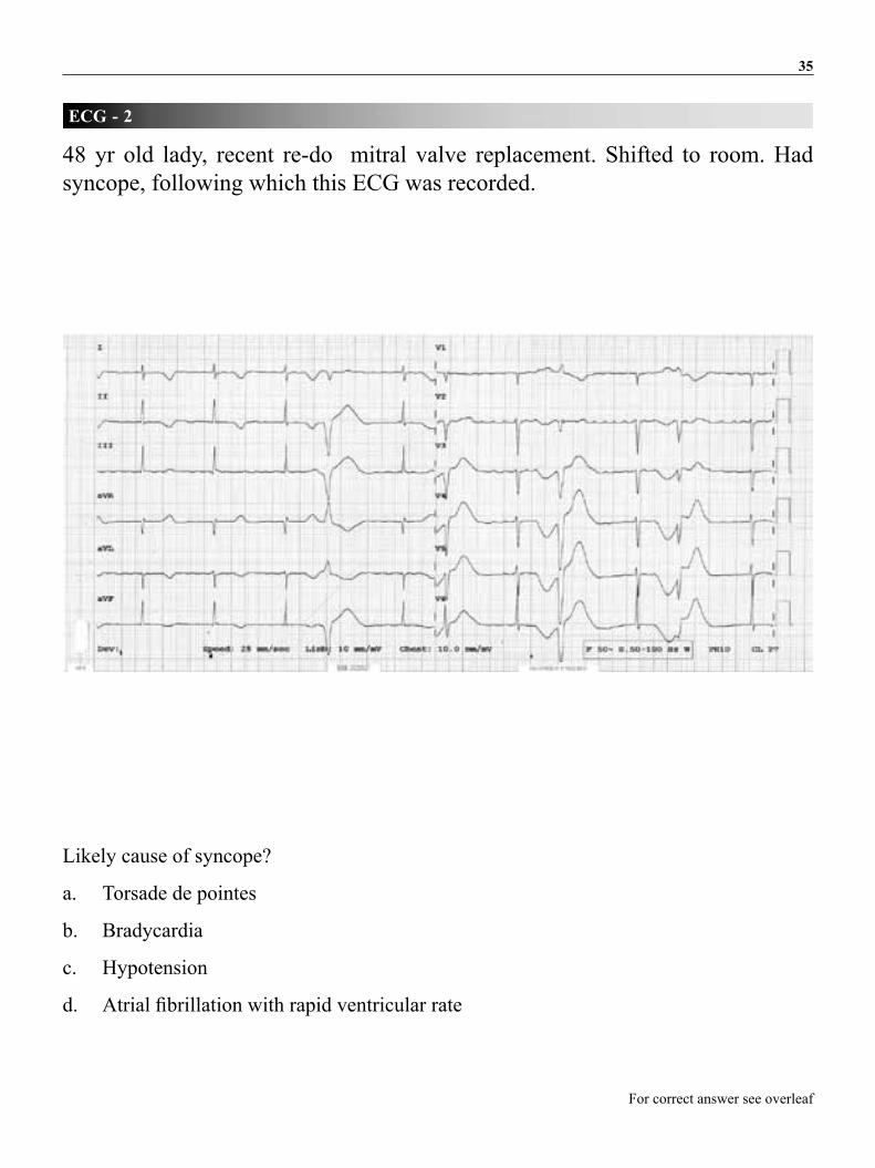
35
Likely cause of syncope?
a. torsade de pointes
b. Bradycardia
c. Hypotension
d. Atrial fibrillation with rapid ventricular rate
ECG - 2
for correct answer see overleaf
48 yr old lady, recent re-do mitral valve replacement. shifted to room. Had syncope, following which this eCg was recorded.
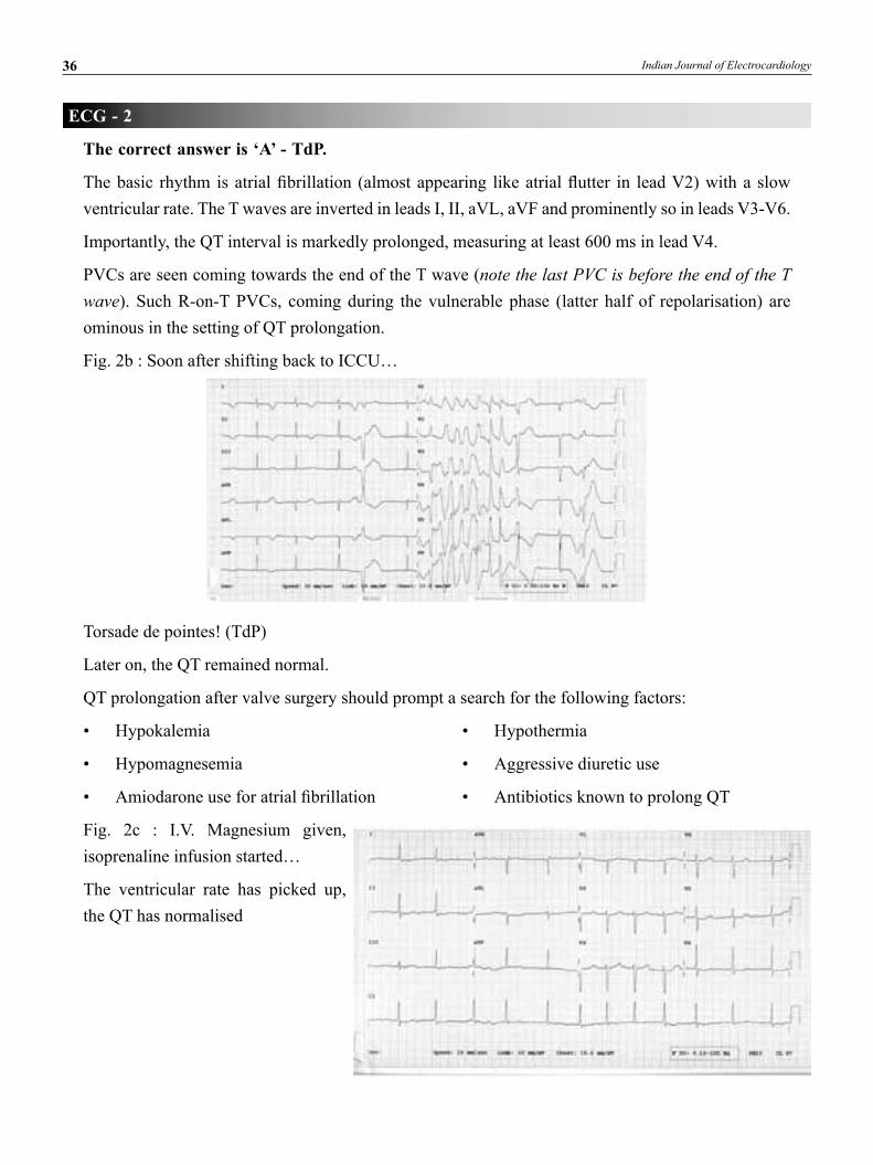
36 Indian Journal of Electrocardiology
ECG - 2
The correct answer is ‘A’ - TdP.
The basic rhythm is atrial fibrillation (almost appearing like atrial flutter in lead V2) with a slow ventricular rate. the t waves are inverted in leads I, II, aVl, aVf and prominently so in leads V3-V6.
Importantly, the Qt interval is markedly prolonged, measuring at least 600 ms in lead V4.
PVCs are seen coming towards the end of the t wave (note the last PVC is before the end of the T wave). such r-on-t PVCs, coming during the vulnerable phase (latter half of repolarisation) are ominous in the setting of Qt prolongation.
fig. 2b : soon after shifting back to ICCU…
torsade de pointes! (tdP)
later on, the Qt remained normal.
Qt prolongation after valve surgery should prompt a search for the following factors:
• Hypokalemia
• Hypomagnesemia
• Amiodarone use for atrial fibrillation
• Hypothermia
• Aggressive diuretic use
• Antibiotics known to prolong QT
fig. 2c : I.V. Magnesium given, isoprenaline infusion started…
the ventricular rate has picked up, the Qt has normalised

37
ECG - 3
17 yr old girl
a. sVt
b. Vt with Va dissociation
c. Preexcited tachycardia
d. none of the above
for correct answer see overleaf

38 Indian Journal of Electrocardiology
ECG - 3
The correct answer is ‘D’ – None of the above
there is a regular wide Qrs tachycardia @ 180/min. the Qrs shows a qr pattern (rBBB-like) in lead V1. the rs pattern in V6 in this setting favours Vt. this diagnosis is strengthened by the northwest axis of -100 0 (dominant broad r in aVr).
the P waves can be picked out carefully in lead III (fig. 3b).
INDIAN SOCIETY OF ELECTROCARDIOLOGYAPPLICATION FORM FORLIFE MEMBERSHIP/FELLOWSHIP
Dear Sir,
I wish to become the Life Member* / Fellow** of the Indian Society of Electrocardiology. I promise to abide by the rules and regulations of the Society.
My particulars are as follows :
_______________________________________________________________________
__________________________________________________________________________________
University (Post-Graduation obtained) _______________________________________________________________
_______________________________________________________________
Mailing Address ________________________________________________________________________________
______________________________________________________________________________________________
Tel. No. Hospital ___________________ Clinic __________________ Residence ___________________________
Fax ____________________________ E-Mail ______________________________________________________
Enclosed a cheque/draft of Rs. 2000/- (for outstation cheques add Rs.100/- more) towards Membership of the Society
No. ___________________________ Dated ________________________________ of __________________
___________________________________________________________________ (Bank), drawn in favour of
“Indian Society of Electrocardiology”, payable at Mumbai.
�anking you,
Yours sincerely,
Signature of the ApplicantProposed by (the Member of the Society)
Name
__________________________________________
Address
__________________________________________
Signature
__________________________________________
FOR OFFICE USE ONLYRecommendations of the
Executive Body / Credential Committee
Accepted / Not Accepted
Life Membership No.
__________________________
Hon. Secretary, ISE
�
S. B. GUPTASECRETARIAT
Indian Society of Electrocardiology102, Rail Mitra, Plot # 125, Sector I, Charkop, Kandivali (W), Mumbai 400067.Mobile : 0 98213 64565 • e-mail : [email protected] • www.iseindia.org

RULES/REGULATIONS OF THE SOCIETY REGARDING ADMISSION OF LIFE MEMBERS/FELLOWSHIP
*Life Members : 1. Person should be a Post-Graduate in Medicine/Pediatrics/Anaesthesia/ Physiology or other allied subjects from an University recognised by Medical Council of India, with interest in Cardiology / Electrocardiology.
2. Candidates are requested to submit Xerox copies of the PG Certificate and Medical Council of India Registration Certificate alongwith Application Form.
**Fellowship: 1. Person should be a Member of the Society.
2. He/She should be of atleast 7 years of standing after Post-Graduation.
3. He/She should have minimum 3 publications In Cardiology In Indexed Journals (Not Abstracts)
4. List of Publications to be submitted for the Fellowship.
5.
*Subject to approval of the Executive Body of the Society**Subject to the approval of the Credential Committee of the Society.
Fellowship Fees: Rs.2,000/- (+Rs.100/- for outstation cheque) only. Incase, fellowship not approved by the Credential Committee, the cheque / draft will be returned.

www.iseindia.orgfor more information &
latest updates

SECRETaRIaTS. B. GUPTAPaST PRESIDENT
Indian Society of Electrocardiology102, Rail Mitra, Plot # 125, Sector I, Charkop, Kandivali (W), Mumbai 400067. Mobile : 0 98213 64565 • e-mail : [email protected] • www.iseindia.org U
RVI C
OM
PUG
Ra
PHIC
S
022-
2494
586
3
urvi
@ur
vi.c
c