increased oxidative modification of albumin when illuminated in vitro in the presence of a common...
TRANSCRIPT

Original Contribution
INCREASED OXIDATIVE MODIFICATION OF ALBUMIN WHENILLUMINATED IN VITRO IN THE PRESENCE OF A COMMON SUNSCREEN
INGREDIENT: PROTECTION BY NITROXIDE RADICALS
ELISABETTA DAMIANI , PATRICIA CARLONI, CRISTIANO BIONDI, and LUCEDIO GRECI
Dipartimento di Scienze dei Materiali e della Terra, Universita` degli Studi di Ancona, Ancona, Italy
(Received28 June1999;Revised16 September1999;Accepted12 October1999)
Abstract—We previously reported on the ability of dibenzoylmethane (DBM) and a relative, Parsol 1789, usedas a ultraviolet A (UVA)-absorbing sunscreen, to generate free radicals upon illumination, and as a consequence,to inflict strand breaks in plasmid DNA in vitro. This study has now been extended to determine the effects ofParsol 1789 and DBM on proteins, under UVA illumination, with the sole purpose of gaining more knowledge onthe photobiological effects of sunscreen chemicals. Parsol 1789 (100mM) caused a 2-fold increase in proteincarbonyl formation (an index of oxidative damage) in bovine serum albumin (BSA) when exposed to illumination,and this damage was both concentration- and time-dependent. The degree of protein damage was markedly reducedby the presence of free radical scavengers, namely piperidinic and indolinonic nitroxide radicals, in accordancewith our previous study. Vitamin E had no effect under the conditions used. The results obtained corroborate thefact that Parsol 1789 generates free radicals upon illumination and that these are, most probably, responsible forthe protein damage observed under the conditions used in our system. However, at present, we cannot extrapolatefrom these results the relevance to human use of sunscreens; therefore, further studies should be necessary todetermine the efficacy at the molecular and cellular level of this UVA-absorber in order to ascertain protectionagainst photocarcinogenic risk. © 2000 Elsevier Science Inc.
Keywords—Protein damage, Indolinonic and piperidinic nitroxides, Parsol 1789, Dibenzoylmethane, Antioxidants,Free radicals
INTRODUCTION
Skin cancers are among the most common humancancers and the incidence rate, in particular during thelast couple of decades, has been increasing dramati-cally [1–3]. Whether this increase is due to behavioralchanges (increased exposure to solar radiation, eithernatural or artificial, in pursuit of a tan), depletion ofthe ozone layer, or increased use of sunscreens, is stillvery much under debate. What is widely accepted,though, is that the ultraviolet (UV) radiation compo-nents of sunlight play a major role in skin tumorinduction and development because the most criticalcellular target, DNA, becomes damaged [4 – 6]. Con-sequently, ample attention has been given to effective
photoprotection against the injurious effects of over-exposure to solar radiation. To this end, among thedifferent defensive measures, the use of sunscreens isprobably the most popular one. It is widely acceptedthat sunscreen agents prevent, or at least delay, theonset of UV radiation–induced erythema and theirefficiency is usually tested in these terms [7,8]. How-ever, more data should possibly be available on theirefficacy at the molecular and cellular level in order toascertain protection against photocarcinogenic risk.This is of paramount importance considering the ap-parent connection between the increased usage of sun-screens and the concomitant rise in skin cancer[9 –11], although there is some evidence that regularuse of sunscreens helps prevent the formation of ac-tinic keratoses, the precursors of squamous cell carci-noma [12].
To this purpose, we previously tested the ability ofdibenzoylmethane (DBM) and a relative, Parsol 1789,
Address correspondence to: Dr. Elisabetta Damiani, Dipartimento diScienze dei Materiali e della Terra, Universita` degli Studi di Ancona,Via Brecce Bianche, I-60131 Ancona, Italy; Tel:139 (071) 220-4416;Fax: 139 (071) 220-4714; E-Mail: [email protected].
Free Radical Biology & Medicine, Vol. 28, No. 2, pp. 193–201, 2000Copyright © 2000 Elsevier Science Inc.Printed in the USA. All rights reserved
0891-5849/00/$–see front matter
PII S0891-5849(99)00221-X
193

used as a UVA-blocking agent in many formulations, togenerate free radicals upon illumination, and as a conse-quence, to inflict strand breaks in plasmid DNA in vitro[13]. This study has now been extended to determine theeffects of Parsol 1789 and DBM on other importantbiological marcromolecules such as proteins, under UVAillumination, with the sole purpose of gaining moreknowledge on the photobiological effects of sunscreenchemicals. Therefore, bovine serum albumin (BSA) waschosen as a simple model protein system and subjectedto UVA irradiation in the presence and absence of thesunscreen ingredients and the degree of oxidative mod-ification was measured by monitoring the carbonyl con-tent–an index of oxidative damage to proteins [14]. Inaddition, in analogy with our previous study concerningthe ability of nitroxide radicals (a new emerging class ofantioxidants) to protect DNA from damage inflicted invitro by Parsol 1789 [13], the antioxidant behavior of thesame nitroxides was examined and compared in thisprotein system. Similarly, Vitamin E was also includedin this study.
MATERIALS AND METHODS
Materials
Indolinonic nitroxides1a and1b and2a and2b (Fig.1) were synthesized according to literature methods [15].The nitroxides used were 1,2-dihydro-2-methyl-2-phe-nyl-3H-indole-3-one-1-oxyl (Fig. 1,1a), 1,2-dihydro-2-methyl-2-phenyl-3H-indole-3-phenylimino-1-oxyl (Fig.1, 1b), 1,2-dihydro-2-ethyl-2-phenyl-3H-indole-3-one-1-oxyl (Fig. 1, 2a), and 1,2-dihydro-2-ethyl-2-phenyl-3H-indole-3-phenylimino-1-oxyl (Fig. 1,2b). The identityand purity of the compounds were checked by thin layerchromatography and by mass spectroscopy on a CarloErba QMD 1000 spectrometer (Milan, Italy). The tetra-methyl-piperidinic nitroxides TEMPO and TEMPOL, vi-tamin E, dibenzoylmethane, BSA (Fraction V, A4503),guanidine hydrochloride, and 2,4-dinitrophenylhydr-azine were obtained from Sigma Chemical Co. (Milan,Italy), while other chemicals and solvents were pur-chased from Aldrich Chemical Co. (Milan, Italy) andwere of the highest purity; 4-tert-butyl-49-methoxydiben-
Fig. 1. Chemical structures of: 4-tert-butyl-49-methoxydibenzoylmethane (Parsol 1789); dibenzoylmethane (DBM); TEMPO; TEM-POL; 1a, 1,2-dihydro-2-methyl-2-phenyl-3H-indole-3-one-1-oxyl;1b, 1,2-dihydro-2-methyl-2-phenyl-3H-indole-3-phenylimino-1-oxyl; 2a, 1,2-dihydro-2-ethyl-2-phenyl-3H-indole-3-one-1-oxyl;2b, 1,2-dihydro-2-ethyl-2-phenyl-3H-indole-3-phenylimino-1-oxyl.
194 E. DAMIANI et al.

zoylmethane (Parsol 1789) was obtained in the form ofEusolex 9020 from Merck (Darmstadt, Germany), and itsidentity was confirmed by NMR. As UVA irradiatingsource, a Philips Original Home Solarium (model HB406/A; Philips, Groningen, Holland) equipped with a400 watt ozone-free Philips HPA lamp, UV type 3, wasused. The fluence rate of the light between 300 and 400nm is 0.75 mW/cm2. It was always pre-run for 15 min toallow the output to stabilize.
Assay of oxidative modification of protein
The protein samples were prepared by dissolving 3mg/ml of BSA in 50 mM potassium phosphate buffer,0.1 mM ethylenediaminetetraacetic acid (EDTA), pH7.4. The samples were then exposed to UVA illumina-tion, in the presence or absence of Parsol 1789 and/orantioxidants. Parsol 1789, DBM, or antioxidants wereadded to the protein as methanol solutions (5ml) at thefinal concentrations desired, and the mixture was vor-texed for thorough incorporation. The samples were thentransferred into 3-ml quartz cuvettes, closed and exposedto UVA irradiation at a distance of 2.5 cm from the lightsource.
The degree of protein oxidation was monitored by themethod of Levine et al. [14], which uses the reaction of2,4-dinitrophenylhydrazine (DNPH) with the carbonylgroups of oxidized protein. At the end of UVA exposure,0.5 ml of 20 mM DNPH dissolved in 2.5 M HCl wasadded to 0.5 ml of each sample and incubated for 1 h atroom temperature under continuous shaking; the remain-ing 0.5 ml of sample were used as blanks and lacked onlyDNPH. The protein was then precipitated by addition of1 ml 20% trichloroacetic acid and centrifuged at 30003g for 10 min. The supernatant was discarded and theresidual pellet was washed with ethanol/ethylacetate(1:1) followed by centrifugation at 30003 g for 10 min.These last three operations: (i) elimination of superna-tant, (ii) washing with ethanol/ethylacetate, and (iii) cen-trifugation, were repeated twice before the residual pelletwas dissolved in 1 ml 6 M guanidine HCl (pH 6.5).Protein carbonyls were then read at 370 nm and evalu-ated using a molar absorption coefficient of 22,000M21cm21.
Appropriate controls were carried out throughout andthe results reported are an average of at least threeindependent experiments each performed in duplicate.Statistical comparisons between: (i) samples with andwithout Parsol 1789 or DBM (control), and (ii) sampleswith Parsol 1789 (control) and with nitroxide addition,under UVA irradiation, were performed using the Stu-dent t-test. Differences were regarded as statisticallysignificant whenp values were, .05 (*), p , .01(**),andp , .001 (***).
EPR measurements
Electron paramagnetic resonance (EPR) measure-ments were carried out at room temperature and thespectra were recorded on a Varian E4 spectrometer (PaloAlto, CA, USA) interfaced with a computer. A 1 mlsolution of nitroxide in CH3OH (10ml of a 15 mM stocksolution) and 1 ml BSA (10 mg) in 50 mM potassiumphosphate buffer, 0.1 mM EDTA, pH 7.4, were intro-duced in an inverted flat quartz U-cell and degassed witha stream of argon. The cell was then illuminated in themicrowave cavity for 25 min using the source describedabove, placed at a distance of 10 cm, and the spectrawere recorded after 10 and 25 min of illumination. TheEPR experiments were performed three times and theresults were reproducible in every case.
RESULTS
The main aim of this study was to determine theeffects of the UVA chemical sunscreen, Parsol 1789, andDBM on the oxidative status of BSA when exposed toUVA and the antioxidant behavior of two differentclasses of nitroxide radicals.
As starting point, we first compared the effects ofDBM with its derivative Parsol 1789. Figure 2 clearlyshows that in the presence of 100mM DBM, the degreeof protein carbonyl formation in BSA is far less than thatobserved in the presence of 100mM Parsol 1789 after 20min of UVA exposure. Secondly, experiments were per-formed with Parsol 1789 to see whether its oxidative
Fig. 2. Protein carbonyl levels in BSA exposed to 20 min of illumina-tion (unless otherwise specified in the graph) in the absence or presenceof a final concentration of 100mM Parsol 1789 or 100mM DBM (errorbars represent SD).
195In vitro protein damage by a UVA-sunscreen

effect on BSA after UVA exposure is concentration- andtime-dependent. Figure 3 shows that on increasing theconcentration of Parsol 1789 (50 to 150mM), the level ofprotein oxidation also rises. The presence of 100mMParsol 1789 in BSA exposed to UVA for 20 min resultsin a 2-fold increase of protein carbonyl formation com-pared with the level of oxidation observed in its absence.In the same graph it can be noticed that methanol, usedto dissolve the sunscreen ingredient, leads to slight vari-ation in the oxidative status of BSA when exposed toUVA, but this variation is negligible in comparison tothat produced by the presence of Parsol 1789.
The results reported in Fig. 4 were obtained by ex-posing BSA with and without 100mM of UVA-filter to10, 20, and 30 min of illumination. One can observe thatthere is a direct time-dependent increment in proteinoxidation and this effect is remarkably greater whenParsol 1789 is present in the protein system.
In order to exclude the possibility that the variation inthe degree of protein oxidation in BSA in the presence ofthe sunscreen ingredient was not caused by any possiblethermal reactions caused by the emitted heat from thelamp (the temperature of the samples after 20 min ofirradiation reached 556 3°C), experiments were per-formed by incubating BSA with and without Parsol 1789for 20 min in a thermostatted water bath at 55°C. Theresults obtained after determination of the protein car-bonyl content were the same as those obtained afterincubating the samples in the dark at room temperature
for 20 min. This proved that the oxidative effects ob-served when BSA was incubated with the sunscreeningredient and exposed to UVA were due to the presenceof Parsol 1789 under illumination.
Once the increased oxidative modification on BSAinduced by Parsol 1789 under UVA irradiation was af-firmed, it was of interest to determine the effects of thepresence of different antioxidants, namely nitroxide rad-icals, of the piperidinic and indolinonic type (the lattersynthesized by us) and vitamin E. Figure 5 shows theresults of the experiments conducted with TEMPO (100mM). One can observe the level of oxidation of BSAexposed to UVA for 20 min, the increment induced bythe presence of Parsol 1789 and the antioxidant effect ofTEMPO when it is added to the system BSA-Parsol1789. An additional consideration that can be made onobserving the results reported in this graph is that theoxidative level of BSA incubated with only TEMPO isthe same as that observed in its absence when exposed toUVA alone; this shows that TEMPO has no effect onprotein carbonyl formation induced by UVA.
The same experiments in the same conditions as thosedescribed above for TEMPO were performed with all theother nitroxides and vitamin E, except that for the indo-linonic nitroxides, the maximum concentration, whichcould be tested for solubility reasons, was 75mM. Theresults are summarized in Fig. 6, which compares theantioxidant efficacy of the different compounds tested atthe highest concentrations used. As can be observed,TEMPO, TEMPOL and nitroxides1b and2b remarkablysuppress carbonyl group formation, all to practically the
Fig. 3. Protein carbonyl levels in BSA exposed to 20 min of illumina-tion (unless otherwise specified in the graph) in the absence or presenceof increasing final concentrations of Parsol 1789 (P). Parsol 1789 wasadded to BSA as a 5ml methanol solution as described in Materials andMethods (error bars represent SD).
Fig. 4. Protein carbonyl levels determined on BSA after different UVAexposure times, in the absence or presence of a final concentration of100 mM Parsol 1789 (error bars represent SD).
196 E. DAMIANI et al.
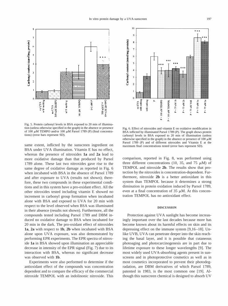
same extent, inflicted by the sunscreen ingredient onBSA under UVA illumination. Vitamin E has no effect,whereas the presence of nitroxides1a and 2a lead tomore oxidative damage than that produced by Parsol1789 alone. These last two nitroxides gave rise to thesame degree of oxidative damage as reported in Fig. 6when incubated with BSA in the absence of Parsol 1789and after exposure to UVA (results not shown); there-fore, these two compounds in these experimental condi-tions and in this system have a pro-oxidant effect. All theother nitroxides tested including vitamin E showed noincrement in carbonyl group formation when incubatedalone with BSA and exposed to UVA for 20 min withrespect to the level observed when BSA was illuminatedin their absence (results not shown). Furthermore, all thecompounds tested including Parsol 1789 and DBM in-duced no oxidative damage to BSA when incubated for20 min in the dark. The pro-oxidant effect of nitroxides1a, 2a with respect to1b, 2b when incubated with BSAalone upon UVA exposure, was also demonstrated byperforming EPR experiments. The EPR spectra of nitrox-ide 1a in BSA showed upon illumination an appreciabledecrease in intensity of the EPR signal (Fig. 7) due to itsinteraction with BSA, whereas no significant decreasewas observed with1b.
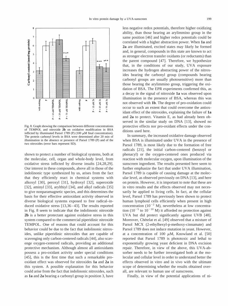
Experiments were also performed to determine if theantioxidant effect of the compounds was concentrationdependent and to compare the efficacy of the commercialnitroxide TEMPOL with an indolinonic nitroxide. This
comparison, reported in Fig. 8, was performed usingthree different concentrations (10, 35, and 75mM) ofTEMPOL and nitroxide2b. The results show that pro-tection by the nitroxides is concentration-dependent. Fur-thermore, nitroxide2b is a better antioxidant in thissystem than TEMPOL because it determines a strongdiminution in protein oxidation induced by Parsol 1789,even at a final concentration of 35mM. At this concen-tration TEMPOL has no antioxidant effect.
DISCUSSION
Protection against UVA sunlight has become increas-ingly important over the last decades because more hasbecome known about its harmful effects on skin and itsdepressing effect on the immune system [9,16–18]. Un-like UVB, UVA can penetrate deeper into the skin reach-ing the basal layer, and it is possible that cutaneousphotoaging and photocarcinogenesis are in part due tolifetime exposure to these longer wavelengths [9]. Themost widely used UVA-absorbing agents present in sun-screens and in photoprotective cosmetics as well as inmost cosmetics incorporated to prevent their photodeg-radation, are DBM derivatives of which Parsol 1789,patented in 1983, is the most common one [19]. Al-though this sunscreen chemical is designed to absorb UV
Fig. 6. Effect of nitroxides and vitamin E on oxidative modification inBSA inflicted by illuminated Parsol 1789 (P). The graph shows proteincarbonyl levels in BSA exposed to 20 min of illumination (unlessotherwise specified in the graph) in the absence or presence of 100mMParsol 1789 (P) and of different nitroxides and Vitamin E at themaximum final concentrations tested (error bars represent SD).
Fig. 5. Protein carbonyl levels in BSA exposed to 20 min of illumina-tion (unless otherwise specified in the graph) in the absence or presenceof 100 mM TEMPO and/or 100mM Parsol 1789 (P) (final concentra-tions) (error bars represent SD).
197In vitro protein damage by a UVA-sunscreen

energy, photostability assessment studies conducted byDeflandre and Lang [20] on Parsol 1789 in very thin filmlayers of an emulsion, found degradation rates up to 50%after only 1 h of irradiation. Schwack and Rudolf [21] intheir photochemical studies, suggested that Parsol 1789breaks down via the initial formation of two carbon-centered free radicals (benzoyl and phenacyl; Fig. 1)to give an array of products. We recently confirmedthe formation of carbon-centered radicals generatedupon UVA illumination of Parsol 1789 via EPR exper-iments using nitroxide radicals as spin traps [13]. Fur-ther, their formation is probably responsible for the directstrand breaks found when plasmid DNA is illuminated invitro in the presence of both Parsol 1789 or DBM. Theresults presented in this article on the oxidative modifi-cation of BSA by Parsol 1789 are in reasonable agree-ment with, and extend our previous observations of the invitro damage inflicted by this sunscreen ingredient uponillumination [13].
Among the covalent changes caused by both directand indirect oxidative modification of proteins by freeradical generation, one of the most common is the intro-duction of carbonyl groups into the side chains [22].Oxidative modification can, therefore, be measured bymonitoring the carbonyl content of a specific protein orof a tissue. The results reported here show that whenBSA is exposed to UVA in the presence of Parsol 1789,an increment in BSA oxidation is observed, stronglysupporting the view that free radicals are generated and
that these could be responsible for the oxidative modifi-cations observed. The increased protein oxidation byParsol 1789 is dependent on both the length of UVexposure and the concentration used. In addition, Parsol1789 determines far more oxidative damage than itscorresponding unsubstituted derivative does and this re-sult is consistent with our previous data which showedthat Parsol 1789 had a greater plasmid nicking abilitythan DBM [13]. This might be attributed to the fact thatDBM is probably more photostable than Parsol 1789,which dissociates faster into carbon-centered radicals.Substituents on the benzene rings such as those presentin Parsol 1789 can have significant effects and this couldbe supported by the fact that p-methoxy-acetophenone ismore labile to light than acetophenone [23].
The results obtained here show the suppression ofoxidative modification by nitroxide radicals in the BSA-Parsol 1789 system and are in general agreement withthose obtained in our previous study on naked DNA [13].This lends support, and confirms the rising interest, onthe versatile antioxidant ability of nitroxide radicals [24].These are a unique group of compounds bearing anunpaired electron on the N-O function that are remark-ably stable and persistent as free radicals due mainly tothe absence of dimerization and disproportionation [25].Other than their use as biophysical probes and labels formembrane and protein studies [25], and as contrastagents for nuclear magnetic [26], and electron param-agentic [27] resonance imaging, they have recently been
Fig. 7. EPR spectra recorded at room temperature before and after 25 min of illumination of 75mM nitroxide1a in BSA (see Materialsand Methods for experimental details).
198 E. DAMIANI et al.
shown to protect a number of biological systems, both atthe molecular, cell, organ and whole-body level, fromoxidative stress inflicted by diverse insults [24,28,29].Our interest in these compounds, above all in those of theindolinonic type synthesized by us, arises from the factthat they efficiently react in chemical systems withalkoxyl [30], peroxyl [31], hydroxyl [32], superoxide[32], aminyl [33], arylthiyl [34], and alkyl radicals [35]to give nonparamagnetic species, and this determines thebasis for their effective antioxidant activity observed indiverse biological systems exposed to free radical–in-duced oxidative stress [13,36–43]. The results reportedin Fig. 8 seem to indicate that the indolinonic nitroxide2b is a better protectant against oxidative stress in thissystem compared to the commercial piperidinic nitroxideTEMPOL. One of reasons that could account for thisbehavior could be due to the fact that indolinonic nitrox-ides, unlike piperidinic nitroxides that are capable ofscavenging only carbon-centered radicals [44], also scav-enge oxygen-centered radicals, providing an additionalprotective mechanism. Although almost all antioxidantspossess a pro-oxidant activity under special conditions[45], this is the first time that such a remarkable pro-oxidant effect was observed for nitroxides1a and2a inthis system. A possible explanation for this behaviorcould arise from the fact that indolinonic nitroxides, suchas1aand2abearing a carbonyl group in position 3, have
less negative redox potentials, therefore higher oxidizingability, than those bearing an arylimmino group in thesame position [46] and higher redox potentials could becorrelated with a higher abstraction power. When1a and2a are illuminated, excited states may likely be formedand, in general, compounds in this state are known to actas stronger electron transfer oxidants (or reductants) thanthe parent compound [47]. Therefore, we hypothesizethat, in the conditions of our study, UVA exposureincreases the hydrogen abstracting power of the nitrox-ides bearing the carbonyl group (compounds bearingcarbonyl groups are usually photosensitive) more thanthose bearing the arylimmino group, triggering the oxi-dation of BSA. The EPR experiments confirmed this, asa decay in the signal of nitroxide1a was observed uponillumination in the presence of BSA, whereas this wasnot observed with1b. The degree of pro-oxidation couldoccur to such an extent that could overcome the antiox-idant effect of the nitroxides, explaining the failure of1aand 2a to protect. Vitamin E, as had already been ob-served in the similar study on DNA [13], showed noprotective effects nor pro-oxidant effects under the con-ditions used here.
In summary, the increased oxidative damage observedwhen BSA is illuminated under UVA in the presence ofParsol 1789, is most likely due to the formation of freeradicals [21], the initial carbon-centered (benzoyl orphenacyl) or the oxygen-centered ones produced viareaction with molecular oxygen, upon illumination of thesunscreen ingredient. The results presented here seem tofurther emphasize the fact that under UVA illumination,Parsol 1789 is capable of causing damage at the molec-ular level, as observed previously on DNA [13], and hereon protein. However, it is important to note that these arein vitro results and the effects observed may not neces-sarily be applied to living cells. In fact, at the cellularlevel, Parsol 1789 has previously been shown to protecthuman lymphoid cells efficiently when present in highconcentration (1022 M), nevertheless at low concentra-tion (1025 to 10210 M) it afforded no protection againstUVA but did protect significantly against UVB [48].Moreover, Che´telat et al. [49] observed that a mixture ofParsol MCX (2-ethylhexyl-p-methoxy-cinnamate) andParsol 1789 does not induce mutation in yeast. However,at a concentration of 100mM, Knowland et al. [50]reported that Parsol 1789 is phototoxic and lethal toexponentially growing yeast deficient in DNA excisionrepair. Therefore, in view of the above, this UVA-ab-sorber needs to be further investigated both at the mo-lecular and cellular level in order to understand better theeffects observed in vitro and in vivo with the ultimatescope of determining whether the results obtained over-all, are relevant to human use of sunscreens.
Finally, in view of the potential applications of ni-
Fig. 8. Graph showing the comparison between different concentrationsof TEMPOL and nitroxide2b on oxidative modification in BSAinflicted by illuminated Parsol 1789 (P) (100mM final concentration).The protein carbonyl levels in BSA were determined after 20 min ofillumination in the absence or presence of Parsol 1789 (P) and of thetwo nitroxides (error bars represent SD).
199In vitro protein damage by a UVA-sunscreen

troxide radicals in human skin [51], and their low poten-tial to cause acute or subacute skin toxicity [52], partic-ularly the pyrrolidinic and imidazolinic ones, the resultsobtained in this study further corroborate the potentialuse of these compounds as antioxidants, possibly incream formulations too, and highlight the importance fortailoring antioxidants to their appropriate use.
Acknowledgements— The authors thank the University of Ancona andthe Italian MURST (Ministero dell’Universita` e della Ricerca Scienti-fica e Tecnologica) for financial support.
REFERENCES
[1] Boyle, P.; Maisonneuve, P.; Dore, J.-F. Epidemiology of malig-nant melanoma.Br. Med. Bull.51:523–547; 1995.
[2] Miller, D. L.; Weinstock, M. A. Nonmelanoma skin cancer in theUnited States: incidence.J. Am. Acad. Dermatol.30:774–78;1994.
[3] Preston, D.; Stern, R. Nonmelanoma cancers of the skin.NewEngl. J. Med.327:1649–1661; 1992.
[4] Lee, J. A. H. The relationship between malignant melanoma ofskin and exposure to sunlight.Photochem. Photobiol.50:493–496; 1989.
[5] Ziegler, A.; Jonason, A.; Leffell, D.; Simon, J.; Sharma, H.;Kimmelman, J.; Remington, L.; Jacks, T., Brash, D. Sunburn andp53 in the onset of skin cancer.Nature372:773–776; 1994.
[6] Davies, R. J. H. Ultraviolet radiation damage in DNA.Biochem.Soc. Trans.23:407–418; 1995.
[7] Council on Scientific Affairs. Harmful effects of ultraviolet radi-ation.J. Am. Med. Assoc.262:380–384; 1989.
[8] Kaidbey, K.; Gange, R. W. Comparison of methods of assessingphotoprotection against ultraviolet A in vivo.J. Am. Acad. Der-matol.16:336–353; 1987.
[9] Garland, C.; Garland, F.; Gorham, E. Could sunscreens increasemelanoma risk?Am. J. Public Health82:614–615; 1992.
[10] Westerdahl, J.; Olsson, H.; Masbaak, A.; Ingvar, C.; Jonsson, N.Is the use of sunscreens a risk factor for malignant melanoma?Melanoma Res.5:59–65; 1995.
[11] Autier, P.; Dore, J.-F.; Schifflers, E.; Cesarini, J.-P.; Bollaerts, A.;Koelmel, K.; Gefeller, O.; Liabeuf, A.; Lejeune, F.; Lienard, D.;Joarlette, M.; Chemaly, P.; Kleeberg, U. Melanoma and use ofsunscreens: an EORTC case control study in Germany, Belgiumand France.Int. J. Cancer61:749–755; 1995.
[12] Dover, J. S.; Arndt, K. A. Dermatology.J. Am. Med. Assoc.271:1662–1663; 1994.
[13] Damiani, E.; Greci, L.; Parsons, R.; Knowland, J. Nitroxideradicals protect DNA from damage when illuminated in vitro inthe presence of dibenzoylmethane and a common sunscreen in-gredient.Free Radic. Biol. Med.26:809–816; 1999.
[14] Levine, R. L.; Garland, K.; Oliver, C. N.; Amici, A.; Climent, I.;Lenz, A. G.; Ahn, B. W.; Shaltiel, S.; Stadtman, E. R. Determi-nation of carbonyl content in oxidatively modified proteins.Meth-ods Enzymol.186:464–478; 1990.
[15] Berti, C.; Colonna, M.; Greci, L.; Marchetti, L. Stable nitroxideradicals from phenyl-isatogen and arylimino derivatives with or-ganometallic compounds.Tetrahedron31:1745–1753; 1975.
[16] Drobetsky, E.; Turcotte, J.; Chateauneuf, A. A role for ultravioletA in solar mutagenesis.Proc. Natl. Acad. Sci. USA92:2350–2354; 1995.
[17] Sterenborg, H.; Van der Leun, J. Tumorigenesis by a long wave-length UVA source.Photochem. Photobiol.51:325–330; 1990.
[18] Fuller, C. J.; Faulkner, H.; Bendich, A.; Parker, R. S.; Roe, D. A.Effect of beta-carotene supplementation on photosuppression ofdelayed-type hypersensitivity in normal young men.Am. J. Clin.Nutr. 56:684–690; 1992.
[19] De Polo, K. F. Dibenzoylmethane derivative and its use in sun-screening compositions.Chem. Abs.93:185967g; 1980.
[20] Deflandre, A.; Lang, G. Photostability assessment of sunscreens.Benzylidene camphor and dibenzoylmethane derivatives.Int. J.Cosmet. Sci.10:53–62; 1988.
[21] Schwack, W.; Rudolph, T. Photochemistry of dibenzoylmethaneUVA filters. Part 1.J. Photochem. Photobiol.28:229–234; 1995.
[22] Amici, A.; Levine, R. L.; Tsai, L.; Stadtman E. R. Conversion ofaminoacid residues in proteins and aminoacid homopolymers tocarbonyl derivatives by metal-catalyzed oxidation.J. Biol. Chem.264:3341–3346; 1989.
[23] Murov, S. L.; Carmichael, I.; Hug, G. L., eds.Handbook ofphotochemistry.New York: Marcel Dekker, Inc.; 1993.
[24] Samuni, A.; Krishna, M. C. Antioxidant properties of nitroxidesand nitroxide SOD mimics. In: Packer, L.; Cadenas, E., eds.Handbook of synthetic antioxidants.New York: Marcel DekkerInc.; 1997:351–373.
[25] Berliner, L. J. Spin labeling II: theory and applications.NewYork: Academic Press Inc.; 1979.
[26] Swartz, H. M.; Feig, E.Advances in magnetic resonance imaging.Norwood, NJ: Ablex Publishing Co.; 1988.
[27] Berliner, L. J.; Fujii, H. Magnetic resonance imaging of biologicalspecimens by electron paramagnetic resonance of nitroxide spinlabels.Science227:517–519; 1985.
[28] Krishna, M. C.; Samuni, A. Nitroxides as antioxidants.MethodsEnzymol.234:580–589; 1994.
[29] Kocherginsky, N.; Swartz, H. M.Nitroxide spin labels: reactionsin biology and chemistry.Boca Raton: CRC Press; 1995.
[30] Greci, L. Homolytic substitution in indolinone nitroxide radicals.Part III. Reactions with terbutoxy and methyl radicals.Tetrahe-dron 38:2435–2439; 1982.
[31] Cardellini, L.; Carloni, P.; Greci, L.; Stipa, P.; Faucitano, A.Homolytic substitution in indolinone nitroxide radicals. Part 5.Reactions with tert-butylperoxy radicals.Gazz. Chim. Ital.119:621–625; 1989.
[32] Damiani, E.; Carloni, P.; Stipa, P.; Greci, L. Reactivity of anindolinonic aminoxyl with superoxide anion and hydroxyl radi-cals.Free Rad. Res.31:113–121; 1999.
[33] Alberti, A.; Greci, L.; Stipa, P.; Sgarabotto, P.; Ugozzoli, F.Homolytic substitution in indolinone nitroxide–IV. Reaction withaminyl radicals. Spectroscopic and crystallographic study.Tetra-hedron43:3031–3040; 1987.
[34] Damiani, E.; Carloni, P.; Stipa, P.; Iacussi, M.; Greci, L. Reac-tivity of sulphur-centered radicals with indolinonic and quinolinicaminoxyls.Eur. J. Org. Chem.2405–2412; 1999.
[35] Stipa, P.; Carloni, P.; Greci, L.; Damiani, E. Synthesis and ther-mal stability of alkoxyamines.Polym. Deg. Stab.55:323–327;1997.
[36] Antosiewicz, J.; Bertoli, E.; Damiani, E.; Greci, L.; Popinigis, J.;Przybylski, S.; Tanfani, F.; Wozniak, M. Indolinonic and quino-linic aminoxyls as protectants against oxidative stress.FreeRadic. Biol. Med.15:203–208; 1993.
[37] Damiani, E.; Paganga, G.; Greci, L.; Rice-Evans, C. Inhibition ofcopper-mediated low density lipoprotein peroxidation by quino-line and indolinone nitroxide radicals.Biochem. Pharmacol.48:1155–1161; 1994.
[38] Tanfani, F.; Carloni, P.; Damiani, E.; Greci, L.; Wozniak, M.;Kulawiak, D.; Jankowski, K.; Kaczor, J.; Matuskiewics, A.Quinolinic aminoxyl protects albumin against peroxyl radicalmediated damage.Free Radic. Res.21:309–315; 1994.
[39] Antosiewicz, J.; Popinigis, J.; Wozniak, M.; Damiani, E.; Carloni,P.; Greci, L. Effects of indolinic and quinolinic aminoxyls onprotein and lipid peroxidation of rat liver microsomes.FreeRadic. Biol. Med.18:913–917; 1995.
[40] Antosiewicz, J.; Damiani, E.; Jassem, W.; Wozniak, M.; Orena,M.; Greci, L. Influence of structure on the antioxidant activity ofindolinic nitroxide radicals.Free Radic. Biol. Med.22:249–255;1997.
[41] Gabbianelli, R.; Falcioni, G.; Santroni, A.; Caulini, G.; Greci, L.;Damiani, E. Effect of aromatic nitroxides on hemolyis of humanerythrocytes entrapped with isolated hemoglobin chains.FreeRadic. Biol. Med.23:278–284; 1997.
[42] Villarini, M.; Moretti, M.; Damiani, E.; Greci, L.; Santroni,
200 E. DAMIANI et al.

A. M.; Fedeli, D.; Falcioni, G. Detection of DNA damage instressed trout nucleated erythrocytes using the comet assay: pro-tection by nitroxide radicals.Free Radic. Biol. Med.24:1310–1315; 1998.
[43] Falcioni, G.; Gabbianelli, R.; Damiani, E.; Santroni, A.M.; Fedeli,D.; Wozniak, M.; Greci, L. The effect of indolinic and quinolinicnitroxide radicals on trout erythrocytes exposed to oxidativestress.Free Radic. Res.28:507–516; 1998.
[44] Beckwith, A. L. J.; Bowry, V. W.; Ingold, K. U. Kinetics ofnitroxyl radical trapping1. Solvent effects.J. Am. Chem. Soc.114:4983–4992; 1992.
[45] Bast, A.; Haenen, G. R. M. M.; Doelman, D. J. A. Oxidants andantioxidants: state of the art.Am. J. Med.91:3C–13S; 1991.
[46] Carloni, P.; Damiani, E.; Greci, L.; Stipa, P.; Marrosu, G.;Petrucci, R.; Trazza, A. Chemical and electrochemical study onthe interactions of aminoxyls with superoxide anion.Tetrahedron53:12445–12452; 1996.
[47] Eberson, L.Electron transfer reactions in organic chemistry.Berlin: Springer-Verlag; 1987.
[48] Andrae, I.; Bringhen, A.; Bohn, F.; Gonzenbach, T.; Hill, T.;Mulroy, L.; Truscott, T. G. A UVA filter (4-tert-butyl-49-me-thoxydibenozoylmethane): photoprotection reflects photophysicalproperties.J. Photochem. Photobiol.37:147–150; 1997.
[49] Chetelat, A.; Albertini, J. H.; Dresp, J. H.; Strobel, R.; Gocke, E.Photomutagenesis test development: I. 8-Methoxypsoralen, chlo-ropromazine and sunscreen compounds in bacterial and yeastassays.Mutat. Res.292:241–250; 1993.
[50] Knowland, J.; McKenzie, P. J.; McHugh, P. J.; Cridland, N. A.Sunlight-induced mutagenicity of common sunscreen ingredients.FEBS Lett.324:309–313; 1993.
[51] Cuscela, D.; Coffin, D.; Lupton, G. P.; Cook, J. A.; Krishna,M. C.; Bonner, R. F.; Mitchell, J. B. Protection from radiation-induced alopecia with topical application of nitroxides: fraction-ated studies.Cancer J. Sci. Am.2:273–278; 1996.
[52] Fuchs, J.; Croth, N.; Herrling, T. Cutaneous tolerance to nitroxidefree radicals in human skin.Free Radic. Biol. Med.24:643–648;1998.
201In vitro protein damage by a UVA-sunscreen