increased expression of epha7 correlates with adverse outcome in
TRANSCRIPT

BioMed CentralBMC Cancer
ss
Open AcceResearch articleIncreased expression of EphA7 correlates with adverse outcome in primary and recurrent glioblastoma multiforme patientsLin-Fang Wang1,3, Emmanouil Fokas1, Janko Juricko1, An You1, Frank Rose1, Axel Pagenstecher2, Rita Engenhart-Cabillic1 and Han-Xiang An*1Address: 1Department of Radiotherapy and Radiation Oncology, Philipps-University Marburg, Baldingerstr. D-35043 Marburg, Germany, 2Department of Neuropathology, Philipps-University Marburg, Baldingerstr. D-35043 Marburg, Germany and 3Department of Emergency Surgery, Union Hospital, Tongji Medical College, Huazhong University of Science and Technology, 430030 Wuhan, People's Republic of China
Email: Lin-Fang Wang - [email protected]; Emmanouil Fokas - [email protected]; Janko Juricko - [email protected]; An You - [email protected]; Frank Rose - [email protected]; Axel Pagenstecher - [email protected]; Rita Engenhart-Cabillic - [email protected]; Han-Xiang An* - [email protected]
* Corresponding author
AbstractBackground: Malignant gliomas are lethal cancers, highly dependent on angiogenesis andtreatment options and prognosis still remain poor for patients with recurrent glioblastomamultiforme (GBM). Ephs and ephrins have many well-defined functions during embryonicdevelopment of central nervous system such as axon mapping, neural crest cell migration, hindbrainsegmentation and synapse formation as well as physiological and abnormal angiogenesis.Accumulating evidence indicates that Eph and ephrins are frequently overexpressed in differenttumor types including GBM. However, their role in tumorigenesis remains controversial, as bothtumor growth promoter and suppressor potential have been ascribed to Eph and ephrins while thefunction of EphA7 in GBM pathogenesis remains largely unknown.
Methods: In this study, we investigated the immunohistochemical expression of EphA7 in a seriesof 32 primary and recurrent GBM and correlated it with clinical pathological parameters andpatient outcome. In addition, intratumor microvascular density (MVD) was quantified byimmunostaining for endothelial cell marker von Willebrand factor (vWF).
Results: Overexpression of EphA7 protein was predictive of the adverse outcome in GBMpatients, independent of MVD expression (p = 0.02). Moreover, high density of MVD as well ashigher EphA7 expression predicted the disease outcome more accurately than EphA7 variablealone (p = 0.01). There was no correlation between MVD and overall survival or recurrence-freesurvival (p > 0.05). However, a statistically significant correlation between lower MVD and tumorrecurrence was observed (p = 0.003).
Conclusion: The immunohistochemical assessment of tissue EphA7 provides importantprognostic information in GBM and would justify its use as surrogate marker to screen patients fortyrosine kinase inhibitor therapy.
Published: 25 March 2008
BMC Cancer 2008, 8:79 doi:10.1186/1471-2407-8-79
Received: 8 August 2007Accepted: 25 March 2008
This article is available from: http://www.biomedcentral.com/1471-2407/8/79
© 2008 Wang et al; licensee BioMed Central Ltd. This is an Open Access article distributed under the terms of the Creative Commons Attribution License (http://creativecommons.org/licenses/by/2.0), which permits unrestricted use, distribution, and reproduction in any medium, provided the original work is properly cited.
Page 1 of 9(page number not for citation purposes)

BMC Cancer 2008, 8:79 http://www.biomedcentral.com/1471-2407/8/79
BackgroundThe incidence of brain tumors worldwide is about 7 in100,000 per year [1,2]. Glioblastoma multiforme (GBM),the most aggressive tumor among malignant gliomas, isthe most common primary brain tumor in adults and rep-resents a significant source of cancer-related death. GBMusually recurs despite the most aggressive treatment viasurgical resection of the tumor followed by radiation and/or chemotherapy [1,2]. The poor prognosis of patientswith GBM (median survival ranging from 9 to 12 months,5-year survival rate close to 0%) mandates the explorationof novel molecular mechanisms that might contribute tothe pathogenesis of this disease and its resistance to ther-apy with the purpose of therapeutic targeting [1-3].
Receptor tyrosine kinases (RTKs) are known to be impor-tant regulators of cellular growth controlling cell prolifer-ation, differentiation and migration [4,5]. The Ephreceptors and their ligands, ephrins, represent the largestknown family of RTKs. Their role has been largely studiedduring the development of nervous system. They areinvolved in the development of central nervous system,including axon guidance, axon fasciculation, neural crestcell migration, hindbrain segmentation, vasculogenesisand neuronal cell survival during embryonic develop-ment [6-13]. Eph receptors and ephrin ligands are classi-fied into A and B subfamily, on the basis of theirsequence, homologies, structures, and binding affinities.EphA receptors bind the glycosylphosphatidylinositol(GPI)-anchored ephrin-A ligands, whereas EphB receptorsbind the transmembrane ephrin-B ligands, whose cyto-plasmic domain is capable to engage in various signalingactivities; an exception is the EphA4 receptor that bindsephrin-B2 and ephrin-B3 as well as ephrin-A ligands [14-16]. Moreover, these RTKs have the ability to induce bothforward and reverse (bi-directional) signaling betweenadjacent interacting cells.
To date, various studies have investigated the involvementof the Eph-RTKs in several pathogenetic processes in thenervous system. EphB2 and ephrin-B2 signaling partici-pate in the glial scarring process after spinal cord trauma[17]. The phosphorylation ratio of R-Ras was closelylinked to the phosphorylation ratio of EphB2 in glioblas-toma tissues [18]. Additionally, the phosphorylation ratioof EphB2 is an important mechanism that mediates gli-oma cell migration and invasion [19]. Ephrin-B2 andEphB4 were overexpressed by endothelial cells of humanmalignant gliomas [20]. Ephrin-B3 was also demon-strated as an important factor regulating glioma cell inva-sion through Rac1 GTPase [21]. EphA2 protein wasoverexpressed in GBM and anaplastic astrocytoma tissuesand was identified as a novel target for the developmentof glioma vaccines [22,23]. Another group confirmedoverexpression of EphA2 expression in GBM cells, proba-
bly through decreased interaction between EphA2 recep-tor and its inhibitory ligand ephrin-A1 in malignant cells[19].
EphA7 (formerly known as Mdk1/Ebk/Ehk) is highly con-served in vertebrates from fish to human [24]. It is widelyexpressed in embryonic tissues, especially developing cen-tral nervous system [25]. EphA7 cooperates with otherEphA receptors in cell signaling, but in contrast to otherEph receptors, it contains two developmentally regulatedisoforms: a full-length version containing the intracellulartyrosine kinase domain and a truncated form that lacksthis domain [26]. Immunoreactivity for the full-lengthwild type receptor is found in all cell populations express-ing EphA7 mRNA in mouse embryo heads and develop-ing brain, while the truncated EphA7 is absent in theembryos. Interestingly, both isoforms show striking distri-butions in adult mouse brain [27]. The full-length EphA7is strongly expressed in neuropil; in contrast the truncatedEphA7 is conspicuous on cell bodies and proximal den-drites of a limited number of neuronal types [28]. Thetruncated form of EphA7 acts as a dominant negativeantagonist, suppressing tyrosine phosphorylation of thefull-length EphA7 receptor and shifts the cellular responsefrom repulsion to adhesion. Additionally, EphA7 is prob-ably essential during closure of the neural folds, sinceEphA7-null mice displayed lack of the neural folds resem-bling anencephaly in man [29]. Moreover, EphA7 hasbeen identified as an important molecular cue expressedafter spinal cord injury, implicated in glial apoptosis [30].Recent work indicated EphA7 as an important mediator ofneural progenitor apoptosis during brain development[31]. However, little is currently known about its role inbrain tumor angiogenesis and pathogenesis.
In the present study, we investigated the immunohisto-chemical expression of EphA7 and correlated it with clin-ical pathological parameters and tumor vascularity. Weprovide evidence that EphA7 is overexpressed in GBM andsuggest that this receptor might be used as a new diagnos-tic and prognostic marker for further Eph/ephrin targetedmolecular cancer therapy.
MethodsTissuesTumor samples of 32 patients with histologically con-firmed GBM, WHO IV, (26 primary GBM, 6 recurrentGBMs and 10 normal brain samples) were obtained fromthe Department of Neuropathology, Marburg UniversityHospital, Germany. Approval for immunohistochemicalstudy conduct in this GBM tissue bank had been obtainedby the university authorities together with the signed con-sent of the patients. The patients underwent surgery andreceived adjuvant radiation therapy combined with chem-otherapy using the schema ACNU and VM-26. The first
Page 2 of 9(page number not for citation purposes)
BMC Cancer 2008, 8:79 http://www.biomedcentral.com/1471-2407/8/79
follow-up occurred 6 weeks after therapy was completed.Subsequent follow-ups were scheduled every 3 months. Inaddition to clinical investigations and monitoring of indi-ces of recurrence, a radiological examination was per-formed to detect possible relapses. Disease progressionwas defined according to WHO criteria by either anincrease of at least 25% in tumor size or any new tumoridentified by CT or MRI scan. Normal brain samples,which included cortex and white matter, were obtainedfrom autopsy cases without any evidences of brain tumoror other brain disease. Totally, five cases of male and fivecases of female (age ranging from 27 to 70 years, average:48.6 years) were obtained.
ImmunohistochemistryImmunohistochemical studies were performed on forma-lin fixed, paraffin-embedded tissue. Samples slides werepassed through a sequence of Roti-histol (Carl Roth, Karl-sruhe, Germany) and graded alcohol and then rinsed inphosphate-buffered saline (PBS). After rinsing with PBS,the slides were treated with 3% hydrogen peroxide in PBSfor 15 min at room temperature in order to abolishendogenous peroxidase activity. Subsequently, the slideswere treated with 5% blocking serum for 1 hour. Follow-ing this, slides were incubated overnight at 4°C with arabbit anti-human EphA7 polyclonal antibody (H-55)against amino acids of human EphA7 (1:100 dilution;Santa Cruz Biotechonology, Heidelberg, Germany), orrabbit anti-human Factor VIII (von Willebrand Factor,vWF) polyclonal antibody (1:400 dilution; Dako Cytoma-tion, Carpinteria, CA). In negative controls, the primaryantibody was replaced with 1 × PBS. The signal wasenhanced by using biotinylated polyclonal goat-anti-rab-bit IgG with streptavidin-HRP (Dako Cytomation,Carpinteria, CA) for 30 minutes. The colour was devel-oped after 5 minutes incubation with 3,3-diaminobenzi-dine (DAB) solution and sections were weaklycounterstained with hematoxylin for 10 seconds.
Evaluation of EphA7 expressionThe membranous and cytoplasmic expression of EphA7on tumor cells was assessed at a ×400 magnification. Theassigned score first reflects the staining intensity A (0, neg-ative; 1, weak; 2, moderate; 3, high) and second the per-centage of positive cells B (0, no positive cells; 1, <25%positive cells; 2, 25 to 50% positive cells; 3, >50% positivecells). An overall score of 3 is defined as positive staining.The scoring was performed separately by two independentobservers who were blinded to the clinical data. Any dis-crepancies were resolved on the conference microscope.
Evaluation of MVD at "hot spot" of tumor angiogenesisTumor angiogenesis can be reflected by MVD in the mostvascularised areas of tumor tissue. MVD, as highlighted byfactor VIII-related antigen immunostaining, was assessed
without knowledge of the patient's clinical outcome, asdescribed by Weidner et al [32]. Briefly, each slide wasscanned at low magnification (×100) to identify fourareas with the highest density of microvessel (hot-spots).Each hot-spot was then evaluated at high power magnifi-cation (×200) for the number of stained microvessels perfield in a 0.7386 mm2 surface area. vWF-positive stainedblood vessels with a complete lumen as well as cell clus-ters without lumina were considered as individual micro-vessels. The final microvessel score was the average ofvessel counts from four fields assessed by a high powermagnification field (×200).
Statistical analysisSurvival curves were estimated using the Kaplan-Meiermethod. The distributions of survival were comparedusing the log rank test. The chi-square test was employedto determine the association between EphA7 expressionintensity on tumor cells and MVD. A p-value < 0.05 wasconsidered to be statistically significant. All statisticalanalysis was performed using SPSS software.
ResultsDemographic factors32 patients with histologically confirmed primary andrecurrent GBM, WHO grade IV, were studied. The meanage at diagnosis was 54.3 years (range 31–71). No signifi-cant difference in age distribution between male (21cases) and female (11 cases) was detected. All of the 32patients showed a relapse between 1 and 22 months aftersurgery and subsequently died of the disease (median sur-vival 15 months).
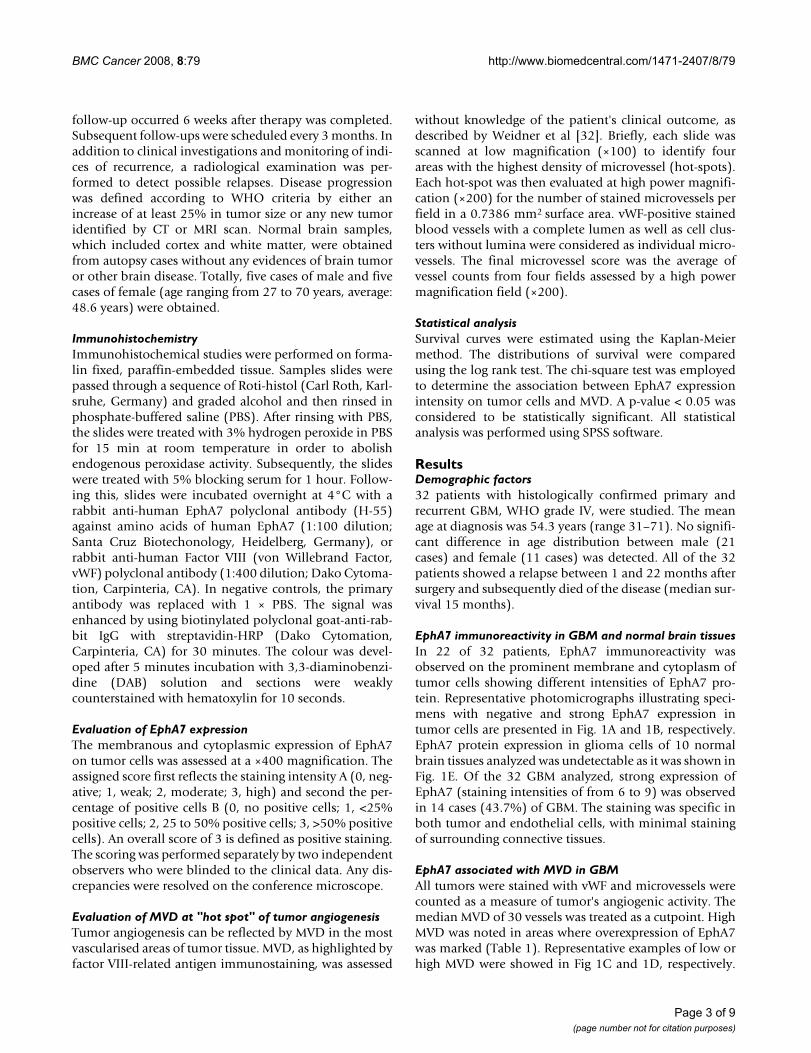
EphA7 immunoreactivity in GBM and normal brain tissuesIn 22 of 32 patients, EphA7 immunoreactivity wasobserved on the prominent membrane and cytoplasm oftumor cells showing different intensities of EphA7 pro-tein. Representative photomicrographs illustrating speci-mens with negative and strong EphA7 expression intumor cells are presented in Fig. 1A and 1B, respectively.EphA7 protein expression in glioma cells of 10 normalbrain tissues analyzed was undetectable as it was shown inFig. 1E. Of the 32 GBM analyzed, strong expression ofEphA7 (staining intensities of from 6 to 9) was observedin 14 cases (43.7%) of GBM. The staining was specific inboth tumor and endothelial cells, with minimal stainingof surrounding connective tissues.
EphA7 associated with MVD in GBMAll tumors were stained with vWF and microvessels werecounted as a measure of tumor's angiogenic activity. Themedian MVD of 30 vessels was treated as a cutpoint. HighMVD was noted in areas where overexpression of EphA7was marked (Table 1). Representative examples of low orhigh MVD were showed in Fig 1C and 1D, respectively.
Page 3 of 9(page number not for citation purposes)

BMC Cancer 2008, 8:79 http://www.biomedcentral.com/1471-2407/8/79
There was a statistically significant correlation betweenexpression of EphA7 and MVD in the tumors (P = 0.004,Table 1).
EphA7 immunoreactivity predicted overall survival but not recurrence-free survivalThe median survival of patients with positive EphA7expression was reduced in comparison with patients withnegative EphA7 expression. EphA7 protein expressionshowed an inverse correlation with the overall survival (p
= 0.02, Fig. 2A). However, the level of EphA7 expressiondid not emerge as a prognostic factor for recurrence-freesurvival of GBM patients (p = 0.51, Fig. 2B). There was nocorrelation between MVD and overall survival or recur-rence-free survival (p > 0.05, data not shown). We furtherexplored the prognostic relationship using EphA7 in com-bination with MVD. The study cohort could be dividedinto 3 groups based on expression for EphA7 and MVDcombination: EphA7(+)/high MVD (n = 12), EphA7(+)/low MVD (n = 10), EphA7(-)/high MVD (n = 0), EphA7(-
Immunohistochemical demonstration of EphA7 protein expression and blood vessels in GBMFigure 1Immunohistochemical demonstration of EphA7 protein expression and blood vessels in GBM. Representative examples of GBM showing negative staining in tumor cells (A) or strong membranous and cytoplasmic staining in both tumor cells and endothelial cells (B). MVD in GBM by immunohistochemical staining for vWF, microvessels are represented by brown clusters, which stand out sharply from other tissues. Low tumor vascularity (C) in GBM with low expression of EphA7 as shown A. In contrast, microvessel density was relatively high (D) in GBM with high expression of EphA7 as shown B. Negative staining in normal brain tissue (E). Original magnification, ×400 (A, B, E) and ×200 (C, D).
A
C
E
B
D
Page 4 of 9(page number not for citation purposes)
BMC Cancer 2008, 8:79 http://www.biomedcentral.com/1471-2407/8/79
)/low MVD (n = 10). As shown in Fig. 2C, high density ofMVD as well as EphA7 expression predicted for the diseaseoutcome more accurately than Eph variable alone (p =0.01).
Clinical features associated with EphA7 or MVDStatistical correlation was detected between the expressionlevels of EphA7 or MVD and clinical pathological param-eters such as age, gender and tumor status (Table 2). A sta-tistically significant correlation between higher MVD andtumor recurrence was observed (p = 0.003). In addition,positive EphA7 expression was associated with increasedage of patients (>55 years, p = 0.003).
DiscussionThere is currently an urgent need for development of alter-native, effective diagnostic and therapeutic approaches toGBM. The survival of patients with GBM may depend onthe identification of novel targets. EphA2 receptor hasalready been recognized as a potential molecular markerand target in GBM for the development of novel biologi-cal therapeutic agents [22,23,33]. Whereas several studiesin recent years have clearly indicated that altered expres-sion of Eph receptors and ephrin ligands is associatedwith increased potential for tumor growth, angiogenesis,metastasis and adverse outcome [34-42], few studies haveaddressed the role of EphA7 in tumor pathogenicity. Byemploying immunohistochemical techniques we havefound that EphA7 protein is predictive for the outcome ofpatients with GBM, independent of MVD expression. Thedata in the present study revealed for the first time a strongcorrelation between EphA7 overexpression and patientsurvival.
Hafner C, et al. found that EphA7 is highly expressed inkidney vasculature [43]. The mRNA of EphA7 is stronglyupregulated in hepatocellular carcinoma as comparedwith healthy liver tissue and is downregulated in coloncarcinomas. EphA7 is also transcriptionally activated inlung cancer [44]. Furthermore, overexpression of EphA7protein is frequently found in younger patients and inpatients with advanced gastric carcinoma [45]. EphA7expression is frequently silenced in human colorectal car-
cinoma by aberrant promoter methylation [46]. EphA7 islocated on 6q16.1, a region in close proximity to the chro-mosome 6 breakpoint found in various types of cancer[47]. Although our findings are not consistent with Wanget al, who found a significant downregulation of EphA7 incolorectal carcinoma [46], they are in line with previousreports reporting a tumor promoter role in lung cancerand hepatocellular carcinoma [43,44], implicating tumortype-specific function for different Eph family members.Eph receptors expressed in different cell types may haveopposite effects due to cell-type specific intracellular sign-aling pathway [4]. Indeed, EphB4 receptor has been iden-tified as a tumor suppressor in breast cancer, throughactivation of Abl-Crk antioncogenic pathway [48], whilethe same receptor presented a tumorigenic potential inmesothelioma, favoring uncontrolled cell growth, migra-tion, and tumor progression [49]. Moreover, membrane-bound ephrins trigger Eph receptor phosphorylation,while soluble forms can bind to Eph receptor, but do nottrigger receptor activation [50]. Murine and humanperipheral lymphocytes secrete a truncated form of EphA7[51]. Truncated Eph receptors retaining their ligand-bind-ing capacity have been shown to block activation of thefull-length receptor [52]. Promoter hypermethylation andsilencing of EphA7 in mature B-cell lymphomas may serveto eliminate the inhibitory activity of secreted EphA7 ontumor-promoting EphA7 receptor signaling, thus enhanc-ing tumor cell spread and recruitment of accessory cellsable to promote tumor growth [51]. A recent study on sig-naling pathways involved in EphA7 RTK reported thatdirect EphA7 knockdown can result in attenuation ofERK1/2 phosphorylation and induce apoptosis of leuke-mia cells, suggesting the impact of EphA7 on the growthof tumor cells [45]. It is of interest that positive EphA7expression was closely associated with increased age ofpatients (>55 years). Whether this is a random finding ornot deserves further investigation.
The unfavorable prognostic influence of EphA7 in GBMcould be attributed to the well-recognised role of EphRTKs in tumor angiogenesis. Indeed, in this study a statis-tically significant correlation between expression ofEphA7 and MVD was noted in GBM specimens. Anotherimportant observation was EphA7 overexpression in bothvasculature as well as tumor cells. The process of angio-genesis plays a central role in tumor growth and in thedevelopment of distant metastases by facilitating the entryof cells into circulation [52-55]. A vast biochemical andgenetic evidence has implicated the critical role of Eph/ephrin signaling in angiogenesis, despite of VEGFR2 andTie2 receptors long been recognized as key players in thisprocess [53,54]. Angiogenetic activity can be measuredhistologically by MVD, which has been shown to be anindependent prognostic parameter in various malignan-cies including gliomas [56-58]. However, other studies on
Table 1: Relationship between EphA7 expression and intratumor microvessel density in 32 primary and recurrent GBM.
EphA7 expression P value
negative positiveMVD
Low 10 10High 0 12 0.004
MVD: microvessel density, N: number of patients. The median MVD of 30 vessels was used as a cutpoint.
Page 5 of 9(page number not for citation purposes)

BMC Cancer 2008, 8:79 http://www.biomedcentral.com/1471-2407/8/79
Page 6 of 9(page number not for citation purposes)
Kaplan-Meier curve for overall survival and recurrence-free survival in 32 patients based on EphA7 expression and MVD indexFigure 2Kaplan-Meier curve for overall survival and recurrence-free survival in 32 patients based on EphA7 expression and MVD index. (A) Increased EphA7 expression was significantly associated with dead of disease (P = 0.02 by log-rank test), when positive EphA7 (score = 4–9) expressing tumors were plotted against negative EphA7 expressing tumors (score = 0–3). (B) EphA7 expression revealed no significance for recurrence-free survival. (C) EphA7 expression in combination with high MVD showed an inverse outcome (p = 0.01).
A
B
C
BMC Cancer 2008, 8:79 http://www.biomedcentral.com/1471-2407/8/79
angiogenesis of glioblastomas suggested the limited usageof MVD as prognostic parameter due to the complexity ofthe microvascular network in GBM [59,60]. Although nocorrelation between MVD and overall survival or recur-rence-free survival was found in our study, we observed astatistically significant correlation between lower MVDand tumor recurrence. Further prospective studies withlarge numbers of patients are, however, needed to fullyclarify the clinical implications of MVD in GBM recur-rence.
ConclusionTaken together, our data illustrated that EphA7 could be apotential candidate as a prognostic tumor marker and anew targeted therapeutic assessment in primary and recur-rent GBM. Based on our findings, there might be a possi-ble relationship between EphA7 and tumorneovascularization. Recent data demonstrating inhibitionof angiogenesis through EphA receptor blockade in twodifferent animal tumor models are consistent with ourobservation [52]. Additional experimental work is neces-sary to unveil the biologic pathway linking Eph/ephrinswith tumor growth in cancer cells and tumor-associatedvessels of GBM and further studies are needed beforeEphA7 becomes established as an important prognosticand predictive tool in GBM. Ultimately, specific EphA7inhibitors may prove to be of therapeutic value.
AbbreviationsGBM, glioblastoma multiforme; RTKs, Receptor tyrosinekinases; MVD, microvascular density; vWF, von Wille-brand factor.
Competing interestsThe author(s) declare that they have no competing inter-ests.
Authors' contributionsLFW, EF and JJ carried out the Immunohistochemicalstudies. AY and FR participated in the design of the studyand performed the statistical analysis. AP and HXA partic-
ipated the evaluation of analysed parameters and tumorpathological characteristics. REC conceived of the studyand participated in the design and coordination as well ashelped to draft the manuscript. All authors read andapproved the final manuscript.
AcknowledgementsWe are very grateful to our technicians, Mrs. B. Kleb, H. Geißel and R. Wassmuth for their expert technical assistance.
References1. Ohgaki H, Kleihues P: Population-based studies on incidence,
survival rates, and genetic alterations in astrocytic and oli-godendroglial gliomas. J Neuropathol Exp Neurol 2005,64:479-489.
2. Reifenberger G, Collins VP: Pathology and molecular genetics ofastrocytic gliomas. J Mol Med 2004, 82:656-670.
3. Zhu Y, Parada LF: The molecular and genetic basis of neurolog-ical tumours. Nat Rev Cancer 2002, 2:616-626.
4. Schlessinger J, Ullrich A: Growth factor signaling by receptortyrosine kinases. Neuron 1992, 9:383-391.
5. van der Geer P, Hunter T, Lindberg RA: Receptor protein-kinasesand their signal transduction pathways. Annu Rev Cell Biol 1994,10:251-337.
6. Flanagan JG, Vanderhaeghen P: The ephrins and Eph receptors inneural development. Annu Rev Neurosci 1998, 21:309-345.
7. Holder N, Klein R: Eph receptors and ephrins: effectors ofmorphogenesis. Development 1999, 126:2033-2044.
8. Kullander K, Klein R: Mechanisms and functions of Eph andephrin signaling. Nat Rev Mol Cell Biol 2002, 3:475-486.
9. Pasquale EB: Eph receptor signalling casts a wide net on cellbehaviour. Nat Rev Mol Cell Biol 2005, 6:462-475.
10. Tessier-Lavigne M: Eph receptor tyrosine kinases, axon repul-sion, and the development of topographic maps. Cell 1995,82:345-348.
11. Winslow JW, Moran P, Valverde J, Shih A, Yuan JQ, Wong SC, TsaiSP, Goddard A, Henzel WJ, Hefti F, Beck KD, Caras IW: Cloning ofAL-1, a ligand for an Eph-related tyrosine kinase receptorinvolved in axon bundle formation. Neuron 1995, 14:973-981.
12. Wang HU, Anderson DJ: Eph family transmembrane ligandscan mediate repulsive guidance of trunk neural migrationand motor axon outgrowth. Neuron 1997, 18:383-396.
13. Martinez A, Soriano E: Functions of ephrin/Eph interactions inthe development of the nervous system: emphasis on thehippocampal system. Brain Res Brain Res Rev 2005, 49:211-226.
14. Lemke G: A coherent nomenclature for Eph receptors andtheir ligands. Mol Cell Neurosci 1997, 9:331-332.
15. Holland SJ, Gale NW, Mbamalu G, Yancopoulos GD, Henkemeyer M,Pawson T: Bidirectional signaling through the EPH-familyreceptor Nuk and its transmembrane ligands. Nature 1996,383:722-725.
16. Davis S, Gale NW, Aldrich TH, Maisonpierre PC, Lhotak V, PawsonT, Goldfarb M, Yancopoulos GD: Ligands for EPH-related recep-
Table 2: Relationship between EphA7 expression or microvessel count and clinicopathological features of 32 patients with GBM.
EphA7 expression P value MVD P value
Age N positive (%) N high MVD N (%)<= 50 10 3 (30) 11 6 (54.55)>50 22 19 (86.36) 0.003 21 16 (76.19) ns
Gendermale 21 15 (71.43) 21 15 (71.43)female 11 7 (63.64) ns 11 8 (72.72) ns
Tumor statusPrimary 26 20 (76.92) 26 22 (84.62)Recurrence 6 2 (33.33) ns 6 1 (16.67) 0.003
MVD: microvessel density, N: number of patients, ns: not significant. The median MVD of 30 vessels was used as a cutpoint.
Page 7 of 9(page number not for citation purposes)

BMC Cancer 2008, 8:79 http://www.biomedcentral.com/1471-2407/8/79
tor tyrosine kinases that require membrane attachment orclustering for activity. Science 1994, 266:816-819.
17. Bundesen LQ, Scheel TA, Bregman BS, Kromer LF: Ephrin-B2 andEphB2 regulation of astrocyte-meningeal fibroblast interac-tions in response to spinal cord lesions in adult rats. J Neurosci2003, 23:7789-7800.
18. Nakada M, Niska JA, Tran NL, McDonough WS, Berens ME: EphB2/R-Ras signaling regulates glioma cell adhesion, growth, andinvasion. Am J Pathol 2005, 167:565-576.
19. Nakada M, Niska JA, Miyamori H, McDonough WS, Wu J, Sato H,Berens ME: The Phosphorylation of EphB2 Receptor Regu-lates Migration and Invasion of Human Glioma Cells. CancerRes 2004, 64:3179-3185.
20. Erber R, Eichelsbacher U, Powajbo V, Korn T, Djonov V, Lin J,Hammes HP, Grobholz R, Ullrich A, Vajkoczy P: EphB4 controlsblood vascular morphogenesis during postnatal angiogen-esis. EMBO J 2006, 25:628-641.
21. Nakada M, Drake KL, Nakada S, Niska JA, Berens ME: Ephrin-B3Ligand Promotes Glioma Invasion through Activation ofRac1. Cancer Res 2006, 66:8492-8500.
22. Hatano M, Eguchi J, Tatsumi T, Kuwashima N, Dusak JE, Kinch MS,Pollack IF, Hamilton RL, Storkus WJ, Okada H: EphA2 as a glioma-associated antigen: a novel target for glioma vaccines. Neo-plasia 2005, 7:717-722.
23. Liu FH, Park PJ, Lai W, Maher E, Chakravarti A, Durso L, Jiang X, YuY, Brosius A, Thomas M, Chin L, Brennan C, DePinho RA, Kohane I,Carroll RS, Black PM, Johnson MD: A genome-wide screenreveals functional gene clusters in the cancer genome andidentifies EphA2 as a mitogen in glioblastoma. Cancer Res2006, 66:10815-10823.
24. Taneja R, Thisse B, Rijli FM, Thisse C, Bouillet P, Chambon P: Theexpression patter of the mouse receptor tyrosine kinasegene MDK1 is conserved through evolution and requiresHoxa-2 for rhombomere-specific expression in mouseembryos. Dev Biol 1996, 177:397-412.
25. Ciossek T, Millauer B, Ulrich A: Identification of alternativelyspliced mRNAs encoding variants of MDKs, a novel receptortyrosine kinase expressed in the murine nervous system.Oncogene 1995, 10:97-108.
26. Valenzuela DM, Rojas E, Griffiths JA, Compton DL, Gisser M, Ip NY,Goldfarb M, Yancopoulos GD: Identification of full length andtruncated forms and Ehk-3, a novel member of the Ephreceptor tyrosine kinase family. Oncogene 1995, 10:1573-1580.
27. Ciossek T, Ullrich A, West E, Rogers JH: Segregation of thereceptor EphA7 form its tyrosine kinase-negative isoform onneurons in adult mouse brain. Molecular Brain Res 1999,74:231-236.
28. Rogers JH, Ciossek T, Ullrich A, West E, Hoare M, Muir EM: Distri-bution of the receptor EphA7 and its ligands in developmentof the mouse nervous system. Molecular Brain Res 1999,74(1–2):225-230.
29. Holmberg J, Clarke DL, Frisén J: Regulation of repulsion versusadhesion by different splice forms of an Eph receptor. Nature2000, 408:203-206.
30. Figueroa JD, Benton RL, Velazquez I, Torrado AI, Ortiz CM, Hernan-dez CM, Diaz JJ, Magnuson DS, Whittemore SR, Miranda JD: Inhibi-tion of EphA7 up-regulation after spinal cord injury reducesapoptosis and promotes locomotor recovery. J NeuroscienceRes 2006, 84(7):1438-1451.
31. Depaepe V, Suarez-Gonzalez N, Dufour A, Passante L, Gorski JA,Jones KR, Ledent C, Vanderhaeghen P: Ephrin signaling controlsbrain size by regulation apoptosis of neural progenitors.Nature 2005, 435:1244-1250.
32. Weidner N, Semple JP, Welch WR, Folkman J: Tumor angiogen-esis and metastasis-correlation in invasive breast carcinoma.N Engl J Med 1991, 324:1-8.
33. Wykosky J, Gibo DM, Stanton C, Debinski W: EphA2 as a novelmolecular marker and target in glioblastoma multiforme.Mol Cancer Res 2005, 3:541-551.
34. Walker-Daniels J, Coffman K, Azimi M, Rhim JS, Bostwick DG, SnyderP, Kerns BJ, Waters DJ, Kinch MS: Overexpression of the EphA2tyrosine kinase in prostate cancer. Prostate 1999, 41:275-280.
35. Kinch MS, Moore MB, Harpole DH Jr: Predictive Value of theEphA2 Receptor Tyrosine Kinase in lung cancer recurrenceand survival. Clin Cancer Res 2003, 9:613-618.
36. Miyazaki T, Kato H, Fukuchi M, Nakajima M, Kuwano H: EphA2overexpression correlates with poor prognosis in esopho-geal squamous cell carcinoma. Int J Cancer 2003, 103:657-663.
37. Easty DJ, Hill SP, Hsu MY, Fallowfield ME, Florenes VA, Herlyn M,Bennett DC: Up-regulation of ephrin-A1 during melanomaprogression. Int J Cancer 1999, 84:494-501.
38. Thaker PH, Deavers M, Celestino J, Thornton A, Fletcher MS, LandenCN, Kinch MS, Kiener PA, Sood AK: EphA2 expression is associ-ated with aggressive features in ovarian carcinoma. Clin Can-cer Res 2004, 10:5145-5150.
39. Wu D, Suo Z, Kristensen GB, Li S, Troen G, Holm R, Nesland JM:Prognostic value of EphA2 and EphrinA-1 in squamous cellcervical carcinoma. Gynecol Oncol 2004, 94:312-319.
40. Zelinski DP, Zantek ND, Stewart JC, Irizarry AR, Kinch MS: EphA2overexpression causes tumorigenesis of mammary epithelialcells. Cancer Res 2001, 61:2301-2306.
41. Nakamura R, Kataoka H, Sato N, Kanamori M, Ihara M, Igarashi H,Ravshanov S, Wang YJ, Li ZY, Shimamura T, Kobayashi T, Konno H,Shinmura K, Tanaka M, Sugimura H: EPHA2/EFNA1 expression inhuman gastric cancer. Cancer Sci 2005, 96:42-47.
42. Kataoka H, Igarashi H, Kanamori M, Ihara M, Wang JD, Wang YJ, LiZY, Shimamura T, Kobayashi T, Maruyama K, Nakamura T, Arai H,Kajimura M, Hanai H, Tanaka M, Suqimura H: Correlation ofEphA2 overexpression with high microvessel count inhuman primary colorectal cancer. Cancer Sci 2004, 95:136-141.
43. Hafner C, Schmitz G, Meyer S, Bataille F, Hau P, Langmann T, Diet-maier W, Landthaler M, Vogt T: Differential gene expression ofEph receptors and ephrins in benign human tissues and can-cers. Clin Chem 2004, 50:490-499.
44. Surawska H, Ma PC, Salgia R: The role of ephrins and Eph recep-tors in cancer. Cytokine Growth Factor Rev 2004, 15:419-433.
45. Nakanishi H, Nakamura T, Canaani E, Croce CM: ALL1 fusion pro-teins induce deregulation of EphA7 and ERK phosphoryla-tion in human acute leukemias. Proc Natl Acad Sci USA 2007,104:14442-14447.
46. Wang JD, Kataoka H, Suzuki M, Sato N, Nakamura R, Tao H, Maru-yama K, Isogaki J, Kanaoka S, Ihara M, Tanaka M, Kanamori M, Naka-mura T, Shinmura K, Sugimura H: Downregulation of EphA7 byhypermethylation in colorectal cancer. Oncogene 2005,24:5637-5647.
47. Mitelman F, Johansson B, Mertens F: Mitelman Database of Chro-mosome Aberrations in Cancer. [http://cgap.nci.nih.gov/Chromosomes/Mitelman].
48. Noren NK, Foos G, Hauser CA, Pasquale EB: The EphB4 receptorsuppresses breast cancer cell tumorigenicity through an Abl-Crk pathway. Nat Cell Biol 2006, 8:815-825.
49. Xia G, Kumar SR, Masood R, Koss M, Templeman C, Quinn D, ZhuS, Reddy R, Krasnoperov V, Gill PS: Up-regulation of EphB4 inmesothelioma and its biological significance. Clin Cancer Res2005, 11:4305-4315.
50. Davis S, Gale NW, Aldrich TH, Maisonpierre PC, Lhotak V, PawsonT, Goldfarb M, Yancopoulos GD: Ligands for EPH-related recep-tors that require membrane attachment or clustering foractivity. Science 1994, 266:816-819.
51. Dawson DW, Hong JS, Shen RR, French SW, Troke JJ, Wu YZ, ChenSS, Gui D, Regelson M, Marahrens Y, Morse HC III, Said J, Plass C,Teitell MA: Global DNA methylation profiling reveals silenc-ing of a secreted form of EphA7 in mouse and human germi-nal center B-cell lymphomas. Oncogene 2007, 26:4243-4252.
52. Brantley DM, Cheng N, Thompson EJ, Lin Q, Brekken RA, Thorpe PE,Muraoka RS, Cerretti DP, Pozzi A, Jackson D, Lin C, Chen J: SolubleEph A receptors inhibit tumor angiogenesis and progressionin vivo. Oncogene 2002, 21:7011-7026.
53. Gale NW, Yancopoulos G: Growth factors acting via endothe-lial cell specific receptor tyrosine kinases: VEGFs, angiopoie-tins, and ephrins in vascular development. Genes 1999,13(9):1055-1066.
54. Yancopoulos GD, Davis S, Gale NW, Rudge JS, Wiegand SJ, Holash J:Vascular-specific growth factors and blood vessel formation.Nature 2000, 407:242-248.
55. Ogawa K, Pasqualini R, Lindberg RA, Kain R, Freeman Al, Pasquale EB:The ephrin-A1 ligand and its receptor, EphA2, are expressedduring tumor neovascularization. Oncogene 2000,19:6043-6052.
Page 8 of 9(page number not for citation purposes)

BMC Cancer 2008, 8:79 http://www.biomedcentral.com/1471-2407/8/79
Publish with BioMed Central and every scientist can read your work free of charge
"BioMed Central will be the most significant development for disseminating the results of biomedical research in our lifetime."
Sir Paul Nurse, Cancer Research UK
Your research papers will be:
available free of charge to the entire biomedical community
peer reviewed and published immediately upon acceptance
cited in PubMed and archived on PubMed Central
yours — you keep the copyright
Submit your manuscript here:http://www.biomedcentral.com/info/publishing_adv.asp
BioMedcentral
56. Mitra AP, Datar RH, Cote RJ: Molecular Pathways in InvasiveBladder Cancer: New Insights Into Mechanisms, Progres-sion, and Target Identification. J Clin Oncol 2006, 24:5552-5564.
57. Chantrain CF, DeClerck YA, Groshen S, McNamara G: Computer-ized quantification of tissue vascularization using high-reso-lution slide scanning of whole tumor sections. J HistochemCytochem 2003, 51:151-158.
58. Uzzan B, Nicolas P, Cucherat M, Perret GY: Microvessel density asa prognostic factor in women with breast cancer: a system-atic review of the literature and meta-analysis. Cancer Res2004, 64:2941-2955.
59. Preusser M, Heinzl H, Gelpi E, Schonegger K, Haberler C, Birner P,Marosi C, Hegi M, Gorlia T, Hainfellner JA, European Organization forResearch and Treatment of Cancer Brain Tumor Group: His-topathologic assessment of hot-spot microvessel density andvascular patterns in glioblastoma: Poor observer agreementlimits clinical utility as prognostic factors: a translationalresearch project of the European Organization for Researchand Treatment of Cancer Brain Tumor Group. Cancer 2006,107:162-170.
60. Korkolopoulou P, Patsouris E, Kavantzas N, Konstantinidou AE,Christodoulou P, Thomas-Tsagli E, Pananikolaou A, Eftychiadis C,Pavlopoulos PM, Angelidakis D, Rologis D, Davaris P: Prognosticimplications of microvessel morphometry in diffuse astro-cytic neoplasms. Neuropathol Appl Neurobiol 2002, 28:57-66.
Pre-publication historyThe pre-publication history for this paper can be accessedhere:
http://www.biomedcentral.com/1471-2407/8/79/prepub
Page 9 of 9(page number not for citation purposes)