incontinence-associated dermatitis · • hydrate dry skin with a hydrating topical product •...
TRANSCRIPT

18/01/2019
1
www.epuap.org | www.pressureulcermaster.org | [email protected]
INCONTINENCE-ASSOCIATED DERMATITISProf. Dimitri BeeckmanGhent University, Belgium
www.epuap.org | www.pressureulcermaster.org | [email protected]
Learning objectives
At the end of this lecture, students will be able to:
• Describe the pathogenesis of incontinence-associated dermatitis (IAD)
• Articulate key risk factors for the development of IAD
• Describe and use the Ghent Global IAD categorization Tool (GLOBIAD)
• Describe key IAD prevention and treatment principles

18/01/2019
2
INTRODUCTION
3
INTRODUCTION
Clinical pictures of IAD
18/01/2019
3
INTRODUCTION
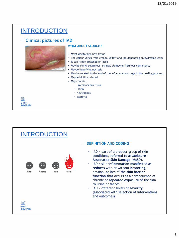
Clinical pictures of IADWHAT ABOUT SLOUGH?
• Moist devitalized host tissue
• The colour varies from cream, yellow and tan depending on hydration level
• It can firmly attached or loose
• May be slimy, gelatinous, stringy, clumpy or fibrinous consistency
• Maybe liquefying necrosis
• May be related to the end of the inflammatory stage in the healing process
• Maybe biofilm related
• May contain:
• Proteinaceous tissue
• Fibrin
• Neutrophils
• bacteria
INTRODUCTION
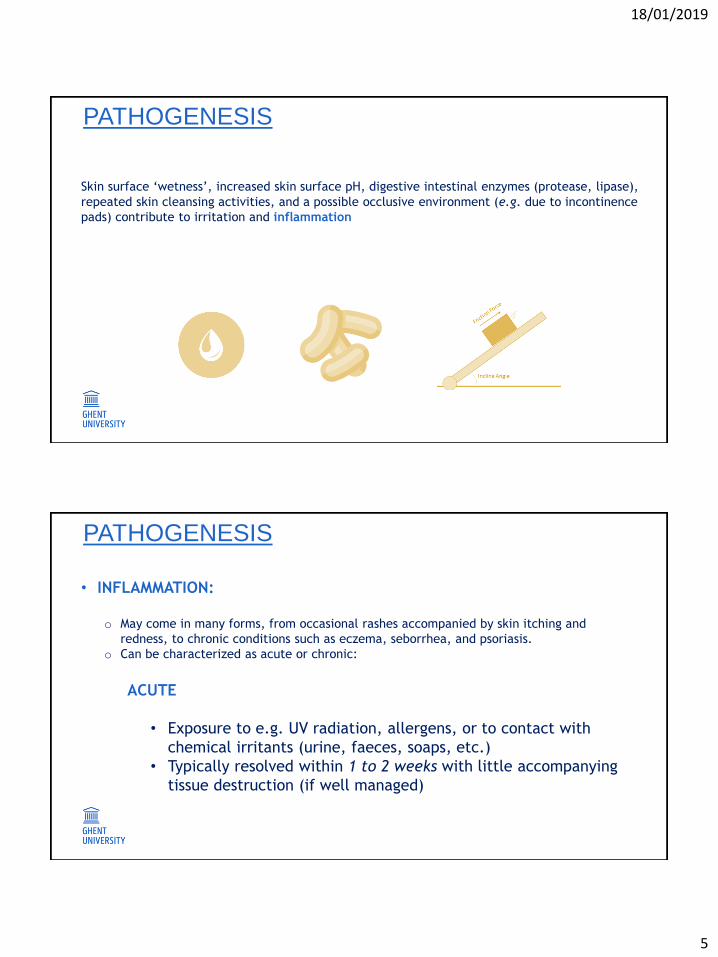
DEFINITION AND CODING
• IAD = part of a broader group of skin
conditions, referred to as Moisture-
Associated Skin Damage (MASD).
• IAD = skin inflammation manifested as
redness with or without blistering,
erosion, or loss of the skin barrier
function that occurs as a consequence of
chronic or repeated exposure of the skin
to urine or faeces.
• IAD = different levels of severity
(associated with selection of interventions
and outcomes)

18/01/2019
4
PATHOGENESIS
7
• An important function of the skin is to protect the body against pathogens.
• The stratum corneum (outermost layer of the epidermis) provides this critical barrier by prohibiting the invasion of micro-organisms
• Stratum corneum = 70% protein, 15% lipids, 15% water
• Lipids and water are important components in the skin’s barrier function
• In older patients, the volume of water decreases to less than 10%.
PATHOGENESIS
18/01/2019
5
Skin surface ‘wetness’, increased skin surface pH, digestive intestinal enzymes (protease, lipase),
repeated skin cleansing activities, and a possible occlusive environment (e.g. due to incontinence
pads) contribute to irritation and inflammation
PATHOGENESIS
• INFLAMMATION:
o May come in many forms, from occasional rashes accompanied by skin itching and
redness, to chronic conditions such as eczema, seborrhea, and psoriasis.
o Can be characterized as acute or chronic:
ACUTE
• Exposure to e.g. UV radiation, allergens, or to contact with
chemical irritants (urine, faeces, soaps, etc.)
• Typically resolved within 1 to 2 weeks with little accompanying
tissue destruction (if well managed)
PATHOGENESIS
18/01/2019
6
• INFLAMMATION:
o May come in many forms, from occasional rashes accompanied by skin itching and
redness, to chronic conditions such as eczema, seborrhea, and psoriasis.
o Can be characterized as acute or chronic:
CHRONIC
• Sustained immune cell mediated inflammatory response within
the skin itself
• Long lasting and can cause significant and serious tissue
destruction
PATHOGENESIS
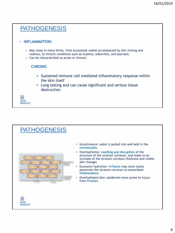
• Incontinence: water is pulled into and held in the corneocytes.
• Overhydration: swelling and disruption of the structure of the stratum corneum, and leads to an increase of the stratum corneum thickness and visible skin changes
• Excessive hydration: irritants may more easily penetrate the stratum corneum to exacerbate inflammation.
• Overhydrated skin: epidermis more prone to injury from friction.
PATHOGENESIS

18/01/2019
7
• Urease transforms urea into ammonium thus increasing the skin surface pH
• Increased skin surface pH: decreased stratum corneum cohesion and decreased recovery capacity ofskin barrier function, micro-organisms to thrive and increase the risk of skin infection
• Impaired skin barrier and occlusive skin conditions: facilitate the infiltration of the stratum corneum by the Candida Albicans, from the gastrointestinal tract, and Staphylococcus, from the perineal skin
• Lipases and proteases attack the the stratum corneum proteins and lipids.
PATHOGENESIS
ABOUT WHAT DO EXPERTS AGREE WHEN THEY OBSERVE IAD?
• Erythema and edema of the skin
• Sometimes accompanied by
bullae with serous exudate,
erosion, and infection
• No common language
PATHOGENESIS

18/01/2019
8
PATHOGENESIS
INDEPENDENT RISK FACTORS IN ICU
16

18/01/2019
9
INDEPENDENT RISK FACTORS
• Knowledge of risk factors is helpful to tailor IAD
prevention and management.
• IAD prevalence studies identified following key risk
factors for IAD:
o Liquid stool
o Critical illness
o Fever
o Diminished perfusion and oxygenation
o Poor skin condition (e.g. steroid use/diabetes)
o Restricted mobility and activity
o Higher score on care dependency
o Poor nutritional status
o Risk of friction and shear
o Restricted cognitive awareness
INDEPENDENT RISK FACTORS
WHAT DO WE KNOW FROM ICU PATIENTS?
• Matched case-control study in critically ill patients
• 30 hospitals (46 ICU/CCU wards) in Belgium
• 206 patients
• Fecal incontinent
Clinical trial registration:
https://clinicaltrials.gov/ct2/show/NCT02996357
18/01/2019
10
INDEPENDENT RISK FACTORS
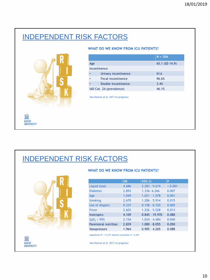
WHAT DO WE KNOW FROM ICU PATIENTS?
N = 206
Age 65.1 (SD 14.9)
Incontinence
• Urinary incontinence N/A
• Fecal incontinence 96.6%
• Double incontinence 3.4%
IAD Cat. 2A (prevalence) 46.1%
Van Damme et al. 2017 (in progress)
INDEPENDENT RISK FACTORS
WHAT DO WE KNOW FROM ICU PATIENTS?
OR 95% CI P
Liquid stool 4.686 2.283 – 9.619 < 0.001
Diabetes 2.893 1.336- 6.266 0.007
Age 1.049 1.021 – 1.078 0.001
Smoking 2.670 1.206 – 5.914 0.015
Use of diapers 0.337 0.158 – 0.720 0.005
Fever 2.603 1.226 – 5.528 0.013
Inotropics 4.109 0.845 – 19.970 0.080
SpO2 < 95% 2.154 1.034 – 4.484 0.040
Parenteral nutrition 2.839 1.000 – 8.055 0.050
Vasopressors 1.964 0.905 – 4.265 0.088
Nagelkerke R² = 0.377; Hosmer-Lemeshow P = 0.301
Van Damme et al. 2017 (in progress)
18/01/2019
11
OBSERVATION OF IADWITH THE GLOBIAD
21
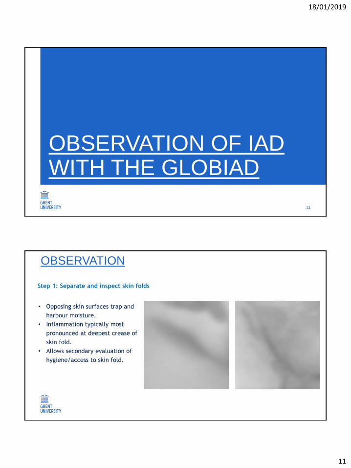
• Opposing skin surfaces trap and
harbour moisture.
• Inflammation typically most
pronounced at deepest crease of
skin fold.
• Allows secondary evaluation of
hygiene/access to skin fold.
Step 1: Separate and inspect skin folds
OBSERVATION

18/01/2019
12
• Opposing skin surfaces trap and
harbour moisture.
• Inflammation typically most
pronounced at deepest crease of
skin fold.
• Allows secondary evaluation of
hygiene/access to skin fold.
Step 1: Separate and inspect skin folds
OBSERVATION
• May present initially as islands of
partial thickness erosion.
• Often see multiple areas of erosion
closely spaced.
• Entire dermis may be eroded in severe
cases.
• Natural history not well defined.
Step 2: Assess for skin erosion
OBSERVATION

18/01/2019
13
• Opportunistic infection with Candida
Albicans.
• Thrives in warm, moist environment and
damages stratum corneum.
• Seen in 18% of one group of 608 acute care
inpatients (Junkin & Selekof, 2007).
Step 3: Inspect for secondary cutaneous infection, especially candidiasis
OBSERVATION
• A few tools have been developed for
assessment of IAD.
• While some of these have been
investigated for validity, reliability and
their use in day-to-day practice
remains limited.
• This is due in part to the lack of
evidence that these tools improve
clinical decision making and care.
WHY ANOTHER NEW TOOL?
18/01/2019
14
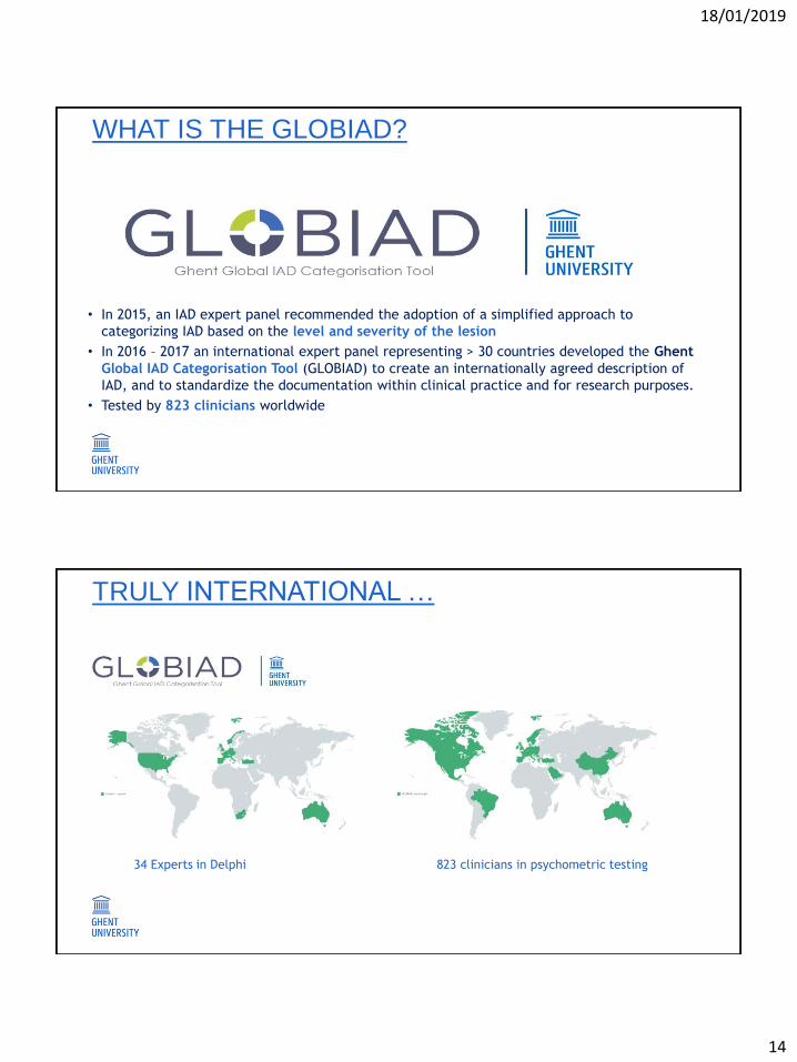
• In 2015, an IAD expert panel recommended the adoption of a simplified approach to
categorizing IAD based on the level and severity of the lesion
• In 2016 – 2017 an international expert panel representing > 30 countries developed the Ghent
Global IAD Categorisation Tool (GLOBIAD) to create an internationally agreed description of
IAD, and to standardize the documentation within clinical practice and for research purposes.
• Tested by 823 clinicians worldwide
WHAT IS THE GLOBIAD?
TRULY INTERNATIONAL …
34 Experts in Delphi 823 clinicians in psychometric testing

18/01/2019
15
29
CATEGORY 1A
30
18/01/2019
16
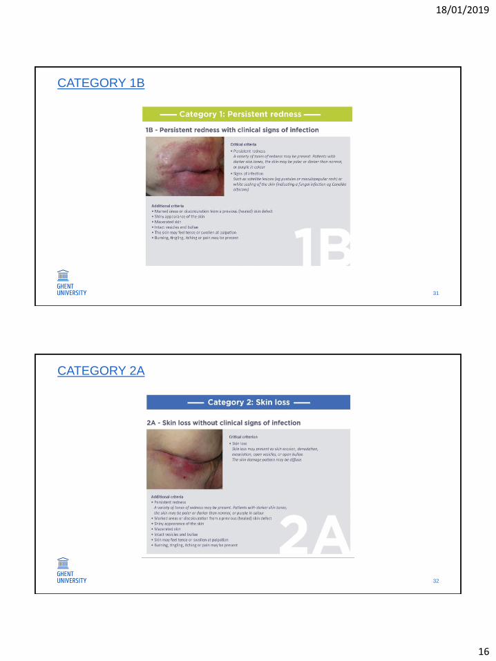
CATEGORY 1B
31
CATEGORY 2A
32
18/01/2019
17
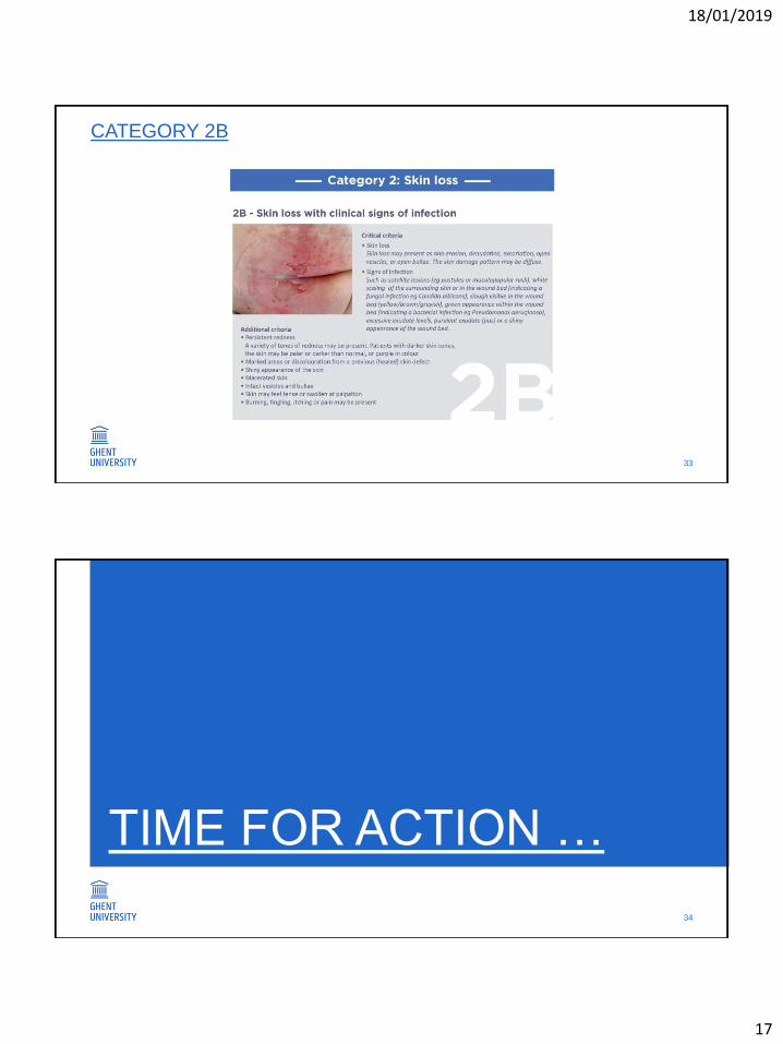
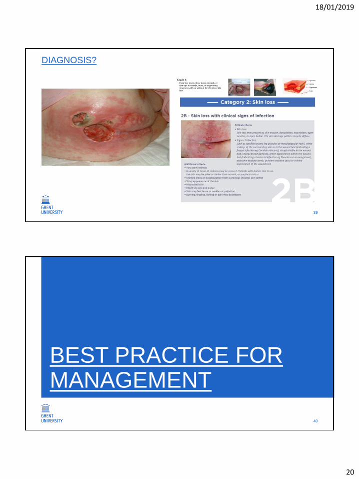
CATEGORY 2B
33
TIME FOR ACTION …
34

18/01/2019
18
DIAGNOSIS?
35
DIAGNOSIS?
36

18/01/2019
19
DIAGNOSIS?
37
DIAGNOSIS?
38
18/01/2019
20
DIAGNOSIS?
39
BEST PRACTICE FOR MANAGEMENT
40
18/01/2019
21
MANAGEMENT OF IAD
Incontinence care
• If incontinence is not treatable, use suitable incontinence material and replace
as frequent as needed
• Consider a bladder and/or bowl program and implement in close collaboration
with continence specialists
• Revise the incontinence approach regularly as changes will occur over time
• Provide a holistic approach to incontinence with attention to financial impact
and psychosocial well-being
MANAGEMENT OF IAD
Gentle cleansing of the genital, peri- anal and groin region
• Use gentle cleansing techniques (cfr. neonates)
• Reduce frequency of skin cleansing
• Avoid traditional soap (alkaline pH)
• Avoid influence on bacterial flora, leading to a selection of pathogens
• Consider using bath oil or shower oil without perfume, a pH neutral cleansing
foam, soft disposable washcloths
• Do not rub the skin after washing, simple patting of the skin is sufficient

18/01/2019
22
MANAGEMENT OF IAD
Skin hydration
• Hydrate dry skin with a hydrating topical product
• Consider the application of a cream (emulsions of water and oil), ointments
are greasy and tend to be more occlusive
o Dry skin: oil in water cream
o Very dry skin: rich cream (water in oil emulsion)
• Limit the amount of cream, especially in the folds, to avoid softening of the
skin and maceration
• Apply the cream in a gentle way, avoid rubbing
MANAGEMENT OF IAD
Application of a barrier leave-on product
• Aim is to protect the skin against further adverse effects of moisture and
incontinence
• Formulation often include Dimethicone or zinc oxide
• Overall formula vary widely, the product selection should depend on the skin
status.
• Most of the barrier products are creams combining barrier with hydrating
properties and are to be used on intact skin
• Some of the formula are liquid and able to desiccate open lesions

18/01/2019
23
MANAGEMENT OF IAD
• Pressure ulcer prevention remains important and is even more important in
patients with IAD. There is growing evidence that incontinence and IAD are risk
factors for the development of pressure ulcers.
• Control the nutritional status of the patient, more particular the protein and
vitamin intake, especially in patients with erosions or ulcerations
COMBINING GLOBIADAND MANAGEMENT
46
18/01/2019
24
COMBINING GLOBIAD AND MANAGEMENT
PREVENTION
• Cleans the perineal region
o Use X® foam and soft (single- use) washcloths
o Do not use water or soap
• Always dry the skin thoroughly after cleansing, if still moist
o Use soft towels
o Do not rub, apply a gentle patting technique
o Be sure that the skin is completely dry before applying other products and diaper/underpads
• Hydration
o Apply a thin layer of Y® hydrating barrier cream 2X / day
o Do not use any other topicals
• Evaluate the skin at least daily
o Whenever persistent redness is observed: apply the category 1 protocol
Beele H, Smet S, Van Damme N, Beeckman D. Incontinence-Associated Dermatitis: Pathogenesis, Contributing Factors, Prevention and Management
Options. Drugs Aging. 2018 Jan;35(1):1-10.
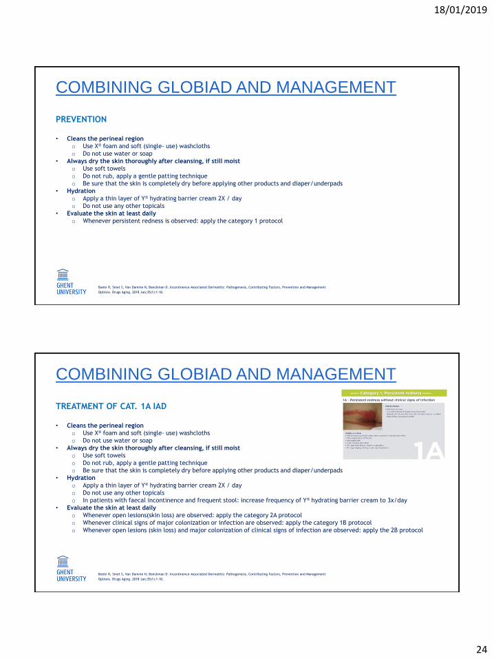
COMBINING GLOBIAD AND MANAGEMENT
TREATMENT OF CAT. 1A IAD
• Cleans the perineal region
o Use X® foam and soft (single- use) washcloths
o Do not use water or soap
• Always dry the skin thoroughly after cleansing, if still moist
o Use soft towels
o Do not rub, apply a gentle patting technique
o Be sure that the skin is completely dry before applying other products and diaper/underpads
• Hydration
o Apply a thin layer of Y® hydrating barrier cream 2X / day
o Do not use any other topicals
o In patients with faecal incontinence and frequent stool: increase frequency of Y® hydrating barrier cream to 3x/day
• Evaluate the skin at least daily
o Whenever open lesions(skin loss) are observed: apply the category 2A protocol
o Whenever clinical signs of major colonization or infection are observed: apply the category 1B protocol
o Whenever open lesions (skin loss) and major colonization of clinical signs of infection are observed: apply the 2B protocol
Beele H, Smet S, Van Damme N, Beeckman D. Incontinence-Associated Dermatitis: Pathogenesis, Contributing Factors, Prevention and Management
Options. Drugs Aging. 2018 Jan;35(1):1-10.

18/01/2019
25
COMBINING GLOBIAD AND MANAGEMENT
TREATMENT OF CAT. 2A IAD
• Cleans the perineal region
o Use X® foam and soft (single- use) washcloths
o Do not use water or soap
• Always dry the skin thoroughly after cleansing, if still moist
o Use soft towels
o Do not rub, apply a gentle patting technique
o Be sure that the skin is completely dry before applying other products and diaper/underpads
• Treatment of exuding lesions
o Apply Y® barrier lotion 2x/ day on the exudation zones
o Shake the lotion and apply a small amount on a sterile gauze and apply it to the lesion
• Hydration
o Apply a thin layer of Y® hydrating barrier cream 2x/ day
o Faecal incontinence and frequent stool: increase frequency of Y® barrier lotion or Y® hydrating barrier cream to 3x/day
• After desiccation of the exudation lesions
o Replace the Y® barrier lotion by Y® hydrating barrier cream
• Evaluate the skin on at least daily base
o Whenever the erosion becomes ulceration: clean the wound with saline and apply the wound care protocol
o Whenever clinical signs of major colonization or infection are observed: apply the category 2B protocol
Beele H, Smet S, Van Damme N, Beeckman D. Incontinence-Associated Dermatitis: Pathogenesis, Contributing Factors, Prevention and Management
Options. Drugs Aging. 2018 Jan;35(1):1-10.
COMBINING GLOBIAD AND
MANAGEMENT
TREATMENT OF CAT. 1B IAD
• Apply the 1A IAD protocol
• Evaluate the infection according to table 1
o In case of local signs of fungal infection:
Apply a paste containing Miconazol, 1 to 2 times a day. The paste should not
be removed before applying a new layer
Replace the paste containing Miconazol by Y® hydrating barrier cream if there
are no local signs of fungal infection anymore.
Beele H, Smet S, Van Damme N, Beeckman D. Incontinence-Associated Dermatitis: Pathogenesis, Contributing Factors, Prevention and Management
Options. Drugs Aging. 2018 Jan;35(1):1-10.

18/01/2019
26
COMBINING GLOBIAD AND MANAGEMENT
TREATMENT OF CAT. 2B IAD
• Apply the 2A IAD protocol
• Evaluate the infection according to table 1
o In the case of local signs of bacterial infection:
o Treatment of exudation lesions:
Apply povidon iodine lotion
o After desiccation of the exudation lesions:
Replace the povidon iodine lotion by Y® hydrating barrier cream
o Whenever there is a more profound ulceration: apply the wound care protocol for infected wounds
• In the case of local signs of fungal infection:
• Treatment of exudation lesions:
Apply povidon iodine lotion on the exudation lesions
• After desiccation of the exudation lesions:
Apply a paste containing Miconazol on the surrounding skin, 1 to 2 times a day, if there are still local signs of
fungal infection. The paste should not be removed before applying a new layer
Replace the povidon iodine lotion by Y® hydrating barrier cream if there are no local signs of fungal infection
anymore
• Whenever there is a more profound ulceration: apply the wound care protocol for infected wounds
Beele H, Smet S, Van Damme N, Beeckman D. Incontinence-Associated Dermatitis: Pathogenesis, Contributing Factors, Prevention and Management
Options. Drugs Aging. 2018 Jan;35(1):1-10.
COMBINING GLOBIAD AND
MANAGEMENT
TREATMENT OF CAT. 2B IAD
• In case of loco-regional clinical signs of infection:
o Apply the measures mentioned above for local signs of bacterial and/or fungal infection
o Start oral systemic antibiotics in an ambulatory setting:
If there is no penicillin allergy: use a penicillinase resistant penicillin or
amoxyclavulanic acid
If there is penicillin allergy: use clindamycin
• Whenever there is no decrease of the loco- regional clinical signs of infection after 3 days:
Adapt he antibiotics according to the results of the bacterial and fungal culture
and/or
Consider hospitalization of the patient
• Whenever there is an aggravation with systemic clinical signs of infection
Consider hospitalization of the patient
• In the case of systemic clinical signs of infection:
• Consider hospitalization of the patient
Beele H, Smet S, Van Damme N, Beeckman D. Incontinence-Associated Dermatitis: Pathogenesis, Contributing Factors, Prevention and Management
Options. Drugs Aging. 2018 Jan;35(1):1-10.

18/01/2019
27
TO CONCLUDE
53
• IAD is associated with weakened skin,
moisture/maceration, bacterial growth, and friction
leading to an inflammatory reaction and (in severe cases)
skin loss
• Key risk factors in ICU patients with fecal incontinence
include liquid stool, diabetes, fever, not using diapers,
age, history/current smoking and reduced perfusion and
oxygenation
• GLOBIAD is a valid tool to assess IAD severity, but training
is needed
• Next step: monitoring and minimum data-set (in progress)
TO CONCLUDE …

18/01/2019
28
REFERENCES & RESOURCES
55
REFERENCES & RESOURCESVan Damme N, Van den Bussche K, De Meyer D, Van Hecke A, Verhaeghe S, Beeckman D. Independent risk factors for the
development of skin erosion due to incontinence (incontinence-associated dermatitis category 2) in nursing home residents:
results from a multivariate binary regression analysis. Int Wound J. 2016 Dec 9.
Beeckman D, Van Damme N, Schoonhoven L, Van Lancker A, Kottner J, Beele H, Gray M, Woodward S, Fader M, Van den
Bussche K, Van Hecke A, De Meyer D, Verhaeghe S. Interventions for preventing and treating incontinence-associated dermatitis
in adults. Cochrane Database Syst Rev. 2016 Nov 10;11:CD011627. Review.
Van den Bussche K, De Meyer D, Van Damme N, Kottner J, Beeckman D. CONSIDER - Core Outcome Set in IAD Research:
study protocol for establishing a core set of outcomes and measurements in incontinence-associated dermatitis research. J Adv
Nurs. 2016 Sep 28.
Kottner J, Beeckman D. Core Outcome Sets (COS) for clinical trials in health- and nursing science: the case of Incontinence-
Associated Dermatitis (IAD). J Adv Nurs. 2016 Sep 14.
Beeckman D. A decade of research on Incontinence-Associated Dermatitis (IAD): Evidence, knowledge gaps and next steps. J
Tissue Viability. 2017
Feb;26(1):47-56.
Campbell JL, Coyer FM, Mudge AM, Robertson IM, Osborne SR. Candida albicans colonisation, continence status and
incontinence-associated dermatitis in the acute care setting: a pilot study. Int Wound J. 2016 Aug 1. doi: 10.1111/iwj.12630.

18/01/2019
29
REFERENCES & RESOURCESBeele H, Smet S, Van Damme N, Beeckman D. Incontinence-Associated Dermatitis: Pathogenesis, Contributing Factors,
Prevention and Management Options. Drugs Aging. 2018 Jan;35(1):1-10.
Gefen A. Tissue changes in patients following spinal cord injury and implications for wheelchair cushions and tissue loading: a
literature review.
Ostomy Wound Manage. 2014 Feb;60(2):34-45.
Kottner J, Beeckman D. Incontinence-associated dermatitis and pressure ulcers in geriatric patients. G Ital Dermatol Venereol.
2015 Dec;150(6):717-29.
Beeckman D., Schoonhoven L., European Pressure Ulcer Advisory Panel. PuClas3 eLearning Module. University Centre for
Nursing & Midwifery and European Pressure Ulcer Advisory Panel. 2015
Brennan MR, Milne CT, Agrell-Kann M, Ekholm BP. Clinical Evaluation of a Skin Protectant for the Management of Incontinence-
Associated Dermatitis: An Open-Label, Nonrandomized, Prospective Study. J Wound Ostomy Continence Nurs. 2017
Mar/Apr;44(2):172-180.
Bliss DZ, Mathiason MA, Gurvich O, Savik K, Eberly LE, Fisher J, Wiltzen KR, Akermark H, Hildebrandt A, Jacobson M, Funk T,
Beckman A, Larson R. Incidence and Predictors of Incontinence-Associated Skin Damage in Nursing Home Residents With New-
Onset Incontinence. J Wound Ostomy Continence Nurs. 2017
Prof. dr. Dimitri BeeckmanProfessor of Skin Integrity and Clinical Nursing
University Centre for Nursing and Midwifery
Skin Integrity Research Group (SKINT)
DEPARTMENT OF PUBLIC HEALTH
T +32 9 332 83 48
www.UGent.be
Ghent University
@ugent, @DimitriBeeckman, @UCVVGent
Ghent University