incidence of cerebellar tonsillar ectopia in idiopathic ... research incidence of cerebellar...
TRANSCRIPT

ORIGINALRESEARCH
Incidence of Cerebellar Tonsillar Ectopia inIdiopathic Intracranial Hypertension: A Mimic ofthe Chiari I Malformation
A.H. AikenJ.A. Hoots
A.M. SaindaneP.A. Hudgins
BACKGROUND AND PURPOSE: IIH is a syndrome of elevated intracranial pressure without hydroceph-alus, mass, or identifiable cause. Diagnosis is made by clinical presentation, intracranial pressuremeasurement, and supportive imaging findings. A subset of patients with IIH may have tonsillarectopia, meeting the criteria for Chiari malformation type I but not responding to surgical decompres-sion for Chiari I. The purpose of this study was to determine the incidence and morphology ofcerebellar tonsillar ectopia in patients with IIH.
MATERIALS AND METHODS: Forty-three patients with clinically confirmed IIH and 44 age-matchedcontrols were included. Two neuroradiologists with CAQs reviewed sagittal T1-weighted MRI in ablinded fashion and measured cerebellar tonsil and obex positions relative to the foramen magnum andprepontine cistern width at the level of the midpons.
RESULTS: Nine of 43 patients with IIH and 1/44 controls had cerebellar tonsillar ectopia of �5 mm. Fiveof 9 of patients with IIH with ectopia of �5 mm also had a “peglike” tonsil configuration. Patients withIIH had a significantly lower tonsillar position (2.1 � 2.8 mm) than age-matched controls (0.7 �1.9 mm,P � .05). The obex position was significantly lower in patients with IIH versus controls (�7.9 mm[above the FM] versus �9.4 mm [above the FM], P � .05). The prepontine width was not significantlydifferent between the groups.
CONCLUSIONS: Cerebellar tonsil position in patients with IIH was significantly lower than that inage-matched controls, often times peglike, mimicking Chiari I. A significantly lower obex positionsuggests an inferiorly displaced brain stem and cerebellum. When tonsillar ectopia of �5 mm isidentified, imaging and clinical consideration of IIH are warranted to avoid misdiagnosis as Chiari I.
ABBREVIATIONS: CAQ � Certificate of Added Qualification; CM � Chiari malformation; FM �foramen magnum; ICP � intracranial pressure; IIH � idiopathic intracranial hypertension
IIH, previously known as pseudotumor cerebri, is a syn-drome characterized by elevated ICP with normal CSF com-
position and no other identifiable cause.1 It has been proposedthat the elevated ICP may be related to decreased CSF resorp-tion due to impaired venous outflow and elevated venouspressure; however, controversy still surrounds the significanceof venous sinus stenosis in IIH as the cause or the result ofelevated ICP.2 IIH predominantly affects young overweight(body mass index �25) women with a reported incidence of19/100,000 in this population.2,3 Patients with IIH most com-monly present with headaches, occurring in 68%–98%.2,4
Other clinical features include pain, pulsatile tinnitus, and vi-sual disturbance, which can lead to blindness.2,5 Treatmentconsists of weight reduction, acetazolamide, and surgical in-tervention, including CSF shunt surgery.6-8
Although IIH is a clinical diagnosis based on normal CSFcomposition with an elevated opening pressure (�20 cm H2Oin nonobese patients and �25 cm H2O in obese patients withbody mass index �30), supportive neuroimaging findings
have been described. These include flattening of the posteriorsclera, tortuosity of the optic nerve sheath, empty sella syn-drome, and stenosis of the transverse venous sinuses.9,10
Therefore, imaging can aid in making or supporting the clin-ical diagnosis in some cases, especially if clinicians are not asfamiliar with the diagnosis. The incidence and morphology ofcerebellar tonsillar ectopia in IIH has not been previously de-scribed in the radiology literature, to our knowledge. Whenpresent, tonsillar ectopia in IIH may confuse the radiographicpicture and mimic other entities more commonly associatedwith tonsillar ectopia, such as Chiari I malformation andspontaneous intracranial hypotension.
Chiari I malformation is characterized by caudal protru-sion of “peg-shaped” cerebellar tonsils below the foramen.11,12
Chiari I malformation is defined radiographically as an infe-rior displacement of the cerebellar tonsils of �5 mm belowthe opisthion-basion line.13,14 In the healthy adult, cerebellartonsils are rarely �3 mm below the foramen magnum. Pa-tients with the radiographic appearance of Chiari I malforma-tion can be asymptomatic, but the most common clinicalsymptoms include headache, neck pain, vertigo, sensorychanges, and poor coordination. Therefore, clinical symptomsmay overlap IIH.11 Chiari I malformation is also associatedwith abnormal CSF flow, which can lead to syringomyelia.Treatment of Chiari I consists primarily of surgical hindbraindecompression with suboccipital craniectomy to restore nor-mal flow at the foramen magnum.15
Previous studies in the surgical literature describe a subset
Received December 1, 2011; accepted after revision January 12, 2012.
From the Department of Radiology and Imaging Sciences, Emory University, Atlanta,Georgia.
Paper previously presented at: 49th Annual Meeting of the American Society of Neurora-diology, June 4 –9, 2011; Seattle, Washington.
Please address correspondence to Ashley H. Aiken, MD, Radiology Department, EmoryUniversity Hospital, 1364 Clifton Rd, Suite BG 26, Atlanta, GA 30322; e-mail:[email protected]
http://dx.doi.org/10.3174/ajnr.A3068
BRA
INORIGIN
ALRESEARCH
AJNR Am J Neuroradiol 33:1901– 06 � Nov 2012 � www.ajnr.org 1901

of pretreatment patients with IIH with cerebellar tonsillar ec-topia, meeting the criteria for Chiari I.16-19 Fagan et al18 de-scribed a “Chiari pseudotumor cerebri syndrome” to highlightthe coexistence of Chiari I and IIH in some patients and thedifficulty in treatment. Most of these patients with IIH andpresumed Chiari I were initially treated with surgical decom-pression with no clinical improvement (Fig 1). However,many ultimately did respond to CSF shunt surgery, one of thetreatments for IIH.18 There is evidence in the clinical and sur-gical literature that Chiari and IIH may coexist. A previousstudy by Banik et al20 observed tonsillar ectopia of �2 mm in24% of patients with IIH and tonsillar ectopia of �5 mm (ra-diographic criterion of Chiari I) in only 11% of patients withIIH. Another study by Johnston et al21 found only 6% of pa-tients with IIH with radiographic criteria of Chiari I. Unfortu-nately, a cause and effect relationship has not been proved. Inother words, it is not clear whether patients with congenitalChiari I develop elevated intracranial pressures and a second-ary diagnosis of IIH or whether patients with IIH secondarilydevelop tonsillar ectopia, which may be mislabeled Chiari I.Therefore, treatment considerations may be complex. Regard-less of the cause and effect relationship, it has been shown thatpatients with IIH may be classified and treated as havingChiari I and may undergo surgical hindbrain decompression,yielding little to no benefit in this subset of patients.18,22
This study aimed to determine the incidence and morphol-ogy of cerebellar tonsillar ectopia in patients with IIH to fur-ther clarify the relationship between a clinical diagnosis of IIHand the presence of cerebellar tonsillar ectopia of �5 mm. Thisstudy emphasizes the importance of clinical history and sup-plementary radiographic evidence to distinguish cerebellartonsillar ectopia of �5 mm in the setting of IIH from the“typical” Chiari I.
Materials and Methods
Patient PopulationAfter obtaining institutional review board approval, a retrospective
search for the terms “idiopathic intracranial hypertension” or “pseu-
dotumor cerebri” in MR imaging reports from 2008 to 2010 yielded
90 patients. After a comprehensive chart review, only 46 patients
(mean age, 36 � 12 years; range, 18 – 61 years; 45 female) had a clin-
ically confirmed diagnosis of IIH with ICP measurements (�20 cm
H2O for nonobese patients and �25 cm H2O for obese patients). An
inclusion criterion was also MR imaging with a sagittal T1-weighted
sequence. Three female patients were excluded because MR images
did not include sagittal T1-weighted images. Forty-four age-matched
control patients were selected (mean age, 40 � 12 years; range, 18 – 61
years; 27 female).
Image ReviewTwo neuroradiologists with CAQs reviewed selected sagittal T1-
weighted MR images of the 43 patients with IIH and 44 age-matched
controls in a blinded fashion. The cerebellar tonsil and obex positions
relative to the foramen magnum were measured by drawing a line
from the basion to the opisthion to define the plane of the foramen
magnum.23 If the tonsils were above this reference line, the measure-
ment was assigned a negative number; if the tonsils were below the
line, the measurement was a positive number. All obex positions were
above this reference line and were assigned a positive number (Fig 2).
In addition, the prepontine cistern width at the level of the midpons
was measured. Measurements from patients with IIH and control
subjects were compared with standard 2-sample t tests, assuming
equal variances. P values �.05 were considered statistically
significant.
ResultsCerebellar tonsillar ectopia of �5 mm was found in 9/43 pa-tients with IIH (20.9%) and in only 1/44 control subjects(2.2%). Five of the 9 patients with IIH with tonsillar ectopiaof �5 mm also had a peglike configuration of their cerebellartonsils, closely mimicking CM (Fig 3). Of the 9 patients withIIH with tonsillar ectopia of �5 mm, 8/9 responded to treat-ment for IIH alone. One patient ultimately underwent a sur-gical decompression for CM. Patients with IIH had signifi-cantly lower mean tonsillar positions (2.1 � 2.8 mm) thanage-matched controls (0.7 � 1.9 mm, P � .05) (Fig 4). Therewas no statistical difference in mild cerebellar tonsillar ectopia(2– 4 mm) between patients with IIH and controls. Twelve of
Fig 1. A 43-year-old woman initially diagnosed with Chiari I and treated with surgical decompression. The patient had persistent headaches and recurrent pseudomeningoceles at thesurgical site. The patient was ultimately diagnosed with IIH and underwent ventriculoperitoneal shunt surgery with symptomatic relief. A, Sagittal T2-weighted image shows a peglikeherniation of the cerebellar tonsils (arrow). However, a partially empty sella is also noted (arrow), which could have been a clue to the underlying or coexistent IIH. B and C, Sagittal andaxial T2WI shows the postsurgical changes from suboccipital craniectomy and a complex extracranial fluid collection, compatible with pseudomeningocele (arrow).
1902 Aiken � AJNR 33 � Nov 2012 � www.ajnr.org
43 patients with IIH (28%) and 12 of 44 control patients(27%) had tonsillar ectopia between 2 and 4 mm.
The prepontine cistern width was not significantly differ-
ent between the 2 groups (P � .3). The mean obex positionwas significantly lower in patients with IIH versus healthy con-trols (�7.9 � 2.8 mm [above the opisthion-basion line] versus�9.4 � 2.9 mm; P � .05) (Fig 4). When we compared the obexposition, the subset of patients with cerebellar tonsillar ectopiain the IIH group also had a significantly lower obex positionthan in the control patients (�5.6 � 2.0 mm [above theopisthion-basion line] versus �9.4 � 2.9 mm; P � .05). Re-sults are also summarized in the Table. Note that negativenumbers denote a position above and positive numbers, a po-sition below the opisthion-basion line.
DiscussionThe cerebellar tonsillar position is below the foramen mag-num in Chiari I and may or may not be low-lying in IIH.Cerebellar tonsillar ectopia is not diagnostic of Chiari I only,and one should take care when interpreting cross-sectionalimaging because all cerebellar tonsillar ectopia does not equalChiari I malformation. In this study, a statistically significantnumber of patients with IIH were found to have tonsillar ec-topia of �5 mm, mimicking a Chiari I malformation. Wefound a higher incidence (21%) of tonsillar ectopia of �5 mm
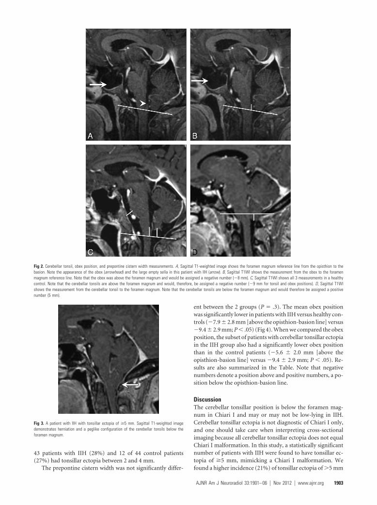
Fig 2. Cerebellar tonsil, obex position, and prepontine cistern width measurements. A, Sagittal T1-weighted image shows the foramen magnum reference line from the opisthion to thebasion. Note the appearance of the obex (arrowhead) and the large empty sella in this patient with IIH (arrow). B, Sagittal T1WI shows the measurement from the obex to the foramenmagnum reference line. Note that the obex was above the foramen magnum and would be assigned a negative number (�8 mm). C, Sagittal T1WI shows all 3 measurements in a healthycontrol. Note that the cerebellar tonsils are above the foramen magnum and would, therefore, be assigned a negative number (�9 mm for tonsil and obex positions). D, Sagittal T1WIshows the measurement from the cerebellar tonsil to the foramen magnum. Note that the cerebellar tonsils are below the foramen magnum and would therefore be assigned a positivenumber (5 mm).
Fig 3. A patient with IIH with tonsillar ectopia of �5 mm. Sagittal T1-weighted imagedemonstrates herniation and a peglike configuration of the cerebellar tonsils below theforamen magnum.
AJNR Am J Neuroradiol 33:1901– 06 � Nov 2012 � www.ajnr.org 1903
in patients with IIH than the 10% previously reported byBanik et al.20 Banik et al used different terminology than thecurrent authors. We have not used the term Chiari I malfor-mation in the setting of IIH. Instead, we have used cerebellartonsillar ectopia to refer to tonsil position in patients with IIHthat may be acquired rather than a malformation but meetsthe radiographic criteria for Chiari I. We believe that using theterm CM in the setting of IIH can be confusing and could evenresult in inappropriate treatment until a relationship has beenproved. Banik et al reported that 24% of patients with IIH hadtonsillar ectopia of �2 mm, but only 10% had tonsillar ectopiaof �5 mm.20 The difficulty in managing this subset of patientswith a diagnosis of IIH and cerebellar tonsillar ectopia of �5mm (meeting the criterion for Chiari I) has been previouslyreported in the surgical literature, though there is little discus-sion in the radiologic literature.17,18,20,22
Chiari I is considered a disorder of the paraxial mesodermwith hindbrain maldevelopment and small posterior fossa vol-ume. IIH has evidence of elevated intracranial pressure, al-tered CSF absorption, and intracranial compliance.21,22 Therelationship between these 2 diagnoses has been establishedbut is poorly understood. It is possible that elevated intracra-nial pressure in IIH may cause cerebellar tonsils to herniatethrough the foramen magnum, manifesting imaging criteriaof Chiari I. Alternatively, it is possible that patients with Chiari
I have abnormal CSF dynamics, which predispose to elevatedintracranial pressure and IIH. It is postulated that patientswith coexistence of Chiari I and IIH may have relief after pos-terior fossa decompression, which alters compliance. How-ever, symptoms often recur, and these patients with IIH maysubsequently require a CSF shunt surgery procedure (Fig 1).
However, the reverse sequence of events has also been re-ported and was encountered in our cohort. Patients with IIHwith cerebellar tonsillar ectopia initially treated with CSFshunt surgery may go on to require decompression. One of the9 patients with IIH with tonsillar ectopia of �5 mm ultimatelyrequired a surgical decompression. During the course of 10years, this patient was treated with both a ventriculoperitonealshunt and a lumbar peritoneal shunt. Initially, the patient re-sponded well to these treatments with dramatic relief of herIIH symptoms. Her course was complicated by low-pressureheadaches treated with a shunt revision, which may have con-tributed to the appearance of her tonsils. Ultimately, she de-veloped refractory suboccipital headaches that were treatedwith suboccipital craniectomy. Her surgeon noted that hertonsillar ectopia was not a typical Chiari I but was likely ac-quired tonsillar ectopia from longstanding IIH and a func-tioning lumbar peritoneal shunt. Indeed acquired tonsillar ec-topia meeting the radiographic criteria for Chiari I and evensyringomyelia has been described in patients with IIH afterlumboperitoneal shunt surgery.24 Nevertheless, our surgeonconcluded that her symptoms and refractory suboccipitalheadache were due to crowding at the foramen magnum. Sheunderwent surgical decompression for a presumed diagnosisof Chiari I with a complicated course (Fig 5).
Although there is no consensus about the ideal manage-ment for these patients, it is clear that careful consideration,upfront measurement of intracranial pressure, and inclusion/exclusion of classic IIH are important before surgically treat-ing any patient with Chiari I or IIH with secondary tonsillar
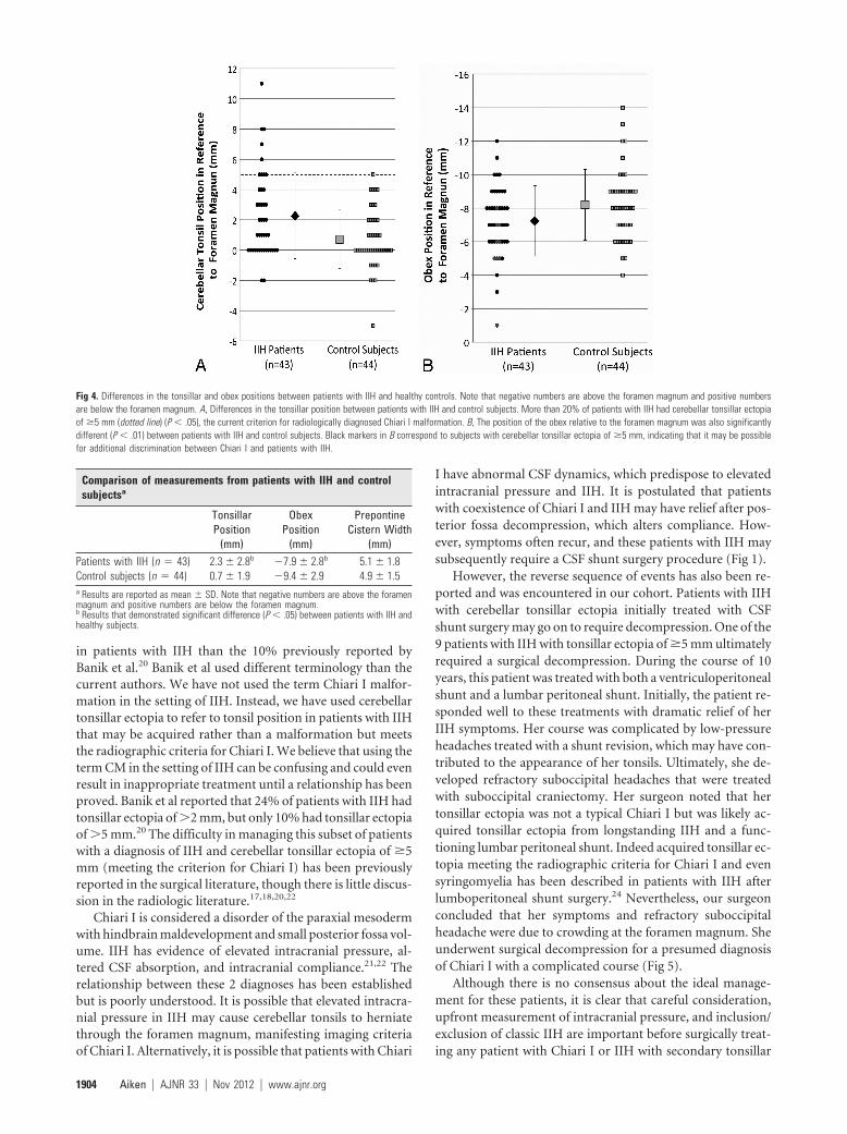
Fig 4. Differences in the tonsillar and obex positions between patients with IIH and healthy controls. Note that negative numbers are above the foramen magnum and positive numbersare below the foramen magnum. A, Differences in the tonsillar position between patients with IIH and control subjects. More than 20% of patients with IIH had cerebellar tonsillar ectopiaof �5 mm (dotted line) (P � .05), the current criterion for radiologically diagnosed Chiari I malformation. B, The position of the obex relative to the foramen magnum was also significantlydifferent (P � .01) between patients with IIH and control subjects. Black markers in B correspond to subjects with cerebellar tonsillar ectopia of �5 mm, indicating that it may be possiblefor additional discrimination between Chiari I and patients with IIH.
Comparison of measurements from patients with IIH and controlsubjectsa
TonsillarPosition
(mm)
ObexPosition
(mm)
PrepontineCistern Width
(mm)Patients with IIH (n � 43) 2.3 � 2.8b �7.9 � 2.8b 5.1 � 1.8Control subjects (n � 44) 0.7 � 1.9 �9.4 � 2.9 4.9 � 1.5a Results are reported as mean � SD. Note that negative numbers are above the foramenmagnum and positive numbers are below the foramen magnum.b Results that demonstrated significant difference (P � .05) between patients with IIH andhealthy subjects.
1904 Aiken � AJNR 33 � Nov 2012 � www.ajnr.org
ectopia mimicking Chiari I. For these reasons, radiologists canplay a critical role in suggesting IIH instead of Chiari I, on thebasis of additional imaging features such as transverse sinusstenosis, empty sella, and tortuous optic nerves (Fig 6).25
Patients with IIH also had a significantly lower mean ton-sillar position compared with healthy controls, again suggest-ing that a low cerebellar tonsil position may be acquired inpatients with IIH, possibly due to chronically elevated ICP.However, there was no statistical difference in mild tonsillarectopia (2– 4 mm) between the patients with IIH and controls.The incidence of mild cerebellar tonsillar ectopia in our pa-tients with IIH (28%) was similar to that previously reportedby Banik et al (24%).20 Unlike the Banik et al, we also com-pared our patients with IIH with healthy controls. Initially wewere surprised that the incidence of mild tonsillar ectopia wassimilar in our patients with IIH and healthy controls. Thereason for this similarity was that the mean tonsillar positionin our healthy controls (0.7 � 1.9 mm below the foramenmagnum) was significantly lower relative to the opisthion-basion line than previously reported. Previously Barkovich
et al13 and Aboulezz et al14 reported a mean tonsillar positionof 1 and 2.9 mm, respectively, above the foramen magnum.
Two other measurements were performed during thisstudy: 1) prepontine cistern width and 2) obex position rela-tive to the foramen magnum. There was no statistical differ-ence in prepontine cistern width between the controls andpatients with IIH. The obex position, defined as where thefourth ventricle becomes the central canal of the cervical spi-nal cord, is a marker for the cervicomedullary junction. Theobex position relative to the foramen magnum was found tobe significantly different in patients with IIH relative to con-trols (7.9 � 2.8 mm above the FM versus 9.4 � 2.9 mm aboveFM; P � .01). This difference implies that there is a downwardshift of the brain stem in patients with IIH. Patients withChiari I are expected to have a normal obex position, thoughthere have been reports of mild descent of the fourth ventricleand medulla.26 If one considered only the subset of 9 patientswith IIH with tonsillar ectopia of �5 mm, the obex positionwas even more significantly different from that in control sub-jects (5.0 mm � 2.0 mm versus 9.4 mm � 2.9 mm, P � .005).Hence, it may be possible to select a threshold for the obexposition (Fig 4B) that could be used as an additional radiologicfinding to separate 2 distinct patient populations that cur-rently overlap. These findings are intriguing, but a futurestudy of the obex position in patients with IIH with tonsillarectopia of �5 mm versus Chiari I would be needed to clarifythe meaning of this finding.
Limitations and Future DirectionsIn the current study, patients with IIH were compared withhealthy controls to determine the incidence and morphologyof cerebellar tonsillar ectopia in IIH. Control cases were ran-domly selected from patients with normal MRI findingsscanned for other reasons, rather than healthy volunteers.23
The use of control subjects with an average tonsillar ectopia of0.7 � 1.9 mm (range, �5 to 5 mm, 5/44 subjects having mildcerebellar tonsillar ectopia between 2 and 4 mm) may under-estimate the difference in incidence of mild tonsillar ectopiabetween patients with IIH versus healthy controls because our
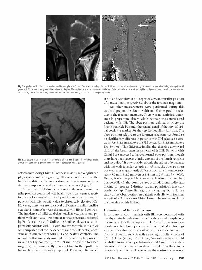
Fig 5. A patient with IIH with cerebellar tonsillar ectopia of �5 mm. This was the only patient with IIH who ultimately underwent surgical decompression after being managed for 12years with CSF shunt surgery procedures alone. A, Sagittal T2-weighted image demonstrates herniation of the cerebellar tonsils with a peglike configuration and crowding at the foramenmagnum. B, Cine CSF flow study shows loss of CSF flow posteriorly at the foramen magnum (arrow).
Fig 6. A patient with IIH with tonsillar ectopia of �5 mm. Sagittal T1-weighted imageshows herniation and a peglike configuration of cerebellar tonsils (arrow).
AJNR Am J Neuroradiol 33:1901– 06 � Nov 2012 � www.ajnr.org 1905

control patients had a lower tonsillar position than previouslyreported for normal controls (as stated in the Discussion sec-tion). This secondary observation is intriguing because it mayreflect geographic differences in the position of cerebellar ton-sils relative to the FM, possibly reflecting a difference in aver-age body mass index and preclinical IIH. However, a widevariation of normal is expected, and our sample size of healthycontrols was small so that a conclusion regarding the variationof tonsil position among healthy controls cannot be made andwas not the purpose of the current study.
A larger study of tonsillar position in healthy controls withclose attention to body mass index, sex, and age would beneeded to determine the validity and significance of this sec-ondary observation. The measurement of the obex position inpatients with IIH compared with controls showed a statisticaldifference, but no patients with Chiari I were included forcomparison; therefore, the true meaning of this finding fordistinguishing among the etiologies of cerebellar tonsillar ec-topia is unclear. Future study of the obex position in patientswith IIH and Chiari I would be helpful to determine the sig-nificance of this finding to distinguish among the differentpathophysiologic etiologies of cerebellar tonsillar ectopia.
ConclusionsThe cerebellar tonsillar position in patients with IIH is signif-icantly lower than that in age-matched controls. A significantproportion (21%) of our patients with IIH had cerebellar ton-sillar ectopia of �5 mm, which was often peglike, mimickingChiari I. The obex position was significantly lower in patientswith IIH, suggesting that the brain stem and cerebellum areboth shifted inferiorly. When cerebellar tonsillar ectopia isidentified, careful radiographic and clinical consideration ofIIH is warranted to avoid misdiagnosis as Chiari I alone.
Disclosures: Patricia Hudgins—UNRELATED: Consultancy, Royalties, and Stock/Stock Op-tions: Amirsys, Comments: medical education company.
References1. Friedman DI, Jacobson DM. Diagnostic criteria for idiopathic intracranial
hypertension. Neurology 2002;59:1492–952. Ball AK, Clarke CE. Idiopathic intracranial hypertension. Lancet Neurol 2006;
5:433– 423. Durcan FJ, Corbett JJ, Wall M. The incidence of pseudotumor cerebri: popu-
lation studies in Iowa and Louisiana. Arch Neurol 1988;45:875–774. Radhakrishnan K, Thacker AK, Bohlaga NH, et al. Epidemiology of idiopathic
intracranial hypertension: a prospective and case-control study. J Neurol Sci1993;116:18 –28
5. Mensah A, Milea D, Jensen R, et al. Persistent visual loss in malignant idio-pathic intracranial hypertension. Acta Ophthalmol 2009;87:934 –36
6. Hammers R, Prabhu VC, Sarker S, et al. Laparoscopic-assisted lumboperito-neal shunt placement for idiopathic intracranial hypertension. Semin Oph-thalmol 2008;23:151–55
7. Abubaker K, Ali Z, Raza K, et al. Idiopathic intracranial hypertension: lumbo-peritoneal shunts versus ventriculoperitoneal shunts— case series and liter-ature review. Br J Neurosurg 2011;25:94 –99
8. Ball AK, Howman A, Wheatley K, et al. A randomised controlled trial of treat-ment for idiopathic intracranial hypertension. J Neurol 2011;258:874 – 81
9. Suzuki H, Takanashi J, Kobayashi K, et al. MR imaging of idiopathic intracra-nial hypertension. AJNR Am J Neuroradiol 2001;22:196 –99
10. Gibby WA, Cohen MS, Goldberg HI, et al. Pseudotumor cerebri: CT findingsand correlation with vision loss. AJR Am J Roentgenol 1993;160:143– 46
11. Aitken LA, Lindan CE, Sidney S, et al. Chiari type I malformation in a pediatricpopulation. Pediatr Neurol 2009;40:449 –54
12. Milhorat TH, Chou MW, Trinidad EM, et al. Chiari I malformation redefined:clinical and radiographic findings for 364 symptomatic patients. Neurosurgery1999;44:1005–17
13. Barkovich AJ, Wippold FJ, Sherman JL, et al. Significance of cerebellar tonsillarposition on MR. AJNR Am J Neuroradiol 1986;7:795–99
14. Aboulezz AO, Sartor K, Geyer CA, et al. Position of cerebellar tonsils in thenormal population and in patients with Chiari malformation: a quantitativeapproach with MR imaging. J Comput Assist Tomogr 1985;9:1033–36
15. Attenello FJ, McGirt MJ, Gathinji M, et al. Outcome of Chiari-associated sy-ringomyelia after hindbrain decompression in children: analysis of 49 con-secutive cases. Neurosurgery 2008;62:1307–13, discussion 1313
16. Kurschel S, Maier R, Gellner V, et al. Chiari I malformation and intra-cranialhypertension: a case-based review. Childs Nerv Syst 2007;23:901– 05
17. Bejjani GK, Cockerham KP, Rothfus WE, et al. Treatment of failed adult Chiarimalformation decompression with CSF drainage: observations in six pa-tients. Acta Neurochir (Wien) 2003;145:107–16, discussion 116
18. Fagan LH, Ferguson S, Yassari R, et al. The Chiari pseudotumor cerebrisyndrome: symptom recurrence after decompressive surgery for Chiari mal-formation type I. Pediatr Neurosurg 2006;42:14 –19
19. Sinclair N, Assaad N, Johnston I. Pseudotumour cerebri occurring in associa-tion with the Chiari malformation. J Clin Neurosci 2002;9:99 –101
20. Banik R, Lin D, Miller NR. Prevalence of Chiari I malformation and cerebellarectopia in patients with pseudotumor cerebri. J Neurol Sci 2006;247:71–75
21. Johnston I, Hawke S, Halmagyi M, et al. The pseudotumor syndrome: disor-ders of cerebrospinal fluid circulation causing intracranial hypertensionwithout ventriculomegaly. Arch Neurol 1991;48:740 – 47
22. Bejjani GK. Association of the adult Chiari malformation and idiopathic in-tracranial hypertension: more than a coincidence. Med Hypotheses 2003;60:859 – 63
23. O’Connor S, du Boulay G, Logue V. The normal position of the cerebellartonsils as demonstrated by myelography. J Neurosurg 1973;39:387– 89
24. Padmanabhan R, Crompton D, Burn D, et al. Acquired Chiari 1 malformationand syringomyelia following lumboperitoneal shunting for pseudotumourcerebri. J Neurol Neurosurg Psychiatry 2005;76:298
25. Farb RI, Vanek I, Scott JN, et al. Idiopathic intracranial hypertension: theprevalence and morphology of sinovenous stenosis. Neurology 2003;60:1418 –24
26. Meadows J, Kraut M, Guarnieri M, et al. Asymptomatic Chiari type I malfor-mations identified on magnetic resonance imaging. J Neurosurg 2000;92:920 –26
1906 Aiken � AJNR 33 � Nov 2012 � www.ajnr.org