in vivo efficacy of a dry powder formulation of
TRANSCRIPT
HAL Id: inserm-02615177https://www.hal.inserm.fr/inserm-02615177
Submitted on 22 May 2020
HAL is a multi-disciplinary open accessarchive for the deposit and dissemination of sci-entific research documents, whether they are pub-lished or not. The documents may come fromteaching and research institutions in France orabroad, or from public or private research centers.
L’archive ouverte pluridisciplinaire HAL, estdestinée au dépôt et à la diffusion de documentsscientifiques de niveau recherche, publiés ou non,émanant des établissements d’enseignement et derecherche français ou étrangers, des laboratoirespublics ou privés.
In vivo Efficacy of a Dry Powder Formulation ofCiprofloxacin-Copper Complex in a Chronic LungInfection Model of Bioluminescent Pseudomonas
aeruginosaFrederic Tewes, Tania Bahamondez-Canas, Daniel Moraga-Espinoza, Hugh
Smyth, Alan Watts
To cite this version:Frederic Tewes, Tania Bahamondez-Canas, Daniel Moraga-Espinoza, Hugh Smyth, Alan Watts. Invivo Efficacy of a Dry Powder Formulation of Ciprofloxacin-Copper Complex in a Chronic LungInfection Model of Bioluminescent Pseudomonas aeruginosa. European Journal of Pharmaceutics andBiopharmaceutics, Elsevier, 2020, 152, pp.201-217. �10.1016/j.ejpb.2020.05.014�. �inserm-02615177�

1
In vivo Efficacy of a Dry Powder Formulation of Ciprofloxacin-Copper
Complex in a Chronic Lung Infection Model of Bioluminescent
Pseudomonas aeruginosa
Frederic Tewes1,2,*, Tania F. Bahamondez-Canas2,3,4, Daniel Moraga-Espinoza2,3,4,
Hugh D.C. Smyth2, Alan B. Watts2
1 INSERM U1070, UFR de Médecine Pharmacie, Université de Poitiers, 1 rue Georges Bonnet,
TSA 51106, 86073 Poitiers Cedex 9, France
2 College of Pharmacy, The University of Texas at Austin, 2409 West University Avenue, PHR
4.214, Austin, TX 78712, USA
3 Escuela de Farmacia, Universidad de Valparaiso, Gran Bretaña 1093, Playa Ancha, Valparaíso,
Chile
4 Centro de Investigación Farmacopea, Universidad de Valparaíso, Santa Marta 183, Playa
Ancha, Valparaíso, Chile
* Corresponding author: [email protected]

2
ABSTRACT
A significant limitation of locally delivered treatments for chronic pulmonary infections
is often the short residence time within the airways. Ciprofloxacin (CIP), for example, undergoes
rapid absorption from the airway lumen. Previously, we demonstrated that the complexation of
CIP with copper (CIP-Cu) reduces its apparent epithelial permeability and pulmonary absorption
rate without affecting antimicrobial activity against Pseudomonas aeruginosa grown
planktonically or as biofilms. This study aimed to evaluate the in vivo efficacy of CIP-Cu,
prepared as a dry powder, in a chronic lung infection model. The powders were prepared by jet
milling (CIP-HCl) and by spray drying (CIP-Cu). A bioluminescent strain of P. aeruginosa
(PAO1::p16Slux) was used to prepare bacteria-loaded agar beads that were inoculated
intratracheally to rats. The dynamics of the infection were monitored using luminometry. The
bacteria/beads ratio was optimized to allow the highest luminescence signal and animal survival
for 8 days. The efficacy of the treatment was evaluated by luminometry in addition to the end-
point (Day 8) where colony counting was performed after lung harvesting. Luminescent P.
aeruginosa entrapped in agar beads were useful to monitor the spatial development of the
chronic lung infection in rats. The rats were treated with the dry powders in a nose-only
inhalation exposure system (NOIES). CIP-Cu and CIP-HCl powders showed similar
aerodynamic properties and comparable CIP lung deposition. However, treatment with CIP-Cu
significantly (p<0.01) reduced by 4-log the number of CFU of P. aeruginosa per lung in the
chronic infection model, whereas CIP-HCl effect was not different from the untreated control
group.
3
1. INTRODUCTION
Effective therapy using pulmonary delivery of antibiotics can be limited by the short drug
residence time at the site of infection, but a novel complexed form of ciprofloxacin may help
overcome this obstacle while maintaining antimicrobial activity against resistant biofilm
bacterial infections [1, 2]. Pulmonary delivery of antibiotics in solution form has demonstrated
clinical benefit in the treatment of chronic bacterial lung infections, as it can allow for
significantly higher drug concentrations at the site of infection with reduced systemic levels
compared to i.v. administration [3]. This is mainly true for antibiotics having very low lung-
blood barrier permeability, such as the currently used tobramycin, colistin methanesulfonate and
aztreonam lysine. However, this approach is less suitable for molecules that readily diffuse
through the lung epithelial barrier and rapidly equilibrate with blood concentrations, such as in
the case of ciprofloxacin (CIP) [4-7]. Thus, for antimicrobial molecules having relatively high
permeability across the lung-blood barrier, advanced formulations can be developed to retain the
drug in the airways by slowing down the drug flux across this barrier. These advanced
formulations generally act either by reducing and sustaining the drug release rate from
formulation [8-13], or by reducing the apparent permeability of the drug across the epithelium [2,
14-17]. In order to increase CIP lung residency time after its aerosolization, we recently
developed a novel positively charged complex made of CIP and copper [2, 14, 18]. This CIP-
copper complex (CIP-Cu) allowed a 90% decrease in the apparent permeability of CIP across a
Calu-3 cell line model of pulmonary epithelium [2, 18]. This decrease in permeability translated
in vivo as a decrease in the lung-blood absorption rate of CIP and a sustained high CIP
concentration in the lung epithelial lining fluid (ELF) after intratracheal delivery to rats [2].
Additionally, the in vitro antibacterial activities of CIP and CIP-Cu evaluated against P.
4
aeruginosa grown in planktonic cultures [14, 18] or as biofilms [1] were also found to be
equivalent.
The aim of this study was, therefore, to evaluate the in-vivo efficacy of CIP-Cu complex-
loaded particles after pulmonary inhalation in model rats that developed chronic pulmonary
infection by instillation of Pseudomonas aeruginosa-loaded agar beads [19, 20]. This approach
has been shown to model the chronic infection and inflammation that affect cystic fibrosis
patients [19, 21, 22]. In order to assess temporal effects of the treatments on the infection, we
also evaluated time resolved luminescence of a bioluminescent P. aeruginosa, PAO1 strain to
non-invasively monitor the chronic lung infections in rats.

5
2. MATERIALS AND METHODS
2.1. Materials
The bioluminescent PAO1::p16Slux, a PAO1 strain (gift from Professor Shawn Lewenza
from the university of Calgary, Canada) tagged by chromosomal integration of p16Slux
construction [23], was used to prepare bacteria-loaded agar beads and monitor the chronic lung
infection. Ciprofloxacin base (CIP) powder (purity ≥ 98,0 %), copper hydroxide Cu(OH)2,
hyaluronic acid (HA) sodium salt from Streptococcus equi, calcium hydroxide Ca(OH)2, formic
acid and ammonium carbonate (NH4)2CO3 were purchased from Sigma Aldrich (St Louis, USA).
Ciprofloxacin hydrochloride monohydrate USP grade was purchased from Letco Medical
(Decatur, AL, USA)
2.2. Preparation and characterization of the dry powder formulations
2.2.1. Preparation of ciprofloxacin-copper loaded microparticles
Microparticles composed of amorphous calcium carbonate and CIP-Cu complex (Spray
dried CIP-Cu) were prepared by spray-drying as previously described [14]. Briefly, two aqueous
solutions were prepared. One solution was made of CIP, calcium hydroxide, sodium hyaluronate,
formic acid, and copper hydroxide. The other one was made of ammonium carbonate. These
solutions were prepared separately and mixed during the microparticle preparation process using
a Y-tube connected to the feeding tube of the B-290 mini spray dryer (Büchi, Labortechnik AG,
Switzerland). The spray dryer was operated in open mode with 27 mL/min peristaltic pump rate,
15 L/min spraying air flow rate, 630 L/h drying air flow rate, and 120°C inlet temperature. Spray
dried CIP-Cu powder was stored at 4°C in desiccated condition.
6
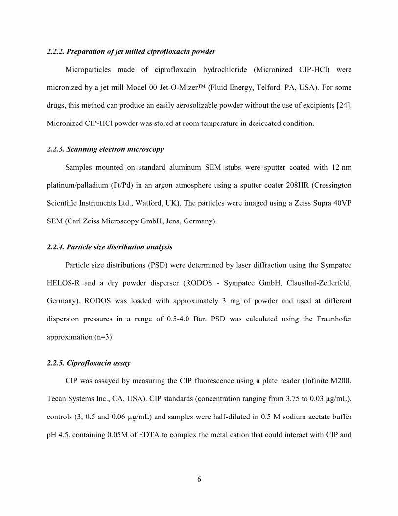
2.2.2. Preparation of jet milled ciprofloxacin powder
Microparticles made of ciprofloxacin hydrochloride (Micronized CIP-HCl) were
micronized by a jet mill Model 00 Jet-O-Mizer™ (Fluid Energy, Telford, PA, USA). For some
drugs, this method can produce an easily aerosolizable powder without the use of excipients [24].
Micronized CIP-HCl powder was stored at room temperature in desiccated condition.
2.2.3. Scanning electron microscopy
Samples mounted on standard aluminum SEM stubs were sputter coated with 12 nm
platinum/palladium (Pt/Pd) in an argon atmosphere using a sputter coater 208HR (Cressington
Scientific Instruments Ltd., Watford, UK). The particles were imaged using a Zeiss Supra 40VP
SEM (Carl Zeiss Microscopy GmbH, Jena, Germany).
2.2.4. Particle size distribution analysis
Particle size distributions (PSD) were determined by laser diffraction using the Sympatec
HELOS-R and a dry powder disperser (RODOS - Sympatec GmbH, Clausthal-Zellerfeld,
Germany). RODOS was loaded with approximately 3 mg of powder and used at different
dispersion pressures in a range of 0.5-4.0 Bar. PSD was calculated using the Fraunhofer
approximation (n=3).
2.2.5. Ciprofloxacin assay
CIP was assayed by measuring the CIP fluorescence using a plate reader (Infinite M200,
Tecan Systems Inc., CA, USA). CIP standards (concentration ranging from 3.75 to 0.03 µg/mL),
controls (3, 0.5 and 0.06 µg/mL) and samples were half-diluted in 0.5 M sodium acetate buffer
pH 4.5, containing 0.05M of EDTA to complex the metal cation that could interact with CIP and

7
modify its spectroscopic properties. The maximum excitation wavelength of CIP was 302 nm
and its maximum fluorescence emission wavelength was 456 nm.
2.3. Preparation and characterization of P. aeruginosa-loaded agar beads
2.3.1. Preparation of the P. aeruginosa-loaded beads
Sterile and bacteria-loaded agar beads were prepared using an adapted version of the
method described by Growcott et al. [22]. Fresh suspension of bioluminescent P. aeruginosa
(strain PAO1::p16Slux) prepared in BHI broth was cultured to exponential growth phase and
then adjusted to an OD600 of 0.3. Two milliliters of this suspension were washed and
concentrated in 1 mL of PBS by centrifugation. The bacteria were embedded into agar beads by
mixing 1 mL of this concentrated suspension with 9 mL of molten 2% w/v LB agar (Fisher
Scientific, NJ, USA). The mixture was then emulsified into 100 mL of warmed (48 °C) paraffin
oil (Sigma Aldrich, USA) containing 0.01% v/v of sorbitan monooleate (SPAN®80, Sigma
Aldrich, USA) based on the report of Growcott et al. [22]. The emulsion was cooled, the beads
centrifuged at 10,000 g for 10 min and then 4-times washed with PBS pH 7.4. Ten milliliters of
the bead suspension in PBS were stored up to 2 weeks at 4°C. For sterile agar beads, the volume
of bacterial suspension was replaced with PBS.
The number of bacteria per bead was determined from a CFU count performed with 100
µL of the bead suspension diluted in 5 mL of PBS and homogenized for 1 min (Polytron
PT2100, Kinematica AG, Switzerland). Ten-fold serial dilutions of this suspension were spread
on LB agar plates (Hardy Diagbostics, CA, USA) for colony counts. Bead concentrations were
determined using a hemocytometer that allowed calculating the number of bacteria per beads.
Size distribution of blank beads was determined by laser diffraction using the Sympatec
HELOS-R equipped with a Cuvette module (Sympatec GmbH, Clausthal-Zellerfeld, Germany).
8
A reference measurement was performed on the cell filled with 50 mL of PBS. After the
reference measurement, an aliquot of sterile beads suspension was added to the cell to achieve
optical concentrations between 10-20 % (n=3).
2.3.2. Time-luminescence intensity kinetics of bacteria-loaded agar beads
In vitro behavior of the luminescent P. aeruginosa in the agar beads was evaluated by
following the luminescence produced by the bacteria within the beads versus time in the
presence of various CIP or CIP-Cu complex concentrations. One hundred and eighty microliters
of agar bead suspension at 1.5 x 10⁵ beads/mL and containing 25 bacteria/bead were added into
the wells of a black 96-well plate with flat clear bottom (Corning Inc. NC, US). Wells were
supplemented with 20 μL of 10-times concentrated two-fold dilution of CIP or CIP-Cu complex
in BHI. Final CIP concentrations ranged from 64 to 0.062 µg/mL. Then, the plates were covered
using an air-permeable membrane and incubated at 37 °C in the plate reader (SpectraMax M3,
Molecular Devices LLC, CA, USA). Luminescence intensity emitted from the bottom of the
wells was measured during 1 second per well every 20 min. Plates were shaken before each
measurement (n=3).
2.4. Development of the chronic lung infection model
2.4.1. Animal model
The protocol for the animal study was approved by the Institutional Animal Care and Use
Committee (IACUC) at the University of Texas at Austin, Austin, TX. Sprague–Dawley rats
weighting between 225 and 275 g were purchased from Charles River Laboratories (Wilmington,
MA). Animals were housed one per cage, subjected to 12 h/12 h light and darkness cycles with
access to food and water ad libitum.
9
2.4.2. Inoculation of rats with bioluminescent P. aeruginosa-loaded agar beads
On day 1, anesthetized (Ketamine/xylazine (80/6 mg/Kg) i. p.) male Sprague–Dawley rats
were inoculated with various dose of P. aeruginosa (0 to 5 x 10⁷ CFU) embedded into agar
beads. Intratracheal (i.t.) inoculation was performed by instilling 100 µL of agar bead suspension
loaded in a syringe connected to a dry powder insufflator cannula (Penn-Century Inc,
Philadelphia, USA). This cannula has a blunt-tip designed to pass between the vocal cords
without cutting blood vessels or damaging the epithelium. The vocal cords were visualized using
a special laryngoscope suitable for rodent (Penn-Century Inc, Philadelphia, USA). Animals were
kept on a warming pad until full recovery from anesthesia. Body weight and luminescence
emitted from the rats anesthetized with isoflurane were measured daily for 8 days using an in
vivo imaging system IVIS® Spectrum (PerkinElmer, Houston, TX, USA) with an exposure time
of 3 min per rat.
2.5. In vivo evaluation of ciprofloxacin-copper inhalation treatment
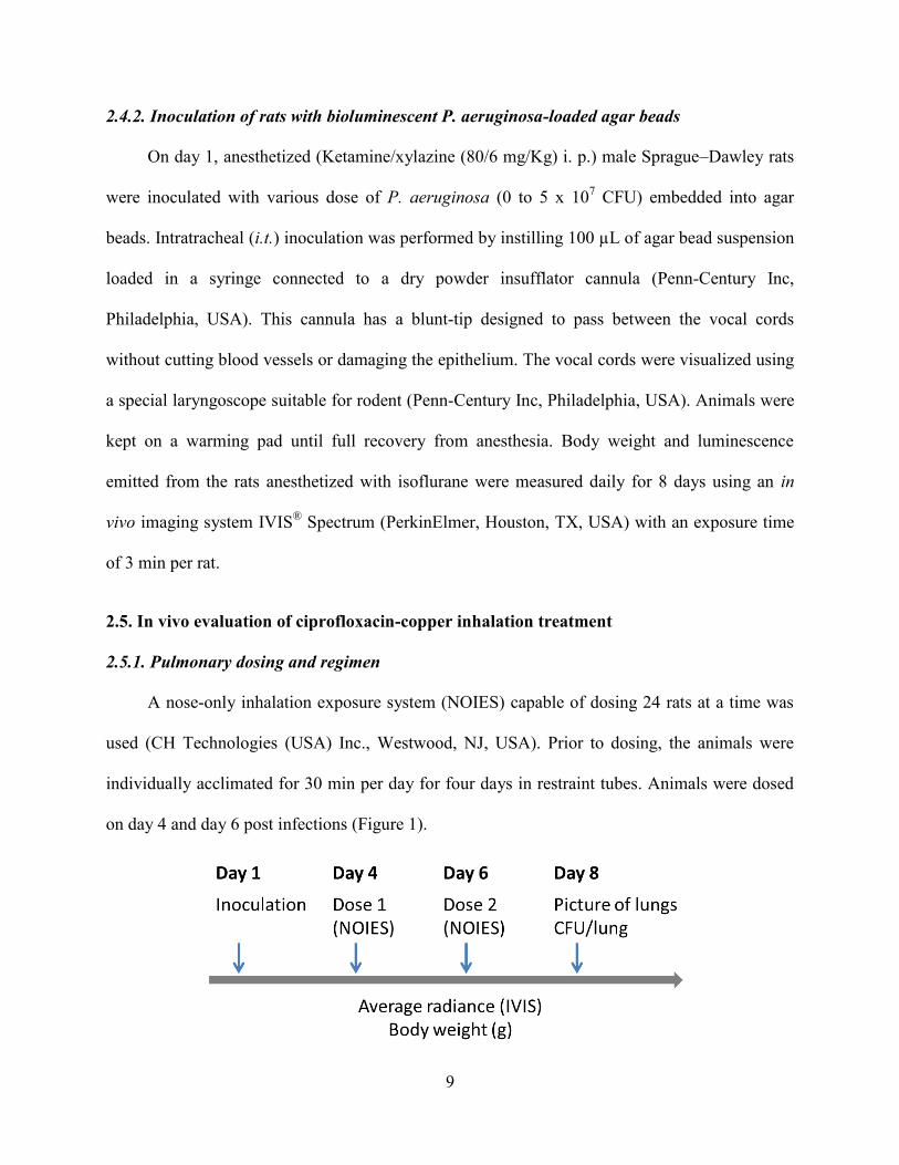
2.5.1. Pulmonary dosing and regimen
A nose-only inhalation exposure system (NOIES) capable of dosing 24 rats at a time was
used (CH Technologies (USA) Inc., Westwood, NJ, USA). Prior to dosing, the animals were
individually acclimated for 30 min per day for four days in restraint tubes. Animals were dosed
on day 4 and day 6 post infections (Figure 1).

10
Figure 1: Dosing regimen after i.t. inoculation of agar beads loaded with bioluminescent
PA01. Ciprofloxacin doses were set at 1.65 mg of CIP (base) per animal.
Powders for inhalation were loaded into a feed cylinder connected to a rotating brush
aerosol generator (RGB 1000). At an airflow rate of 0.5 L/min, animals treated with spray dried
CIP-Cu were dosed for 35 min and animals treated with micronized CIP-HCl were dosed for 20
min, so that equivalent doses of CIP (1.65 mg/animal) were given. The purpose of the 0.5 L/min
flow was to transport the aerosolized particles to the animal at a flow slightly higher than the
minute ventilation of a rat. Following dosing, animals were placed back into their respective
cages. These times of exposures were calculated to provide the same Total inhaled dose (TID -
mg) by rats and determined according to Eq. (1) as described by Alexander et al. [25].
Eq. (1)
where C is the measured particle concentration in air (mg/L), RMV is the species-specific
respiratory minute volume or the volume of air inhaled in one minute per animal (L/min), D is
the duration of exposure (min), and P is the particle drug potency (mass of CIP base per mass of
particles – 0.4 for CIP-Cu and 0.86 for CIP·HCl). The RMV for rats was calculated according to
the Eq. (2) [25].
Eq. (2)
where BW (kg) is the body weight of the rats. The aerosol concentration in air C, was calculated
based on the average mass of samples deposited on filters (n = 3) taken from the NOIES
inhalation chamber throughout the exposure using a vacuum flow of 0.5 L/min. To determine the
portion of the TID that was deposited in the lungs, the percent deposition in relationship to the
aerodynamic PSD was estimated based on rodent dosimetry findings reported by Kuehl et
al. [26]. Thus, a Mercer-style mini cascade impactor (CH Technologies (USA) Inc., Westwood,
NJ, USA) was used to determine the aerodynamic PSD of both aerosolized powders present
11
within the nose-only exposure chamber. This impactor is a seven-stage cascade impactor that has
been manufactured to be plugged directly to the nose-only exposure system, in exactly the same
position as the animal restraint tubes used to connect the animal to the inhalation chamber [26,
27]. The particles were collected at a rate of 0.5 L/min on this seven impactor stages having the
following cut-off diameters at this specific airflow rate (5.14, 3.04, 1.89, 1.16, 0.77, 0.48, 0.35
µm). The MMADs were determined by differential weight analysis of the stages before and after
impaction using a microbalance (METTLER TOLEDO, Columbus, OH, USA) and plotting the
inverse of the standard normal cumulative mass distribution less than stated size cut-off against
the natural logarithm of the cut-off diameter of the respective stages. The MMAD of the particles
distribution was defined as the particle size at which the line crosses the 50% mark.
2.5.2. Lung harvest for microbiological assessment
Eight days after infection, the animals in each group were euthanized by CO2 inhalation,
right after measuring the luminescence. Animals were cleaned with 70% v/v ethanol solution
before lungs collection. Lungs were homogenized in PBS and serial dilutions of these
homogenates were plated onto LB agar plates to measure the number of CFU per lung. (n = 4 for
the untreated group; n = 4 for the CIP-HCl group, and n = 7 for the CIP-Cu group)
2.5.3. Statistical analysis
Statistical analyses were performed using GraphPad Prism 6.0 (GraphPad Software Inc.). All
data were expressed as means ± SD and analysed with the Dunn's Kruskal-Wallis multiple
comparisons test. p < 0.05 was considered statistical significance.

12
3. RESULTS
3.1. Preparation and characterization of the dry powder formulations
The SEM micrographs of spray dried CIP-Cu and micronised CIP-HCl powders showed
different morphologies (Figure 2A). Spray dried CIP-Cu particles were spherical hollow brittle
particles while jet milled CIP-HCl particles were solid and non-spherical. The RODOS dry
powder disperser was used to compare the dispersibility of the powders at different pressures
(Figure 2B). The parameters D10, D50 and D90, represent the 10%, 50% and 90% point in the
cumulative undersize PSD, respectively. The increase in the dispersion pressure decreased
mainly the D90 and D50 of the powders. This behavior was more noticeable for the spray dried
CIP-Cu powder than for the jet milled CIP-HCl. However, both powders had similar PSD when
they were dispersed with a pressure of 3 bar or above.
Figure 2: Characterization of spray dried CIP-Cu and jet milled CIP-HCl powders. A.
SEM micrographs, B. Evolution of the PSD parameters D10, D50, and D90 versus dispersion
pressure. Solid symbols are for spray dried CIP-Cu, and open symbols are for micronized
CIP-HCl. Values are means ± SD (n=3).
13
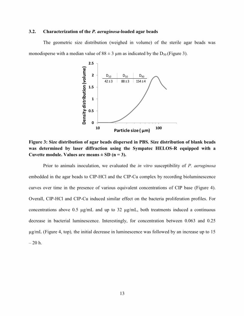
3.2. Characterization of the P. aeruginosa-loaded agar beads
The geometric size distribution (weighed in volume) of the sterile agar beads was
monodisperse with a median value of 88 ± 3 µm as indicated by the D50 (Figure 3).
Figure 3: Size distribution of agar beads dispersed in PBS. Size distribution of blank beads
was determined by laser diffraction using the Sympatec HELOS-R equipped with a
Cuvette module. Values are means ± SD (n = 3).
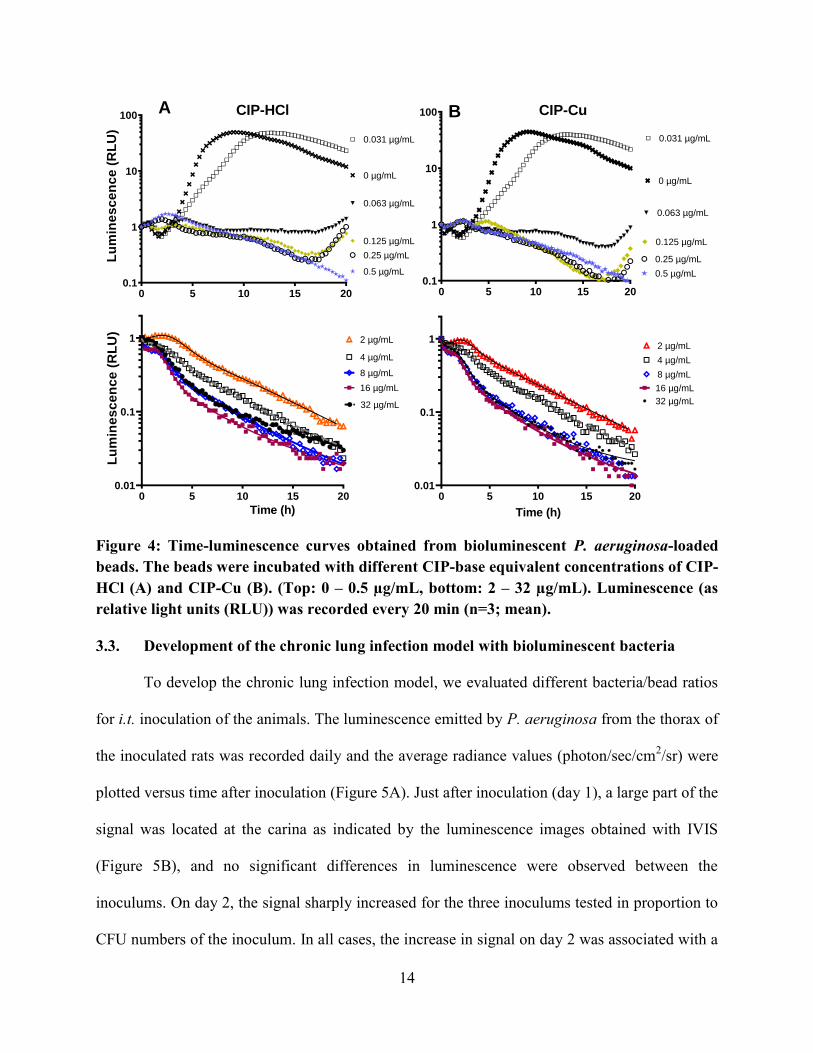
Prior to animals inoculation, we evaluated the in vitro susceptibility of P. aeruginosa
embedded in the agar beads to CIP-HCl and the CIP-Cu complex by recording bioluminescence
curves over time in the presence of various equivalent concentrations of CIP base (Figure 4).
Overall, CIP-HCl and CIP-Cu induced similar effect on the bacteria proliferation profiles. For
concentrations above 0.5 µg/mL and up to 32 µg/mL, both treatments induced a continuous
decrease in bacterial luminescence. Interestingly, for concentration between 0.063 and 0.25
µg/mL (Figure 4, top), the initial decrease in luminescence was followed by an increase up to 15
– 20 h.
0
0.5
1
1.5
2
2.5
10 100
De
nsi
ty d
istr
ibu
tio
n (v
olu
me
)
Particle size ( μm)
D10 D50 D90
42 ± 3 88 ± 3 154 ± 4
14
Figure 4: Time-luminescence curves obtained from bioluminescent P. aeruginosa-loaded
beads. The beads were incubated with different CIP-base equivalent concentrations of CIP-
HCl (A) and CIP-Cu (B). (Top: 0 – 0.5 µg/mL, bottom: 2 – 32 µg/mL). Luminescence (as
relative light units (RLU)) was recorded every 20 min (n=3; mean).
3.3. Development of the chronic lung infection model with bioluminescent bacteria
To develop the chronic lung infection model, we evaluated different bacteria/bead ratios
for i.t. inoculation of the animals. The luminescence emitted by P. aeruginosa from the thorax of
the inoculated rats was recorded daily and the average radiance values (photon/sec/cm2/sr) were
plotted versus time after inoculation (Figure 5A). Just after inoculation (day 1), a large part of the
signal was located at the carina as indicated by the luminescence images obtained with IVIS
(Figure 5B), and no significant differences in luminescence were observed between the
inoculums. On day 2, the signal sharply increased for the three inoculums tested in proportion to
CFU numbers of the inoculum. In all cases, the increase in signal on day 2 was associated with a
Lu
min
es
ce
nc
e (
RL
U)
0 5 10 15 200.01
0.1
1
32 µg/mL
16 µg/mL
8 µg/mL
4 µg/mL
2 µg/mL
Time (h)
0 5 10 15 200.1
1
10
100
0.5 µg/mL
0.25 µg/mL
0.125 µg/mL
0.063 µg/mL
0.031 µg/mL
0 µg/mL
CIP-HClL
um
ine
sc
en
ce
(R
LU
)
Time (h)
0 5 10 15 200.01
0.1
1
32 µg/mL
16 µg/mL
8 µg/mL
4 µg/mL
2 µg/mL
0 5 10 15 200.1
1
10
100
0.5 µg/mL
0.25 µg/mL
0.125 µg/mL
0.063 µg/mL
0.031 µg/mL
0 µg/mL
CIP-CuA B

15
loss of the animals’ body weight. By day 3, the CIP-HCl group had lost 11.8% of body weight
and the CIP-Cu group had lost 6.9% relative to average body weight on day 1. By day 8, the
average body weight of the groups was similar to the body weight on day 1, with CIP-HCl group
reaching 99.7% of the initial weight and CIP-Cu reaching 97.6% of the initial body weight.
Figure 5: In vivo chronic lung infection model. Sprague-Dawley rats were inoculated
intratracheally with various ratios of luminescent P. aeruginosa entrapped in agar beads
(CFU/beads ratios): 20 CFU/bead (6 x 10⁶
CFU/animal), 250 CFU/bead (1.5 x 10⁷
CFU/animal), and 500 CFU/bead (5 x 10⁷
CFU/animal). A. Variation of luminescence (in
photons s-1
cm-2
steradian-1
) per day (n=2-4; mean ± S.E.M.). B. Luminescence imaging
obtained in IVIS. C. Pictures of the infected lungs on day 8 and number of CFU recovered
from the lungs (group 6 x 10⁶
CFU/animal).
In the highest inoculum (5 x 10⁷ CFU; 500 CFU/bead), the signal increased 100-times
compared to day 1, however, none of the animals survived until day 3. For the 2 lower inoculums
(1.5 x 10⁷ and 6 x 10
6 CFU/animal), after a sharp luminescence increase on day 2, the signals
decreased on day 3 to values close to these obtained on day 1 and then were stable from day 3 to

16
day 8. The inoculum with 6 x 10⁶ CFU (20 CFU/bead) gave the best survival rate associated to
the highest and stable signal for 3 days. On day 8, the lungs of these rats had visible nodules
(yellow arrows, Figure 5C) that correlated with values of luminescence and the number of CFU
recovered from the lungs after plating. This inoculum was selected to evaluate the in vivo
efficacy of the CIP powders after pulmonary inhalation.
3.4. In vivo evaluation of ciprofloxacin-copper inhalation treatment
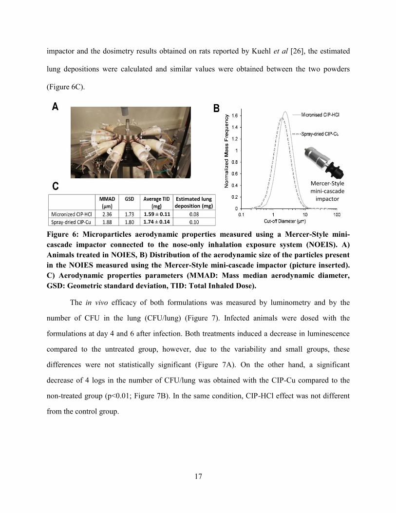
Powders were administered to rats by the inhalation route using the NOIES that allowed dosing
multiple animals in a time (Figure 6A). In order to compare the efficacy of the CIP-HCl and CIP-
Cu powders in vivo, the same equivalent dose of CIP, with a similar lung deposition, had to be
delivered to rats. Therefore, we evaluated the aerodynamic properties of the powders present in
the inhalation chamber using a Mercer-Style mini cascade impactor directly connected to the
NOIES. Aerodynamic PSD of the powders’ particles present in the inhalation chamber are
presented in Figure 6B. Both formulations had a narrow PSD and similar MMADs in the 1 to 3
µm range. Particles having an MMAD in this range generally give a same percentage of lung
deposition in rats of approximately 20% as shown by Kuehl et al.[26]. Moreover, both
formulation particles in the NOIES inhalation chamber have close GSD indicating a narrow
dispersity of the particles from the MMAD value. Therefore, the same lung deposition should
occur with both formulations. The other parameters evaluated before dosing the animals were the
Total Inhaled Dose (TID) and the time needed to achieve the same TID in each group of animals
(D in Eq. 1). The potency of CIP-Cu was lower than the CIP-HCl due to the presence of more
excipients (CaCO3 and hyaluronic acid). Thus, animals were dosed for 35 min with CIP-Cu and
for 20 min with CIP-HCl to achieve comparable TID of around 1.65 mg. Using this TID, the
aerodynamic properties of the powder particles evaluated with the Mercer-Style mini-cascade
17
impactor and the dosimetry results obtained on rats reported by Kuehl et al [26], the estimated
lung depositions were calculated and similar values were obtained between the two powders
(Figure 6C).
Figure 6: Microparticles aerodynamic properties measured using a Mercer-Style mini-
cascade impactor connected to the nose-only inhalation exposure system (NOEIS). A)
Animals treated in NOIES, B) Distribution of the aerodynamic size of the particles present
in the NOIES measured using the Mercer-Style mini-cascade impactor (picture inserted).
C) Aerodynamic properties parameters (MMAD: Mass median aerodynamic diameter,
GSD: Geometric standard deviation, TID: Total Inhaled Dose).
The in vivo efficacy of both formulations was measured by luminometry and by the
number of CFU in the lung (CFU/lung) (Figure 7). Infected animals were dosed with the
formulations at day 4 and 6 after infection. Both treatments induced a decrease in luminescence
compared to the untreated group, however, due to the variability and small groups, these
differences were not statistically significant (Figure 7A). On the other hand, a significant
decrease of 4 logs in the number of CFU/lung was obtained with the CIP-Cu compared to the
non-treated group (p<0.01; Figure 7B). In the same condition, CIP-HCl effect was not different
from the control group.
Mercer-Style mini-cascade
impactorEstimated lungdeposition (mg)
1.59 0.11
1.74 0.14

18
Figure 7: In vivo efficacy in a P. aeruginosa chronic lung infection model. Rats were infected
with 6 x 10⁶
CFU/animal, then treated on days 4 and 6 with CIP-Cu or CIP-HCl, and the lungs
were harvested on day 8. CIP dose was 1.65 mg/animal. A) Changes in the luminescence
(photons per s-1
cm-2
steradian-1
). B) Box and whisker plot of P. aeruginosa surviving colony
forming units (CFU) per lung on day 8. Whiskers represent the minimal and maximal values (n =
4 for the untreated group; n = 4 for the CIP-HCl group, and n = 7 for the CIP-Cu group).

19
4. DISCUSSION
4.1. Pulmonary delivery of ciprofloxacin-copper complex significantly reduced the P.
aeruginosa lung infection in the in vivo chronic lung infection model.
Microparticles loaded with CIP-Cu produced a significant reduction in bacterial lung
burden compared to untreated rats (4-log reduction, Figure 7B), while the micronized powder of
CIP-HCl did not change it significantly. Yet, with both powders, the total and peripheral doses of
CIP delivered into the lungs of rats were identical (Figure 6). Moreover, it has been previously
demonstrated that CIP or CIP-Cu solutions have the same efficacy against P. aeruginosa PA01
cultured either planktonically or as biofilms [1, 18]. The better efficacy observed with the CIP-
Cu over the CIP-HCl formulation could be attributed to a higher CIP lung residence time
obtained with the CIP-Cu microparticles. Indeed, a previous study has shown that intratracheal
(i.t.) administration to rats of CIP-Cu-loaded particles provides a mean AUC ratio of the CIP
concentration kinetic profiles in ELF on the CIP concentration kinetic profiles in plasma 100-
fold higher than after administration of a CIP solution [2]. After CIP-Cu i.t. administration, CIP
half-life in rat lung ELF is higher than 2 h, while it is lower than 1 h after a CIP solution
nebulization [2]. At the opposite, previous studies showed that CIP-HCl has poor lung targeting
following oral inhalation; the drug is rapidly absorbed into the systemic circulation with a half-
life of less than 1 h in the rat lungs, making CIP-HCl suboptimal for pulmonary delivery [5, 13,
28]. To improve the lung targeting of CIP by inhalation, Bayer AG also aimed at prolonging the
residence time of CIP in the lungs. To do this, they have developed a slow-dissolving CIP
formulation using the neutral (zwitterionic) solid form of the drug (CIP-betaine) instead of the
faster dissolving CIP-HCl [13]. When used as a dry powder, this neutral form of CIP enhances
the efficacy of CIP in the treatment of a model of pulmonary P. aeruginosa infection in the rat.
20
After i.t. administration at a dose of 10 mg/kg, which is 2 times the total dose used in this study,
a 4 to 10-log reduction in P. aeruginosa CFU count was observed with the neutral form of CIP
versus a 1.1 to 2.8-log reduction for CIP-HCl [13]. In this approach, CIP lung targeting and
efficacy was improved only by controlling the dissolution rate of the CIP. Indeed, after
dissolution, CIP-betaine and CIP-HCl lead to the presence of the same zwitterionic CIP in
solution at the pH of the pulmonary fluids. In the approach presented in this study, CIP lung
targeting and efficacy were improved by reducing CIP apparent permeability across the blood-
lung barrier [18].
P. aeruginosa growing as aggregates, or biofilms, is the main responsible of the recurrent
lung infections that affect cystic fibrosis patients [29, 30]. Biofilms can be highly resistant to
antibiotics compared to free swimming planktonic bacteria [31]. For example, about ten- to
hundred-times more CIP is necessary to inhibit the growth of bacteria as biofilms compared to
planktonic bacteria [20, 32, 33]. P. aeruginosa-entrapped in agar beads once in the lungs for
several days resembles P. aeruginosa from biofilms observed in chronic infections [30, 34, 35].
The results of the present study indicate that complexation of CIP with Cu2+
can effectively lead
to enhanced local concentration in the lung necessary to reduce P. aeruginosa biofilm infection.
4.2. The in vivo chronic lung infection model with bioluminescent PA01 was highly
resistant and allowed for monitoring of the progression of infection in the same animal.
A non-invasive technique was used to monitor the progression of the pulmonary infection
caused by a luminescent strain of P. aeruginosa (PAO1::p16Slux) entrapped in agar beads
(Figure 5). Riedel et al. demonstrated that the transformation of P. aeruginosa with the p16Slux
system does not affect bacterial pathogenesis in rats after intranasal inoculation [23]. They also

21
demonstrated that after acute PAO1::p16Slux infection, luminescence from dissected organs
linearly correlated with their bacterial loads, including lungs. Animal inoculation with bacteria-
laden beads has been extensively used to study novel therapeutic approaches to fight chronic
lung infections [36] as it resembles the lung disease observed in humans with P. aeruginosa
chronic pneumonia with formation of pulmonary biofilm [37]. The P. aeruginosa-laden beads
used in this study had a median diameter D50 of 88 ± 3 µm. Growcott et al. showed that beads
with a D50 of 155 µm were widely distribute in the lungs of rats, and were found as aggregates in
the airways and alveolar spaces after i.t. inoculation [22]. The development of the infection in the
whole lungs was confirmed in the present study by luminescence measurements from the
thoracic region of the rats (Figure 5B) as well as pulmonary lesions observed after 8 days post-
inoculation (Figure 5C).
P. aeruginosa-entrapped in agar beads present in the lungs resembles P. aeruginosa from
biofilms observed in chronic infections [30, 34, 35]. Biofilms bacterial population is
heterogeneous and often harbor several subpopulations of bacteria that are resistant, persistent
and/or tolerant to antimicrobial [38, 39]. Tolerance and persistence are phenomena of increased
bacterial survival in the presence of an antibiotic without an increase in the MIC [40]. Tolerant
and persistent bacteria often are in dormancy. They do not grow, or very slowly, and have
reduced metabolism activity when compared with growing cells [40]. To distinguish between the
in-vivo antimicrobial activity of CIP-Cu and CIP-HCl, the number of colonies appearing in 24
hours from lung homogenate was counted (Figure 7.B). This method allows the quantification
mainly of the surviving bacterial population able to grow, but is not adapted for bacteria in
dormancy. Similarly, luminescence is mainly emitted by metabolically active bacteria and helps
to quantify bacterial proliferation [41] but also allows a more direct measurement. Riedel et al.
22
[23] showed that luminescence produced by PAO1::p16Slux remained relatively stable
throughout the entire growth curve, while luminescence in other species such as Listeria
monocytogenes and other gram-positive bacteria decreased dramatically in stationary phase. In
stationary phase, planktonic bacteria have lower metabolic activity and lower susceptibility to
CIP comparable to those of certain phases of biofilm growth [42]. This suggests that, with
enough sensitivity, luminescence could be used to monitor slow growing bacteria during
bacterial biofilm infection. In the present study, an optimal bacteria/beads ratio was selected to
inoculate the rats for achieving the best survival and a high luminescence signal (Figure 5A-B).
For the two lower inoculums (1.5 x 10⁷ CFU and 6 x 10
6 CFU), after a sharp luminescence
increase on day 2, the signals decreased on day 3 to values close to these obtained on day 1 and
then were stable from day 5 to day 8. These profiles were very similar to those usually obtained
by measuring the CFU numbers in the lungs [19, 21]. However, the initial luminescence level
seemed too low to accurately assess the effect of antibiotics. In addition, bacterial loads below
1.107 CFU per lung gave bioluminescence values that were close to the detection limit of the
IVIS equipment. Thus, no correlation between bioluminescence and the number of CFU per lung
measured on day 8 after infection could be obtained because most of the bacterial loads were less
than 1.107 CFU per lung on that day (Fig. 7B). The use of a less virulent strain could help
increase the inoculum used for infection and thereby increase the stable basal signal observed 4
days after infection. In addition, using a strain with a higher luminescence yield or increasing the
exposure time could also increase the sensitivity of the method and make it possible to quantify
the degree of infection according to the value of luminescence, not just to assess its spatial
distribution.
23
4.3. Spray dried and micronized powders properties were suitable for inhalation
The RODOS unit has long been used to characterize powders for inhalation across a
range of settings on the instrument. Over the past 15 years or so, many powders of different
physical and chemical properties have been analyzed by different groups revealing some
consistent trends. In particular, it has been generally shown that RODOS dispersion pressures
above 3 kPa result in full dispersion and allows sizing of the primary particle sizes of a powder
[43, 44]. Similarly, dispersion pressures used in the RODOS of between 0.5-3 kPa can indicate
the dispersion performance of a powder [43] agnostic to proprietary inhaler designs and even
allow matching to novel inhaler dispersion performance [44]. Thus, in these preclinical in vivo
feasibility studies we have characterized the powder particle size distributions to indicate how
they will be dispersed as a powder and also specifically their performance for nose-only
inhalation testing. Future studies will couple the powder with an inhaler system and be
characterized using compendia methods to support eventual clinical testing.
5. CONCLUSION
Pulmonary delivery of ciprofloxacin-copper complex significantly reduced the P.
aeruginosa in a rat chronic lung infection model. Luminescent P. aeruginosa entrapped in agar
beads were useful to spatially monitor the development of the lung infection in rats.

24
6. REFERENCES
[1] F. Tewes, T.F. Bahamondez-Canas, H.D.C. Smyth, Efficacy of Ciprofloxacin and Its Copper
Complex against Pseudomonas aeruginosa Biofilms, AAPS PharmSciTech, (2019).
[2] B. Lamy, F. Tewes, D.R. Serrano, I. Lamarche, P. Gobin, W. Couet, A.M. Healy, S.
Marchand, New aerosol formulation to control ciprofloxacin pulmonary concentration, Journal
of Controlled Release, 271 (2018) 118-126.
[3] Q.T. Zhou, S.S.Y. Leung, P. Tang, T. Parumasivam, Z.H. Loh, H.-K. Chan, Inhaled
formulations and pulmonary drug delivery systems for respiratory infections, Advanced drug
delivery reviews, 85 (2015) 83-99.
[4] H. Stass, J. Nagelschmitz, S. Willmann, H. Delesen, A. Gupta, S. Baumann, Inhalation of a
dry powder ciprofloxacin formulation in healthy subjects: a phase I study, Clinical drug
investigation, 33 (2013) 419-427.
[5] H. Stass, B. Weimann, J. Nagelschmitz, C. Rolinck-Werninghaus, D. Staab, Tolerability and
pharmacokinetic properties of ciprofloxacin dry powder for inhalation in patients with cystic
fibrosis: a Phase I, randomized, dose-escalation study, Clinical therapeutics, 35 (2013) 1571-
1581.
[6] A.V.L. Gontijo, J. Brillault, N. Grégoire, I. Lamarche, P. Gobin, W. Couet, S. Marchand,
Biopharmaceutical characterization of nebulized antimicrobial agents in rats: 1. Ciprofloxacin,
moxifloxacin, and grepafloxacin, Antimicrobial agents and chemotherapy, 58 (2014) 3942-3949.
[7] R. Endermann, H. Labischinski, C. Ladel, U. Petersen, B. Newton, Treatment of bacterial
diseases of the respiratory organs, in, Bayer Pharma Aktiengesellschaft, 2011.
[8] M.C. Gaspar, N. Grégoire, J.J. Sousa, A.A. Pais, I. Lamarche, P. Gobin, J.-C. Olivier, S.
Marchand, W. Couet, Pulmonary pharmacokinetics of levofloxacin in rats after aerosolization of
immediate-release chitosan or sustained-release PLGA microspheres, European Journal of
Pharmaceutical Sciences, 93 (2016) 184-191.
[9] D.J. Serisier, D. Bilton, A. De Soyza, P.J. Thompson, J. Kolbe, H.W. Greville, D. Cipolla, P.
Bruinenberg, I. Gonda, O.-. investigators, Inhaled, dual release liposomal ciprofloxacin in non-
cystic fibrosis bronchiectasis (ORBIT-2): a randomised, double-blind, placebo-controlled trial,
Thorax, 68 (2013) 812-817.
[10] S.N. Nurbaeti, J. Brillault, F. Tewes, J.-C. Olivier, Sustained-release microparticle dry
powders of chloramphenicol palmitate or thiamphenicol palmitate prodrugs for lung delivery as
aerosols, European Journal of Pharmaceutical Sciences, (2019) 105028.
[11] D. Cipolla, J. Blanchard, I. Gonda, Development of liposomal ciprofloxacin to treat lung
infections, Pharmaceutics, 8 (2016) 6.
[12] N.G. Türeli, A. Torge, J. Juntke, B.C. Schwarz, N. Schneider-Daum, A.E. Türeli, C.-M.
Lehr, M. Schneider, Ciprofloxacin-loaded PLGA nanoparticles against cystic fibrosis P.
aeruginosa lung infections, European Journal of Pharmaceutics and Biopharmaceutics, 117
(2017) 363-371.
[13] P.J. McShane, J.G. Weers, T.E. Tarara, A. Haynes, P. Durbha, D.P. Miller, T. Mundry, E.
Operschall, J.S. Elborn, Ciprofloxacin Dry Powder for Inhalation (ciprofloxacin DPI): Technical
25
design and features of an efficient drug–device combination, Pulmonary Pharmacology &
Therapeutics, 50 (2018) 72-79.
[14] F. Tewes, J. Brillault, B. Lamy, P. O'Connell, J.C. Olivier, W. Couet, A.M. Healy,
Ciprofloxacin-Loaded Inorganic-Organic Composite Microparticles To Treat Bacterial Lung
Infection, Molecular pharmaceutics, 13 (2016) 100-112.
[15] D.C. Griffith, M.N. Dudley, M.W. Surber, K.A. Bostian, O. Rodny, Aerosol
fluoroquinolone formulations for improved pharmacokinetics, in, Patents, 2014.
[16] B. Lamy, D. Remedios Serrano, P. O’Connell, W. Couet, S. Marchand, A.M. Healy, F.
Tewes, Use of leucine to improve aerodynamic properties of ciprofloxacin-loaded maltose
microparticles for inhalation, European Journal of Pharmaceutical Research, 1 (2019) 2-11.
[17] J. Brillault, F. Tewes, Control of the Lung Residence Time of Highly Permeable Molecules
after Nebulization: Example of the Fluoroquinolones, Pharmaceutics, 12 (2020) 387.
[18] J. Brillault, F. Tewes, W. Couet, J. Olivier, In vitro biopharmaceutical evaluation of
ciprofloxacin/metal cation complexes for pulmonary administration, European Journal of
Pharmaceutical Sciences, 97 (2017) 92-98.
[19] I. Kukavica-Ibrulj, R. Levesque, Animal models of chronic lung infection with
Pseudomonas aeruginosa: useful tools for cystic fibrosis studies, Laboratory animals, 42 (2008)
389-412.
[20] B.G.S. Torres, R. Awad, S. Marchand, W. Couet, F. Tewes, In vitro evaluation of
Pseudomonas aeruginosa chronic lung infection models: Are agar and calcium-alginate beads
interchangeable?, European Journal of Pharmaceutics and Biopharmaceutics, 143 (2019) 35-43.
[21] A. Bragonzi, D. Worlitzsch, G.B. Pier, P. Timpert, M. Ulrich, M. Hentzer, J.B. Andersen,
M. Givskov, M. Conese, G. Döring, Nonmucoid Pseudomonas aeruginosa expresses alginate in
the lungs of patients with cystic fibrosis and in a mouse model, Journal of Infectious Diseases,
192 (2005) 410-419.
[22] E. Growcott, A. Coulthard, R. Amison, E. Hardaker, V. Saxena, L. Malt, P. Jones, A.
Grevot, C. Poll, C. Osborne, Characterisation of a refined rat model of respiratory infection with
Pseudomonas aeruginosa and the effect of ciprofloxacin, Journal of Cystic Fibrosis, 10 (2011)
166-174.
[23] C.U. Riedel, P.G. Casey, H. Mulcahy, F. O'Gara, C.G. Gahan, C. Hill, Construction of
p16Slux, a novel vector for improved bioluminescent labeling of gram-negative bacteria,
Applied and environmental microbiology, 73 (2007) 7092-7095.
[24] A.D. Brunaugh, H.D. Smyth, Formulation techniques for high dose dry powders,
International journal of pharmaceutics, 547 (2018) 489-498.
[25] D.J. Alexander, C.J. Collins, D.W. Coombs, I.S. Gilkison, C.J. Hardy, G. Healey, G.
Karantabias, N. Johnson, A. Karlsson, J.D. Kilgour, P. McDonald, Association of Inhalation
Toxicologists (AIT) Working Party Recommendation for Standard Delivered Dose Calculation
and Expression in Non-Clinical Aerosol Inhalation Toxicology Studies with Pharmaceuticals,
Inhalation Toxicology, 20 (2008) 1179-1189.
[26] P.J. Kuehl, T.L. Anderson, G. Candelaria, B. Gershman, K. Harlin, J.Y. Hesterman, T.
Holmes, J. Hoppin, C. Lackas, J.P. Norenberg, H. Yu, J.D. McDonald, Regional particle size
26
dependent deposition of inhaled aerosols in rats and mice, Inhalation Toxicology, 24 (2012) 27-
35.
[27] S.R. Carvalho, A.B. Watts, J.I. Peters, S. Liu, S. Hengsawas, M.S. Escotet-Espinoza, R.O.
Williams III, Characterization and pharmacokinetic analysis of crystalline versus amorphous
rapamycin dry powder via pulmonary administration in rats, European Journal of Pharmaceutics
and Biopharmaceutics, 88 (2014) 136-147.
[28] J. Weers, Comparison of Phospholipid-Based Particles for Sustained Release of
Ciprofloxacin Following Pulmonary Administration to Bronchiectasis Patients, Pulmonary
Therapy, (2019).
[29] M.L. Barclay, E.J. Begg, S.T. Chambers, P.E. Thornley, P.K. Pattemore, K. Grimwood,
Adaptive resistance to tobramycin in Pseudomonas aeruginosa lung infection in cystic fibrosis,
Journal of Antimicrobial Chemotherapy, 37 (1996) 1155-1164.
[30] T. Bjarnsholt, P.Ø. Jensen, M.J. Fiandaca, J. Pedersen, C.R. Hansen, C.B. Andersen, T.
Pressler, M. Givskov, N. Høiby, Pseudomonas aeruginosa biofilms in the respiratory tract of
cystic fibrosis patients, Pediatric pulmonology, 44 (2009) 547-558.
[31] N. Høiby, O. Ciofu, T. Bjarnsholt, Pseudomonas aeruginosa biofilms in cystic fibrosis,
Future microbiology, 5 (2010) 1663-1674.
[32] M.C. Walters, F. Roe, A. Bugnicourt, M.J. Franklin, P.S. Stewart, Contributions of
antibiotic penetration, oxygen limitation, and low metabolic activity to tolerance of
Pseudomonas aeruginosa biofilms to ciprofloxacin and tobramycin, Antimicrobial agents and
chemotherapy, 47 (2003) 317-323.
[33] H. Bandara, D. Nguyen, S. Mogarala, M. Osiñski, H. Smyth, Magnetic fields suppress
Pseudomonas aeruginosa biofilms and enhance ciprofloxacin activity, Biofouling, 31 (2015)
443-457.
[34] M. Sønderholm, K.N. Kragh, K. Koren, T.H. Jakobsen, S.E. Darch, M. Alhede, P.Ø. Jensen,
M. Whiteley, M. Kühl, T. Bjarnsholt, Pseudomonas aeruginosa Aggregate Formation in an
Alginate Bead Model System Exhibits In Vivo-Like Characteristics, Applied and Environmental
Microbiology, 83 (2017).
[35] D.P. Gnanadhas, M. Elango, A. Datey, D. Chakravortty, Chronic lung infection by
Pseudomonas aeruginosa biofilm is cured by L-Methionine in combination with antibiotic
therapy, Scientific reports, 5 (2015) 16043.
[36] P. Meers, M. Neville, V. Malinin, A. Scotto, G. Sardaryan, R. Kurumunda, C. Mackinson,
G. James, S. Fisher, W. Perkins, Biofilm penetration, triggered release and in vivo activity of
inhaled liposomal amikacin in chronic Pseudomonas aeruginosa lung infections, Journal of
Antimicrobial Chemotherapy, 61 (2008) 859-868.
[37] H. Cash, D. Woods, B. McCullough, W. Johanson Jr, J. Bass, A rat model of chronic
respiratory infection with Pseudomonas aeruginosa, American Review of Respiratory Disease,
119 (1979) 453-459.
[38] K. Lewis, Persister cells, Annual review of microbiology, 64 (2010) 357-372.
[39] H.-C. Flemming, J. Wingender, U. Szewzyk, P. Steinberg, S.A. Rice, S. Kjelleberg,
Biofilms: an emergent form of bacterial life, Nature Reviews Microbiology, 14 (2016) 563.

27
[40] N.Q. Balaban, S. Helaine, K. Lewis, M. Ackermann, B. Aldridge, D.I. Andersson, M.P.
Brynildsen, D. Bumann, A. Camilli, J.J. Collins, Definitions and guidelines for research on
antibiotic persistence, Nature Reviews Microbiology, 17 (2019) 441-448.
[41] S. Duncan, L.A. Glover, K. Killham, J.I. Prosser, Luminescence-based detection of activity
of starved and viable but nonculturable bacteria, Appl. Environ. Microbiol., 60 (1994) 1308-
1316.
[42] T.-F.C. Mah, G.A. O'Toole, Mechanisms of biofilm resistance to antimicrobial agents,
Trends in Microbiology, 9 (2001) 34-39.
[43] S. Jaffari, B. Forbes, E. Collins, D.J. Barlow, G.P. Martin, D. Murnane, Rapid
characterisation of the inherent dispersibility of respirable powders using dry dispersion laser
diffraction, International journal of pharmaceutics, 447 (2013) 124-131.
[44] A.H. de Boer, P. Hagedoorn, E.M. Westerman, P.P. Le Brun, H.G. Heijerman, H.W.
Frijlink, Design and in vitro performance testing of multiple air classifier technology in a new
disposable inhaler concept (Twincer®) for high powder doses, European journal of
pharmaceutical sciences, 28 (2006) 171-178.