in vivo cryotechnique· for paradigm shift to … · department of anatomy, interdisciplinary...
TRANSCRIPT

Founded and printed in Varna, Bulgar iaISSN 1310-392X
Biomedical Reviews 2004; 15: 1-19.
”IN VIVO CRYOTECHNIQUE· FOR PARADIGM SHIFT TO ”LIVING MORPHOLOGY· OF ANIMAL ORGANS
Nobuhiko Ohno, Nobuo Terada, Yasuhisa Fujii, Takeshi Baba, and Shinichi Ohno Department of Anatomy, Interdisciplinary Graduate School of Medicine and Engineering, University of Yamanashi, Tamaho, Yamanashi, Japan
The morphological study has been one of the major approaches in medical and biological fields. For the last century, the con-ventional chemical fixation and alcohol dehydration were commonly used as an easy preparation method, but it was frequentlypointed out that they usually yield many structural artifacts during their preparation processes. Although both conventional quick-freezing and high-pressure freezing methods, by which animal tissues are resected and frozen for physical fixation, can reducesuch structural artifacts, the tissues have to be removed from living animal organs for the freezing. Therefore, such specimens are inevitably exposed to noxious stresses of anoxia and ischemia, exhibiting only dead morphological states of animal tissues without blood circulation. To the contrary, our “in vivo cryotechnique”, by which all cells and tissues in animal bodies are cryofixed in vivo, can prevent such artifacts of resected specimens. By means of the cryotechnique, it is now possible to reveal the in vivo morphology of cells and tissues in living animal organs. Actually, it has been already applied to several animal organs, such as kidney, liver, intestine, cerebellum, eye ball, blood vessel, and joint cartilage, and brought new morphological findings, reflectingtheir physiological significance, which had been difficult to demonstrate by the conventional preparation methods. Moreover, itsapplication to immunohistochemistry has also revealed more precise immunolocalizations of dynamically changing molecules in living animal organs, easily translocated by ischemic stresses and anoxia caused during the tissue resection. The “in vivo cryotechnique” allows us to perform novel morphological investigations of “living” morphological states, and develops new medical and biological fields with “living morphology” during this 21st century.
Received 17 November 2004 and accepted 10 December 2004. Correspondence and reprint requests to Dr Shinichi Ohno, Professor and Chairman, Department of Anatomy, Interdisciplinary Graduate School of Medicine and Engineering, University of Yamanashi, 1110 Shimokato, Tamaho, Yamanashi 409-3898, Japan. Tel./Fax: 81 55 273 6743, E-mail: [email protected]
INTRODUCTION
The morphological study had been one of the indispensable approaches to understand the physiological and pathologi-cal features of living animals for the last century. Although substantial progress of research techniques in the molecular biology has been realized to establish new biological fieldsduring the last decade (1,2), morphological techniques were
also necessary for more precise understanding in the field,because structures of cells and tissues reflect some functionalaspects of living animal organs. In such morphology, the electron microscopy, developed in the middle 20th century, has yielded the enormous progress of ultrastructural analyses, and it is now indispensable in the morphological field withmany applications (3). Other progress in light microscopic approaches was also remarkable during the last decades, and

2
Biomed Rev 15, 2004
N. Ohno, Terada, Fujii, Baba, and S. Ohno
new fluorescence technologies have enabled us to get dynamicimages of signal molecules and their molecular interactions in living cells (4,5). However, each technique always has its own merit and demerit, and so it would be necessary to understand such technical features for choosing appropriate techniques in various morphological experiments.
Since 1995, we have been developing the “in vivo cryo-technique” to clarify functioning morphology of living animal organs, and reported new findings of dynamically changingstructures and molecular immunolocalization in cells and tis-sues by both electron and light microscopy (6-16). Although this cryotechnique has some unique merits for the morpho-logical analyses of living animal organs, there has been no systematic report about its comparison with other specimen preparation methods until now. Therefore, the purpose of this review is to describe some significant merits of the “in vivo cryotechnique”, and review the new findings obtained by thecryotechnique.
PROBLEMS OF CONVENTIONAL PREPARATION METHODS
Although new technologies have greatly expanded our abil-ity to examine structures of molecules, cells and tissues, they are still technically limited at the present time (5,17). In most morphological studies of animal organs, therefore, their prepa-ration procedures are usually composed of chemical fixation,dehydration in organic solvents, embedding in paraffin wax orepoxy resin, sectioning, and dye or metal staining steps, fol-lowed by observation with a light or electron microscope (18). It has been known that various kinds of inevitable artifacts influence their morphological findings, especially owing to theconventional fixation and dehydration steps (19,20).
The first step, chemical fixation, is usually performed withroutine chemical fixatives, such as paraformaldehyde, glutar-aldehyde and osmium tetroxide (21). However, some technical problems of the chemical fixation have been already pointedout during the last century. One of the problems is the rapid molecular movement and structural changes occurring during the fixation step (20). For example, small soluble molecularcomponents in cells and tissues are inevitably redistributed before the cross-linking effect is completed as the fixationmechanism (19). In addition, the chemical fixatives cansometimes modify the molecular conformation (22). Another technical problem is that transient or dynamic structures of cells and tissues are difficult to capture by the chemical fixa-tion, because certain time intervals are necessary to complete it, particularly by the immersion fixation (23).
There are also other technical problems during the follow-ing steps, such as alcohol dehydration, embedding in paraffin
wax or epoxy resin, sectioning and staining with dye or metal, although some of them are not restricted to the conventional preparation procedure. It has been frequently pointed out that the alcohol dehydration step can cause tissue shrinkage or additional morphological artifacts, which sometimes lead to misunderstanding of functional significance (24,25). In thecase of embedding in epoxy resin, the embedding step needs heating up over 50oC, which might cause additional heat dam-age on biological tissues. The thickness of cut sections during the sectioning step depends on the embedded specimens and following observation devices, but the sectioning usually results in two-dimentional viewing of cells and tissues. Next, various kinds of staining processes are available during the dye or metal staining, but every staining technique enhances some aspects of the specimen structures and inversely obscures the others. Even during observation in an electron microscope, ultrathin sections stained with metals have to be put into highly vacuumed chambers and undergo heating damage induced by electron beams.
Although the conventional preparation techniques have several problems with morphological preservation of cells and tissues, the morphological studies have yielded impor-tant findings during the last century, and also contributed toacademic achievement in biological and medical fields. Onthe other hand, lots of effort has been made to reduce such morphological artifacts commonly induced during the conven-tional preparation steps. For that purpose, the quick-freezing (QF) method was introduced for biological specimens at the middle of 20th century, and gradually improved during the late 20th century.
QUICK-FREEZING METHOD AND ITS TECHNICAL PROBLEMS
The QF method, usually termed as “cryofixation”, is one ofthe physical fixation methods, in which biological specimensare not chemically fixed, but quickly embedded in vitrified ice(26). The term “vitrification” means ice formation without anyice crystals seen by electron microscopy, which often destroy morphological structures of cells and tissues. In the case of fresh resected tissues without cryoprotectants (e.g. glycerol or sucrose solution), the good freezing to obtain the vitrificationhas to be performed either at very high cooling-rate (usually more than 105 oC/sec) or under very high pressures (a few thousand times higher than atmospheric pressure) (26). For the last century, various QF methods have been developed for the purpose (reviewed in 26,27). One example is called the “slamming QF” method, by which resected tissues are slammed onto copper blocks cooled down in liquid helium
3
Biomed Rev 15, 2004
In vivo cryotechnique for living morphology
(-269oC) or liquid nitrogen (-196oC). Another example is called the “plunging QF” method, by which they are plunged into liquid cryogens, such as propane alone or isopentane-propane mixture (-193oC) cooled in liquid nitrogen. The slamming or plunging QF method prevents formation of detectable ice crystals in areas less than 10 µm deep from the contacted tissue surface. To the contrary, the high-pressure freezing is a different method from the other QF methods, because it is always performed under very high pressures to achieve the vitrification in relatively broad tissue areas.
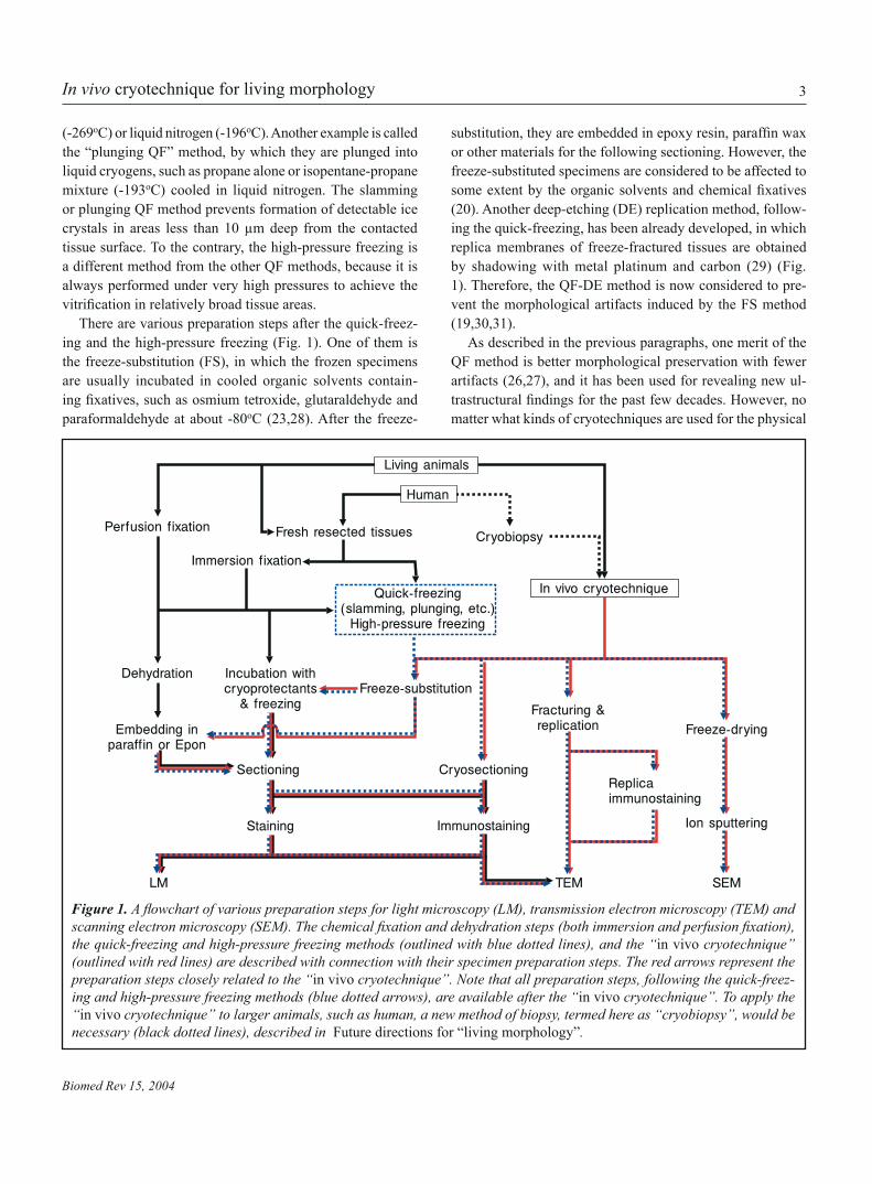
There are various preparation steps after the quick-freez-ing and the high-pressure freezing (Fig. 1). One of them is the freeze-substitution (FS), in which the frozen specimens are usually incubated in cooled organic solvents contain-ing fixatives, such as osmium tetroxide, glutaraldehyde andparaformaldehyde at about -80oC (23,28). After the freeze-
substitution, they are embedded in epoxy resin, paraffin waxor other materials for the following sectioning. However, the freeze-substituted specimens are considered to be affected to some extent by the organic solvents and chemical fixatives(20). Another deep-etching (DE) replication method, follow-ing the quick-freezing, has been already developed, in which replica membranes of freeze-fractured tissues are obtained by shadowing with metal platinum and carbon (29) (Fig. 1). Therefore, the QF-DE method is now considered to pre-vent the morphological artifacts induced by the FS method (19,30,31).
As described in the previous paragraphs, one merit of the QF method is better morphological preservation with fewer artifacts (26,27), and it has been used for revealing new ul-trastructural findings for the past few decades. However, nomatter what kinds of cryotechniques are used for the physical
Figure 1. A flowchart of various preparation steps for light microscopy (LM), transmission electron microscopy (TEM) andscanning electron microscopy (SEM). The chemical fixation and dehydration steps (both immersion and perfusion fixation),the quick-freezing and high-pressure freezing methods (outlined with blue dotted lines), and the “in vivo cryotechnique” (outlined with red lines) are described with connection with their specimen preparation steps. The red arrows represent the preparation steps closely related to the “in vivo cryotechnique”. Note that all preparation steps, following the quick-freez-ing and high-pressure freezing methods (blue dotted arrows), are available after the “in vivo cryotechnique”. To apply the “in vivo cryotechnique” to larger animals, such as human, a new method of biopsy, termed here as “cryobiopsy”, would be necessary (black dotted lines), described in Future directions for “living morphology”.
4
Biomed Rev 15, 2004
N. Ohno, Terada, Fujii, Baba, and S. Ohno
cryofixation, pieces of tissues have to be always taken outfrom living animal organs, and it has long been suggested that morphology of the resected tissues would be inevitably changed, because of loss of blood supply causing ischemia and anoxia (8,32). Thus, dynamic morphology of living animals, changing with various blood pressures, is hardly investigated by the conventional cryotechniques (7,8,11). Furthermore, immunohistochemical analyses of dynamically changing sig-nal molecules would be more difficult by such conventionalcryotechniques, because animal tissues have to be exposed to ischemia and anoxia during the resection process (16). To overcome these problems, it is necessary to avoid the removal of animal tissues, and directly freeze living animal organs in vivo under normal blood circulation. Although a few pioneer-ing studies challenged to solve them (33,34), it was difficult toestablish a new cryotechnique, which could yield satisfactory results for various organs of living animals.
DEVELOPMENT OF OUR NEW ”IN VIVO CRYOTECHNIQUE·
The “in vivo cryotechnique” is a technique to directly cryo-fix living animal organs in vivo without resecting tissues. Briefly, after exposing the animal organs under anesthesia,it is performed by cutting them with a cryoknife precooled in liquid nitrogen (-196oC) and simultaneously pouring the isopentane-propane cryogen (-193oC) over them, followed by liquid nitrogen (8). By combination of the cryoknife and the cryogen, good vitrification of the frozen tissues can be obtainedwithin the areas of several micrometers deep from the tissue surface first contacted with the cryoknife (8,14,35). Damagedareas caused by the cryoknife are usually limited within less than 0.5 µm from the contacted tissue edge (8).
The most significant point of the “in vivo cryotechnique” is preservation of blood circulation into living animal organs at the exact moment of freezing, in addition to the very high time-resolution of QF (26). Hence, it is possible to cryofix their
cells and tissues with fewest artifacts induced by ischemia or anoxia (16). Furthermore, dynamic morphological changes of kidney or lung induced by different blood pressures or respiratory ventilation can be also examined in the specimens prepared by the “in vivo cryotechnique” (7-9). Therefore, it enables us to capture the cellular structures and molecular distributions closer to dynamic “living” states, which can’t be detected by the other cryotechniques. Then, the preparation procedures following the other QF methods can be used after the “in vivo cryotechnique”, as summarized in Figure 1. It was also shown that the cryotechniques reduce several steps of antigen retrieval treatments required for the chemically fixedand dehydrated tissues (6).
However, a few comments on the “in vivo cryotechnique” have to be made in this paragraph. First, well-frozen areas prepared by the “in vivo cryotechnique” are usually restricted to less than 10 µm depth from the surface tissue at an elec-tron microscopic level, which is almost similar to that by the conventional QF methods (8,14,35-38). Therefore, it might be necessary to collect morphological data on ultrathin sections from the small areas of each specimen. Such troublesome ef-forts can be minimized by cutting the tissue sections in paral-lel to the cryoknife contact face, which usually increases the tissue areas with good freezing. Additionally, the tissue areas without detectable ice crystals are wider at a light microscopic level, because the spatial resolution of the light microscope is lower to detect the tiny ice crystals in cells and tissues, formed by slow freezing (6,16). To maximize the well-frozen tissue areas, it would be necessary to use several tips for the better freezing. These tips will be discussed in the paragraphs below. Second, to perform the “in vivo cryotechnique” and especially achieve the vitrification in wider areas, we have to expose theorgans of living animals to pour the cryogen. Thus it is chal-lenging to perform the “in vivo cryotechnique” if the target organs, such as lung under negative pressures, are difficult
Table 1. Merits and demerits of “in vivo cryotechnique”.
Merits Demerits
1. Preservation of blood supply into organs; hemodynamically changing morphological data and molecular immunolocaliza-tion in vivo can be obtained from cells and tissues in living animals.
2. Highest time-resolution by immediate cryofixation, withoutischemic or anoxic stresses; dynamically functioning signal, channel and receptor molecules can be precisely captured in various cells of living animal organs.
1. Small well-preserved tissue areas, as similar to those obtained by the QF methods.
2. Surgical exposure of target organs to air atmosphere, especially in the case of electron microscopic specimens, while they are resected for the QF methods.

5
Biomed Rev 15, 2004
In vivo cryotechnique for living morphology
Figure 2. An overview of the “in vivo cryoap-paratus”, which is composed of the main part on a table (a) and a compressor (b, xi) and a foot switch (b, x) under the table. A schematic drawing of “in vivo cryotechnique” applied to the mouse cerebellum (c). The “in vivo cryo-technique” can be performed with this “in vivo cryoapparatus”, as described in Table 2. (i) a balancer, (ii) a reservoir for liquid nitrogen, (iii) a reservoir for isopentane-propane cryo-gen, (iv) a valve for the isopentane-propane cryogen, (v) a nozzle, (vi) a cryoknife, (vii) a valve for liquid nitrogen, (viii) a nozzle for liquid nitrogen, (ix) an electrical controller, (x) a foot switch, (xi) a compressor, and (xii) a piece of sponge attached to the cryoknife. Triple arrowheads, movement track of the cryoknife; arrowhead, flowing direction of the isopentane-propane cryogen.
to expose in vivo. Some merits and demerits of the “in vivo cryotechnique” are listed in Table 1.
HOW TO PERFORM THE ”IN VIVO CRYOTECHNQUE·
The “in vivo cryotechnique” was originally performed without the new “in vivo cryoapparatus”, which will be described be-low (Fig. 2). For the original method, the isopentane-propane cryogen is manually poured over the exposed animal organs with or without cutting with a cryoknife. The cryogen must be poured over the target organs immediately after they are cut with the cryoknife. After pouring the cryogen for several seconds, the in vivo frozen tissues are cracked off from animal bodies and plunged into liquid nitrogen for preservation. When it is difficult to crack off the frozen tissues, like cerebellar tis-
sues, the whole animal body with the target organ is first put in liquid nitrogen, and thereafter some tissue pieces can be taken out in the liquid nitrogen.
For the past several years, it was sometimes difficult to per-form all procedures manually by one operator and constantly obtain well-frozen tissue specimens. To achieve them more easily, a new “in vivo cryoapparatus” is now commercially available all over the world (IV-12, Eiko engineering, Hitachi-naka, Ibaraki, Japan) (Fig. 2a,b). The operation procedure of the “in vivo cryoapparatus” is described in Table 2. After per-forming the “in vivo cryotechnique” and removing the frozen tissues, various preparation steps can follow (Fig. 1).
To achieve the better freezing, there are a few tips in the performance of the “in vivo cryotechnique”. First, the surgi-cally exposed organ of living animals should not be dried up
(i)
(ii)
(iii)
(xi)
(x)
b
(iv)
(v)
(vi)
(vii)
(viii)
(ix)
а
c
(xii)
(v)Exposedcerebellum
(vi)

6
Biomed Rev 15, 2004
N. Ohno, Terada, Fujii, Baba, and S. Ohno
in the air atmosphere. For this purpose, physiological saline is often poured over the target organ. However, the excessive saline should be always absorbed with small pieces of filterpaper just before the freezing, because the vitrification is usu-ally obtained from the directly contact surface areas covered with much less saline. Second, it is recommended to place small and thin plates, made of plastic or rubber and wrapped with aluminum sheets, under target organs in living animal bodies, to make it easy to recover the frozen target tissues, because the frozen organs are sometimes attached to other adjacent organs in the ice block or pushed down into the deeper position without such plates.
VARIOUS APPLICATIONS OF ”IN VIVO CRYOTECHNIQUE·
A. Flowing erythrocytes in blood vesselIt has been well known that human or rodent erythrocytes ex-hibit a biconcave discoid shape in blood suspension, and their shapes can be changed by different hemodynamic conditions, various drugs and experimental calcium-loading (39-42). How-ever, there had been few studies about morphology of flowing
erythrocytes in large blood vessels and capillaries of living ani-mal organs, because of technical difficulty in their preparation. Their morphology is particularly important to be examined, considering that deformability of flowing erythrocytes wouldaffect the efficiency of blood flow in microcirculation andthen oxygen supply into the organs (40). Therefore, the “in vivo cryotechnique”, which can directly cryofix the flowingerythrocytes and preserve their native shapes and structures, has been already applied to the morphological investigations in various mouse organs, including abdominal aorta, inferior vena cava, liver and spleen (11,13,14).
The erythrocytes flowing in the inferior vena cava exhibitedthe typical shapes similar to well-known biconcave discoid ones, but those in the abdominal aorta showed rather ellip-soidal or irregular shapes (Fig. 3a,b) (14). This difference of the erythrocyte shapes was caused by hemodynamic factors of blood flowing speeds and pressures, which are much higherin the abdominal aorta. To the contrary, flowing erythrocytesin mouse hepatic sinusoids exhibited various shapes in the branching blood vessels under normal circulation, as similar
Table 2. How to operate the “in vivo cryoapparatus”.
Step 1. Pour some amounts of liquid nitrogen into the reservoirs [Fig. 2(ii), (iii)] to be cooled down. Once the cooling of the reservoirs starts, they should be continuously cooled with liquid nitrogen. If they are warmed up, some water produced by melting of attached frost covers the valves. When the reservoirs are cooled down again, the valves are completely frozen and immobilized.
Step 2. Set timers of the controller [Fig. 2(ix)] and press the foot switch [Fig. 2(x)] to check if the liquid nitrogen in the reservoirs correctly comes through the nozzles [Fig. 2(v), (viii)]. This trial is important to prevent machinery accidents.
Step 3. Pour the isopentane-propane cryogen into the reservoir [Fig. 2(iii)]. The isopentane-propane cryogen is prepared beforehand by bubbling propane gas in liquid isopentane precooled in liquid nitrogen and always agitated with a magnet stirrer. The ratio of isopentane to propane should be 1:2 to 1:3 to achieve the maximal cooling ability at -193oC (63).
Step 4. Expose a target organ of living animals surgically under anesthesia.
Step 5. Set timers of the controller [Fig. 2(ix)], and precool the cryoknife [Fig. 2(vi)] in liquid nitrogen pooled in another container. A piece of sponge [Fig. 2(xii)] is usually attached to the cryoknife [Fig. 2(vi)], to absorb some liquid nitrogen and keep the cryoknife cooled during transfering over the exposed organ.
Step 6. From this step, see Fig. 2c. Bring the cooled cryoknife [Fig. 2(vi)] onto the exposed target organ, and press the foot switch [Fig. 2(x)]. Immediately after the pressing, cut the organ manually with the cryoknife (triple arrowheads in Fig. 2c), then followed by pouring of the isopentane-propane cryogen through the nozzle (Fig. 2(v), arrowhead in Fig. 2c), which was already initiated by pressing the foot switch and automatically regulated by the controller. In several seconds after pour-ing of the isopentane-propane cryogen, liquid nitrogen is also automatically poured onto the frozen organ through another nozzle [Fig. 2(viii)].
Step 7. Put the frozen organ as a whole in liquid nitrogen, and preserve it until removal.
Step 8. Get necessary tissue parts of the frozen organ in the liquid nitrogen by a dental electric drill.

7
Biomed Rev 15, 2004
In vivo cryotechnique for living morphology
to those in the splenic red pulp (Fig. 3c) (11,13). They were changed into the biconcave discoid shape after the heart ar-rest (Fig. 3d). Therefore, a variety of erythrocyte shapes are considered to be caused by dynamic turbulence of blood flow,which is presumably induced by complicated networks of blood vessels in some organs, such as liver and spleen. From these findings, the “in vivo cryotechnique” is useful for mor-phological analyses of flowing erythrocytes in vivo. Moreover, it can be applied to immunohistochemical examination of other blood cells and components flowing in blood vessels ofliving animal organs.
B. Abdominal organsThe “in vivo cryotechnique” can be also applied to examin-
ing various abdominal organs, such as kidney, intestine and liver, but a few technical points should be considered for each preparation step. First, as already mentioned above, the abdominal organs are easily moved by the external press force and pushed into deeper position by the cryoknife. Therefore, small plastic plates should be placed under the target organ in advance to remove it more easily after freezing. Second, in the case of retroperitoneal organs, such as kidney and abdominal aorta, the parietal peritoneum and surrounding connective tissues are carefully removed to achieve the better freezing. Third, as the surface parts of abdominal organs are usually covered with a little ascites and kept moist in living animals, careful attention should be paid to keep them from drying up, with physiological saline or moist filter paper, as described in
Figure 3. Electron micrographs of flowing erythrocytes in abdominal aorta (AAo) (a), inferior vena cava (IVC) (b) and hepatic sinusoid (c,d) under normal blood circulation (a-c) or the heart arrest condition (d). Two reconstructed images of flowing erythrocytes in AAo (a) and IVC (b) are obtained on serial ultrathin sections by TEM. Although erythrocytes flowingin IVC (b, inset) exhibit shapes resembling traditional biconcave discoid shapes, which are more evident in the reconstructed image (b), those flowing in AAo (a, inset) have ellipsoidal shapes (a). Erythrocytes flowing in hepatic sinusoids exhibitvarious shapes, as observed by SEM (c). The free spaces among erythrocytes, which are considered to be filled with plasmain the living mouse, are well preserved under normal blood circulation (c), although such free spaces become collapsed under the heart arrest condition, resulting in typical biconcave discoid shapes of congested erythrocytes (d). H, hepatocyte; R, erythrocyte. Bars: 10 µm in insets, and 1µm in c and d.
а b
R
c
R
R
RR
R R
R
RR
R
H
Hd
8
Biomed Rev 15, 2004
N. Ohno, Terada, Fujii, Baba, and S. Ohno
the previous section. However, the excessive fluid coveringthe surface tissues of target organs should be wiped off just before the freezing.
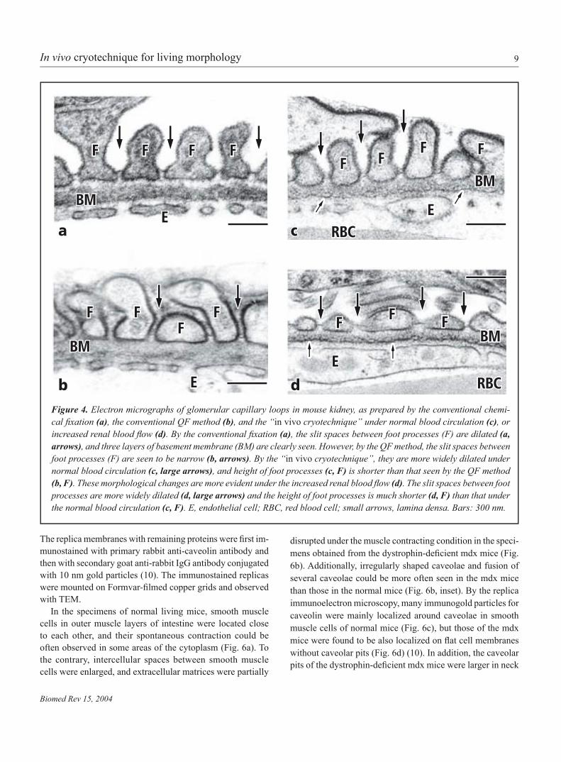
Morphological analyses of kidneyIt is generally accepted that the glomerular basement mem-brane constitutes a major part of filtration barriers betweenblood capillaries and urinary spaces. Although morphology of the basement membrane would be affected by hemody-namic factors, probably causing modification of normal barrierfunctions (43-48), the precise ultrastructures of glomerular capillary loops couldn’t be analyzed by the conventional preparation methods, because of some technical problems. For example, the morphology of glomerular capillary loops can be easily modified by artificial pressures during the perfusionfixation and also components of fixatives (49,50). In addition,the glomerular ultrastructures can be also changed by renal ischemia during the resection step (44), which is inevitable in the conventional QF methods. To overcome these problems, we have applied the “in vivo cryotechnique” to the morphological analyses of glomerular capillary loops of living mouse kidneys under different hemodynamic conditions.
A mouse kidney was exposed under pentobarbital an-esthesia, and then the “in vivo cryotechnique” was routinely performed, as already described above. Additionally, to inves-tigate morphological changes of glomerular capillary loops by either increase or decrease of renal blood flow, it was alsoperformed after the ligation of abdominal aorta at the point just distal to branching renal arteries (a hyperflow state), or after theheart arrest by intraperitoneal injection of excessive amounts of pentobarbital (a heart arrest state). After resecting the frozen kidney tissues, some of them were freeze-substituted in acetone containing 2% osmium tetroxide, and routinely processed for scanning electron microscopy (SEM) (7,15). Other specimens were embedded in epoxy resin after the freeze-substitution, sectioned under an ultramicrotome, stained with uranyl acetate and lead citrate, and finally observed with a transmission elec-tron microscope (TEM) (8). For comparison, mouse kidney tissues were prepared by the conventional chemical fixationwith glutaraldehyde and osmium tetroxide, followed by the routine dehydration with a graded series of alcohol (8,15). The conventional QF method was also performed, in which fresh kidney tissues were resected and quickly frozen by plunging into the isopentane-propane cryogen (8).
The slit spaces between foot processes were widely open in the specimens prepared by the conventional chemical fixa-tion (Fig. 4a). In addition, such foot processes exhibited their irregular surface contours, indicating that they were shrunken
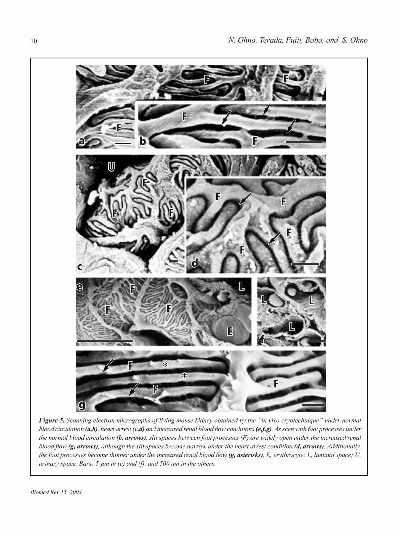
during the chemical fixation and dehydration steps, as reportedbefore (Fig. 4a) (8). To the contrary, in the specimens prepared by the conventional QF method (Fig 4b), the width of the slit spaces became narrow, because the shrinkage artifacts could be hardly produced with the cryofixation. Moreover, in thespecimens prepared by the “in vivo cryotechnique” (Fig. 4c), the slit spaces between foot processes were more widely open under normal blood circulation, and their height was a little shorter than that as revealed by the conventional QF method. In the specimens of mouse kidneys under the hyperflow state(Fig. 4d), they were much more widely open, and their height was dramatically shorter, which might reflect extension offoot processes around blood capillaries due to the increase of renal blood flow (8). By SEM (Fig. 5), both elongation of footprocesses and wideness of their slit spaces could be more obvi-ously confirmed in the view of three-dimension (Fig. 5e-f), ascompared with those under normal blood circulation (Fig. 5a, b) (7). On the other hand, deep interdigitation of compact foot processes was seen in the specimens of mouse kidney under the heart arrest, and the slit spaces between foot processes became narrower (Fig. 5c,d).
The widely open slit spaces between foot processes in the specimens prepared by the conventional chemical fixation wereconsidered to be artifacts induced by perfusion pressures of chemical fixatives and shrinkage of foot processes during thealcohol dehydration (8). The new findings obtained by the “in vivo cryotechnique” indicate that the renal glomerular capil-lary loops can be dynamically changed in vivo, depending on the renal blood flow condition. The “in vivo cryotechnique” would be useful for physiological and pathological analyses of dynamic morphology in kidney and other organs, which has been difficult to be examined with the conventional chemicalfixation or QF methods.
Dynamic ultrastructures of smooth muscle cellsIntestinal organs of normal mice or dystrophin-deficient mdxmice under pentobarbital anesthesia were surgically exposed in their abdomen and put on a thin plastic plate. The “in vivo cryotechnique” was similarly performed, as described above. Some of the frozen specimens were routinely freeze-substi-tuted in acetone containing osmium tetroxide, embedded in epoxy resin, sectioned, stained with uranyl acetate and lead citrate, and observed with TEM. Others were freeze-fractured, deeply etched under high vacuum conditions and replicated with platinum and carbon in the freeze-fracture apparatus (Eiko FD-3AS, Eiko Engineering, Ibaraki, Japan), as reported before (10). Some tissue components attached to the prepared replica membranes were partially dissolved by incubation in both 5% sodium dodecyl sulfate (SDS) and 0.5% collagenase.
9
Biomed Rev 15, 2004
In vivo cryotechnique for living morphology
Figure 4. Electron micrographs of glomerular capillary loops in mouse kidney, as prepared by the conventional chemi-cal fixation (a), the conventional QF method (b), and the “in vivo cryotechnique” under normal blood circulation (c), or increased renal blood flow (d). By the conventional fixation (a), the slit spaces between foot processes (F) are dilated (a, arrows), and three layers of basement membrane (BM) are clearly seen. However, by the QF method, the slit spaces between foot processes (F) are seen to be narrow (b, arrows). By the “in vivo cryotechnique”, they are more widely dilated under normal blood circulation (c, large arrows), and height of foot processes (c, F) is shorter than that seen by the QF method (b, F). These morphological changes are more evident under the increased renal blood flow (d). The slit spaces between foot processes are more widely dilated (d, large arrows) and the height of foot processes is much shorter (d, F) than that under the normal blood circulation (c, F). E, endothelial cell; RBC, red blood cell; small arrows, lamina densa. Bars: 300 nm.
The replica membranes with remaining proteins were first im-munostained with primary rabbit anti-caveolin antibody and then with secondary goat anti-rabbit IgG antibody conjugated with 10 nm gold particles (10). The immunostained replicas were mounted on Formvar-filmed copper grids and observed with TEM.
In the specimens of normal living mice, smooth muscle cells in outer muscle layers of intestine were located close to each other, and their spontaneous contraction could be often observed in some areas of the cytoplasm (Fig. 6a). To the contrary, intercellular spaces between smooth muscle cells were enlarged, and extracellular matrices were partially
disrupted under the muscle contracting condition in the speci-mens obtained from the dystrophin-deficient mdx mice (Fig. 6b). Additionally, irregularly shaped caveolae and fusion of several caveolae could be more often seen in the mdx mice than those in the normal mice (Fig. 6b, inset). By the replica immunoelectron microscopy, many immunogold particles for caveolin were mainly localized around caveolae in smooth muscle cells of normal mice (Fig. 6c), but those of the mdx mice were found to be also localized on flat cell membranes without caveolar pits (Fig. 6d) (10). In addition, the caveolar pits of the dystrophin-deficient mdx mice were larger in neck
F F F F
EBM
а c
b d
F F FF
BM
EE
FF F
BM
RBC
BM
FFFF
RBC
E
10
Biomed Rev 15, 2004
N. Ohno, Terada, Fujii, Baba, and S. Ohno
Figure 5. Scanning electron micrographs of living mouse kidney obtained by the “in vivo cryotechnique” under normal blood circulation (a,b), heart arrest (c,d) and increased renal blood flow conditions (e,f,g). As seen with foot processes under the normal blood circulation (b, arrows), slit spaces between foot processes (F) are widely open under the increased renal blood flow (g, arrows), although the slit spaces become narrow under the heart arrest condition (d, arrows). Additionally, the foot processes become thinner under the increased renal blood flow (g, asterisks). E, erythrocyte; L, luminal space; U, urinary space. Bars: 5 µm in (e) and (f), and 500 nm in the others.
Fа
F F
F
F
b
F
F
FF
FF
U
Fdc
e
f
g
F
F F
F
F F
E
LL
L
L
**

11
Biomed Rev 15, 2004
In vivo cryotechnique for living morphology
size than those of the normal mice (Fig. 6c,d).Thus, the “in vivo cryotechnique” has revealed morpho-
logical and immunohistochemical features of spontaneously contracting smooth muscle cells in the normal living mouse intestine and clarified their clear difference of extracellularmatrix and caveolae from those in the dystrophin-deficientmdx mice. From these findings, the “in vivo cryotechnique” combined with the replica immunoelectron microscopy would be highly useful for examining functional ultrastructures of living animal cells and in situ immunodistribution of signal molecules.
Light microscopic analyses of liver specimensMouse liver tissues were prepared by the “in vivo cryotech-nique”, as compared with those frozen by the plunging QF method with isopentane-propane cryogen. The both frozen specimens were freeze-substituted in acetone containing 2% PFA and embedded in the paraffin wax. Then thin sections wereroutinely stained with hematoxylin-eosin (HE) and observed with a light microscope. Other sections were immunostained with goat anti-mouse albumin or IgG antibody and visualized by the routine immunoperoxidase method.
Figure 6. Electron micro-graphs of intestinal smooth muscle cells in normal mice (a,c) or dystrophin-deficientmdx mice (b,d). They were prepared by the “in vivo cryotechnique”, followed by either embedding in epoxy resin (a,b) or replica immunoelectron micros-copy (c,d). The intercellular matrix (a, arrows) between contracting smooth muscle cells (SM) is tightly at-tached to each other in the normal mice, but it is widely dilated in the dys-trophin-deficient mdx mice(b, arrows). Additionally, irregularly shaped or fus-ing caveolae are also seen in the mdx mice (b, inset, arrowheads). In the rep-lica immunoelectron micro-graphs (c,d), immunogold particles for caveolin are seen around caveolae in the normal mice (c, arrows), although they are also seen on flat cell membranes inthe mdx mice (d, arrows). M, mitochondria. Bars: 500 nm in (a) and (b); 100 nm in (c) and (d).
M
a
SM
SM
SM
M
M
b
c d

12
Biomed Rev 15, 2004
N. Ohno, Terada, Fujii, Baba, and S. Ohno
Figure 7. Light micrographs of living mouse liver sections with hematoxylin-eosin staining (a,b) and immunohisto-chemical staining for IgG (c,e) and albumin (d), as prepared by the “in vivo cryotechnique” (a,c,d) or the plunging QF method (b,e). Sinusoidal cavities with flowing erythrocytesare well preserved and widely open in the specimens pre-pared by the “in vivo cryotechnique” (a, arrowheads), although such sinusoidal cavities are collapsed in the resected tissues prepared by the QF method (b). Addition-ally, the immunoreactivities of IgG and albumin in the sinusoids are clearly detected in the specimens prepared by the “in vivo cryotechnique” (c,d, arrows), although the immunoreactivity of IgG is also seen diffusely throughout the cytoplasm of some hepatocytes in the resected tissues prepared by the QF method (e, arrows). Asterisks, central vein. Bars: 50 µm.
As compared with the resected liver tissues, sinusoidal cavities between hepatocytes in living mouse liver prepared by the “in vivo cryotechnique” were maintained to be widely open, accompanied by many flowing erythrocytes (Fig. 7a).The serum albumin and IgG were clearly immunolocalized in the sinusoidal cavities of living mouse liver (Fig. 7c,d), and cytoplasmic immunoreactivity of synthesized albumin could be also detected in almost all hepatocytes. However, in the resected liver tissues prepared by the plunging QF method, both albumin and IgG immunoreactivities could be detected in the cytoplasm of some hepatocytes, in addition to their immunolocalization in the collapsed sinusoids (Fig. 7b, e). These findings indicate that anoxia and ischemic stresseswith the tissue resection might induce rapid changes of cell membrane permeability of hepatocytes and distribution of blood components in the liver tissues. Therefore, the “in vivo cryotechnique” would be also useful for immunohistochemi-cal analyses on such rapid translocation of serum proteins in other organs.
Visualization of injected fluorescent dyeA living mouse kidney was surgically exposed under pentobar-bital anesthesia, and directly injected inside its renal capsule with FITC-conjugated goat IgG (green). They were quickly frozen in a few seconds immediately after the injection by the “in vivo cryotechnique”. The frozen tissues were routinely freeze-substituted in acetone containing 2% PFA, incubated with 30% sucrose, and sectioned in a cryostat machine after freezing. In the cryostat sections, the green fluorescent dyecould be observed in glomelular capillary loops in addition to blood vessels around proximal or distal tubules (Fig. 8). Therefore, the “in vivo cryotechnique” in combination with direct injection of fluorescent dyes would be useful for dy-namic analyses of time-dependent changes of blood flow inliving animal organs.
C. Central nervous system (cerebellum)The central nervous regions, including cerebellum and cere-brum, are covered with the cranial bone, which must be care-fully opened in the case of living mice under pentobarbital anesthesia. Some devices, such as dental electric drills, are useful for opening the cranial bone. When highly concentrated attention has been paid to minimizing their bleeding and pre-venting them from drying, the “in vivo cryotechnique” can be successfully applied to their morphological and immunohis-tochemical analyses.
ba
c
e
*
d
**
*

13
Biomed Rev 15, 2004
In vivo cryotechnique for living morphology
Figure 8. Light micrographs of fluorescence (a) or DIC image (b) of a living mouse kidney after direct injection of a green fluorescent dye, as prepared by the “in vivo cryotechnique”. The fluorescent dye circulating in an afferent or efferent arteri-ole (large arrows), glomerular capillary loops (arrowheads), and blood vessels among renal tubules (small arrows) can be clearly observed under normal blood circulation. Bar: 100 µm.
Morphological analyses of molecular layerThe mouse cerebella frozen by the “in vivo cryotechnique” were freeze-substituted in acetone containing 2% osmium tetroxide and embedded in epoxy resin. Other cerebella, which had been routinely perfusion-fixed with glutaraldehyde, werepostfixed with osmium tetroxide, dehydrated in a graded seriesof alcohol, and embedded in the epoxy resin. Both embedded cerebellar tissues were cut on an ultramicrotome, routinely stained with uranyl acetate and lead citrate and observed with
TEM. Extracellular spaces among neuronal and glial cells in molecular layers were well maintained to be widely open in the specimens prepared by the “in vivo cryotechnique” (Fig. 9a). Some synaptic densities, where the intercellular spaces became narrow, could be observed in the widely open extracellular spaces (Fig. 9a, arrowhead). To the contrary, the extracellular spaces were not well preserved and lost in the specimens pre-pared by the conventional fixation and dehydration method(Fig. 9b). It was reported that the ischemia of cerebral tissues
Figure 9. Electron micrographs of molecular layers in mouse cerebellum, as prepared by either the “in vivo cryotechnique” (a) or the conventional chemical fixation and dehydration method (b). In the specimens prepared by the “in vivo cryo-technique”, lots of extracellular spaces (a, asterisks) are well preserved, and synaptic clefts become narrow near synaptic densities (a, arrowheads), although such extracellular spaces are eliminated by the conventional method (b). Arrowheads, synaptic density. Bars: 2 µm.
ba
a
** *
b*

14
Biomed Rev 15, 2004
N. Ohno, Terada, Fujii, Baba, and S. Ohno
might cause some structural changes, resulting in a decrease of electrical conductivity (51-53). However, the real ultrastructur-al changes could never been clarified by the conventional QFmethods. In our study, the “in vivo cryotechnique” can avoid such artificial modifications due to ischemia. Actually, extra-cellular spaces filled with intercellular fluid are widely openunder normal blood circulation, which are easily collapsed by the resection of cerebellar tissues and stop of blood circulation. As such extracellular spaces are suggested to be important for physiological functions of remodeling neural or glial cells, the “in vivo cryotechnique” would be useful as a morphological approach for functional analyses of dynamically changing cells and tissues in the central nervous system.
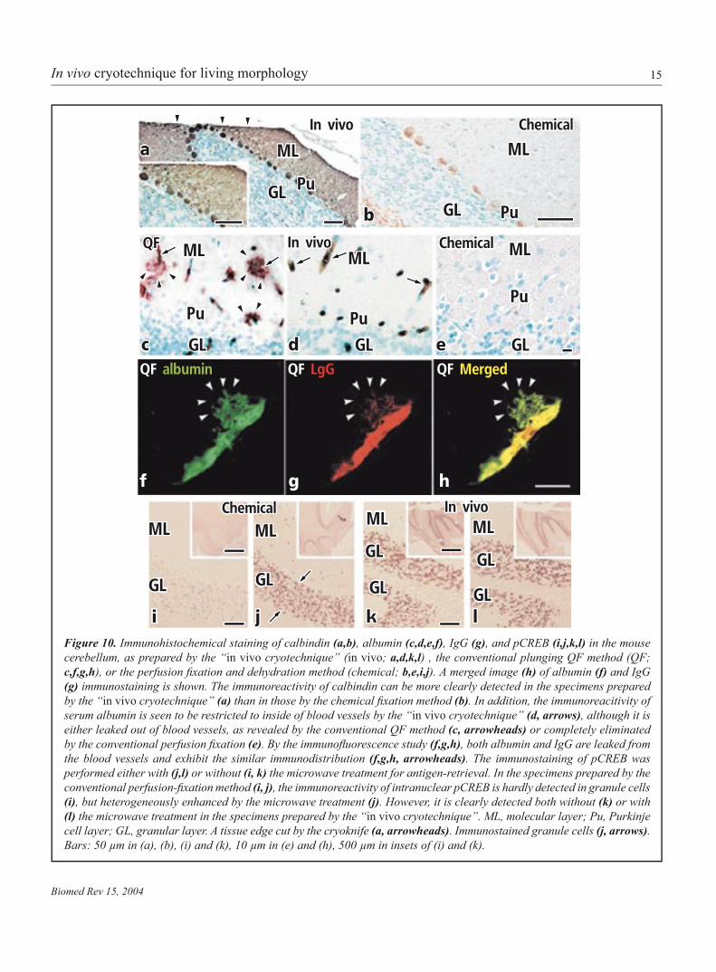
Immunolocalization of functional proteinsMouse cerebellar tissues under normal blood circulation were cryofixed by the “in vivo cryotechnique”, freeze-substituted in acetone containing 2% PFA and routinely embedded in the paraffin wax. The conventional QF method was alsoperformed by resecting cerebellar tissues and then plung-ing them into the isopentane-propane cryogen. The frozen specimens were similarly embedded in the paraffin wax. Theconventional chemical fixation was performed by perfusionwith buffered 2% PFA, followed by alcohol dehydration, and the cerebellar tissues were embedded in the paraffin wax. Allspecimens were sectioned at 4~5µm thickness, mounted on glass slides and immunostained with four kinds of antibodies against calcium-binding calbindin, serum albumin, mouse IgG and phosphorylated cyclic-AMP responsive element binding protein (pCREB) by either immunoperoxidase or immunofluo-rescence method (6,16).
The immunoreactivity of calbindin in the cytoplasm of Purkinje cells could be more clearly detected in the specimens prepared by the “in vivo cryotechnique” than in those by the conventional perfusion-fixation method (Fig. 10a,b). Ad-ditionally, the immunoreactivities of soluble serum proteins, albumin and IgG, could be detected exclusively within blood vessels in the specimens prepared by the “in vivo cryotech-nique” (Fig. 10d), although such immunoreactivities were completely eliminated in the specimens by the conventional perfusion-fixation method (Fig. 10e). They were detectedoutside of blood vessels in the resected specimens prepared by the plunging QF method (Fig. 10c,f-h) (16). Furthermore, the immunoreactivity of intranuclear pCREB in granule cells was clearly detected in the specimens prepared by the “in vivo cryotechnique” even without the microwave treatment for antigen-retrieval (Fig. 10k,l). However, the immunore-activity of pCREB was not clearly detected in the specimens
prepared by the conventional fixation method (Fig. 10i), andits enhancement was heterogeneously obtained with the mi-crowave treatment (Fig. 10j) (6).
The leakage of serum proteins might be caused by the dis-ruption of blood-brain barriers, immediately after the ischemic stress and anoxia (16). The “in vivo cryotechnique” would be useful for immunohistochemical analyses of intranuclear signal molecules, soluble serum proteins and calcium-binding proteins, which can be easily washed out during the preparation steps or translocated by various biological stresses. Moreover, the time-resolution of cryotechniques for physical fixation,depending on their cooling rates, is very high (26), and they were used for analyses of dynamic phenomena, such as neu-rotransmitter release or exocytotic processes (54,55). As the “in vivo cryotechnique” can directly cryofix the living animaltissues without ischemia or anoxia, it would be more useful for examining dynamically changing components in vivo, such as signal molecules, at an ultimate time-resolution.
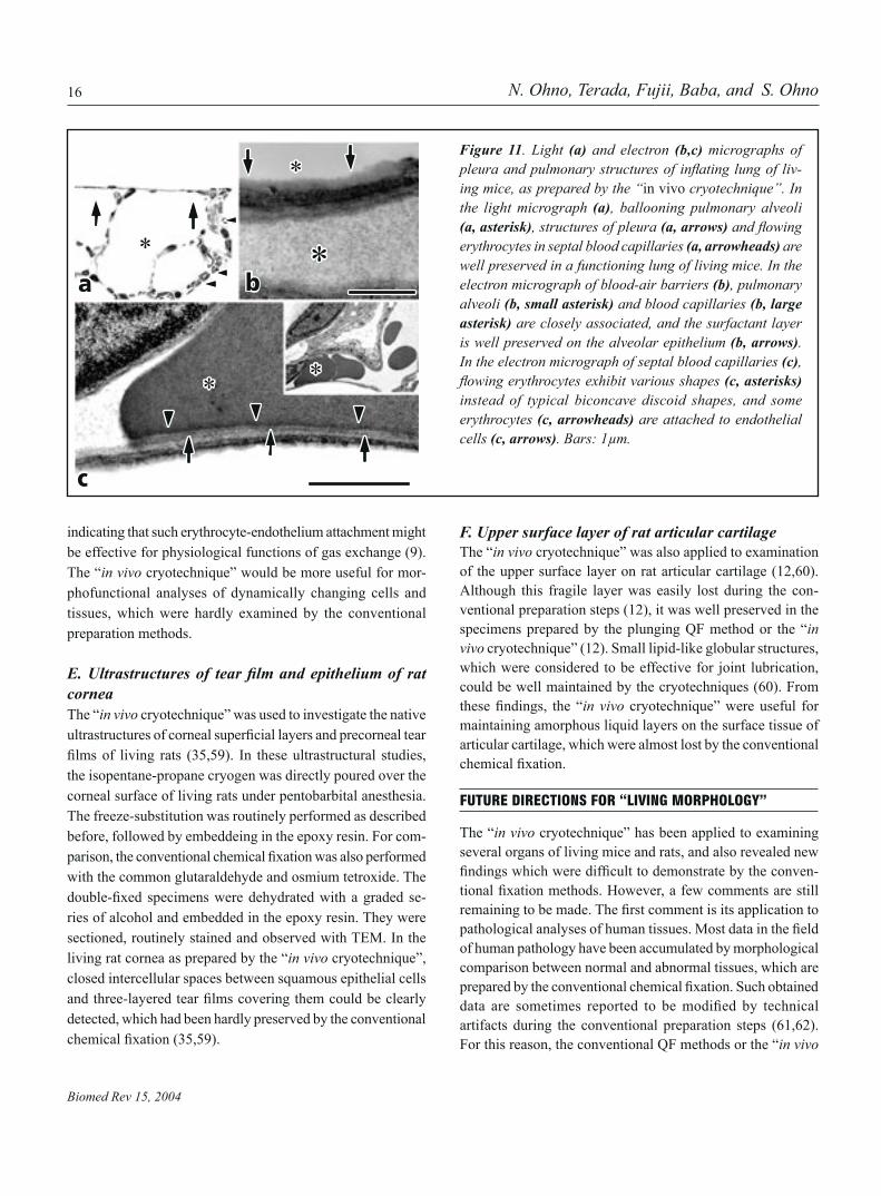
D. Alveolar structures of living mice during respira-tionAlthough some ultrastructural features of animal lungs were described in the previous reports (56-58), they could provide not enough information about physiological functions of the lungs. This is because the conventional electron microscopy couldn’t reveal pulmonary ultrastructures of living animals during functional respiration. Therefore, the “in vivo cryo-technique” was applied to morphological analyses of mouse lungs inflated by mechanical ventilation (9). Some BALB/cmice were intubated under pentobarbital anesthesia, and then mechanical ventilation was started with air. Thereafter, their lungs were surgically exposed and cryofixed at the end ofinspiration by the “in vivo cryotechnique”. The frozen tissues were freeze-substituted in acetone containing 2% osmium tetroxide, embedded in epoxy resin and routinely processed for light or electron microscopy.
In thick sections stained with toluidine blue as observed with a light microscope (Fig. 11a), dynamic structures of pleura and subpleural regions of the mouse lung, including inflatedalveoli, interstitial or alveolar epithelial cells and flowingerythrocytes in blood vessels, were well preserved without any remarkable ice crystal damage. Moreover, close associa-tion between pulmonary alveoli and blood capillaries with flowing erythrocytes could be detected in ultrathin sections,as observed with TEM (Fig. 11b). The flowing erythrocytesexhibited various shapes different from the typical biconcave discoid (Fig. 11c, inset), as seen in other organs (11,13). Some of them were partially attached to endothelial cells (Fig. 11c),
15
Biomed Rev 15, 2004
In vivo cryotechnique for living morphology
Figure 10. Immunohistochemical staining of calbindin (a,b), albumin (c,d,e,f), IgG , and pCREB (i,j,k,l) in the mouse cerebellum, as prepared by the “in vivo cryotechnique” (in vivo; a,d,k,l) , the conventional plunging QF method (QF; c,f,g,h), or the perfusion fixation and dehydration method (chemical; b,e,i,j). A merged image (h) of albumin (f) and IgG (g) immunostaining is shown. The immunoreactivity of calbindin can be more clearly detected in the specimens prepared by the “in vivo cryotechnique” (a) than in those by the chemical fixation method (b). In addition, the immunoreacitivity of serum albumin is seen to be restricted to inside of blood vessels by the “in vivo cryotechnique” (d, arrows), although it is either leaked out of blood vessels, as revealed by the conventional QF method (c, arrowheads) or completely eliminated by the conventional perfusion fixation (e). By the immunofluorescence study (f,g,h), both albumin and IgG are leaked from the blood vessels and exhibit the similar immunodistribution (f,g,h, arrowheads). The immunostaining of pCREB was performed either with (j,l) or without (i, k) the microwave treatment for antigen-retrieval. In the specimens prepared by the conventional perfusion-fixation method (i, j), the immunoreactivity of intranuclear pCREB is hardly detected in granule cells (i), but heterogeneously enhanced by the microwave treatment (j). However, it is clearly detected both without (k) or with (l) the microwave treatment in the specimens prepared by the “in vivo cryotechnique”. ML, molecular layer; Pu, Purkinje cell layer; GL, granular layer. A tissue edge cut by the cryoknife (a, arrowheads). Immunostained granule cells (j, arrows). Bars: 50 µm in (a), (b), (i) and (k), 10 µm in (e) and (h), 500 µm in insets of (i) and (k).
a
b
c d e
hgf
i j k l
ML
GL Pu
PuGL
Pu
GL
Pu
GLGL
Pu
ML ML ML
In vivo Chemical
ChemicalIn vivoQF
QF albumin QF LgG QF Merged
Chemical In vivoML MLGL GL
GL
MLML
GL GL GL
ML
16
Biomed Rev 15, 2004
N. Ohno, Terada, Fujii, Baba, and S. Ohno
indicating that such erythrocyte-endothelium attachment might be effective for physiological functions of gas exchange (9). The “in vivo cryotechnique” would be more useful for mor-phofunctional analyses of dynamically changing cells and tissues, which were hardly examined by the conventional preparation methods.
E. Ultrastructures of tear film and epithelium of ratcorneaThe “in vivo cryotechnique” was used to investigate the native ultrastructures of corneal superficial layers and precorneal tearfilms of living rats (35,59). In these ultrastructural studies,the isopentane-propane cryogen was directly poured over the corneal surface of living rats under pentobarbital anesthesia. The freeze-substitution was routinely performed as described before, followed by embeddeing in the epoxy resin. For com-parison, the conventional chemical fixation was also performedwith the common glutaraldehyde and osmium tetroxide. The double-fixed specimens were dehydrated with a graded se-ries of alcohol and embedded in the epoxy resin. They were sectioned, routinely stained and observed with TEM. In the living rat cornea as prepared by the “in vivo cryotechnique”, closed intercellular spaces between squamous epithelial cells and three-layered tear films covering them could be clearlydetected, which had been hardly preserved by the conventional chemical fixation (35,59).
F. Upper surface layer of rat articular cartilageThe “in vivo cryotechnique” was also applied to examination of the upper surface layer on rat articular cartilage (12,60). Although this fragile layer was easily lost during the con-ventional preparation steps (12), it was well preserved in the specimens prepared by the plunging QF method or the “in vivo cryotechnique” (12). Small lipid-like globular structures, which were considered to be effective for joint lubrication, could be well maintained by the cryotechniques (60). From these findings, the “in vivo cryotechnique” were useful for maintaining amorphous liquid layers on the surface tissue of articular cartilage, which were almost lost by the conventional chemical fixation.
FUTURE DIRECTIONS FOR ”LIVING MORPHOLOGY·
The “in vivo cryotechnique” has been applied to examining several organs of living mice and rats, and also revealed new findings which were difficult to demonstrate by the conven-tional fixation methods. However, a few comments are stillremaining to be made. The first comment is its application topathological analyses of human tissues. Most data in the fieldof human pathology have been accumulated by morphological comparison between normal and abnormal tissues, which are prepared by the conventional chemical fixation. Such obtaineddata are sometimes reported to be modified by technicalartifacts during the conventional preparation steps (61,62). For this reason, the conventional QF methods or the “in vivo
Figure 11. Light (a) and electron (b,c) micrographs of pleura and pulmonary structures of inflating lung of liv-ing mice, as prepared by the “in vivo cryotechnique”. In the light micrograph (a), ballooning pulmonary alveoli (a, asterisk), structures of pleura (a, arrows) and flowingerythrocytes in septal blood capillaries (a, arrowheads) are well preserved in a functioning lung of living mice. In the electron micrograph of blood-air barriers (b), pulmonary alveoli (b, small asterisk) and blood capillaries (b, large asterisk) are closely associated, and the surfactant layer is well preserved on the alveolar epithelium (b, arrows). In the electron micrograph of septal blood capillaries (c), flowing erythrocytes exhibit various shapes (c, asterisks) instead of typical biconcave discoid shapes, and some erythrocytes (c, arrowheads) are attached to endothelial cells (c, arrows). Bars: 1µm.
a
*
b
c
*
**
*

17
Biomed Rev 15, 2004
In vivo cryotechnique for living morphology
cryotechnique” would be useful for solving some technical problems in such pathological cases. As already discussed above, however, the conventional QF methods also yield some morphological and immunohistochemical artifacts induced by the tissue resection, resulting in ischemia and anoxia. The “in vivo cryotechnique” would be more suitable for these cases than the QF methods. For its human application, we need to develop a new device or improve the “in vivo cryoapparatus” for the human biopsy system, named as “cryobiopsy” (Fig. 1).
Another comment will be a technical advancement, which is necessary not only for its human application as described in the previous paragraph, but also for obtaining more data with the cryotechniques. Especially in the case of electron micros-copy, the “in vivo cryotechnique” can quickly freeze only small limited areas. Moreover, its application is restricted to living animal organs exposed to air with minimal bleeding or less mechanical stresses. For its further effective application, such limitations should be overcome. It might be partially realized by making use of cryogens with much lower temperature, such as liquid helium.
CONCLUSION
By the “in vivo cryotechnique”, it is now possible to make morphological and immunohistochemical studies of living animal organs and tissues without any major stresses of anoxia and ischemia. We have already obtained various new findings,which had never been demonstrated with the other conven-tional methods. As the “in vivo cryotechnique” can reveal the dynamic morphology closer to “living” states with normal blood circulation, it will develop a new morphological fieldof “living morphology” during this 21th century, and allow us to understand physiology and pathology of cells and tissues in living animal organs.
REFERENCES
1. Collins FS, Morgan M, Patrinos A. The Human Genome Project: lessons from large-scale biology. Science 2003; 300: 286-290.
2. Novina CD, Sharp PA. The RNAi revolution. Nature 2004; 430: 161-164.
3. McIntosh JR. Electron microscopy of cells: a new begin-ning for a new century. J Cell Biol 2001; 153: F25-F32.
4. Selvin PR. The renaissance of fluorescence resonanceenergy transfer. Nat Struct Biol 2000; 7: 730-734.
5. Stephens DJ, Allan VJ. Light microscopy techniques for live cell imaging. Science 2003; 300: 82-86.
6. Ohno N, Terada N, Murata S, Katoh R, Ohno S. Ap-plication of cryotechniques with freeze-substitution for the immunohistochemical demonstration of intranuclear pCREB and chromosome territory. J Hictochem Cytochem 2005; 53: 55-62.
7. Ohno S, Kato Y, Xiang T, Terada N, Takayama I, Fujii Y, et al. Ultrastructural study of mouse renal glomeruli under various hemodynamic conditions by an “in vivo cryotech-nique”. Ital J Anat Embryol 2001; 106: 431-438.
8. Ohno S, Terada N, Fujii Y, Ueda H, Takayama I. Dynamic structure of glomerular capillary loop as revealed by an in vivo cryotechnique. Virchows Arch 1996; 427: 519-527.
9. Takayama I, Terada N, Baba T, Ueda H, Fujii Y, Kato Y, et al. Dynamic ultrastructure of mouse pulmonary alveoli revealed by an in vivo cryotechnique in combination with freeze-substitution. J Anat 2000; 197: 199-205.
10. Takayama I, Terada N, Baba T, Ueda H, Kato Y, Fujii Y, et al. “In vivo cryotechnique” in combination with rep-lica immunoelectron microscopy for caveolin in smooth muscle cells. Histochem Cell Biol 1999; 112: 443-445.
11. Terada N, Kato Y, Fujii Y, Ueda H, Baba T, Ohno S. Scan-ning electron microscopic study of flowing erythrocytes inhepatic sinusoids as revealed by ‘in vivo cryotechnique’. J Electron Microsc 1998; 47: 67-72.
12. Watanabe M, Leng CG, Toriumi H, Hamada Y, Aka-matsu N, Ohno S. Ultrastructural study of upper surface layer in rat articular cartilage by “in vivo cryotechnique” combined with various treatments. Med Electron Microsc 2000; 33: 16-24.
13. Xue M, Baba T, Terada N, Kato Y, Fujii Y, Ohno S. Morphological study of erythrocyte shapes in red pulp of mouse spleens revealed by an in vivo cryotechnique. Histol Histopathol 2001; 16: 123-129.
14. Xue M, Kato Y, Terada N, Fujii Y, Baba T, Ohno S. Morphological study by an ‘in vivo cryotechnique’ of the shape of erythrocytes circulating in large blood vessels. J Anat 1998; 193: 73-79.
15. Yu Y, Leng CG, Terada N, Ohno S. Scanning electron microscopic study of the renal glomerulus by an in vivo cryotechnique combined with freeze-substitution. J Anat 1998; 192: 595-603.
16. Zea-Aragon Z, Terada N, Ohno N, Fujii Y, Baba T, Ohno S. Effects of anoxia on serum immunoglobulin and albumin leakage through blood-brain barrier in mouse cerebellum as revealed by cryotechniques. J Neurosci Methods 2004; 138: 89-95.
17. Fricker MD, Meyer AJ. Confocal imaging of metabolism in vivo: pitfalls and possibilities. J Exp Bot 2001; 52: 631-640.

18
Biomed Rev 15, 2004
N. Ohno, Terada, Fujii, Baba, and S. Ohno
18. Brandtzaeg P. Tissue Preparation Methods for Immuno-histochemistry. Techniques in Immunocytochemistry, 1st ed., 1982, Academic Press, London.
19. Hippe-Sanwald S. Impact of freeze substitution on bio-logical electron microscopy. Microsc Res Tech 1993; 24: 400-422.
20. Kellenberger E. The potential of cryofixation and freezesubstitution: observations and theoretical considerations. J Microsc 1991; 161: 183-203.
21. Hopwood D. Fixatives and fixation: a review. Hitochem J 1969; 1: 323-360.
22. Kellenberger E, Johansen R, Maeder M, Bohrmann B, Stauffer E, Villiger W. Artefacts and morphological changes during chemical fixation. J Microsc 1992; 168: 181-201.
23. Shiurba R. Freeze-substitution: origins and applications. Int Rev Cytol 2001; 206: 45-96.
24. Chan FL, Inoue S. Lamina lucida of basement membrane: an artifact. Microsc Res Tech 1994; 28: 48-59.
25. Leu FJ, Chen CF, Sun AM. A new method of tissue pro-cessing that causes no shrinkage or distortion. Lab Invest 1993; 69: 121-130.
26. Plattner H, Bachmann L. Cryofixation: a tool in biologi-cal ultrastructural research. Int Rev Cytol 1982; 79: 237-304.
27. Menco BP. A survey of ultra-rapid cryofixation methodswith particular emphasis on applications to freeze-frac-turing, freeze-etching, and freeze-substitution. 1986; 4: 177-240.
28. Nicolas MT, Bassot JM. Freeze substitution after fast-freeze fixation in preparation for immunocytochemistry.Microsc Res Tech 1993; 24: 474-487.
29. Chandler DE. Rotary shadowing with platinum-carbon in biological electron microscopy: a review of methods and applications. J Electron Microsc Tech 1986; 3: 305-335.
30. Miller KR, Prescott CS, Jacobs TL, Lassignal NL. Arti-facts associated with quick-freezing and freeze-drying. J Ultrastruct Res 1983; 82: 123-133.
31. Sleytr UB, Robards AW. Understanding the artefact prob-lem in freeze-fracture replication: a review. J Microsc 1982; 126: 101-122.
32. Terracio L, Schwabe KG. Freezing and drying of biologi-cal tissues for electron microscopy. J Hitochem Cytochem 1981; 29: 1021-1028.
33. Chang SH, Mergner WJ, Pendergrass RE, Bulger RE, Berezesky IK, Trump BF. A rapid method of cryofixationof tissues in situ for ultracryomicrotomy. J Histochem Cytochem 1980; 28: 47-51.
34. von Zglinicki T, Rimmler M, Purz HJ. Fast cryofixationtechnique for X-ray microanalysis. J Mirosc 1986; 141: 79-90.
35. Chen HB, Yamabayashi S, Ou B, Ohno S, Tsukahara S. Ul-trastructural studies on the corneal superficial epitheliumof rats by in vivo cryofixation with freeze substitution.Ophthalmic Res 1995; 27: 286-295.
36. Dempsey GP, Bullivant S. A copper block method for freezing non-cryoprotected tissue to produce ice-crystal-free regions for electron microscopy. I. Evaluation using freeze-substitution. J Microsc 1976; 106: 251-260.
37. van Harreveld A, Trubatch J, Steiner J. Rapid freezing and electron microscopy for the arrest of physiological processes. J Microsc 1974; 100: 189-198.
38. von Schack ML, Fakan S, Villiger W, Muller M. Cryo-fixation and cryosubstitution: a useful alternative in theanalyses of cellular fine structure. Eur J Histochem 1993; 37: 5-18
39. Blank ME, Diedrich DF. Erythrocyte shape and volume changes caused by an inhibitor of the glucose and anion transporters. Biorheology 1990; 27: 345-355.
40. Maeda N. Erythrocyte rheology in microcirculation. Jpn J Physiol 1996; 46: 1-14.
41. Murakami J, Maeda N, Kon K, Shiga T. A contribution of calmodulin to cellular deformability of calcium-loaded human erythrocytes. Biochim Biophys Acta 1986; 863: 23-32.
42. Nishiguchi E, Ozono S, Takakuwa Y, Hamasaki N. Mechanism of the change in shape of human erythrocytes induced by lidocaine. Cell Struct Funct 1995; 20: 71-79.
43. Brenner BM, Bohrer MP, Baylis Ch, Deen WM. Determi-nants of glomerular permselectivity: Insights derived from observations in vivo. Kidney Int 1977; 12: 229-237.
44. Griffith LD, Bulger RE, Trump BF. The ultrastructure ofthe functioning kidney. Lab Invest 1967; 16: 220-246.
45. Kanwar YS. Biophysiology of glomerular filtration andproteinuria. Lab Invest 1984; 51: 7-21.
46. Kriz W, Hackenthal E, Nobiling R, Sakai T, Elger M, Hahnel B. A role for podocytes to counteract capillary wall distension. Kidney Int 1994; 45: 369-376.
47. Olivetti G, Kithier K, Giacomelli F, Wiener J. Glomerular permeability to endogenous proteins in the rat: effects of acute hypertension. Lab Invest 1981; 44: 127-137.
48. Ryan GB, Karnovsky MJ. Distribution of endogenous albumin in the rat glomerulus: role of hemodynamic fac-tors in glomerular barrier function. Kidney Int 1976; 9: 36-45.

19
Biomed Rev 15, 2004
In vivo cryotechnique for living morphology
49. Ohno S, Baba T, Terada N, Fujii Y, Ueda H. Cell biology of kidney glomerulus. Int Rev Cytol 1996; 166: 181-230.
50. Yu Y, Leng CG, Kato Y, Ohno S. Ultrastructural study of glomerular capillary loops at different perfusion pres-sures as revealed by quick-freezing, freeze-substitution and conventional fixation methods. Nephron 1997; 76: 452-459.
51. van Harreveld A, Biber MP. Conductivity changes in some organs after circulatory arrest. Am J Physiol 1962; 203: 609-614.
52. van Harreveld A, Crowell J, Malhotra SK. A study of extracellular space in central nervous tissue by freeze-substitution. J Cell Biol 1965; 25: 117-137.
53. van Harreveld A, Malhotra SK. Extracellular space in the cerebral cortex of the mouse. J Anat 1967; 101: 197-207.
54. Chandler DE, Heuser J. Membrane fusion during secre-tion: cortical granule exocytosis in sex urchin eggs as studied by quick-freezing and freeze-fracture. J Cell Biol 1979; 83: 91-108.
55. Heuser JE, Reese TS, Dennis MJ, Jan Y, Jan L, Evans L. Synaptic vesicle exocytosis captured by quick freezing and correlated with quantal transmitter release. J Cell Biol 1979; 81: 275-300.
56. Bastacky J, Lee CY, Goerke J, Koushafar H, Yager D, Kenaga L, et al. Alveolar lining layer is thin and continu-
ous: low-temperature scanning electron microscopy of rat lung. J Appl Physiol 1995; 79: 1615-1628.
57. Ham AW, Cormack DH. Histology, 8th ed., 1978, JB Lippincott, Philadelphia.
58. Weiss L, Greep RO. Histology, 4th ed., 1977, McGraw-Hill, New York.
59. Chen HB, Yamabayashi S, Ou B, Tanaka Y, Ohno S, Tsu-kahara S. Structure and composition of rat precorneal tear film.Astudy by an in vivo cryofixation. Invest Ophthalmol Vis Sci 1997; 38: 381-387.
60. Zea-Aragon Z, Terada N, Ohno N, Fujii Y, Baba T, Yoshida M, et al. Replica immunoelectron microscopic study of the upper surface layer in rat mandibular condylar cartilage by a quick-freezing method. Histochem Cell Biol 2004; 121: 255-259.
61. Kiyono T, Katagiri M, Harada T. The incidence of ground glass nuclei in thyroid diseases. Thyroidology 1994; 6: 43-48.
62. Naganuma H, Murayama H, Ohtani N, Takaya K, Mori Y, Sakai N, et al. Optically clear nuclei in papillary carci-noma of the thyroid: demonstration of one of the fixationartifacts and its practical usefulness. Pathol Int 2000; 50: 113-118.
63. Jehl B, Bauer R, Dorge A, Rick R. The use of propane/isopentane mixtures for rapid freezing of biological speci-mens. J Microsc 1981; 123: 307-309.