in vivo analysis of human lhx3 gene regulation rachel
TRANSCRIPT

IN VIVO ANALYSIS OF HUMAN LHX3 GENE REGULATION
Rachel Diane Mullen
Submitted to the faculty of the University Graduate School in partial fulfillment of the requirements
for the degree Doctor of Philosophy
in the Department of Biochemistry and Molecular Biology, Indiana University
February 2011

ii
Accepted by the Faculty of Indiana University, in partial fulfillment of the requirements for the degree of Doctor of Philosophy.
Simon J. Rhodes Ph.D., Chair
B. Paul Herring Ph.D.
Doctoral Committee
David G. Skalnik Ph.D.
December 7, 2010
Debbie C. Thurmond Ph.D.
Emily C. Walvoord M.D.

iii
ACKNOWLEDGMENTS
I would like to acknowledge the members of my graduate committee: Dr. Simon
Rhodes, Dr. Paul Herring, Dr. Debbie Thurmond, Dr. David Skalnik, and Dr. Emily
Walvoord. Your insight and helpful support has been invaluable. I would like to thank
past and present Rhodes’ lab members: Dr. Chad Hunter, Dr. Jesse Savage, Dr. Stephanie
Colvin, Dr. Zachary Neeb, Tafadzwa Mwashita, Marin Garcia, Brooke West, Krystal
Renner, Soyoung Park, Raleigh Malik, and Dr. Kelly Prince. I would also like to thank
the Department of Biochemistry and Molecular Biology faculty and staff. I especially
wish to acknowledge Dr. Zhong-Yin Zhang, Dr. William Bosron, Dr. Mark Goebl, Dr.
Anna Depaoli-Roach, Sandy McClain and Mary Harden. During my graduate career I
have been fortunate to be a visiting member of the Department of Cellular and Integrative
Physiology and wish to thank the faculty, students and staff for making me feel like a part
of their department. I also wish to extend my gratitude to Indiana University East Biology
professor and friend, Dr. Mary Blakefield, for her encouragement early in my career and
her continued support.
I would like to say a special thanks to my mentor, Dr. Simon Rhodes. Thank you
for allowing me the freedom to figure things out on my own, but helping when I asked.
Thank you for the occasional push when I procrastinated a little too long. Thank you for
your guidance, mentorship and most of all friendship.

iv
ABSTRACT
Rachel Diane Mullen
IN VIVO ANALYSIS OF HUMAN LHX3 GENE REGULATION
LHX3 is a transcription factor important in pituitary and nervous system
development. Patients with mutations in coding regions of the gene have combined
pituitary hormone deficiency (CPHD) that causes growth, fertility, and metabolic
problems. Promoter and intronic elements of LHX3 important for basal gene expression
in vitro have been identified, but the key regulatory elements necessary for in vivo
expression were unknown. With these studies, I sought to elucidate how LHX3 gene
expression is regulated in vivo. Based on sequence conservation between species in non-
coding regions, I identified a 7.9 kilobase (kb) region 3' of the human LHX3 gene as a
potential regulatory element. In a beta galactosidase transgenic mouse model, this region
directed spatial and temporal expression to the developing pituitary gland and spinal cord
in a pattern consistent with endogenous LHX3 expression. Using a systematic series of
deletions, I found that the conserved region contains multiple nervous system enhancers
and a minimal 180 base pair (bp) enhancer that direct expression to both the pituitary and
spinal cord in transgenic mice. Within this minimal enhancer, TAAT/ATTA sequences
that are characteristic of homeodomain protein binding sites are required to direct
expression. I performed DNA binding experiments and chromatin immunoprecipitation
assays to reveal that the ISL1 and PITX1 proteins specifically recognize these elements
in vitro and in vivo. Based on in vivo mutational analyses, two tandem ISL1 binding sites

v
are required for enhancer activity in the pituitary and spine and a PITX1 binding site is
required for spatial patterning of gene expression in the pituitary. Additional experiments
demonstrated that these three elements cannot alone direct gene expression, suggesting a
combination of factors is required for enhancer activity. This study reveals that the key
regulatory elements guiding developmental regulation of the human LHX3 gene lie in this
conserved downstream region. Further, this work implicates ISL1 as a new
transcriptional regulator of LHX3 and describes a possible mechanism for the regulation
of LHX3 by a known upstream factor, PITX1. Identification of important regulatory
regions will also enable genetic screening in candidate CPHD patients and will thereby
facilitate patient treatment and genetic counseling.
Simon J. Rhodes Ph.D., Chair
vi
TABLE OF CONTENTS
LIST OF TABLES ........................................................................................................... viii
LIST OF FIGURES ........................................................................................................... ix
LIST OF ABBREVIATIONS ............................................................................................ xi
CHAPTER ONE – INTRODUCTION
1.1 Pituitary Structure and Function ..............................................................................1
1.2 Early Signaling Events in Pituitary Development ...................................................3
1.3 Transcriptional Regulation of Anterior Pituitary Development .............................4
1.4 LIM-HD Transcription Factors ISL1, LHX3, and LHX4 .....................................10
1.5 Central Hypothesis and Aims ................................................................................17
CHAPTER TWO – MATERIALS AND METHODS
2.1 DNA Cloning and Vector Construction .................................................................20
2.2 Protein Analyses ....................................................................................................28
2.3 Cell Culture and Transient Transfections ..............................................................31
2.4 Generation, Genotyping, and Breeding of Transgenic Mice ................................32
2.5 Histology and Immunohistochemistry ...................................................................33
2.6 Microscopy ............................................................................................................36
2.7 Bioinformatics Analyses ........................................................................................36
2.8 General Molecular Techniques ..............................................................................36
CHAPTER THREE – IN VIVO ANALYSIS OF HUMAN LHX3 GENE
REGULATION
3.1 Results ....................................................................................................................42

vii
CHAPTER FOUR – DISCUSSION ..................................................................................66
REFERENCES ..................................................................................................................79
CURRICULUM VITAE

viii
LIST OF TABLES
Table
3.1 Single nucleotide variations identified in human LHX3 regulatory regions ................65

ix
LIST OF FIGURES
Figure
1.1 Regulation of anterior pituitary gland development by signaling proteins and
transcription factors. .................................................................................................19
2.1 LHX3a promoter-LHX3 exon Ia-LHX3b promoter-nLACZ- Full 3' modified
pWHERE transgene ..................................................................................................39
2.2 Full 3'-HSP68 nLACZ pWHERE transgene ..............................................................40
2.3 R3-HSP68 nLACZ pWHERE transgene ...................................................................41
3.1 Distal downstream regions of the human LHX3 gene direct expression to the
developing pituitary and spinal cord. ........................................................................52
3.2 Expression patterns guided by the 7.9 kb 3' enhancer region ...................................54
3.3 Native LHX3 and enhancer directed beta galactosidase expression co-
localization pattern is similar in the hormone-expressing cell types ........................56
3.4 Deletion analysis of the 3' region reveals several nervous system enhancers
and a pituitary enhancer ............................................................................................57
3.5 UTR R1 (~4500 bp) contains a silencing element for the developing
forebrain ....................................................................................................................59
3.6 A highly conserved 180 bp minimal region (Core R3) is sufficient to direct
expression to the developing pituitary ......................................................................60
3.7 Alignment of human Core R3 enhancer sequences with multiple species ...............61
3.8 ISL and PITX binding sites in the Core R3 enhancer are critical for
expression in the developing pituitary and spinal cord .............................................62

x
3.9 EMSA analysis of PITX2A, LHX3, and LHX4 binding of TAAT elements
in Core R3 .................................................................................................................64
4.1 A schematic summary of findings ............................................................................77
4.2 A hypothetical mechanism for regulation of the spatial expression pattern in
the developing pituitary ............................................................................................78

xi
LIST OF ABBREVIATIONS
Adrenocorticotropic hormone ACTH
Alpha glycoprotein subunit αGSU
Alpha melanocyte-stimulating hormone αMSH
Arginine vasopressin AVP
Base pair bp
Basic helix loop helix bHLH
Bone morphogenetic protein BMP
CCAAT-enhancer-binding protein C/EBP
CCCTC-binding factor CTCF
Central nervous system CNS
Chromatin conformation capture 3C
Combined pituitary hormone deficiency CPHD
Conserved non-coding element CNE
Corticotropin-releasing hormone CRH
Days post coitum dpc
Diaminobenzidine DAB
Diencephalon DIEN
Electrophoretic mobility shift assay EMSA
Fibroblast growth factor FGF
Follicle-stimulating hormone FSH
Follicle-stimulating hormone beta FSHβ

xii
Forkhead box FOX
Forebrain FB
Gonadotropin-releasing hormone GnRH
Gonadotropin-releasing hormone receptor GnRH-R
Growth hormone GH
Growth hormone-releasing hormone GHRH
Growth hormone-releasing hormone receptor GHRHR
High mobility group HMG
Homeodomain HD
Immunohistochemistry IHC
Infundibulum IF
Islet1 ISL1
Islet2 ISL2
Kilobases kb
Luria-Bertani broth LB
LHX3 binding consensus site LBC
LIM homeobox 3 LHX3
LIM homeobox 4 LHX4
LIM homeodomain LIM-HD
Luteinizing hormone LH
Luteinizing hormone beta LHβ
Magnocellular neurons MCN
Medial ganglionic eminence MGE

xiii
Multi-cloning site MCS
Nuclear factor 1 NF1
Nuclear LIM interacting protein NLI
Nuclear localization signal NLS
Oral ectoderm OE
Oxytocin OT
Paired homeobox PAX
Phosphate buffered saline PBS
Polymerase chain reaction PCR
Pro-opiomelanocortin POMC
Prolactin PRL
Prophet of Pit1 PROP1
Rathke’s pouch RP
Sex-determining region Y SRY
Sine oculis homeobox SIX
Single nucleotide variations SNV
Sonic hedgehog SHH
SRY-box SOX
Specificity protein 1 SP1
Steroidogenic factor 1 SF1
Thyroid-stimulating hormone TSH
Thyroid-stimulating hormone beta TSHβ
Wingless/integrated protein WNT

1
CHAPTER ONE
INTRODUCTION
1.1 Pituitary Structure and Function
The pituitary is located near the base of the brain in the sella turcica (a depression
of the sphenoid bone), and secretes hormones which regulate many essential processes
including development, the stress response, growth, reproduction, metabolism, and
lactation. The pituitary has dual embryonic origins consisting of a posterior lobe
originating from the neuroectoderm, or diencephalon, and the intermediate and anterior
lobes developing from an invagination of the oral ectoderm known as Rathke’s pouch.
The release of pituitary hormones in response to physiological conditions is mediated by
signals from the hypothalamus.
Two major hormones are secreted by the posterior lobe: arginine vasopressin
(AVP) and oxytocin (OT). AVP controls osmotic balance by regulating water absorption
in the kidneys and OT is required to stimulate muscle contractions during parturition and
lactation. The posterior lobe connects directly to the hypothalamus via the infundibulum
or pituitary stalk. Magnocellular neurons (MCN) originate in the supraoptic nuclei and
paraventricular nuclei of the hypothalamus and extend through the pituitary stalk into the
posterior lobe. AVP and OT are synthesized in MCN and transported along their axons to
a capillary bed in the posterior lobe where they are secreted into the blood.
The intermediate lobe of the pituitary secretes α-melanocyte-stimulating hormone
(αMSH) from melanotrope cells. αMSH is produced by proteolytic processing of its
prohormone from the pro-opiomelanocortin (POMC) gene. Alpha MSH has functions in
2
skin pigmentation and dark adaptation in lower vertebrates. The human intermediate lobe
is less pronounced than in other vertebrates consisting of only a thin layer of cells.
Because of the diminutive size of the human intermediate lobe, humans produce little
αMSH.
Five hormone-secreting cell types are found in the anterior pituitary:
corticotropes, gonadotropes, thyrotropes, somatotropes, and lactotropes secreting
adrenocorticotropic hormone (ACTH, a product of the POMC gene), follicle-stimulating
hormone (FSH) and luteinizing hormone (LH), thyroid-stimulating hormone (TSH),
growth hormone (GH), and prolactin (PRL), respectively. Glycoprotein hormones TSH,
FSH, and LH are composed of a unique beta subunit (TSHβ, FSHβ and LHβ) and a
common alpha-glycoprotein subunit (αGSU). Hormones secreted from the human
anterior pituitary have key roles in development, the stress response (ACTH),
reproduction (FSH, LH, and PRL), metabolism (TSH), growth (GH), and lactation
(PRL). The hormone-secreting cell types are observed to differentiate in a distinct dorsal
to ventral pattern in the developing pituitary. In the dorsal portion of the anterior
pituitary, corticotropes, somatotropes, lactotropes are observed. Thyrotropes are found in
the rostral tip and central portion of the lobe and gonadotropes arise ventrally (Dasen et
al., 1999; Kioussi et al., 1999; Lin et al., 1994).
Hormone release by secreting cell types in the anterior and intermediate pituitary
is positively and negatively regulated by hypophysiotropic hormones (e.g. release of GH:
GH-releasing hormone, inhibition of GH: somatostatin). Hypophysiotropic hormones
secreted from the median eminence of the hypothalamus are transported via the
hypophyseal portal blood system and bind specific cell surface receptors (e.g. GH-

3
releasing hormone receptor, somatostatin receptor) in the anterior and intermediate
pituitary resulting in hormone (e.g. GH) release or inhibition.
1.2 Early Signaling Events in Pituitary Development
Signaling gradients between multiple factors in the diencephalon and oral
ectoderm result in the invagination of the oral ectoderm to form Rathke’s pouch, the
primordium of the anterior pituitary lobe (Figure 1.1). The first step in the formation of
the anterior pituitary is a thickening of the oral ectoderm and invagination to form
Rathke’s pouch, the primordial structure of the anterior pituitary. Based on findings from
multiple studies in mice, this initial step is dependent on bone morphogenetic protein
(BMP) 4 signals originating in the adjacent ventral diencephalon (Davis and Camper,
2007; Sheng et al., 1997; Takuma et al., 1998). This invagination brings Rathke’s pouch
in close contact with the adjacent ventral diencephalon and promotes further the
proliferation and differentiation signaling events required for the formation of the mature
pituitary gland.
Subsequently, BMP2 and BMP7 expression is initiated in the ventral
mesenchyme adjacent to Rathke’s pouch and expands into the pouch in a ventral to dorsal
pattern (Ericson et al., 1998; Gleiberman et al., 1999). Signaling gradients involving
BMPs and fibroblast growth factors (FGF) 8, FGF10, and FGF18 have key roles in dorsal
to ventral patterning of the pituitary gland. Dorsally, FGFs are thought to maintain
Rathke’s pouch cells in a proliferative state and prohibit cell cycle exit. As cells migrate
ventrally, FGF levels are reduced and cells exit the cell cycle and differentiate into
definitive hormone cell types. Ventrally, BMPs promote ISL1 (described in section 1.4)

4
and αGSU expression and ventral cell types in the anterior pituitary in part by opposing
FGF signaling (Ericson et al., 1998; Kimura et al., 1996; Norlin et al., 2000).
Sonic hedgehog (SHH), expressed both in the ventral diencephalon and
throughout the oral ectoderm, is excluded from Rathke’s pouch (Treier et al., 1998;
Treier et al., 2001). Studies have shown that SHH signaling has important roles in
pituitary development. Blocking the pathway with the SHH antagonist hedgehog
interacting protein in Rathke’s pouch arrested pituitary development, and over expression
of SHH in the developing pituitary of mice resulted in pituitary hyperplasia (Treier et al.,
2001). Other signaling molecules and transcription factors in the ventral diencephalon
important for proper pituitary development include the LIM homeodomain (HD) protein
LHX2, SOX3, WNT5a, and NKX2.1 (Alatzoglou et al., 2009; Cha et al., 2004; Potok et
al., 2008; Takuma et al., 1998; Zhao et al., 2010).
1.3 Transcriptional Regulation of Anterior Pituitary Development
Further differentiation and proliferation events controlled by a cascade of
transcription factors results in development of the anterior pituitary and establishment of
the hormone-secreting cell types (Figure 1.1) [reviewed in (Kelberman et al., 2009; Zhu
et al., 2007)]. Signaling molecules and transcription factors found in the anterior pituitary
required for these developmental events include GLI1, GLI2, EYA1, SIX1, SIX3, SIX6,
PAX6, HESX1, SOX2, PITX1, PITX2, ISL1, LHX3, and LHX4 (described in section
1.4), and PROP1.
GLI1, GLI2, and GLI3 are downstream transcription factors expressed in
Rathke’s pouch in response to SHH signaling. Gli1-/- mice have variable loss of the
pituitary while Gli1-/-/ Gli2-/- double knockout mice have a more severe phenotype; in

5
addition to defects in the ventral diencephalon, all have aplastic pituitaries (Park et al.,
2000). Heterozygous mutations within the human GLI2 gene cause variable forms of
holoprosencephaly with hypoplastic or absent pituitaries and variable defects in facial
structures (Roessler et al., 2003).
The SIX gene family members are mammalian homologs of Drosophila
melanogaster sine oculis homeobox containing genes and act as part of protein
complexes containing the co-repressor recruiter DACH and the EYA phosphatase. SIX1,
SIX3, and SIX6 are expressed in the developing pituitary [reviewed in (Kawakami et al.,
2000)]. Studies in mice and zebrafish have shown that SIX1 and EYA1 have cooperative
functions in pituitary development. Double knockdown of SIX1 and EYA1 in zebrafish
results in a failure to develop corticotropes, melanotropes, and gonadotropes.
Somatotropes and thyrotropes are present but fail to express GH and TSHβ (Nica et al.,
2006). The Six1-/- / Eya1-/- double knockout mice have hypoplastic pituitary glands (Li et
al., 2003). In Six3-/- mice, early inductive events are disrupted and Rathke’s pouch fails to
form and mice double heterozygous for Six3 and Hesx1 null alleles have hypopituitarism
(Gaston-Massuet et al., 2008; Lagutin et al., 2003). Six6 knockout mice have defects in
retinal, optic nerve and pituitary development. SIX6 also represses transcription of cell
cycle inhibitors thereby promoting cellular proliferation in the developing retina and
pituitary (Li et al., 2002).
PAX6 is a paired HD transcription factor important in the development of several
tissues including the eye, nervous system, pancreas, and pituitary (Bentley et al., 1999;
Dohrmann et al., 2000; Terzic and Saraga-Babic, 1999). The Small eye Pax6 mutants and
Pax6-/- mice have defects in dorsal to ventral patterning of the pituitary that results in
6
reduced numbers of somatotropes and lactotropes dorsally and an increase in thyrotropes
and gonadotropes ventrally (Kioussi et al., 1999). Recently the only surviving patient
with a compound heterozygous mutation in the PAX6 gene was described presenting with
severe developmental defects consistent with single heterozygous mutations plus a
hypoplastic pituitary (Solomon et al., 2009).
In mouse and humans, the paired-class HD transcription factor HESX1 is
expressed first in the neural plate and later restricted to the forebrain, ventral
diencephalon and Rathke’s pouch by e9.5 (Hermesz et al., 1996; Sajedi et al., 2008;
Thomas et al., 1995). LHX3 is required during early pituitary development to maintain
HESX1 expression (Sheng et al., 1997). Then as differentiation proceeds of specific
hormone-secreting cell types, Hesx1 is down regulated by the PROP1 paired homeobox
protein (described below) and becomes undetectable by e15.5 (Gage et al., 1996;
Hermesz et al., 1996). HESX1 is capable of repressing Prop1 gene expression by
recruiting co-repressor complexes containing Groucho-like TLE proteins and histone
deacetylases (Brickman et al., 2001; Carvalho et al., 2010; Dasen et al., 2001). Hesx1-
null and human mutation knock-in mouse models have defects in eye, olfactory, and
forebrain development and pituitary dysplasia (Dattani et al., 1998; Sajedi et al., 2008).
Similarly, HESX1 mutations in human patients are associated with septo-optic dysplasia
and pituitary abnormalities (Dattani et al., 1998; Sobrier et al., 2005; Thomas et al.,
2001).
The SRY-related high mobility group box (SOX) 2 transcription factor has
important roles in anterior pituitary development. During pituitary development, SOX2 is
first expressed in the ectoderm and by e11.5 throughout Rathke’s pouch, but as cell
7
differentiation proceeds its expression is down regulated in a manner similar to HESX1.
By e18.5 expression is found only in the lumen of Rathke’s pouch and the mature gland,
in the region thought to contain the adult stem cell population of the pituitary (Fauquier et
al., 2008; Kelberman and Dattani, 2006). Sox2-null mice die shortly after implantation
prior to pituitary development (Avilion et al., 2003). The roles of SOX2 in pituitary
development have been partially elucidated in studies of heterozygous mice and humans.
A portion of surviving Sox2+/- heterozygous mice have mild hypopituitarism and mild
hypoplasia of the anterior pituitary with bifurcations in Rathke’s pouch (Alatzoglou et al.,
2009; Avilion et al., 2003; Ferri et al., 2004; Kelberman and Dattani, 2006). Humans with
heterozygous mutations in SOX2 display pleiotrophic symptoms including bilateral
anophthalmia or severe microphthalmia, anterior pituitary hypoplasia and gonadotropin
deficiency (Fantes et al., 2003; Kelberman et al., 2008; Kelberman et al., 2006;
Williamson et al., 2006). Human mutations in either the SOX2 or LHX3 genes are also
sometimes associated with sensorineural hearing loss in addition to pituitary defects. The
two proteins have overlapping expression patterns in the developing ear and pituitary and
SOX2 can bind and activate the LHX3a promoter in vitro suggesting a possible role in
LHX3 gene regulation (Rajab et al., 2008).
The bicoid-like HD transcription factors PITX1 and PITX2 are required for the
proper development of multiple organs including the heart, limbs, and pituitary. PITX1
was first identified as a protein-protein partner of the pituitary transcription factor, PIT1
(Szeto et al., 1996). PITX1 also regulates expression of the POMC gene in early pituitary
development (Lamonerie et al., 1996). Pitx1-/- mice have morphologically normal
pituitaries; however there are reductions in the number of gonadotropes and thyrotropes

8
present and LHβ and TSHβ levels and an increase in ACTH levels (Szeto et al., 1999).
Both PITX1 and PITX2 recognize and bind the hormone promoters αGSU, TSHβ, LHβ,
FSHβ, GnRHR, PRL, and GH (Tremblay et al., 2000). Knock down of PITX1 in vitro
causes a loss of both Lhx3 and αGSU expression (Tremblay et al., 1998). Further in vivo
experiments show PITX1 or PITX2 are required for activation of Lhx3 during early
pituitary development (Charles et al., 2005).
PITX2 is found in both the developing and adult pituitary gland (Gage and
Camper, 1997; Semina et al., 1996). Pitx2 gene activation is induced by the WNT-
activated beta-catenin pathways during early pituitary development (Baek et al., 2003;
Kioussi et al., 2002) and PITX2 promotes cellular proliferation by activating transcription
of critical cell cycle regulators (Baek et al., 2003; Kioussi et al., 2002). Pitx2-/- mice have
developmental defects in the heart, tooth, eye and pituitary and disruption of normal left-
right asymmetry (Lin et al., 1999; Logan et al., 1998; Lu et al., 1999; Piedra et al., 1998;
Ryan et al., 1998; Yoshioka et al., 1998). The pituitary defects of the Pitx2-null mice are
more severe than the Pitx1-null mice and pituitary development is arrested at e12.5 (Gage
et al., 1999). Further studies of Pitx2 neo/neo hypomorphs demonstrated PITX2 is required
for proper pituitary development and the differentiation of gonadotropes, thyrotropes,
somatotropes, and lactotropes (Suh et al., 2002). Both PITX1 and PITX2 proteins are
found primarily to co-localize with gonadotropes and thyrotropes in the adult pituitary.
However, mice with tissue-specific knock out of Pitx2 in adult gonadotropes are normal
(Charles et al., 2008; Charles et al., 2005). This demonstrates that PITX2 is not required
for gonadotrope function and maintenance. However, similar to the overlapping functions
seen in early development, PITX1 may be compensating for the loss of PITX2 in this

9
mouse model. PITX2 mutations in humans are a known molecular cause of Rieger
syndrome, iridogoniodysgenesis syndrome, type 2 autosomal dominant iris hypoplasia,
and Peter’s anomaly (Alward et al., 1998; Doward et al., 1999; Kulak et al., 1998;
Semina et al., 1996).
The paired-like HD transcription factor, Prophet of PIT1 (PROP1), is expressed
exclusively in the developing pituitary and is required for its proper development and
function (Sloop et al., 2000; Sornson et al., 1996). PROP1 can act as either a
transcriptional activator or repressor (Nasonkin et al., 2004). For example, the PROP1/β-
catenin complex has been shown to activate Pit1 transcription and repress Hesx1
transcription depending which cofactors are present (Olson et al., 2006). PROP1
expression in the developing pituitary is initiated at e10 to e10.5, peaks at e12.5 and then
declines after e14.5 (Sornson et al., 1996). The Ames dwarf mouse is a naturally
occurring mutant mouse found to have a point mutation resulting in a defective DNA
binding HD. Ames and Prop1-null mice have identical phenotypes. Both have
hypoplastic pituitaries with deficiencies in GH, TSH, LH, FSH, and PRL and fail to
express PIT1 (Gage et al., 1996; Sornson et al., 1996; Tang et al., 1993). In these mouse
models, proliferation of progenitors in the perilumenal region is not affected but the cells
fail to migrate. This results in a pituitary which first appears enlarged at e14.5 with
abnormal morphology, and then later as a result of increased apoptosis is hypoplastic
(Ward et al., 2005). This wax and wane in pituitary size has also been observed in some
human patients with PROP1 mutations. PROP1 gene mutations in humans are the most
common known cause of combined pituitary hormone deficiency (CPHD) and patients
have hormone deficiencies like those seen in the Prop1 mutant mouse models (Cushman

10
et al., 2002; Wu et al., 1998). The results of several transgenic mouse over-expression
studies have demonstrated that tight temporal control of Prop1 gene expression is
required for proper pituitary development. Expression of PROP1 early throughout
Rathke’s pouch ablates pituitary development and prolonged expression in gonadotropes
and thyrotropes delays gonadotrope development and leads to pituitary tumors (Cushman
et al., 2001; Dasen et al., 2001; Dasen and Rosenfeld, 2001). Double knockout of Lhx4
and Prop1 in mice more severely affects pituitary development than single knockout of
either gene. Corticotrope differentiation is delayed and the other hormone-secreting cells
fail to develop. This indicates LHX4 and PROP1 together regulate differentiation and
expansion events in the developing pituitary gland (Raetzman et al., 2002).
Further actions by downstream transcription factors including PIT1, SF1 and
TPIT are also required for differentiation and specification of specific hormone-secreting
cell types. PIT1 (also POU1F1, and GHF1) is a POU-HD transcription factor required for
specification of somatotropes, thyrotropes, and lactotropes. Steroidogenic factor (SF) 1 is
essential for gonadotrope development. A T-box class transcription factor, known as
TBX19 or TPIT, has key roles in specification of cortitropes and directly activates POMC
expression with PITX1 (Figure 1.1) [reviewed in (Kelberman et al., 2009; Zhu et al.,
2007)].
1.4 LIM-HD Transcription Factors ISL1, LHX3, and LHX4
ISL1
Islet (ISL) 1 is a member of the LIM-HD family of transcription factors. LIM-HD
transcription factors contain two zinc finger LIM domains important for protein-protein
interactions and a central DNA binding homeodomain domain [reviewed in (Hunter and

11
Rhodes, 2005)]. ISL1 was first found in the pancreas and was shown to regulate insulin
gene expression via the insulin gene enhancer (Karlsson et al., 1990). ISL1 is expressed
in a wide variety of tissues including the pituitary, thyroid, kidney, spinal cord,
hypothalamus, diencephalon, telencephalon, inner ear and pancreas (Dong et al., 1991;
Karlsson et al., 1990; Mitsiadis et al., 2003; Radde-Gallwitz et al., 2004; Thor et al.,
1991). ISL1 is the first LIM-HD protein expressed during mouse pituitary development
and is detectable at e8.5 throughout the oral ectoderm and Rathke’s pouch (Ericson et al.,
1998; Pfaff et al., 1996). Between e10.5 and e11.5 in mouse, Isl1 is repressed dorsally in
response to FGF8 signals from the neuroectoderm and becomes restricted to the ventral
portion of the developing pituitary and is co-expressed with αGSU (Ericson et al., 1998).
Rathke’s pouch is formed but its development is blocked in Isl1-null mice. The pituitary
defect in the Isl1 knockout is similar to Lhx3-null mice and LHX3 expression is absent
from the pituitary. However, ISL1 is thought to block differentiation at an earlier stage
rather than acting directly upstream of LHX3 (Takuma et al., 1998). In Lhx3-null mice,
ISL1 expression is activated normally in the pituitary at e9.5, but is transiently lost at
e12.5. Later ISL1 expression returns but is found ectopically in the dorsal region of the
gland (Ellsworth et al., 2008). These experiments suggest LHX3 may regulate Isl1
expression both positively at e12.5 and later negatively in the dorsal pituitary. ISL1 is
found primarily in the gonadotropes of the adult pituitary and positively regulates FSHβ
and LHβ transcription and mediates leptin regulation of their synthesis (Liu et al., 2005a;
Liu et al., 2005b; Wu et al., 2010). ISL1 and LHX3 act together in gonadotropes to trans-
activate the gonadotropin releasing hormone receptor, GnRH-R promoter (Granger et al.,
2006b).
12
ISL1 also has important roles in neural development. Conditional Isl1
motoneuron knockouts fail to develop motoneurons and a subpopulation of interneurons,
and do not have any markers of motoneuron development (Pfaff et al., 1996). In the
spinal cord, ISL1 functions as a part of a combinatorial code of regulatory transcription
factors, including ISL2, LHX3, and NLI, that direct proper differentiation of neural
progenitor cells into either motoneurons or interneurons (Jurata et al., 1998; Thaler et al.,
2002; Tsuchida et al., 1994). Similarly, ISL1 is necessary for bipolar interneuron
development in the retina. Mice with conditional knockouts of Isl1 in the neural retina
have vision loss and defects in biopolar interneuron differentiation. LHX3 and LHX4 are
also expressed in bipolar interneurons at P9 and partially co-localize with ISL1. In the
neural retina conditional knockout of Isl1, LHX4 expression is maintained however
LHX3 expression is lost (Elshatory et al., 2007).
LHX3
The LHX3 LIM-HD protein consists of two N-terminal tandem repeat zinc finger
LIM motifs followed by a DNA binding homeodomain and a proline rich C-terminus
(Bach et al., 1995; Seidah et al., 1994; Zhadanov et al., 1995). The LHX3 gene has seven
coding exons and six introns, and produces two mRNAs, LHX3a and LHX3b, that result
in three protein isoforms: LHX3a, LHX3b, and M2-LHX3 (Sloop et al., 2001). The two
messages, LHX3a and LHX3b, are produced from alternative splicing of exon Ia and exon
Ib. The LHX3a and LHX3b protein isoforms are translated from the first methionine of
LHX3a and LHX3b mRNAs whereas the M2-LHX3 protein isoform results from
translation from an internal start codon within LHX3a mRNA. The LHX3a and LHX3b
isoforms have identical LIM domains, DNA binding homeodomain, and C-terminus, but

13
different amino termini. M2-LHX3 lacks the LIM domains (Sloop et al., 2001; Sloop et
al., 1999). Transcription of the LHX3 gene results from two TATA-less, GC-rich
promoters upstream of exon Ia and exon Ib and involves the actions of specificity protein
(SP) 1 and nuclear factor (NF) 1 (Yaden et al., 2005).
LHX3 is expressed throughout the developing pituitary at mouse e9.5 (Sheng et
al., 1997). Maximal expression of mRNA in the pituitary is detected by in situ
hybridization at e14. Expression in the anterior lobe decreases after e18, but is
maintained in adult pituitary. The central nervous system shows expression in the ventral
portion of the presumptive pons, the medulla, and the spinal cord in two thin strips along
the longitudinal axis from e9.5-P1 with highest levels of expression at e13 (Bach et al.,
1995; Seidah et al., 1994; Zhadanov et al., 1995). Similar expression patterns are seen in
the developing human nervous system and pituitary (Sobrier et al., 2004).
LHX3 has important roles in the development of both motoneurons and the
pituitary. Acting with ISL1 and LHX4, LHX3 directs axons ventrally from the neural
tube in the developing nervous system (Sharma et al., 1998). LHX3 is required for the
proper development of the anterior and intermediate lobes of the pituitary, and is
necessary for the specification and differentiation of four of the five hormone-secreting
cell types: somatotropes, thyrotropes, lactotropes, and gonadotropes, (Sheng et al., 1997;
Sheng et al., 1996). In Lhx3-null mice, which die shortly after birth, a definitive Rathke’s
pouch forms but fails to develop further and lacks four of the five hormone-secreting cell
types, containing only a small population of corticotropes. Rathke’s pouch appears
normal in the Lhx3-/- mouse at e11.5, but by e12.5, expansion of the pouch is arrested.
The posterior lobe appears normal, however the anterior lobe is missing and the

14
intermediate lobe shows a reduction in size. The Lhx3+/- heterozygous mice have
sufficient LHX3 for normal specification of the cell lineages and pituitary development
(Sheng et al., 1996). Studies of Lhx3 Cre/Cre mice revealed reduced expression of LHX3 in
the pituitary, but near normal expression in the developing nervous system (Sharma et al.,
1998; Zhao et al., 2006). In contrast to the Lhx3+/- mice, the Lhx3 Cre/Cre mice displayed a
pituitary phenotype similar to the null mouse. In these mice with reduced LHX3 action
there is increased cell apoptosis in the ventral portion of Rathke's pouch, but similar
levels of cell proliferation to wild type animals. Increased apoptosis is also noted in
Pitx1/Pitx2-null mice which lack detectable LHX3 expression (Charles et al., 2005).
Several factors including FGF8, PITX1, PITX2, SOX2, LHX4 and FOXP1 have
all been implicated in the regulation of LHX3 gene transcription in pituitary and neural
tissues. Expression of FGF8 in the adjacent diencephalon and Rathke’s pouch is
responsible for activation of Lhx3 and Lhx4. Mice null for T/ebp fail to express FGF8 in
this area and display a phenotype similar to Lhx3/Lhx4 double knockout mice (Takuma et
al., 1998). PITX1 or PITX2 is also required for activation of Lhx3 during early pituitary
development. Pitx1/Pitx2 double knockout mice fail to express Lhx3 and have an
analogous phenotype to Lhx3-null mice (Charles et al., 2005). LHX3 expression is
maintained in both Pitx1-null and Pitx2-null mice suggesting an overlapping function of
the two proteins with expression of either sufficient to activate Lhx3 during pituitary
development (Lanctot et al., 1999; Szeto et al., 1999). SOX2 has been shown to bind and
activate the LHX3a promoter in vitro (Rajab et al., 2008). In vivo studies have shown
LHX4 is required for timely activation of LHX3. In Lhx4 knockout mice, LHX3
expression is delayed but returns to normal by e14.5 (Raetzman et al., 2002). The

15
winged-helix/ forkhead transcription factor, FOXP1, has been shown to repress LHX3
expression in neuroendocrine cell lines and occupy the Lhx3a promoter in cell lines and
e13.5 spinal cords in chromatin immunoprecipitation (ChIP) assays suggesting a possible
role for FOXP1 in the negative regulation of Lhx3 gene transcription during spinal cord
development (Morikawa et al., 2009).
LHX3 is required for activation and expression of FOXL2, a transcription factor
expressed from e10.5 to e12.5 in mouse with suspected roles in promoting differentiation
in the developing pituitary as well as possible maintenance roles in adult pituitary
function (Ellsworth et al., 2006). Other known target genes of LHX3 include αGSU,
TSHβ, Pit1, FSHβ, GnRH-R, and PRL (Granger et al., 2006a; McGillivray et al., 2005;
Savage et al., 2003; West et al., 2004).
To date ten autosomal recessive mutations within the human LHX3 gene have
been described including missense mutations, intragenic deletions, nonsense mutations,
and a complete gene deletion. All characterized patients have combined pituitary CPHD
lacking GH, TSH, FSH, LH, and PRL. Two recently described mutations also have
ACTH deficiency (Rajab et al., 2008). This is similar to the Lhx3-null mice that lose all
hormone-secreting cell types, except a small population of ACTH-secreting
corticotropes. Not unlike the Lhx3+/- mouse, heterozygous family members are
unaffected. The majority of LHX3 mutation patients have rigid cervical spine and limited
neck rotation presumably related to LHX3’s role in motoneuron development. Patient
with LHX3 mutations have variable pituitary morphology ranging from hypoplastic to
enlarged pituitaries (Bhangoo et al., 2006; Kristrom et al., 2009; Netchine et al., 2000;
Pfaeffle et al., 2007; Rajab et al., 2008). In addition to CPHD and limited neck rotation,

16
other neural defects have been observed including mental deficiency and deafness
(Bhangoo et al., 2006; Kristrom et al., 2009; Rajab et al., 2008). Some patients exhibit
CPHD plus spine and neck defects that are similar to patients with LHX3 mutations
despite normal coding regions for the gene. One possible explanation for this phenotype
is mutation of regulatory or enhancer elements of LHX3. Regulatory and enhancer
mutations have been identified previously in other human diseases including
Hirschsprung disease, familial triphalangeal thumb and preaxial polydactyly, and IgA
nephropathy for example (Aupetit et al., 2000; Emison et al., 2005; Gurnett et al., 2007).
LHX4
LHX4 is expressed in the developing hindbrain, cerebral cortex, pituitary gland
and spinal cord (Li et al., 1994; Liu et al., 2002). The highly related proteins, LHX4 and
LHX3, share 63% amino acid identity overall and 75%-95% homology within the LIM
and HD domains (Hunter and Rhodes, 2005; Mullen et al., 2007). At e9.5, LHX4 is
found throughout Rathke’s pouch. In contrast to LHX3 which remains expressed in all
areas of the developing pituitary, LHX4 is transiently expressed and is then restricted by
e12.5 to the future anterior lobe and finally down regulated by e15.5 (Sheng et al., 1997).
Lhx4-/- mice die shortly after birth due to defects in lung development, but similar to
Lhx3+/- mice, Lhx4+/- mice are normal. In Lhx4-/- mice, Rathke’s pouch forms, however it
fails to develop properly resulting in a hypoplastic pituitary. All of the hormone-secreting
cell types are present, but are greatly reduced in number (Li et al., 1994; Sheng et al.,
1997). Although proliferation is also slightly reduced, a wave of apoptosis at e14.5
appears to be the major cause of the hypoplasia (Raetzman et al., 2002). LHX4 with
PROP1 plays a role in cell survival and regulation of the Lhx3 gene. Although delayed in

17
Lhx4-/- and Lhx4/Prop1 double knockout mice, LHX3 expression is normal by e14.5
(Raetzman et al., 2002). Early in development LHX3 and LHX4 have overlapping
functions. The presence of one functional allele of either results in the formation of a
definitive Rathke’s pouch. Pituitaries of mice with complete loss of both LHX3 and
LHX4 proteins do not develop past an early rudimentary stage (Sheng et al., 1997).
LHX4 also has important roles in the development of the ventral motoneurons in the
spinal cord (Sharma et al., 1998). Similar to the LHX3 gene, in vitro studies have shown
that LHX4 transcription is regulated by a TATA-less promoter(s) containing recognition
sites for SP1 (Liu et al., 2008; Yaden et al., 2006). LHX4 binds and activates several
pituitary target genes including αGSU, GH, PRL, PIT-1, and FSHβ (Castinetti et al.,
2008; Kawamata et al., 2002; Machinis and Amselem, 2005; Sloop et al., 2001; West et
al., 2004).
Five heterozygous mutations in the LHX4 gene and a complete gene deletion have
been identified that result in CPHD and other defects including hypoplasia of the anterior
lobe, ectopic posterior pituitary, structural abnormalities of the sella turcica, chiari
malformations in the brain, and respiratory distress syndrome. GH and TSH deficiencies
are common to all patients, but deficiencies in LH, FSH, ACTH, and PRL are variable
(Castinetti et al., 2008; Dateki et al., 2010; Machinis et al., 2001; Pfaeffle et al., 2008;
Tajima et al., 2007; Tajima et al., 2009).
1.5 Central Hypothesis and Aims
The central hypothesis for this study was that enhancers found 3' of the LHX3
gene are necessary for the proper expression of the protein in both the developing
pituitary and spinal cord, and that mutations in these elements can result in CPHD.

18
This hypothesis was based on the following observations. First, a 7.9 kb region 3' of the
LHX3 gene was found that directed expression to the pituitary and nervous system
expression. In addition, this region was found to function independent of its position and
the LHX3 proximal promoters indicating that enhancer elements were contained in this
region. Furthermore, these non-coding regions have a high degree of conservation in
multiple vertebrate species which also often correlates with regulatory function.
Additionally, regulatory and enhancer mutations have been identified previously in other
human diseases (Aupetit et al., 2000; Emison et al., 2005; Gurnett et al., 2007).
Moreover, some CPHD patients with the spine and neck defects that are similar to
patients with LHX3 mutations lack coding-region mutations suggesting an alternate
defect in gene expression.
The key regulatory elements necessary for in vivo expression of LHX3 were
unknown. The overall goal of this study was to uncover the molecular mechanisms of
LHX3 regulation and the possible role of mutations in LHX3 regulatory regions in CPHD.
The specific aims of this study were to: characterize the temporal and spatial expression
patterns of the identified 3' enhancer regions; identify trans-acting factors affecting LHX3
expression; and screen candidate CPHD patients for mutations in the identified regulatory
regions.

19
Figure 1.1. Regulation of anterior pituitary gland development by signaling proteins and
transcription factors. Inductive signals between the ventral diencephalon (DIEN) and the
oral ectoderm/anterior neural ridge (OE) precede formation of a rudimentary Rathke’s
pouch (rRP, the precursor of the adenohypophysis from which the anterior pituitary
develops). Subsequently, a definitive, closed Rathke’s pouch (dcRP) forms. Further
differentiation and proliferation events controlled by a cascade of transcription factors
results in development of the anterior pituitary and establishment of the hormone-
secreting cell types. The mature pituitary gland has three main components: the anterior
pituitary lobe (AP), the intermediate pituitary (IP), and the posterior pituitary (PP).
Adapted from (Colvin et al., 2009)

20
CHAPTER TWO
MATERIALS AND METHODS
2.1 DNA Cloning and Vector Construction
Luciferase Reporter Constructs
The cloning and construction of the human -3.24 kb LHX3a promoter, -1.8 kb
LHX3b promoter, and -2.5 kb LHX3a promoter-LHX3 Exon Ia-LHX3b promoter pGL2-
basic constructs has been previously described (Yaden et al., 2006). To construct the
-3.24 kb LHX3a promoter-LHX3 Exon Ia-LHX3b promoter pGL2-basic vector, a region
from the NdeI restriction site in the LHX3a promoter to the end of the LHX3b promoter,
including LHX3 Exon Ia, was cut from the -2.5 kb LHX3a promoter-LHX3 Exon Ia-
LHX3b promoter pGL2-basic vector with MluI (blunted by incubating with 2 units of
Klenow enzyme (Roche, Indianapolis, IN) for 20 m at room temperature) and NdeI, and
inserted into the -3.24 kb LHX3a promoter pGL2-basic vector cut with BamHI, (blunted)
and NdeI.
To construct the Full 3' enhancer pSC-B cloning vector, the 7.9 kb region 3' prime
of the human LHX3 gene was amplified in two fragments from 700 ng of BAC clone
RP11-83N9/ALI38781 using Pfu Ultra II HS DNA polymerase (Stratagene, La Jolla,
CA) and primers (5'-cgggatccgacccagttctgacctatcc-3' (S) and 5'-gaacagtcggcactttattaa
ccacctgtcagc-3' (AS) for fragment I; 5'-ccaggtcgaaggcggaatttagggag-3' (S) and 5'-acgcg
tcgaccactggcgacatcatctctg-3' (AS) for fragment II). PCR parameters were 2 m at 95°C,
(20 s at 95°C, 20 s at 64°C, 1 m 15 s at 72°C) x 25, and 3 m at 72°C. PCR products were
sub-cloned into pSC-B vector using Strataclone blunt cloning kit (Stratagene). Vector

21
fragment II- pSC-B and insert fragment I- pSC-B were cut at an overlapping NotI site and
ligated together to form the Full 3' enhancer pSC-B vector. Vector was treated with
Antarctic phosphatase (New England Biolabs, Ipswich, MA) prior to ligation.
The -3.24 kb LHX3a promoter-LHX3 Exon Ia-LHX3b promoter-luciferase-Full 3'
pGL2-basic vector was constructed by first excising the 7.9 kb Full 3' LHX3 enhancer
region from Full 3' pSC-B (BamHI and SalI sites) and ligating into the pGL2-basic vector
(BamHI and SalI sites). Next the LHX3a plus LHX3b promoter region was excised from
-3.24 kb LHX3a promoter-LHX3 Exon Ia-LHX3b promoter pGL2-basic (SpeI sites,
blunted) and ligated into luciferase-Full 3' pGL2-basic (BglII site, blunted).
To construct the -3.24 kb LHX3a promoter-LHX3 Exon Ia-LHX3b promoter-
luciferase-R3 pGL2-basic vector, the R3 enhancer was amplified using Pfu Ultra HF
DNA polymerase (Stratagene) and primers (5'-cgggatccctgagactcctaggcctgacg-3' and 5'-
acgcgtcgaccactggcgacatcatctctg-3'). PCR parameters were 4 m at 95°C, (30 s at 95°C,
30 s at 65°C, 30s at 72°C) x 30, and 7 m at 72°C. PCR products were sub-cloned into
pSC-B vector using Strataclone blunt cloning kit (Stratagene). The R3 pituitary enhancer
was excised from R3-pSC-B (BamHI and SalI sites) and ligated into -3.24 kb LHX3a
promoter-LHX3 Exon Ia-LHX3b promoter pGL2-basic vector (BamHI and SalI sites).
The minimal -36 bp rat prolactin promoter was excised from the 3xPRDQ9 -36
PRL luciferase plasmid (described in Sloop et al., 2000) with BglII and HindIII and
ligated into pGL4.1 (Promega, Madison, WI) upstream of the luciferase gene (BamHI
and HindIII sites) to build the -36PRL pGL4.1 vector. The R3 pituitary enhancer was
excised from R3 pSC-B (BamHI and SalI sites) and ligated into -36PRL pGL2-basic
(BamHI and SalI sites) to construct the -36PRL-luciferase-R3 pGL4.1 vector.

22
LHX3 Promoter pWHERE Transgenes
The -3.24 kb LHX3a promoter pWHERE transgene was constructed by inserting
the LHX3a promoter into the multi-cloning site (MCS) of the pWHERE vector
(Invivogen, San Diego, CA). This vector contains a MCS upstream of a beta
galactosidase transcription unit with a nuclear localization signal followed by
untranslated region (UTR) and a polyadenylation signal from human EF1 alpha gene and
flanked by murine H19 insulator regions. The LHX3a promoter was cut from the -3.24 kb
LHX3a promoter pGL2-basic construct (SpeI and BglII) and inserted into the pWHERE
vector (AvrII and BamHI sites).
The -1.8 kb LHX3b promoter pWHERE transgene was constructed by inserting
the LHX3b promoter into the MCS of the pWHERE vector (Invivogen). The LHX3b
promoter was cut from the -1.8 kb LHX3b promoter pGL2-basic construct (SpeI and
HindIII, blunted, sites) and ligated into the pWHERE vector (Invivogen) (AvrII and SmaI
sites). The -3.24 kb LHX3a promoter-LHX3 Exon Ia-LHX3b promoter modified
pWHERE transgene was constructed in two steps. First, the pWHERE vector was
modified to remove an additional PstI site in the MCS, leaving only the PstI site
immediately after the poly-A tail. The pWHERE vector was digested with SdaI (cuts at
the PstI site in the MCS) then blunted and re-ligated to remove the SdaI and PstI sites.
Second, the LHX3 promoter region was excised from the -3.24 kb LHX3a promoter-
LHX3 Exon Ia-LHX3b promoter pGL2-basic construct (BamHI sites) and ligated into the
modified pWHERE vector (AvrII site).
To build the -3.24 kb LHX3a promoter-LHX3 Exon Ia-LHX3b promoter-nLacZ-
Full 3' modified pWHERE transgene (Figure 2.1), first, the Full 3' enhancer was excised

23
from Full 3' pSC-B (BamHI and SalI sites, blunted) and ligated into the modified
pWHERE vector (PstI site, blunted). Next, the LHX3 promoter region was excised from
the -3.24 kb LHX3a promoter-LHX3 Exon Ia-LHX3b promoter pGL2-basic construct
(SpeI sites, blunted) and ligated into the nLacZ- Full 3' modified pWHERE vector
(BamHI site, blunted).
To build the -3.24 kb LHX3a promoter-LHX3 Exon Ia-LHX3b promoter -LacZ-
Full 3' + Far modified pWHERE, first the Far enhancer was amplified using Pfu Ultra II
HS DNA polymerase (Stratagene) and primers (5'-gacagcagtgaagatttgtgac-3' and 5'-gag
tgactgaaacagctccc-3'). PCR parameters were 2 m at 95°C, (20 s at 95°C, 20 s at 57°C, 15
s at 72°C) x 30, and 3 m at 72°C. PCR products were sub-cloned into pSC-B vector using
Strataclone blunt cloning kit (Stratagene). Next, the Far enhancer was excised (EcoRV
and KpnI sites) from pSC-B and ligated into the Full 3' enhancer pSC-B (SmaI and KpnI
sites). The combined enhancer region (Full 3' + Far) was excised (EcoRV and BAMHI,
blunted, sites) and ligated into modified pWHERE vector (PstI site, blunted). Lastly, the
LHX3 promoter region was excised from the -3.24 kb LHX3a promoter-LHX3 Exon Ia-
LHX3b promoter pGL2-basic construct (SpeI sites, blunted) and ligated into the MCS of
nLacZ- Full 3' + Far modified pWHERE (BamHI site, blunted).
Human LHX3 Enhancer HSP68 Promoter pSC-B Transgenes
The HSP68-Hand2-LacZ pSK-Bluescript (a kind gift from Dr. Simon Conway,
Indiana University School of Medicine, Indianapolis, IN) was first modified to remove
the Hand2 control enhancer by digestion with XhoI and HindIII followed by gel
purification and re-ligation. Next, HSP68-LacZ was excised from HSP68-LacZ pSK-
Bluescript (KpnI and HindIII sites, blunted) and ligated into the Full 3' enhancer pSC-B

24
(EcoRV site) to construct the Full 3'-HSP68-LacZ pSC-B vector. HSP68-LacZ (KpnI and
HindIII sites, blunted) was ligated into Full 3' pSC-B (BsaBI and EcoRV sites) to
construct the UTR HSP68-LacZ pSC-B vector. HSP68-LacZ (KpnI and HindIII sites,
blunted) was ligated into Full 3' pSC-B (MluI, blunted, and EcoRV sites) to construct the
UTR R1 HSP68-LacZ pSC-B vector. HSP68-LacZ (KpnI and HindIII sites, blunted) was
ligated into Full 3' pSC-B (NcoI (blunted) and EcoRV sites) to construct the UTR R1 R2
HSP68-LacZ pSC-B vector. The R2 enhancer region was isolated from fragment II of the
Full 3' enhancer by digestion with NaeI and PmlI and ligated into the cloning vector pSC-
B (SmaI and EcoRV sites). HSP68-LacZ (KpnI and HindIII sites, blunted) was ligated
into R2 pSC-B (XhoI site, blunted) to construct R2 HSP68-LacZ pSC-B plasmid. HSP68-
LacZ (KpnI and HindIII sites, blunted) was ligated into Far 3' pSC-B (BamHI and HindIII
sites, blunted) to construct the Far 3' HSP68-LacZ pSC-B vector.
Enhancer HSP68 pWHERE Transgenes
The Full 3' enhancer was excised from Full 3' enhancer pSC-B (BamHI and SalI
sites) and ligated into the MCS of the modified pWHERE vector (BamHI and SalI sites).
Next, the HSP68 promoter was excised (KpnI and NcoI sites, blunted) and ligated into
Full 3' pWHERE (SalI site, blunted) to construct the Full 3'-HSP68 pWHERE (Figure
2.2). The UTR HSP68 pWHERE, UTR R1 HSP68 pWHERE, UTRR1R2 HSP68
pWHERE, and R2 HSP68 pWHERE transgenes were constructed by excising the
respective enhancer-HSP68 region from the pSC-B vector with BamHI and NcoI and
ligating into the MCS of the modified pWHERE plasmid digested with the same
enzymes. The Far 3'-HSP68 pWHERE was constructed by excising the Far 3' enhancer-
HSP68 from the pSC-B vector with NotI (blunted) and NcoI and ligating into the MCS of

25
the modified pWHERE plasmid digested with SmaI and NcoI. To construct the R3-
HSP68 pWHERE transgene (Figure 2.3), the R3 enhancer was excised from the pSC-B
vector with BamHI and SalI and ligated into R2-HSP68 pWHERE digested with BamHI
and SalI, thereby removing the R2 enhancer and replacing it with the R3 enhancer. The
R3 enhancer was excised (SalI sites) and ligated 3' of the R2 region into the R2-HSP68
pWHERE (SalI site) to construct the R2R3-HSP68 pWHERE transgene. The Delta R2-
HSP68 pWHERE transgene was constructed by amplifying a region from directly
downstream of the R2 enhancer to the 3' end of the Full enhancer using Pfu Ultra HF
DNA polymerase (Stratagene) and primers (5'-cgggatccctgagactcctaggcctgacg-3' and 5'-
acgcgtcgaccactggcgacatcatctctg-3'). The primers added MluI and SalI sites to the 5' and 3'
end respectively. PCR parameters were 4 m at 95°C, (30 s at 95°C, 30 s at 65°C, 30 s at
72°C) x 30, and 7 m at 72°C. PCR products were sub-cloned into pSC-B vector using
Strataclone blunt cloning kit (Stratagene). The insert was excised (MluI and SalI sites)
and ligated into the Full 3' pWHERE (MluI and SalI sites) to construct Delta R2
pWHERE. The MluI site is 428 bp upstream of R2 and SalI is at the 3' end of the Full
enhancer. HSP68 (KpnI and NcoI sites, blunted) was then ligated into DeltaR2 pWHERE
(SalI, blunted).
To construct the Core R3-HSP68 pWHERE transgene, the 180 bp Core R3
enhancer region was amplified from the Full 3' enhancer using Pfu Ultra HF DNA
polymerase (Stratagene) and primers (5'-cgggatcccaggcctctgctagggtggg-3' and 5'-
acgcgtcgacatcccaatcccaccgccatc-3') and the PCR parameters 4 m at 95°C, (30 s at 95°C,
30 s at 65°C, 30 s at 72°C) x 30, and 7 m at 72°C. The primers added BamHI and SalI
sites to the 5' and 3' end respectively. PCR products were sub-cloned into pSC-B vector

26
using Strataclone blunt cloning kit (Stratagene). The insert was excised (BamHI and SalI
sites) and ligated into R3-HSP68 pWHERE (BamHI and SalI sites) thereby removing the
R3 enhancer and replacing it with the Core R3 enhancer region.
The Core R3 Fragment I-HSP68 pWHERE was constructed as described for the
Core R3-HSP68 pWHERE transgene. Region was amplified with primers (5'-cgggatccca
gtaatcctcggaatg-3' and 5'-tggtcgacgcgtcattccgaggattac-3'). The Core R3 Fragment II-
HSP68 pWHERE was constructed as described for the Core R3-HSP68 pWHERE
transgene. Region was amplified with primers (5'-cgggatcccagtaatcctcggaatg-3' and
5'-acgcgtcgacgaggagagtttgcg-3'). The Core R3 Fragment III-HSP68 pWHERE was
constructed as described for the Core R3-HSP68 pWHERE transgene. Region was
amplified with primers (5'-cgggatccactctcctcattaaac-3' and 5'-acgcgtcgacatcccaatccc
accgccatc-3').
R3 Binding Site Mutation Transgenes
Site-directed mutagenesis using the QuikChange II system (Stratagene) was used
to mutate the R3 pSC-B construct. Oligonucleotides for mutagenesis were 5'-gctcct
ctccctggcaaacgagtgggtcagagctcagtaatcctcg-3', 5'-cgaggattactgagctctgacccactcgtttg
ccagggagaggagc-3' (“SOX” mutation); 5'-gctttgttcagagctcagtcggcctcggaatgacaagg-3', 5'-
ccttgtcattccgaggccgactgagctctgaacaaagc-3' (TAAT1 site mutation); 5'-cggaatgacaagg
tttaaaatttcggtagcaggctcctcttacgc-3', 5'-gcgtaagaggagcctgctaccgaaattttaaaccttgtcattccg-3'
(TAAT/ATTA2 mutation); 5'-ggtttaaaatttaattagcaggctcctcggacgggtactctcctcattaaactaagtgt
ccc-3', 5'-gggacacttagtttaatgaggagagtacccgtccgaggagcctgctaattaaattttaaacc-3' (“C/EBP”
mutation); 5'-ggctcctcttacgcaaactctcctccggcaactaagtgtcccattagttaaagt-3', 5'-actttaactaat
gggacacttagttgccggaggagagtttgcgtaagaggagcc-3' (ATTA3 mutation); 5'-ctctcctcattaaac

27
taagtgtccccggcgttaaagtgaaacttgatggcggtg-3', 5'-caccgccatcaagtttcactttaacgccggggacactta
gtttaatgaggagag-3' (ATTA4 mutation) (Site mutations are bold underlined). The mutated
R3 region was excised from the pSC-B vector and ligated into HSP68 pWHERE (BamHI
and SalI sites). R3 (ATTA3 Mutation and ATTA4 Mutation)-HSP68 pWHERE was
generated by site-directed mutagenesis of R3 (ATTA3)-pSC-B using the ATTA4
mutation oligonucleotides and ligation into HSP68 pWHERE as described above.
Human Patient Sequencing
The R3 enhancer region was amplified from purified DNA of candidate patients
using Pfu Ultra II HS DNA polymerase (Stratagene) and primers (5'-ctgagactcctaggcctga
cg-3' and 5'-ctcactggcgacatcatctct-3') with the parameters; 2 m at 95°C, (30 s at 95°C,
30 s at 56°C, 1 m at 72°C) x 30, 10 m at 72°C. To sequence the PCR products in bulk,
20% of the total PCR product was digested with 0.5 U of exonuclease I (USB Corp.,
Cleveland, OH) for 60 m followed by heat inactivated for 15 m at 80°C. The PCR
products were then purified by ethanol precipitation and resuspended in nuclease free
water for DNA sequencing.
DNA Sequencing
DNA sequencing was performed with a Perkin Elmer DNA sequencer
(Biochemistry Biotechnology Facility at the Indiana School of Medicine). The DNA
templates were submitted using the recommended guidelines from the sequencing
facility. The sequence alignment and analyses were done with the DNASIS (Hitachi
Software Engineering, San Francisco, CA) software.

28
In Vitro Transcription/Translation
2.2 Protein Analyses
Human(h)LHX3, hLHX4, rat(r)ISL1, hPITX1 and hPITX2 proteins were
synthesized in vitro from 0.5-1.0 μg expression vector substrates (LHX3 and LHX4
expression vectors, described in Pfaeffle et al. 2007 and 2008; rISL1 expression vector, a
kind gift from Dr. Samuel Pfaff, Salk Institute, La Jolla, CA ; hPITX1 a kind gift from
Dr. Marie-Hélène Quentien, Département de Neuroendocrinologie and
Neuroimmunologie, Université de la Méditerranée; hPITX2 a kind gift from Dr. Micheal
Walter, Department of Medical Genetics, University of Alberta) using T7 RNA
polymerase, TNT rabbit reticulocyte lysates (Promega, Madison, WI), and cold or 35S-
cysteine (PerkinElmer, Waltham, MA). Parallel negative controls were programmed with
empty vector. 35S labeled proteins were separated in 12% SDS-PAGE gels, treated with
Amplify fluorography reagent (GE Healthcare Biosciences, Piscataway, NJ) and
visualized by autoradiography or using a Fujifilm FLA-5100 phosphorimager to confirm
their correct size.
Electrophoretic Mobility Shift Assays (EMSAs)
EMSAs were performed using in vitro translated (see above) non-radiolabeled
proteins. For LHX3, LHX4 and rISL1 EMSAs, equivalent amounts (7 μl) of TnT proteins
and empty vector programmed cell lysates were incubated with 12 μl reaction mixture
(5x reaction buffer [60% glycerol, 100 mM Hepes pH 7.9, 20 mM Tris-Cl pH 8.0, 300
mM KCl, and 3 mM EDTA], 1 μg/ml dIdC, 0.1 μg/μl salmon sperm DNA, 10 μg/μl
BSA, and 100 mM DTT) to a final volume of 19 μl. Reactions were pre-blocked on ice
for 10 m and then combined with 1 μl (40,000 cpm) of radiolabeled DNA probes and

29
incubated at 25°C for 30-45 m. Reaction complexes were resolved by gel electrophoresis
in 5% polyacrylamide.
PITX1 and PITX2 EMSA experiments were performed using conditions modified
from Amendt et al., 1999. Briefly, 10 μl of in vitro-translated protein lysate and 32P
labeled probes were incubated in 20 mM Hepes pH 7.5, 5% glycerol, 50 mM NaCl,
1 mM EDTA, 1 mM dithiothreitol, 1.0 μg of poly(dI·dC) on ice for 30 m. The samples
were electrophoresed for 2 ½ h at 250 V in 8% polyacrylamide gel with 0.25× TBE at
4 °C following pre-electrophoresis of the gels for 15 m at 200 V.
All EMSA were dried onto Whatman 3mm paper and visualized by
autoradiography or using a Fujifilm FLA-5100 phosphorimager. DNA probe sequences
were as follows; the LHX3 consensus binding site (LBC): 5'-gcgatcccagaaaattaattaattgtaa
gcg-3'and 5'-cgcttacaattaattaattttctgggatcgc-3', the A3/4 ISL1 binding site in the rat
insulin promoter: 5'-ccttgttaataatctaattacccta-3' and 5'-tagggtaattagattattaacaagg-3', the
tandem bicoid element, Bcd2x5n, previously shown to bind PITX proteins (Saadi et al.,
2003): 5'-atctaatcccgtcgtaatcgcat-3' and 5'-atgcgattacgacgggattagat-3', R3 enhancer site
TAAT1: 5'-gttcagagctcagtaatcctcggaatg-3'and 5'-cattccgaggattactgagctctgaac-3', R3
enhancer site TAAT1 mutated: (Mutations are bold underlined.) 5'-gttcagagctcagtcggcctc
ggaatg-3'and 5'-cattccgaggccgactgagctctgaac-3', R3 enhancer site TAAT/ATTA2: 5'-
aaggtttaaaatttaattagcaggctcc-3'and 5'-ggagcctgctaattaaattttaaacctt-3', R3 enhancer site
TAAT/ATTA2 mutated: 5'-aaggtttaaaatttcggtagcaggctcc-3'and 5'-ggagcctgctaccgaaattttaa
acctt-3', R3 enhancer site ATTA3 and ATTA4: 5'-ctcattaaactaagtgtcccattagtta-3' and 5'-
taactaatgggacacttagtttaatgag-3', R3 enhancer site ATTA3 mutated: 5'-ctcgccgaactaagtgtcc

30
cattagtta-3' and 5'-taactaatgggacacttagttcggcgag-3', R3 enhancer site ATTA4 mutated: 5'-
ctcattaaactaagtgtcccgccggtta-3'and 5'-taaccggcgggacacttagtttaatgag-3', or R3 enhancer
site ATTA3 and ATTA4 mutated: 5'-ctcgccgaactaagtgtcccgccggtta-3' and 5'-taaccggcggg
acacttagttcggcgag-3'.
Chromatin Immunoprecipitation (ChIP)
Chromatin cross-linking and immunoprecipitation (ChIP) analyses were
performed on αT3 mouse pituitary cells with the EZ Chip™ Chromatin
Immunoprecipitation Kit (Millipore, Billerica, MA) following the manufacturers
recommended protocol. Protein DNA chromatin complexes were fragmented by
sonication with conditions optimized to obtain the majority DNA fragments within the
range of 200 to 1000 bp. One million cells were used for each immunoprecipitation.
Precleared protein DNA chromatin complexes were incubated overnight at 4°C with
either 5 µg of PITX1 rabbit polyclonal antibody (Abnova Corporation, Walnut, CA) or a
cocktail of ISL1 monoclonal antibodies (3 µg each) used previously by Du et al., 2009
for ChIP assay (Developmental Studies Hybridoma Bank 39.4D5, 39.3F7, 40.3A4,
40.2D6). Controls were incubated with normal mouse immunoglobulin (Santa Cruz
Biotechnology, Santa Cruz, CA) for ISL1 experiments or normal rabbit immunoglobulin
(Sigma, St. Louis, MO) for PITX1 experiments. Quantitative real-time PCR was
performed on 5 µl of the immunoprecipitated and input DNA using SYBR Green PCR
master mix (Applied Biosystems, Carlsbad, CA) and an ABI Prism 7900 instrument. The
2–ΔΔCt, where ΔΔCt = ΔCt,input - ΔCt,sample, was calculated for each sample. Relative
enrichment was calculated as the fold difference above the 2–ΔΔCt for the control mouse or

31
rabbit normal immunoglobulin samples. Oligonucleotides used for quantitative PCR
were; 5'-agccacccctcccaccatca-3' and 5'-ggagagtttgcataagagaaacctgct-3', or 5'-gcaggtttctc
ttatgcaaactctcct-3' and 5'-tagctccaccccacccccac-3'.
2.3 Cell Culture and Transient Transfections
HEK 293T cells (1.5 x 10⁵ cells/35 mm dish), mouse pituitary gonadotrope αT3
cells (5.0 x 10⁵ cells/35 mm dish), and mouse pituitary gonadotrope LβT2 cells (2.5 x 10⁵
cells/35 mm dish) were cultured in DMEM with 10% FBS (Irvine Scientific, Santa Ana,
CA), 100 U/ml penicillin, and 100 μg/ml streptomycin (Invitrogen). HEK 293T cells
were transiently transfected using the CalPhos™ Mammalian Transfection Kit
(Clontech). LβT2 and αT3 cells were transiently transfected using Lipofectamine 2000
(Invitrogen, Carlsbad, CA) with 0.5-1.0 μg of reporter gene plasmid and 0.1-1.0 μg of
expression vector. Parallel control samples received equivalent amounts of empty
expression vector DNA. All luciferase assays were performed in triplicate. Forty-eight
hours following transfection, cells were lysed in 25 mM Tris pH 7.8, 2 mM DTT, 1%
Triton X-100, 2 mM EDTA, and 10% glycerol assay buffer and luciferase activity was
measured using a Beckman Coulter LD400 plate reader/luminometer (Beckman Coulter,
Fullerton, CA) as described (Sloop et al., 2001). Following determination of total protein
levels by the Bradford method (BioRad), luciferase activities were normalized to protein
concentration. Experiments were repeated a minimum of three times.

32
2.4 Generation, Genotyping, and Breeding of Transgenic Mice
Transgenic Mouse Generation
One hundred micrograms of transgene plasmid DNA was linearized with Pac I
digestion. Digest was submitted either to the Purdue Transgenic Mouse Core Facility
(West Lafayette, IN) or the Indiana University Cancer Center Transgenic and Knock-out
Mouse Core (Indianapolis, IN) for gel purification and microinjection. The linearized
transgenes were microinjected into F2 zygotes from FVB/N or C3H parents at a
concentration of approximately 2-3 ng/μl. After microinjection, two-cell stage embryos
were transferred to 0.5 days post coitum (dpc) pseudopregnant females. Founder
transgenic mice were harvested at embryonic day 12.5 (e12.5) or e14.5 for transient
transgenic studies or used for breeding as adults for the generation of stable transgenic
lines. Harvested embryos were designated e0.5 the day after microinjection of the
transgene.
Breeding and Housing of Transgenic Mice
Transgenic founder animals and their progeny were bred to C3H mice (Harlan
Laboratories, Indianapolis, IN) to generate heterozygotes. The morning after copulation,
indicated by the presence of a vaginal plug, was considered embryonic day 0.5 (e0.5) and
the day of birth was postnatal day 1 (P1).
Mice were housed in a specific pathogen-free environment under controlled
conditions of temperature and light and were provided free access to tap water and
commercial mouse chow. The Indiana University Committee on Use and Care of
Animals approved all procedures done using the mice, and all experiments were

33
performed in agreement with the principles and procedures outlined in the National
Institutes of Health Guidelines for the Care and Use of Experimental Animals.
Genotyping of Transgenic Mice
Genomic DNA was purified from mouse tail snips taken between 14 and 21 days
of age using the genomic DNA solution set kit (Gerard Biotech, Oxford, OH) according
to manufacturer’s instructions. Genotyping for transgenic mouse lines was performed
using a multiplex PCR amplifying the transgenic region and wild type control region with
the following oligonucleotides: 5'-aggactgggtggcttccaactcccagacac-3', 5'-agcttctcatt
gctgcgcgccaggttcagg-3' (wild type control, Rapsn gene) and 5'-tcatcagcagaaagacctacag-3',
5'-tcagaagggaacacataaggg-3' (pWHERE modified beta galactosidase gene). Expected
amplicon sizes were 591 bp and 252 bp respectively. PCR parameters used for
genotyping experiments were 2 m at 94°C, (30 s at 94°C, 30 s at 56 °C, 30 s at 72°C) x
30, and 7 m at 72°C.
2.5 Histology and Immunohistochemistry
Fixation and Sectioning
Embryos for immunohistochemistry were fixed on ice in 4% paraformaldehyde in
1X PBS for 1 to 24 h. Adult pituitaries and embryos used for beta galactosidase staining
were fixed on ice in 2% paraformaldehyde and 0.2% glutaraldehyde in 1X PBS (pH 7.2)
for 1 h. All harvested tissues were washed three times in 1X PBS (pH 7.2; quick rinse, 1
h, and 30 m) and placed in 20% sucrose overnight. Next tissues and embryos were
embedded in O.C.T. compound (Sakura Finetek, Torrence, CA) on dry ice and stored at
-80°C until cryosectioned at a thickness of 7 µm.

34
Whole Mount Beta Galactosidase Activity Staining
After fixation and washing as described above, embryos were incubated at room
temperature overnight with gentle agitation in X-gal solution (in 1X PBS (pH 7.2),
35 mM potassium ferrocyanide, 35 mM potassium ferricyanide, 2 mM MgCl2, 0.2% each
of Triton X-100, Nonidet P-40 and Tween 20, and 1 mg/ml X-gal diluted in
dimethylformamide). Following staining, specimens were washed at room temperature
overnight with gentle agitation in 1X PBS (pH 7.2). After washing, embryos were imaged
for surface staining. To clear, embryos were dehydrated in 70% (30 m), 95% (30 m), and
100% ethanol (30 m twice or overnight) and then incubated with gentle agitation in 100%
methyl salicylate at room temperature for 1 h. Imaging was done immediately following
clearing. Wild type embryos in litters served as negative controls. After whole mount
staining, e12.5 embryos were paraffin embedded and sectioned at a thickness of 6 µm.
Sections were deparaffinized, dehydrated in ethanol washes (95% then 100%; 1 m two
times) and then eosin counterstained (1:1 mixture of 100% ethanol and eosin stain; 30 s)
and then washed in 100% ethanol and allowed to dry. After drying, slides were cover-
slipped using permamount (Fisher Scientific, Pittsburg, PA).
Cryosection Beta Galactosidase Activity Staining
Cryosections were air dried and fixed for 10 m with 0.5% glutaraldehyde in 1X
PBS (pH 7.2) then washed three times in 1X PBS (pH 7.2; quick rinse, 10 m, and 5 m)
followed by staining in X-gal solution (described above) at room temperature in the dark
overnight with gentle agitation. Following staining, the slides were washed overnight in
1X PBS (pH 7.2). After washing, slides were dehydrated in ethanol washes (95% then
100%; 1 m two times) and then eosin counterstained (1:1 mixture of ethanol and eosin

35
stain; 1 time 30 s) and then washed in 100% ethanol and allowed to dry. After drying,
slides were cover-slipped using permamount (Fisher Scientific, Pittsburg, PA).
Immunohistochemistry
Cryosections were rehydrated in 1X PBS (pH 7.2) and antigen unmasked by
citrate boil [(10 m boil in 10 mM citric acid (pH 6.0)] before immunostaining. Double
immunohistochemistry was performed using polyclonal antibodies against E. coli β-
galactosidase (1:500 Fluor.) (MP Biomedicals, Solon, OH) and mouse LIM-3 (1:100
Fluor.) (Chemicon, Temecula, CA) or mouse LIM-3 (1:100 Fluor.) (Chemicon,
Temecula, CA) and human ACTH (1:500 Fluor.) (AFP-39032082), or rat αGSU (1:100
Fluor.) (AFP-66P9986), or rat GH (1:500 Fluor.) (AFP-5672099), or rat LHβ (1:400
Fluor.) (AFP-571292393), or rat TSHβ (1:500 Fluor.) (AFP-1274789) (National
Hormone and Pituitary Program, Torrance, CA). To determine the co-localization pattern
of enhancer directed transgenes and the hormone secreting cell types, cryosections were
first stained for β-galactosidase activity and then immunostained with the antibodies
against human ACTH (1:1000 DAB) (AFP-39032082), or rat αGSU (1:500 DAB) (AFP-
66P9986), or rat GH (1:1000 DAB) (AFP-5672099), or rat LHβ (1:800 DAB) (AFP-
571292393), or rat TSHβ (1:1000 DAB) (AFP-1274789) (National Hormone and
Pituitary Program). Bound biotinylated secondary antibodies were detected using avidin
and biotinylated peroxidase or fluorescent avidin only (Vectastain rabbit and M.O.M.
kits; Vector Laboratories, Burlingame, CA). Diaminobenzidine (DAB) (Sigma, St. Louis,
MO) was used as the chromogen for some immunostaining reactions. Normal serum was
substituted for primary antibody in parallel negative control experiments.
36
2.6 Microscopy
Light and fluorescent images of embryo and pituitary sections were obtained with
a Nikon Eclipse 90i microscope (Nikon Instruments, Inc., Melville, NY) with DAPI,
FITC, and TRITC filter cubes. Canvas (ACD Systems of America, Inc., Miami, FL) and
NIS Elements (Nikon Instruments, Inc.) were used to process the images. Light images of
whole embryos and adult pituitaries were obtained using a Leica MZ 6 microscope and a
CCD camera (PL A662, PixeLINK, Ottawa, Ontario, Canada) with PixeLINK Capture
software.
2.7 Bioinformatics Analyses
NCBI (www.ncbi.nlm.nih.gov/sites/genome) and the Ensembl genome browser
(www.ensembl.org) were used to retrieve the human LHX3 gene and enhancer sequences.
The ECR, UCSC, and VISTA Genome browsers were used to identify conserved non-
coding elements (CNEs) between 10 kb upstream of the transcription start site and 10 kb
downstream of the stop codon of the human LHX3 gene (Couronne et al., 2003; Kent et
al., 2002; Ovcharenko et al., 2004). Putative transcription factor binding sites were
predicted with TRANSFAC and rVISTA tools (Loots et al., 2002; Matys et al., 2003).
2.8 General Molecular Techniques
Ligations and Transformations
DNA fragments to be ligated were visualized for size and concentration by
agarose gel electrophoresis. DNA fragments were joined using 0.5 μl T4 DNA ligase
(Roche) in a total volume of 10 μl buffered solution. Typically, ligation reactions are
allowed to proceed overnight at 16°C. Five μl of the ligation reaction or 2-5 ng of
plasmid preparation were transformed into bacterial cells for preparation of clones.

37
Plasmid DNA was added to 50 μl of chemically competent E. coli DH5α cells
(Invitrogen) on ice. This mixture was heat shocked at 42°C for 45 s and placed on ice
again for a minimum of 2 m. The addition of 800 μl of LB broth supplemented with 0.2%
glucose and 10 mM MgCl2 preceded incubation at 37°C for 45-60 m with shaking at 180
rpm. Transformed cells were plated on LB agar plates containing the appropriate
selective antibiotic.
Small Scale Alkaline Lysis Plasmid Preparation
E. coli DH5α transformants were incubated at 37°C with shaking overnight in LB
broth with appropriate antibiotics. To pellet cells, 1.5 ml of liquid culture was centrifuged
at 16,000 x g for 1 m. The supernatant was aspirated and the pellet was resuspended in
100 µl hypertonic solution 1 (50 mM Tris-HCl, pH 8.0, 0.9% glucose, 10 mM EDTA)
followed by incubation on ice for 5 m. Cells were lysed by the addition of 200 µl solution
2 (0.2 M NaOH, 0.5% SDS), followed by gentle mixing and incubation on ice for 5 m.
One hundred seventy five μl solution 3 (3 M KOAc, 11.5% (v/v) glacial acetic acid) was
added followed by an additional incubation on ice for 5 m. The solution was centrifuged
at 16,000 x g for 10 m followed by the addition of 300 µl of phenol/chloroform (1:1)
solution and centrifugation at 16,000 x g for 10 m. The upper aqueous phase was
removed and 1 volume of room temperature isopropanol was added to precipitate nucleic
acids. The sample was centrifuged at 16,000 x g for 10 m and the pellet washed with 75%
ethanol. The pellet was dried at 37°C for 5 m and resuspended in 40 µl TER (100 μg/ml
RNaseA in 1X TE buffer [10 mM Tris-Cl, pH 7.4, 1 mM EDTA]).

38
One to two microliters of the plasmid preparations were digested with restriction
endonucleases using the recommended buffer conditions (New England Biolabs).
Restriction digests were separated on agarose/1X Tris-borate gels to select properly
constructed plasmids.
Gel Purification of DNA Fragments
DNA fragments to be gel purified were separated on 0.7%, 1.0%, or 1.5%
agarose/Tris-borate gels. Gel slices containing the fragments of interest were excised as
quickly as possible using razor blades and long wavelength ultraviolet light. Gel
purification was performed with regular Qiaquick or MinElute gel extraction columns
(Qiagen, Valencia, CA) according to the manufacturer’s protocol. Elution from the
column was performed with either 10 μl (MinElute) or 30 μl (Qiaquick) of the supplied
buffer.

39
Figure 2.1. LHX3a promoter-LHX3 exon Ia-LHX3b promoter-nLACZ- Full 3' modified
pWHERE transgene. The -3.24 kb LHX3a promoter-LHX3 Exon Ia-LHX3b promoter
region and the full 3' enhancer region were inserted into the modified pWHERE vector as
shown. Beta galactosidase transcription unit with a nuclear localization signal (nLACZ);
polyadenylation signal from human EF1 alpha gene (EF1pAn); murine H19 insulator
regions (H19 ins).
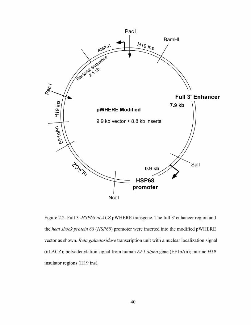
40
Figure 2.2. Full 3'-HSP68 nLACZ pWHERE transgene. The full 3' enhancer region and
the heat shock protein 68 (HSP68) promoter were inserted into the modified pWHERE
vector as shown. Beta galactosidase transcription unit with a nuclear localization signal
(nLACZ); polyadenylation signal from human EF1 alpha gene (EF1pAn); murine H19
insulator regions (H19 ins).

41
Figure 2.3. R3-HSP68 nLACZ pWHERE transgene. The R3 enhancer region and the heat
shock protein 68 (HSP68) promoter were inserted into the modified pWHERE vector as
shown. Beta galactosidase transcription unit with a nuclear localization signal (nLACZ);
polyadenylation signal from human EF1 alpha gene (EF1pAn); murine H19 insulator
regions (H19 ins).

42
CHAPTER THREE
IN VIVO ANALYSIS OF HUMAN LHX3 GENE REGULATION
3.1 Results
Conserved Distal Elements Downstream of the Human LHX3 Gene
Direct Pituitary and Nervous System Expression
Our lab previously characterized two human LHX3a and LHX3b promoters with
basal activity in vitro. To test the function of the promoters in vivo, I used a beta
galactosidase reporter gene mouse model. Transgenes were constructed by placing the
promoters in a vector containing a beta galactosidase reporter gene with a nuclear
localization signal followed by an untranslated region (UTR) and a polyadenylation
signal from the human EF1 alpha gene flanked by murine H19 insulator regions. H19
insulators have been shown to help reduce inappropriate repression or activation of gene
expression caused by the site of integration of the transgene (Touw et al., 2007).
Transient transgenics were analyzed at e14.5, a time point when mouse Lhx3 gene
expression is high in both the developing pituitary and spinal cord (Bach et al., 1995;
Seidah et al., 1994; Zhadanov et al., 1995). Neither promoter alone was able to drive
expression in any tissue including the pituitary and spinal cord (Figure 3.1 B, C; I and II).
To test the hypothesis that interactions between the two upstream promoter regions were
required to guide gene expression in vivo, I also examined the activity of the combined
promoter regions in the mouse model (Figure 3.1 B; III). This transgene was also unable
to direct reporter gene expression (Figure 3.1 C; III). A minimum of six independent
founders were analyzed for each promoter transgene (Figure 3.1 C; I, II, and III).

43
Together, these results indicate that regions outside of the basal promoters are required to
direct LHX3 gene expression in vivo.
Conserved non-coding elements (CNE) are associated with gene enhancer activity
in multiple tissues including neural tissues and the developing heart (Blow et al., 2010;
Visel et al., 2007). To search for regulatory regions, I performed bioinformatic searches
using the ECR browser of sequences ± 10 kb surrounding the human LHX3 gene and
discovered multiple CNEs within a 7.9 kb region directly 3' of the gene (Bray et al.,
2003; Couronne et al., 2003). The CNEs in the 3' region show ≥ 70% conservation
between humans and multiple other vertebrates (Figure 3.1 A). In order to examine the
function of these CNEs in vivo, I next tested the combined human promoter region plus
the 3' 7.9 kb region in the transgenic mouse model. Unlike the promoter constructs, this
construct was able to drive tissue-specific expression in the developing pituitary and
spinal cord at e14.5 (Figure 3.1 C; IV). This result indicates that the 3' region contains
one or more enhancers required for spatial and temporal expression of the LHX3 gene.
An additional CNE located far downstream (Far 3') was also identified from the
VISTA enhancer browser computational dataset and tested in vivo (Visel et al., 2007).
This element was able to direct some expression to the developing spinal cord but not the
pituitary gland (Figure 3.1 C; VI). The Far 3' CNE lies ~63 kb 3' of LHX3 and 31 kb 5' of
the nucleus accumbens-associated protein 2 (NACC2) gene. NACC2 is expressed in the
developing nervous system but not the pituitary (Visel et al., 2004). It is therefore
possible the Far 3' is an enhancer of NACC2 expression. In mouse, Lhx3 expression
begins at approximately e9.5. By e11.5, robust Lhx3 expression is detected in both the
developing spinal cord and pituitary. Additional Far 3' transient transgenic founders were

44
evaluated at e11.5 and no beta galactosidase activity was detected (data not shown). This
result and the fact that the nearest gene, NACC2, is expressed in neural tissues makes the
role of the Far 3' CNE in LHX3 gene regulation difficult to interpret.
One recognized characteristic of an enhancer is the ability to act independent of
position to guide expression from a heterologous basal promoter (Blackwood and
Kadonaga, 1998). Construct IV containing the LHX3 promoters and the 3' region in its
native position exhibits tissue-specific transgene expression. To determine if the 3' region
acts as an independent enhancer, we tested a transgene containing the 3' region upstream
of the HSP68 promoter, a basal promoter which lacks activity in the absence of an
enhancer in transgenic mice (Kothary et al., 1989; Pennacchio et al., 2007) (Figure 3.1 B;
V). The construct V expression pattern recapitulates that seen with the LHX3 construct IV
(Figure 3.1 C; V). Therefore the 3' enhancer region has transferrable activity independent
of its native promoters and genomic position.
Endogenous LHX3 Expression Correlates with the Expression Pattern
Guided by the 3' Enhancer Region During Development
To more completely dissect the spatial and temporal expression patterns of the
enhancer region, stable transgenic lines were generated using the HSP68 promoter
downstream of the full 3' enhancer region (Figure 3.1 B; V). Whole mount beta
galactosidase staining of the embryos showed that the enhancer element was activated at
about e9.5 to e10.0 in Rathke’s pouch, the primordium of the pituitary. Spinal cord
expression preceded pituitary expression slightly and was easily detectable at e9.0 to e9.5
(Figure 3.2 B; and data not shown). In previous studies it has been shown that mouse
Lhx3 is activated by e9.5 in both the pituitary and spinal cord (Bach et al., 1995; Seidah

45
et al., 1994; Zhadanov et al., 1995). To examine whether the 3' enhancer region activation
coincided with endogenous Lhx3 activation, we used serial sections from the same
embryo to control for differences in activation of Lhx3 expression between embryos.
Serial sections were stained for either beta galactosidase activity or
immunohistochemistry was performed using antibodies against mouse LHX3 protein.
LHX3 expression and the spatial pattern of enhancer-directed transgene activity were
similar, consistent with a role of the 3' enhancer region in LHX3 gene activation (Figure
3.2 A). LHX3 protein expression appeared stronger in the developing spinal cord than in
the pituitary matching the pattern of expression seen for the enhancer directed transgene
(Figure 3.2 A).
Whole mount staining at e12.5 revealed high levels of transgene expression in the
developing spinal cord and pituitary in a pattern consistent with the expression pattern of
endogenous mouse LHX3 protein (Figure 3.2 B). Some ectopic expression was found in
the epidermis and the nasal cavity (Figure 3.2 B). LHX3 has not been shown to be
expressed in these areas. This activity may be attributed to effects from the site of
transgene integration or might also be because other regulatory elements not contained
within the transgene are needed to more tightly regulate LHX3 expression.
Expression of the transgene in the pituitary was consistent with a subset of the
endogenous LHX3 expression pattern. The transgene was expressed throughout the
unclosed Rathke’s pouch, recapitulating LHX3 expression patterns, but became restricted
to the ventral portion of the pituitary by e14.5 (Figure 3.2 C). In contrast, LHX3 is more
strongly expressed in the dorsal pituitary with weaker expression in the ventral pituitary

46
by e12.5 (Raetzman et al., 2002). At postnatal day 1 (P1), expression was absent from the
intermediate lobe (Figure 3.2 C).
Next to examine the expression pattern on a cellular level; I wanted to determine
which hormone-secreting cell types also showed enhancer directed expression. Because
the co-expression pattern of endogenous LHX3 had not previously been determined, I
also did co-labeling experiments for the hormone subunits and LHX3. Both native LHX3
and enhancer-directed β-galactosidase co-localize with αGSU expressing cells but not
GH-positive cells in P1 pituitaries (Figure 3.3).
The 3' Region Contains Several Nervous System Enhancers and a Pituitary Enhancer
The identified 3' region contains several conserved sub-regions that were
designated regions R1 (355 bp), R2 (680 bp) and R3 (557 bp) (Figure 3.4 A). To evaluate
the importance of these sequences, a series of systematic deletions was generated.
Regulatory regions R1, R2, R3 were each independently sufficient to direct expression to
the spinal cord, but not transgenes containing the UTR region alone (Figure 3.4 A, B).
In the absence of the UTR R1 region both the R2 and R3 enhancer separately
directed robust expression to the developing cerebral cortex (Figure 3.5 E, F). Cerebral
cortex expression is not detected in transgenics containing the UTR R1 (~ 4500 bp)
element indicating a neural silencing element is contained in this region. Interestingly, at
e14.5 in any of the deletion constructs tested containing either R1, R2, or R3, transgene
expression is detected in the medial ganglionic eminence (MGE) that was not observed in
transgenic mice containing the full 3' enhancer region (Figure 3.5 A, B, C, D; data not
shown). However at e17.5, Full-HSP68 (transgene V) mice have reporter gene expression
in the MGE (data not shown). One possible explanation for these results is an element

47
needed for temporal control of MGE expression is contained or partially contained in the
only non-overlapping region immediately 3' of R2 (Figure 3.4).
Only transgenic founders generated using reporter genes containing the R3
element directed expression to both the developing spinal cord and pituitary (Figure 3.4
A, B; X, XI, and XIII). Transgenes containing the R3 element alone were expressed in
the spinal cord in a similar pattern to the full 7.9 kb region (Figure 3.1; V). However,
pituitary expression was expanded to include the dorsal portion of the developing anterior
pituitary (Figure 3.4 B; XIII). Intriguingly, the pattern of pituitary expression in a
construct with a deleted R2 sub-region (transgene X) shows repression in the dorsal
anterior pituitary thereby recapitulating the expression of the full enhancer element
(Figure 3.4 B). This result suggests that a pituitary silencing or repressive element is
contained within either the UTR R1 region or in the region between R2 and R3.
A 180 Bp Minimal Region Is Sufficient To Direct
Expression to the Developing Pituitary
Sequence alignment of the R3 pituitary enhancer region from several species
using the ECR browser identified a highly conserved region of 180 bp (designated Core
R3, Figure 3.6 A). We generated transgenic reporter gene containing the Core R3 region
(Figure 3.6 A; XIV). Of six transgenic embryos analyzed at e14.5, four had strong levels
of transgene expression and directed expression to the pituitary (Figure 3.6 A, B; XIV).
Of these, two directed weak (compared to the R3 or full 3' enhancer transgenic mice)
expression in the developing spinal cord (Figure 3.6 A, B). Additional transgenic mice
were generated expressing reporter genes containing overlapping fragments of the Core
R3 enhancer. These fragments were unable to direct pituitary expression (Figure 3.6 B;

48
XV, XVI, and XVII). Fragments 2 and 3 directed some weak spinal cord expression in a
pattern similar to the Core R3 enhancer transgenic mice (Figure 3.6 B; XIV, XVI, and
XVII). Overall, these data indicate that key elements required to direct pituitary
expression are contained in the 180 bp Core R3 enhancer.
Core R3 enhancer sequences for multiple species were aligned and conserved
putative trans-acting factor binding sites were identified (Figure 3.7). The Core R3
enhancer contained four conserved TAAT/ATTA sequences that are characteristic of
homeodomain protein recognition sites (Figure 3.7; sites AT1 to AT4). These sites were
compared to the consensus binding sites of candidate factors with suggested roles in
pituitary development and LHX3 gene regulation. AT1 matches the pituitary homeobox
(PITX) consensus binding site. PITX1 and PITX2 are required for activation of Lhx3 in
vitro and in vivo (Charles et al., 2005; Tremblay et al., 1998). AT2 was identified as a
possible binding site for LHX3 and LHX4 proteins (Bridwell et al., 2001). This sequence
might be important for autoregulation by LHX3 or regulation by LHX4. In vivo studies
have shown that Lhx4 is required for proper activation of Lhx3: in Lhx4 knockout mice,
LHX3 expression is delayed but returns to normal by e14.5 (Raetzman et al., 2002). AT3
and AT4 sequences are consistent with a tandem binding site for ISL1 (Karlsson et al.,
1990). Importantly, Isl1 expression precedes and then overlaps Lhx3 expression in the
developing spine and pituitary in a pattern consistent with a possible role in Lhx3
regulation (Ericson et al., 1998; Pfaff et al., 1996). The pituitary defect in the Isl1
knockout is similar to Lhx3-null mice and LHX3 expression is missing; however,
previous studies have suggested ISL1 blocks differentiation at an earlier stage of
development independent of LHX3 gene regulation (Takuma et al., 1998). Conditional

49
motoneuron knockouts of Isl1 do not have any markers of motoneuron development
including Lhx3 (Pfaff et al., 1996). LHX3 and LHX4 are expressed in the developing
bipolar interneurons of the eye at P9 and partially co-localize with ISL1. Loss of ISL1 in
the neural retina causes loss of Lhx3 expression, but Lhx4 expression is maintained
(Elshatory et al., 2007).
Additional transcription factor binding sites within the Core R3 included putative
elements for CCAAT enhancer binding protein alpha (CEBPA) and SRY-box 2 (SOX2)
sites (Figure 3.7). CEBPA has not been implicated in LHX3 regulation, but is important
in differentiation and proliferation events. Mouse Cebpa is expressed in the developing
pituitary and has been shown to regulate the prolactin promoter with PIT1 (Enwright et
al., 2003). SOX2 is known to bind to the human LHX3a promoter (Rajab et al., 2008) and
human patients and mouse models heterozygous for SOX2 gene mutations have anterior
pituitary hormone deficiencies (Kelberman et al., 2006).
To determine whether these elements are required to direct expression in vivo,
transient transgenic founders were generated with individual mutations in the putative
SOX, AT1, AT2 and CEBPA binding sites or a combined mutation of the tandem sites
AT3 and AT4 in the context of the 557 bp R3 enhancer (modeled in Figure 3.8 A).
Founders with mutations in AT1 lost expression in the dorsal portion of the pituitary in
comparison to the wild type R3 expression pattern (Figure 3.8 B). This is similar to the
expression pattern of the full 3' enhancer. This suggests a role for the AT1 element in
spatial control of pituitary LHX3 expression. Mutation of the tandem AT3 and AT4 sites
abolished nearly all expression in both the pituitary and the developing spinal cord
(Figure 3.8 B) demonstrating that AT3 and AT4 are required for enhancer directed
50
expression. However, since fragment 3 of the core R3 containing both AT3 and AT4 did
not alone direct pituitary expression, the AT3 and AT4 elements are not sufficient for
enhancer function (Figure 3.6 B; XVII). Individual mutation of the SOX, AT2, or
CEBPA sites failed to alter the expression pattern of the R3 enhancer at e14.5 (Figure 3.8
B). These sites are not required to direct expression at e14.5 however it is possible that
they may have function at other developmental time points.
To test whether candidate factors could bind to the AT elements in the core R3
enhancer in vitro, electrophoretic mobility shift assays (EMSAs) were performed using in
vitro translated proteins and 32P-labeled enhancer DNA sequences. ISL1 protein was able
to bind probes containing sites AT2, AT3, and AT4 and mutation of these sites abolished
binding (Figure 3.8 C). PITX1 and PITX2 protein bound to probes containing AT1 but
not to the mutated AT1 probe or other AT sites (Figure 3.8 D, and Figure 3.9 A). LHX3
and LHX4 proteins were able to bind probes containing AT2 (Figure 3.9 B, C, D).
Lysates programmed with empty vector did not bind to the probes specifically (Figure 3.8
C, D and Figure 3.9). Similarly, chromatin immunoprecipitation (ChIP) experiments
using alpha T3 pre-gonadotrope cells confirmed in vivo occupancy of PITX1 and ISL1
proteins in a cellular context (Figure 3.8 E). In luciferase reporter gene assays in either α-
T3 or LβT2 pituitary cell lines, addition of the R3 enhancer upstream of the LHX3
promoters or the minimal prolactin promoter failed to increase gene activation above
basal levels. Consistent with this lack of R3 mediated reporter gene activation, co-
transfection of PITX1 and ISL1 proteins did not synergistically activate the construct
(n=1; data not shown). EMSA and ChIP experiments were done in collaboration with
graduate student Soyoung Park.

51
Genetic Analysis of Regulatory Regions in CPHD Patients
Sequencing of the R3 region of thirty three CPHD patient DNA samples
previously screened in our lab for coding region mutations revealed no variations.
Additionally our collaborator from the Hospital for Children and Adolescents at the
University of Leipzig, Dr. Roland Pfaeffle screened an additional 100 candidate CPHD
patients for mutations in the LHX3a and LHX3b promoters and the R2 and R3 enhancers.
Patients with previously identified mutations in the coding regions of PROP1, PIT1 or
LHX3 gene were excluded. Samples from patients with CPHD were screened by
denaturing gradient high pressure liquid chromatography (dHPLC) (WAVE DNA
Fragment Analysis System, Transgenomic, Omaha, NE). Fragments showing abnormal
retention patterns on the HPLC column were subjected to Dideoxy-Sequencing to specify
the sequence abnormality. No homozygous sequence aberrations were detected. Three
heterozygous single nucleotide variations (SNV) were identified (Table 3.1). These SNVs
were not found in 50 unaffected individuals sequenced. The SNV within the R3 enhancer
region lies outside of the minimal Core R3 pituitary enhancer.

52

53
Figure 3.1. Distal downstream regions of the human LHX3 gene direct expression to the
developing pituitary and spinal cord. (A) Comparative genomic analysis. The ECR
browser was used to compare sequences surrounding human LHX3 and a Far 3' region
identified using the VISTA enhancer browser computational dataset to frog, chicken,
opossum, rat, mouse, dog, and cow. Conserved non-coding elements (CNE) were defined
as regions ≥70% identity and ≥100 base pairs: red color indicates CNE; (blue) coding
exons; (salmon) conserved intronic regions; (yellow) UTR; (green) transposable elements
and simple repeats. (B) Reporter gene constructs I - VI used to generate transgenic mice.
(C) Sagittal cryosections of e14.5 founder embryos stained for beta galactosidase activity.
The fraction of transgenic embryos expressing beta galactosidase in the pituitary or spinal
cord is shown below each respective image. The asterisk indicates ectopic expression. D,
dorsal; V, ventral; P, posterior lobe; I, intermediate lobe; A, anterior lobe.

54

55
Figure 3.2. Expression patterns guided by the 7.9 kb 3' enhancer region during
development correlate with endogenous LHX3 expression. (A) Antibody staining of
mLHX3 and beta galactosidase activity staining of serial sections at e9.5 in a stable
transgenic line containing construct line V. The onset of beta galactosidase activity is
consistent with the onset of mLHX3 protein expression detected by antibody staining in
Rathke’s pouch (RP) and in the developing spinal cord. (B) X-gal stained embryos at e9.5
and e12.5 show strong staining in the developing pituitary (arrowhead and circle) and
spinal cord consistent with known Lhx3 expression patterns. (C) X-gal staining of sagittal
cryosections of the developing pituitary at e10.5, e12.5, e14.5, e17.5 and coronal
cryosections at postnatal day 1(P1). The 3' enhancer directs expression through out the
developing anterior pituitary at e10.5 and e12.5. At e14.5, e17.5 and P1 expression is
restricted to the anterior lobe and is absent from the intermediate and posterior lobes. RP,
Rathke’s pouch, IF, infundibulum, H, hypothalamus; P, posterior lobe; I, intermediate
lobe; A, anterior lobe.

56
Figure 3.3. Native LHX3 and enhancer directed beta galactosidase expression co-
localization pattern is similar in the hormone-expressing cell types of P1 mouse
pituitaries. Coronal pituitary sections from newborn (P1) Full-HSP68-nLacZ transgenic
mice (transgene V) were stained by immunohistochemistry (green) for LHX3 (A–E) or
stained for beta galactosidase activity (blue) (F–J) and double-labeled [red (upper panel)
or brown (lower panel)] for pituitary hormones: GH (A and F), ACTH (B and G), αGSU
(C and H), TSH (D and I), LH (E and J). Co-localization was highest (arrowheads) for
gonadotropes and thyrotropes (C, D, E, H, I and J) and nearly absent (arrowheads) for
corticotropes and somatotropes (A, B, F, and G).

57

58
Figure 3.4. Deletion analysis of the 3' region reveals several nervous system enhancers
and a pituitary enhancer. (A) Alignment of the 3' prime region across the indicated
species from the UCSC Genome Browser (http://genome.ucsc.edu). The fraction of
transgenic embryos expressing beta galactosidase in the pituitary or spinal cord is shown
beside each construct. One founder each from lines UTR, UTR R1 and R2 showed
expression in the majority of tissues including the spinal cord and pituitary. The pattern
was inconsistent with other founder lines and was considered ectopic expression and
likely due to affects from the site integration of the transgene. Asterisks indicate non-
specific ectopic expression. (B) Sagittal sections of e14.5 embryos harboring constructs
VIII (UTR R1), XII (R2), X (delta R2), and XIII (R3) stained for beta galactosidase
activity. P, posterior lobe; I, intermediate lobe; A, anterior lobe.

59
Figure 3.5. UTR R1 (~4500 bp) contains a silencing element for the developing
forebrain. (A) hLHX3a/b promoters -7.9 kb 3′ enhancer (B) Full-HSP68 (C) UTR R1 R2-
HSP68 (D) UTR R1-HSP68 (E) R2-HS68 (F) R3-HSP68. Arrows show additional
forebrain expression in the developing cerebral cortex (E, F) and in the medial ganglionic
eminence (C, D). Sagittal sections at e14.5. n≥3 for all lines. FB, forebrain; MGE, medial
ganglionic eminence.

60
Figure 3.6. A highly conserved 180 bp minimal region (Core R3) is sufficient to direct
expression to the developing pituitary. (A) The ECR browser indicated a conserved non-
coding element (CNE, ≥70% identity and ≥100 base pairs); shown in red color.
Constructs XIV through XVII were used to generate transgenic mice and the fraction of
transgenic embryos expressing beta galactosidase in the pituitary or spinal cord is shown
beside each construct. (B) Sagittal cryosections of e14.5 embryos stained for beta
galactosidase activity. P, posterior lobe; I, intermediate lobe; A, anterior lobe.

61
Figure 3.7. Alignment of human Core R3 enhancer sequences with multiple species
reveals conserved putative transcription factor binding consensus sequences. Putative
SOX sites (yellow); four potential homeodomain factor sites (AT1-AT4, green); and a
possible C/EBP site (gray) are indicated. Analyses were performed with ClustalW2.0 and
rVISTA.

62

63
Figure 3.8. ISL and PITX binding sites in the Core R3 enhancer are critical for
expression in the developing pituitary and spinal cord. (A) Potential transcription factor
binding sites in the R3 region are shown in the context of the R3-HSP68-nLACZ
transgenic reporter gene. (B) Mutation analyses in transgenic mice demonstrate that AT3
and AT4 are required for pituitary and spinal cord expression in vivo and AT1 is required
for dorsal pituitary expression. (C) ISL1 binds specifically to AT2, AT3 and AT4 in
electrophoretic mobility shift assays (EMSA). The A3/A4 element from the enhancer of
the rat insulin promoter was used as the positive control (Karlsson et al., 1990). P.C.,
positive control; –, empty vector programmed lysate; +, PITX1 or ISL1 expression vector
programmed lysate; Arrows, specific bound complex; *NS, nonspecific band; F, free
probe. (D) PITX1 binds specifically to AT1 sites in EMSA. A Bcd2x5n bicoid element
was used as the positive control (Saadi et al., 2003). (E) ChIP experiments show
occupancy of the Core R3 enhancer by ISL1 and PITX1 proteins in αT3 pituitary cells.
Pull-down and input DNAs were characterized for the presence of the Core R3 enhancer
element by quantitative PCR. Immunoprecipitation with non-immune species-appropriate
IgG was carried out as a control and relative enrichment was calculated as the fold
difference above the 2–ΔΔCt for the normal immunoglobulin samples. Values are mean ±
s.e.m. for three independent experiments.

64
Figure 3.9. EMSA analysis of PITX2A, LHX3, and LHX4 binding of TAAT elements in
Core R3. (A) PITX2A binds specifically to AT1 sites in EMSA. A Bcd2x5n bicoid
element was used as the positive control (Saadi et al., 2003). (B) LHX3 and LHX4 fail to
bind to AT1 sites in EMSA. (C and D) LHX3 and LHX4 bind to AT2 in EMSA, but not
AT3 or AT4. The LHX3 consensus binding (LCB) element was used as the positive
control (Bridwell et al., 2001). P.C., positive control; –, empty vector programmed lysate;
+, LHX3 or LHX4 expression vector programmed lysate; Arrows, specific bound
complex; *NS, nonspecific band; F, free probe.

65
Table 3.1. Single nucleotide variations identified in human LHX3 regulatory regions
1 Numbering in reference to the LHX3a isoform (genomic DNA (g); bp upstream (-) of
the first start methionine; bp downstream (+) of LHX3 STOP codon); * This patient was
later identified with a compound heterozygous PROP1 mutation.
Patient Region Genetic Variation1 additional SNP
A R3 enhancer g.+7757C>G heterozygous
B LHX3a
Promoter g.-31G>A heterozygous
C LHX3a
Promoter g.-119C>T heterozygous
db SNP rs10858245
heterozygous
D LHX3a
Promoter g.-119C>T* heterozygous
db SNP rs11103377
heterozygous

66
CHAPTER FOUR
DISCUSSION
The pituitary gland is essential for the function and control of many processes in
the body including development, growth, metabolism, reproduction and the stress
response. Numerous studies have demonstrated that LHX3 is required for proper pituitary
development and function. While a great deal is known about the actions of the LHX3
protein, information about the transcriptional regulation of the LHX3 gene was limited to
in vitro studies identifying promoter and intronic elements important for basal gene
expression (Rajab et al., 2008; Yaden et al., 2005). The crucial regulatory elements
required for in vivo expression were unknown. This is the first in vivo description of
LHX3 gene regulation. This study reveals that multiple enhancers 3' of the LHX3 gene are
involved in regulation of LHX3 pituitary and spinal cord expression. Further, this work
presents evidence that ISL1 and PITX1 are direct transcriptional regulators of LHX3,
functioning in part through interactions with the Core R3 enhancer element.
These results show that PITX proteins are capable of binding to the AT1 element
in vitro, occupy the core R3 enhancer in cells, and mutation of the PITX recognized AT1
element affects the spatial pattern of the R3 enhancer in vivo. This suggests a mechanism
for PITX1 in regulation of the spatial pattern of LHX3 expression in the pituitary. This
study showed that ISL1 binds specifically to the AT2, AT3 and AT4 elements in vitro
and occupies the core R3 enhancer in vivo. Mutations of the AT3 and AT4 elements
resulted in almost complete loss of expression in both the pituitary and spinal cord.
However, alone these elements were insufficient to direct any pituitary expression

67
suggesting a combination of homeodomain and perhaps other factors is required for
enhancer activity. This implicates ISL1 for the first time as a regulator of LHX3 gene
expression in the ventral portion of the anterior pituitary and the spinal cord. Results of
this study are summarized in Figure 4.1.
Screening of 100 CPHD candidate patients in the LHX3a and LHX3b promoters
and R2 and R3 enhancers and of 33 additional candidate patients in the R3 enhancer has
revealed no homozygous variations in these regulatory regions to date. There are several
possible explanations for this. First, the cohort of patients screened was relatively small
in number and mutations may yet be found in other patients. It is also possible that
additional enhancers play roles in pituitary development in particular in somatotrope
development. Support for this includes the observation that the R3 enhancer region was
found to be primarily expressed ventrally where gonadotropes and thyrotropes develop
and not dorsally where somatotropes are found. Alternately the low degree of variation
observed suggests a high level of evolutionary constraint and may indicate mutations in
these regions are detrimental to survival. Considering that patients with LHX3 coding
region mutations survive this seems unlikely (Bhangoo et al., 2006; Kristrom et al., 2009;
Netchine et al., 2000; Pfaeffle et al., 2007; Rajab et al., 2008). Although several known
regulatory mutations are associated with human disease, it is a relatively rare cause of
human disease and most have been found in proximal promoters. Less than 1.8% of the
mutations documented in the Human Gene Mutation Database are point mutations in
non-coding regions (Noonan and McCallion, 2010). This percentage likely reflects the
difficulty in correlating mutations often found far from the coding regions of genes to
specific human diseases.

68
Though PITX1 and PITX2 are known upstream factors in the regulation of LHX3
no mechanism had been previously identified (Charles et al., 2005; Tremblay et al.,
1998). PITX1 and PITX2 are expressed strongly in the dorsal pituitary with weak
expression ventrally in a pattern similar to LHX3 during development. However in P1
and adult pituitaries these PITX proteins are mostly found to co-localize predominantly
with the αGSU expressing cells found more ventrally (Charles et al., 2005; Lanctot et al.,
1999). This is nearly identical to the LHX3 expression patterns shown in this study at P1
and previous studies at earlier time points (Bach et al., 1995; Seidah et al., 1994;
Zhadanov et al., 1995).
The full 3' enhancer directs expression throughout Rathke’s pouch at e9.5 but by
e12.5 it is expressed more ventrally and by e14.5 the expression is strongly ventral and
nearly absent dorsally (Figure 3.2). There are a few possible explanations for this
difference between previously observed LHX3 expression patterns and enhancer-directed
reporter gene expression patterns. Because this study uses the human enhancer region, it
is possible that the differences in observed pituitary expression represent differences
between mouse and human expression patterns or differences in control mechanisms
between the two species. Equally plausible is that this enhancer is not responsible for all
regions of LHX3 pituitary expression. Perhaps this points to one or more additional
enhancers that direct expression in the dorsal pituitary. It is possible that pituitary
expression of LHX3 is regulated by multiple enhancers and the identified 3' enhancer
region acts as an activating enhancer with later roles in the ventral anterior pituitary.
Another key difference in the expression pattern directed by the enhancer and the
endogenous gene is intermediate lobe expression. The enhancer fails to direct expression

69
to the intermediate lobe, but both the human and mouse intermediate lobes express LHX3
(Bach et al., 1995; Seidah et al., 1994; Sobrier et al., 2004; Zhadanov et al., 1995).
However, the human intermediate lobe is very small or even absent in adults. The
expression pattern directed by this enhancer may be an indication of the level of human
LHX3 expression in the intermediate lobe. The difference could possibly represent
morphological differences between the human intermediate lobe and that of mice. If there
is a marked reduction in LHX3 levels in the human intermediate lobe it is possible this
could affect its development. It is conceivable that apoptosis as a result of low LHX3
expression in the intermediate lobe is a key factor in loss of all but just a few cells of the
adult human intermediate lobe. Reductions in LHX3 action are known to increase
apoptosis in the ventral portion of Rathke’s pouch during development and the
intermediate lobes of the Lhx3-/- mice and Lhx3 Cre/Cre hypomorph mice are reduced in size
at e15.5 (Charles et al., 2005; Sheng et al., 1996; Zhao et al., 2006). To explore this idea,
first the mouse full 3' enhancer would need to be tested in the reporter gene mouse model.
Then, if marked differences in expression are observed between the human and mouse
enhancer region, a knock-in of the human enhancer region could be used to determine
what affect this change would have on pituitary morphology. Changes in conserved non-
coding elements have been established as one way evolution of morphological change
occurs (Carroll, 2008). This would be a high risk set of experiments because redundancy
is very common with regulatory elements. A number of enhancers with confirmed ability
to direct expression in vivo have been knocked out with no observable phenotype (Ahituv
et al., 2007; Nobrega et al., 2004).

70
In this study, ISL1 was identified as a likely regulator of LHX3 gene transcription
based on ChIP and EMSA analysis. From the in vivo mutational analysis, R3 enhancer-
directed expression in both the developing pituitary and spinal cord was shown to be
dependent on a single tandem ATTA site. ISL is expressed in both these tissues in a
temporal and spatial pattern consistent with a role in activation of the R3 enhancer
(Ellsworth et al., 2008; Ericson et al., 1998).
Several pieces of evidence in the literature point to a role for ISL1 in the
regulation of LHX3 gene expression. ISL1 protein expression precedes LHX3 expression
in Rathke’s pouch and at the time of Lhx3 activation ISL1 is co-localized with Lhx3 in
Rathke’s pouch (Ellsworth et al., 2008; Ericson et al., 1998). ISL1 expression later
primarily co-localizes with αGSU expressing cells similar to what was found in this study
for LHX3 and the enhancer-directed transgene (Liu et al., 2005a; Liu et al., 2005b; Wu et
al., 2010). Is11-/- mice display a similar pituitary phenotype to Lhx3-/- and lack LHX3
expression (Takuma et al., 1998). Tissue specific knock-outs of Isl1 in motoneuron and
neural retina also lack LHX3 expression (Elshatory et al., 2007; Pfaff et al., 1996). So
why has ISL1 not previously been identified as a potential upstream factor of LHX3?
Probably the primary reason for this is that after the early overlap of expression, the two
proteins segregate into a largely inverse expression pattern with ISL1 restricted to the
most ventral region and LHX3 expressed dorsally. This led to the interpretation that the
phenotypes of the Isl1 knock-out in both the pituitary and motoneurons blocked
development at an earlier stage and was not the result of LHX3 loss (Elshatory et al.,
2007; Pfaff et al., 1996; Takuma et al., 1998). Although ISL1 likely has other early roles
in the development of neural retina, pituitary and spinal cord it is also possible it acts as

71
an activator of LHX3. Earlier roles of ISL1 in differentiation do not necessarily exclude a
direct role in the activation of specific factors. The regulatory relationship between LHX3
and ISL1 is likely complex. Loss of LHX3 in mice does not affect PITX1 or PITX2
expression however the spatial and temporal expression pattern of ISL1 is affected. In
Lhx3-/- mice, ISL1 is expressed normally at e9.5, but is transiently lost at e12.5 (Ellsworth
et al., 2008; Zhao et al., 2006). At e16.5 and e18.5, ISL1 expression returns but its
expression is shifted dorsally (Ellsworth et al., 2008). This and the largely inverse
expression pattern of the two proteins after e9.5 have suggested LHX3 is a both a positive
and negative regulator of Isl1. Work presented in this study suggests that ISL1 may also
regulate LHX3 activation and later its activity in αGSU expressing cells.
LHX3 has important roles in somatotrope development based on both the mouse
models and the human CPHD patients with LHX3 coding regions mutations (Colvin et
al., 2009). Although LHX3 is not co-expressed at P1 with somatotropes it seems likely it
is found in somatotrope progenitors based on its expression throughout Rathke’s pouch
and importance in somatotrope development. An alternative hypothesis is that LHX3’s
action on somatotropes is not direct. To more completely answer where enhancer-
directed expression is found during pituitary development and by extension LHX3
expression, a cre recombinase lineage tracing approach is being used. Two mouse lines
have been generated containing the human full 3' region upstream of the HSP68 promoter
directing cre recombinase, and these have been crossed to floxed stop reporter strains.
Unfortunately the expression patterns of the two founders are too variable to draw any
conclusions without examining more transgenic lines. This variability is most easily
explained by site of transgene integration effects. Additional transgenics are being

72
generated. Some early data however indicates the enhancer does not direct expression in
somatotrope progenitors. Unlike a positive result in which enhancer-directed transgene
co-localization would suggest LHX3 co-localization in progenitors, this result gives us no
information as to whether LHX3 is expressed in somatotrope progenitors. Additional
experiments will be needed to determine the lineage specific expression of LHX3.
Possible methods include generating a LHX3 bacterial artificial chromosome-cre mouse
model or co-labeling with somatotrope lineage specific transcription factors. Two
transcription factors PIT1 and MATH3 could be used to identify somatotrope progenitors
prior to GH expression at e15.5. PIT1 is known to be required for somatotrope,
lactotrope, and thyrotrope development. MATH3, downstream factor to PIT1, is
expressed at e13.5 and required for maturation and expansion of somatotropes (Zhu et al.,
2006).
The R1, R2, and R3 enhancers were found to direct nervous system expression in
the developing mouse embryo (Figure 4.1). Whether these enhancers are functionally
redundant or direct unique expression patterns in the developing spinal cord will require
further study. Additional co-localization experiments with lineage specific transcription
factors HB9 (motoneurons) and CHX10 (interneurons) are also needed to determine on a
cellular level where the enhancer regions direct expression in the developing spinal cord
(Thaler et al., 2004).
Another interesting finding from this study is the presence of potential silencing
elements. Future work will be needed to isolate these elements and determine how they
are functioning mechanistically. Insulators or repressors are known to block enhancer
activity or serve as a barrier between areas of open and closed chromatin and in some

73
cases function in both manners (Gaszner and Felsenfeld, 2006). Numerous studies have
shown the CCCTC-binding factor (CTCF) is found at many insulators but exactly how
CTCF functions in enhancer insulating and blocking is unknown (Noonan and
McCallion, 2010). Insulators repressing expression in the dorsal pituitary and in
developing cerebral cortex were contained within either the UTR R1 region or in the
region between R2 and R3 (Figure 4.1). R1 may contain these insulator elements.
Potential CTCF binding sites were identified using TRANSFAC in R1, but these did not
strongly match the known consensus sequence of CTCF. Further experiments are needed
to determine if R1 is an insulator element, what trans-acting factors are needed for its
functions, and if CTCF binding is required for R1 insulator function.
The AT1, PITX binding site, was identified as important in spatial control of the
enhancer-directed pituitary expression. This pattern could indicate a function of this site
in dorsal pituitary repression of the larger full 3' enhancer. A potential mechanism for this
observed activity is that an unknown dorsal factor bound to the insulator element
facilitates chromatin looping between the R3 enhancer and the insulator that is mediated
by PITX protein bound to the AT1 element and this looping isolates the R3 enhancer
away from the proximal promoter (Figure 4.2). In the context of the R3-HSP68
transgene, without the insulator regions, the enhancer is not sequestered away from the
proximal promoter and is able to direct dorsal expression in the pituitary. Chromatin
conformation capture (3C) experiments will be needed to test this hypothesis after a
minimal insulator element is identified. A similar mechanism of function has been
identified for the H19ICR insulator. In 3C experiments, H19ICR enhancers and the

74
promoter region were shown to form loops with the insulator that blocked gene
expression (Yoon et al., 2007).
Several chromatin markers have been associated with active enhancers including
monomethylation on histone H3 lysine 4 and acetylation of histone H3 lysine 27
(Heintzman et al., 2009; Heintzman et al., 2007). Active enhancers have also been
associated with binding of the transcriptional co-activator p300. Recent global ChIP
sequencing experiments using e11.5 mouse forebrain, midbrain, and limb tissues were
able to highly correlate p300 binding with active enhancers which when tested directed
tissue-specific enhancer expression in the same tissue with in vivo occupancy of p300
(Gotea et al., 2010).
Further, this same study, showed homotypic clusters of transcription factor
binding sites were found to be abundant in both promoter regions and developmental
enhancers and were more highly correlated with functional activity than CNEs not
containing homotypic sites (Gotea et al., 2010). Intriguingly, I have found that both the
R2 and R3 enhancers contained multiple conserved TAAT sites (numbering 9 and 4
respectively). A combination of the TAAT elements in R3 was also found to be needed
for proper enhancer activity. This discovery of homotypic TAAT sites within the LHX3
enhancers may have broader implications for the regulation of other LIM-HD
transcription factors. Similar to the LHX3 gene, a 15 kb region 3' of the LHX4 gene
contains conserved regions with multiple TAAT elements (Sequences analyzed using the
ECR browser). Considering the close relationship of these genes it is highly probable this
3' region of the LHX4 gene may also function as an enhancer of neural and pituitary
expression. Additionally, the less related LIM-HD transcription factor LHX2 is expressed

75
primarily in the neural ectoderm and developing cerebral cortex and shows areas of high
conservation 5' of the gene with multiple TAAT elements (Sequences analyzed using
ECR browser) (Hunter and Rhodes, 2005). These findings suggest TAAT homotypic
clusters may play broad and important role in the regulation of LIM-HD genes in
multiple tissues.
Interestingly, a NOBOX binding site [TAATT(G/A)] was highly associated with
forebrain activity in the p300 ChIP sequencing study. NOBOX is not found in the
forebrain. However, LIM-HD transcription factors are important in transcriptional
regulation of multiple systems and have consensus binding sequences similar to the
NOBOX binding site. LHX2 is highly expressed in the developing forebrain and could
potentially be the factor binding to these identified sites (Gotea et al., 2010; Hunter and
Rhodes, 2005). In the absence of the insulator regions, R2 and R3 both have very robust
forebrain expression. This suggests the default for enhancers with homotypic TAAT sites
is to be active in the forebrain, and insulator regions likely play key roles in the
repression of ectopic forebrain expression. This result also highlights an important caveat
to these types of studies. Many studies look at enhancer elements based on conservation
and analyze these in isolation (Visel et al., 2007). It is important to keep in mind the true
activity of the enhancer is dependent on interactions with other regulatory regions in its
native position and many of these interacting regions are found many kb away (Noonan
and McCallion, 2010).
The studies presented in this report describe and characterize the first known
pituitary and spinal cord enhancers of LHX3 and are the first step in uncovering the
mechanisms required for proper spatial and temporal expression of the LHX3 gene. In

76
addition to identifying other important regulatory regions, future research will need to
explore the interactions between the LHX3 proximal promoters, insulator regions, and
enhancers. Uncovering how these elements interact to direct tissue-specific gene
expression will be essential to understanding the mechanisms behind LHX3 gene
regulation and understanding pituitary development on a molecular level. Information
gathered about LHX3 may have implications in the regulation of other LIM-HD genes
and give us insight into how gene regulation works in general. The molecular cause of
most cases of CPHD is unknown (Dattani, 2005). Further understanding and
identification of important LHX3 gene regulatory regions will allow for the identification
of novel genetic defects responsible for CPHD and will facilitate patient treatment and
enable genetic counseling.

77
Figure 4.1. A schematic summary of findings. Regulatory regions R1, R2, and R3 are
sufficient to independently direct expression to the central nervous system (CNS). Only
R3 directs pituitary and CNS expression. PITX proteins bind to the AT1 element in vitro
and occupy the Core R3 enhancer. Mutation of the PITX recognized AT1 element affects
the dorsal pituitary expression pattern of the R3 enhancer in vivo. ISL1 binds specifically
to the AT2, AT3 and AT4 elements in vitro and occupies the Core R3 enhancer in vivo.
Mutations of the AT3 and AT4 elements resulted in almost complete loss of expression in
both the pituitary and spinal cord. Pituitary and neural insulators are contained within
either the UTR R1 region or in the region between R2 and R3.

78
Figure 4.2. A hypothetical mechanism for regulation of the spatial expression pattern in
the developing pituitary. In the dorsal pituitary, an unknown factor bound to the insulator
element facilitates chromatin looping between the R3 enhancer and the insulator that is
mediated by PITX protein bound to the AT1 element. This looping isolates the R3
enhancer away from the proximal promoter and silences dorsal pituitary expression. In
the ventral pituitary, because the dorsal factor is absent the interaction does not occur and
the R3 enhancer is able to loop to the promoter and activate gene expression. Prom,
promoter; Ins, insulator element.

79
REFERENCES
Ahituv, N., Zhu, Y., Visel, A., Holt, A., Afzal, V., Pennacchio, L. A. and Rubin, E. M. (2007). Deletion of ultraconserved elements yields viable mice. PLoS Biol 5, e234.
Alatzoglou, K. S., Kelberman, D. and Dattani, M. T. (2009). The role of SOX proteins in normal pituitary development. J Endocrinol 200, 245-58.
Alward, W. L., Semina, E. V., Kalenak, J. W., Heon, E., Sheth, B. P., Stone, E. M. and Murray, J. C. (1998). Autosomal dominant iris hypoplasia is caused by a mutation in the Rieger syndrome (RIEG/PITX2) gene. Am J Ophthalmol 125, 98-100.
Amendt, B. A., Sutherland, L. B. and Russo, A. F. (1999). Multifunctional role of the Pitx2 homeodomain protein C-terminal tail. Mol Cell Biol 19, 7001-10.
Aupetit, C., Drouet, M., Pinaud, E., Denizot, Y., Aldigier, J. C., Bridoux, F. and Cogne, M. (2000). Alleles of the alpha1 immunoglobulin gene 3' enhancer control evolution of IgA nephropathy toward renal failure. Kidney Int 58, 966-71.
Avilion, A. A., Nicolis, S. K., Pevny, L. H., Perez, L., Vivian, N. and Lovell-Badge, R. (2003). Multipotent cell lineages in early mouse development depend on SOX2 function. Genes Dev 17, 126-40.
Bach, I., Rhodes, S. J., Pearse, R. V., 2nd, Heinzel, T., Gloss, B., Scully, K. M., Sawchenko, P. E. and Rosenfeld, M. G. (1995). P-Lim, a LIM homeodomain factor, is expressed during pituitary organ and cell commitment and synergizes with Pit-1. Proc Natl Acad Sci U S A 92, 2720-4.
Baek, S. H., Kioussi, C., Briata, P., Wang, D., Nguyen, H. D., Ohgi, K. A., Glass, C. K., Wynshaw-Boris, A., Rose, D. W. and Rosenfeld, M. G. (2003). Regulated subset of G1 growth-control genes in response to derepression by the Wnt pathway. Proc Natl Acad Sci U S A 100, 3245-50.
Bentley, C. A., Zidehsarai, M. P., Grindley, J. C., Parlow, A. F., Barth-Hall, S. and Roberts, V. J. (1999). Pax6 is implicated in murine pituitary endocrine function. Endocrine 10, 171-7.
Bhangoo, A. P., Hunter, C. S., Savage, J. J., Anhalt, H., Pavlakis, S., Walvoord, E. C., Ten, S. and Rhodes, S. J. (2006). Clinical case seminar: a novel LHX3 mutation presenting as combined pituitary hormonal deficiency. J Clin Endocrinol Metab 91, 747-53.
Blackwood, E. M. and Kadonaga, J. T. (1998). Going the distance: a current view of enhancer action. Science 281, 60-3.

80
Blow, M. J., McCulley, D. J., Li, Z., Zhang, T., Akiyama, J. A., Holt, A., Plajzer-Frick, I., Shoukry, M., Wright, C., Chen, F. et al. (2010). ChIP-Seq identification of weakly conserved heart enhancers. Nat Genet 42, 806-10.
Brickman, J. M., Clements, M., Tyrell, R., McNay, D., Woods, K., Warner, J., Stewart, A., Beddington, R. S. and Dattani, M. (2001). Molecular effects of novel mutations in Hesx1/HESX1 associated with human pituitary disorders. Development 128, 5189-99.
Bridwell, J. A., Price, J. R., Parker, G. E., McCutchan Schiller, A., Sloop, K. W. and Rhodes, S. J. (2001). Role of the LIM domains in DNA recognition by the Lhx3 neuroendocrine transcription factor. Gene 277, 239-50.
Carroll, S. B. (2008). Evo-devo and an expanding evolutionary synthesis: a genetic theory of morphological evolution. Cell 134, 25-36.
Carvalho, L. R., Brinkmeier, M. L., Castinetti, F., Ellsworth, B. S. and Camper, S. A. (2010). Corepressors TLE1 and TLE3 interact with HESX1 and PROP1. Mol Endocrinol 24, 754-65.
Castinetti, F., Saveanu, A., Reynaud, R., Quentien, M. H., Buffin, A., Brauner, R., Kaffel, N., Albarel, F., Guedj, A. M., El Kholy, M. et al. (2008). A novel dysfunctional LHX4 mutation with high phenotypical variability in patients with hypopituitarism. J Clin Endocrinol Metab 93, 2790-9.
Cha, K. B., Douglas, K. R., Potok, M. A., Liang, H., Jones, S. N. and Camper, S. A. (2004). WNT5A signaling affects pituitary gland shape. Mech Dev 121, 183-94.
Charles, M. A., Mortensen, A. H., Potok, M. A. and Camper, S. A. (2008). Pitx2 deletion in pituitary gonadotropes is compatible with gonadal development, puberty, and fertility. Genesis 46, 507-14.
Charles, M. A., Suh, H., Hjalt, T. A., Drouin, J., Camper, S. A. and Gage, P. J. (2005). PITX genes are required for cell survival and Lhx3 activation. Mol Endocrinol 19, 1893-903.
Colvin, S. C., Mullen, R. D., Pfaeffle, R. W. and Rhodes, S. J. (2009). LHX3 and LHX4 transcription factors in pituitary development and disease. Pediatr Endocrinol Rev 6 Suppl 2, 283-90.
Couronne, O., Poliakov, A., Bray, N., Ishkhanov, T., Ryaboy, D., Rubin, E., Pachter, L. and Dubchak, I. (2003). Strategies and tools for whole-genome alignments. Genome Res 13, 73-80.
Cushman, L. J., Showalter, A. D. and Rhodes, S. J. (2002). Genetic defects in the development and function of the anterior pituitary gland. Ann Med 34, 179-91.

81
Cushman, L. J., Watkins-Chow, D. E., Brinkmeier, M. L., Raetzman, L. T., Radak, A. L., Lloyd, R. V. and Camper, S. A. (2001). Persistent Prop1 expression delays gonadotrope differentiation and enhances pituitary tumor susceptibility. Hum Mol Genet 10, 1141-53.
Dasen, J. S., Barbera, J. P., Herman, T. S., Connell, S. O., Olson, L., Ju, B., Tollkuhn, J., Baek, S. H., Rose, D. W. and Rosenfeld, M. G. (2001). Temporal regulation of a paired-like homeodomain repressor/TLE corepressor complex and a related activator is required for pituitary organogenesis. Genes Dev 15, 3193-207.
Dasen, J. S., O'Connell, S. M., Flynn, S. E., Treier, M., Gleiberman, A. S., Szeto, D. P., Hooshmand, F., Aggarwal, A. K. and Rosenfeld, M. G. (1999). Reciprocal interactions of Pit1 and GATA2 mediate signaling gradient-induced determination of pituitary cell types. Cell 97, 587-98.
Dasen, J. S. and Rosenfeld, M. G. (2001). Signaling and transcriptional mechanisms in pituitary development. Annu Rev Neurosci 24, 327-55.
Dateki, S., Fukami, M., Uematsu, A., Kaji, M., Iso, M., Ono, M., Mizota, M., Yokoya, S., Motomura, K., Kinoshita, E. et al. (2010). Mutation and gene copy number analyses of six pituitary transcription factor genes in 71 patients with combined pituitary hormone deficiency: identification of a single patient with LHX4 deletion. J Clin Endocrinol Metab 95, 4043-7.
Dattani, M. T. (2005). Growth hormone deficiency and combined pituitary hormone deficiency: does the genotype matter? Clin Endocrinol (Oxf) 63, 121-30.
Dattani, M. T., Martinez-Barbera, J. P., Thomas, P. Q., Brickman, J. M., Gupta, R., Martensson, I. L., Toresson, H., Fox, M., Wales, J. K., Hindmarsh, P. C. et al. (1998). Mutations in the homeobox gene HESX1/Hesx1 associated with septo-optic dysplasia in human and mouse. Nat Genet 19, 125-33.
Davis, S. W. and Camper, S. A. (2007). Noggin regulates Bmp4 activity during pituitary induction. Dev Biol 305, 145-60.
Dohrmann, C., Gruss, P. and Lemaire, L. (2000). Pax genes and the differentiation of hormone-producing endocrine cells in the pancreas. Mech Dev 92, 47-54.
Dong, J., Asa, S. L. and Drucker, D. J. (1991). Islet cell and extrapancreatic expression of the LIM domain homeobox gene isl-1. Mol Endocrinol 5, 1633-41.
Doward, W., Perveen, R., Lloyd, I. C., Ridgway, A. E., Wilson, L. and Black, G. C. (1999). A mutation in the RIEG1 gene associated with Peters' anomaly. J Med Genet 36, 152-5.
Du, A., Hunter, C. S., Murray, J., Noble, D., Cai, C. L., Evans, S. M., Stein, R. and May, C. L. (2009). Islet-1 is required for the maturation, proliferation, and survival of the endocrine pancreas. Diabetes 58, 2059-69.
82
Ellsworth, B. S., Butts, D. L. and Camper, S. A. (2008). Mechanisms underlying pituitary hypoplasia and failed cell specification in Lhx3-deficient mice. Dev Biol 313, 118-29.
Ellsworth, B. S., Egashira, N., Haller, J. L., Butts, D. L., Cocquet, J., Clay, C. M., Osamura, R. Y. and Camper, S. A. (2006). FOXL2 in the pituitary: molecular, genetic, and developmental analysis. Mol Endocrinol 20, 2796-805.
Elshatory, Y., Everhart, D., Deng, M., Xie, X., Barlow, R. B. and Gan, L. (2007). Islet-1 controls the differentiation of retinal bipolar and cholinergic amacrine cells. J Neurosci 27, 12707-20.
Emison, E. S., McCallion, A. S., Kashuk, C. S., Bush, R. T., Grice, E., Lin, S., Portnoy, M. E., Cutler, D. J., Green, E. D. and Chakravarti, A. (2005). A common sex-dependent mutation in a RET enhancer underlies Hirschsprung disease risk. Nature 434, 857-63.
Enwright, J. F., 3rd, Kawecki-Crook, M. A., Voss, T. C., Schaufele, F. and Day, R. N. (2003). A PIT-1 homeodomain mutant blocks the intranuclear recruitment of the CCAAT/enhancer binding protein alpha required for prolactin gene transcription. Mol Endocrinol 17, 209-22.
Ericson, J., Norlin, S., Jessell, T. M. and Edlund, T. (1998). Integrated FGF and BMP signaling controls the progression of progenitor cell differentiation and the emergence of pattern in the embryonic anterior pituitary. Development 125, 1005-15.
Fantes, J., Ragge, N. K., Lynch, S. A., McGill, N. I., Collin, J. R., Howard-Peebles, P. N., Hayward, C., Vivian, A. J., Williamson, K., van Heyningen, V. et al. (2003). Mutations in SOX2 cause anophthalmia. Nat Genet 33, 461-3.
Fauquier, T., Rizzoti, K., Dattani, M., Lovell-Badge, R. and Robinson, I. C. (2008). SOX2-expressing progenitor cells generate all of the major cell types in the adult mouse pituitary gland. Proc Natl Acad Sci U S A 105, 2907-12.
Ferri, A. L., Cavallaro, M., Braida, D., Di Cristofano, A., Canta, A., Vezzani, A., Ottolenghi, S., Pandolfi, P. P., Sala, M., DeBiasi, S. et al. (2004). Sox2 deficiency causes neurodegeneration and impaired neurogenesis in the adult mouse brain. Development 131, 3805-19.
Gage, P. J., Brinkmeier, M. L., Scarlett, L. M., Knapp, L. T., Camper, S. A. and Mahon, K. A. (1996). The Ames dwarf gene, df, is required early in pituitary ontogeny for the extinction of Rpx transcription and initiation of lineage-specific cell proliferation. Mol Endocrinol 10, 1570-81.
Gage, P. J. and Camper, S. A. (1997). Pituitary homeobox 2, a novel member of the bicoid-related family of homeobox genes, is a potential regulator of anterior structure formation. Hum Mol Genet 6, 457-64.

83
Gage, P. J., Suh, H. and Camper, S. A. (1999). Dosage requirement of Pitx2 for development of multiple organs. Development 126, 4643-51.
Gaston-Massuet, C., Andoniadou, C. L., Signore, M., Sajedi, E., Bird, S., Turner, J. M. and Martinez-Barbera, J. P. (2008). Genetic interaction between the homeobox transcription factors HESX1 and SIX3 is required for normal pituitary development. Dev Biol 324, 322-33.
Gaszner, M. and Felsenfeld, G. (2006). Insulators: exploiting transcriptional and epigenetic mechanisms. Nat Rev Genet 7, 703-13.
Gleiberman, A. S., Fedtsova, N. G. and Rosenfeld, M. G. (1999). Tissue interactions in the induction of anterior pituitary: role of the ventral diencephalon, mesenchyme, and notochord. Dev Biol 213, 340-53.
Gotea, V., Visel, A., Westlund, J. M., Nobrega, M. A., Pennacchio, L. A. and Ovcharenko, I. (2010). Homotypic clusters of transcription factor binding sites are a key component of human promoters and enhancers. Genome Res 20, 565-77.
Granger, A., Bleux, C., Kottler, M. L., Rhodes, S. J., Counis, R. and Laverriere, J. N. (2006). The LIM-homeodomain proteins Isl-1 and Lhx3 act with steroidogenic factor 1 to enhance gonadotrope-specific activity of the gonadotropin-releasing hormone receptor gene promoter. Mol Endocrinol 20, 2093-108.
Gurnett, C. A., Bowcock, A. M., Dietz, F. R., Morcuende, J. A., Murray, J. C. and Dobbs, M. B. (2007). Two novel point mutations in the long-range SHH enhancer in three families with triphalangeal thumb and preaxial polydactyly. Am J Med Genet A 143, 27-32.
Heintzman, N. D., Hon, G. C., Hawkins, R. D., Kheradpour, P., Stark, A., Harp, L. F., Ye, Z., Lee, L. K., Stuart, R. K., Ching, C. W. et al. (2009). Histone modifications at human enhancers reflect global cell-type-specific gene expression. Nature 459, 108-12.
Heintzman, N. D., Stuart, R. K., Hon, G., Fu, Y., Ching, C. W., Hawkins, R. D., Barrera, L. O., Van Calcar, S., Qu, C., Ching, K. A. et al. (2007). Distinct and predictive chromatin signatures of transcriptional promoters and enhancers in the human genome. Nat Genet 39, 311-8.
Hermesz, E., Mackem, S. and Mahon, K. A. (1996). Rpx: a novel anterior-restricted homeobox gene progressively activated in the prechordal plate, anterior neural plate and Rathke's pouch of the mouse embryo. Development 122, 41-52.
Hunter, C. S. and Rhodes, S. J. (2005). LIM-homeodomain genes in mammalian development and human disease. Mol Biol Rep 32, 67-77.
Jurata, L. W., Pfaff, S. L. and Gill, G. N. (1998). The nuclear LIM domain interactor NLI mediates homo- and heterodimerization of LIM domain transcription factors. J Biol Chem 273, 3152-7.

84
Karlsson, O., Thor, S., Norberg, T., Ohlsson, H. and Edlund, T. (1990). Insulin gene enhancer binding protein Isl-1 is a member of a novel class of proteins containing both a homeo- and a Cys-His domain. Nature 344, 879-82.
Kawakami, K., Sato, S., Ozaki, H. and Ikeda, K. (2000). Six family genes--structure and function as transcription factors and their roles in development. Bioessays 22, 616-26.
Kawamata, N., Sakajiri, S., Sugimoto, K. J., Isobe, Y., Kobayashi, H. and Oshimi, K. (2002). A novel chromosomal translocation t(1;14)(q25;q32) in pre-B acute lymphoblastic leukemia involves the LIM homeodomain protein gene, Lhx4. Oncogene 21, 4983-91.
Kelberman, D. and Dattani, M. T. (2006). The role of transcription factors implicated in anterior pituitary development in the aetiology of congenital hypopituitarism. Ann Med 38, 560-77.
Kelberman, D., de Castro, S. C., Huang, S., Crolla, J. A., Palmer, R., Gregory, J. W., Taylor, D., Cavallo, L., Faienza, M. F., Fischetto, R. et al. (2008). SOX2 plays a critical role in the pituitary, forebrain, and eye during human embryonic development. J Clin Endocrinol Metab 93, 1865-73.
Kelberman, D., Rizzoti, K., Avilion, A., Bitner-Glindzicz, M., Cianfarani, S., Collins, J., Chong, W. K., Kirk, J. M., Achermann, J. C., Ross, R. et al. (2006). Mutations within Sox2/SOX2 are associated with abnormalities in the hypothalamo-pituitary-gonadal axis in mice and humans. J Clin Invest 116, 2442-55.
Kelberman, D., Rizzoti, K., Lovell-Badge, R., Robinson, I. C. and Dattani, M. T. (2009). Genetic regulation of pituitary gland development in human and mouse. Endocr Rev 30, 790-829.
Kent, W. J., Sugnet, C. W., Furey, T. S., Roskin, K. M., Pringle, T. H., Zahler, A. M. and Haussler, D. (2002). The human genome browser at UCSC. Genome Res 12, 996-1006.
Kimura, S., Hara, Y., Pineau, T., Fernandez-Salguero, P., Fox, C. H., Ward, J. M. and Gonzalez, F. J. (1996). The T/ebp null mouse: thyroid-specific enhancer-binding protein is essential for the organogenesis of the thyroid, lung, ventral forebrain, and pituitary. Genes Dev 10, 60-9.
Kioussi, C., Briata, P., Baek, S. H., Rose, D. W., Hamblet, N. S., Herman, T., Ohgi, K. A., Lin, C., Gleiberman, A., Wang, J. et al. (2002). Identification of a Wnt/Dvl/beta-Catenin --> Pitx2 pathway mediating cell-type-specific proliferation during development. Cell 111, 673-85.
Kioussi, C., O'Connell, S., St-Onge, L., Treier, M., Gleiberman, A. S., Gruss, P. and Rosenfeld, M. G. (1999). Pax6 is essential for establishing ventral-dorsal cell boundaries in pituitary gland development. Proc Natl Acad Sci U S A 96, 14378-82.

85
Kothary, R., Clapoff, S., Darling, S., Perry, M. D., Moran, L. A. and Rossant, J. (1989). Inducible expression of an hsp68-lacZ hybrid gene in transgenic mice. Development 105, 707-14.
Kristrom, B., Zdunek, A. M., Rydh, A., Jonsson, H., Sehlin, P. and Escher, S. A. (2009). A novel mutation in the LIM homeobox 3 gene is responsible for combined pituitary hormone deficiency, hearing impairment, and vertebral malformations. J Clin Endocrinol Metab 94, 1154-61.
Kulak, S. C., Kozlowski, K., Semina, E. V., Pearce, W. G. and Walter, M. A. (1998). Mutation in the RIEG1 gene in patients with iridogoniodysgenesis syndrome. Hum Mol Genet 7, 1113-7.
Lagutin, O. V., Zhu, C. C., Kobayashi, D., Topczewski, J., Shimamura, K., Puelles, L., Russell, H. R., McKinnon, P. J., Solnica-Krezel, L. and Oliver, G. (2003). Six3 repression of Wnt signaling in the anterior neuroectoderm is essential for vertebrate forebrain development. Genes Dev 17, 368-79.
Lamonerie, T., Tremblay, J. J., Lanctot, C., Therrien, M., Gauthier, Y. and Drouin, J. (1996). Ptx1, a bicoid-related homeo box transcription factor involved in transcription of the pro-opiomelanocortin gene. Genes Dev 10, 1284-95.
Lanctot, C., Moreau, A., Chamberland, M., Tremblay, M. L. and Drouin, J. (1999). Hindlimb patterning and mandible development require the Ptx1 gene. Development 126, 1805-10.
Li, H., Witte, D. P., Branford, W. W., Aronow, B. J., Weinstein, M., Kaur, S., Wert, S., Singh, G., Schreiner, C. M., Whitsett, J. A. et al. (1994). Gsh-4 encodes a LIM-type homeodomain, is expressed in the developing central nervous system and is required for early postnatal survival. EMBO J 13, 2876-85.
Li, X., Oghi, K. A., Zhang, J., Krones, A., Bush, K. T., Glass, C. K., Nigam, S. K., Aggarwal, A. K., Maas, R., Rose, D. W. et al. (2003). Eya protein phosphatase activity regulates Six1-Dach-Eya transcriptional effects in mammalian organogenesis. Nature 426, 247-54.
Li, X., Perissi, V., Liu, F., Rose, D. W. and Rosenfeld, M. G. (2002). Tissue-specific regulation of retinal and pituitary precursor cell proliferation. Science 297, 1180-3.
Lin, C. R., Kioussi, C., O'Connell, S., Briata, P., Szeto, D., Liu, F., Izpisua-Belmonte, J. C. and Rosenfeld, M. G. (1999). Pitx2 regulates lung asymmetry, cardiac positioning and pituitary and tooth morphogenesis. Nature 401, 279-82.
Lin, S. C., Li, S., Drolet, D. W. and Rosenfeld, M. G. (1994). Pituitary ontogeny of the Snell dwarf mouse reveals Pit-1-independent and Pit-1-dependent origins of the thyrotrope. Development 120, 515-22.

86
Liu, J., He, Y., Wang, X., Zheng, X. and Cui, S. (2005a). Developmental changes of Islet-1 and its co-localization with pituitary hormones in the pituitary gland of chick embryo by immunohistochemistry. Cell Tissue Res 322, 279-87.
Liu, J., Liu, Z., Yi, S. and Cui, S. (2005b). Islet-1 expression and its colocalization with luteinising hormone, thyroid-stimulating hormone and oestrogen receptor alpha in the developing pituitary gland of the sheep foetus. J Neuroendocrinol 17, 773-80.
Liu, S., Luo, H., Liu, J., McNeilly, A. S. and Cui, S. (2008). Specificity protein 1 (Sp1) plays role in regulating LIM homeodomain transcription factor Lhx4 gene expression. Biochem Biophys Res Commun 366, 36-41.
Liu, Y., Fan, M., Yu, S., Zhou, Y., Wang, J., Yuan, J. and Qiang, B. (2002). cDNA cloning, chromosomal localization and expression pattern analysis of human LIM-homeobox gene LHX4. Brain Res 928, 147-55.
Logan, M., Pagan-Westphal, S. M., Smith, D. M., Paganessi, L. and Tabin, C. J. (1998). The transcription factor Pitx2 mediates situs-specific morphogenesis in response to left-right asymmetric signals. Cell 94, 307-17.
Loots, G. G., Ovcharenko, I., Pachter, L., Dubchak, I. and Rubin, E. M. (2002). rVista for comparative sequence-based discovery of functional transcription factor binding sites. Genome Res 12, 832-9.
Lu, M. F., Pressman, C., Dyer, R., Johnson, R. L. and Martin, J. F. (1999). Function of Rieger syndrome gene in left-right asymmetry and craniofacial development. Nature 401, 276-8.
Machinis, K. and Amselem, S. (2005). Functional relationship between LHX4 and POU1F1 in light of the LHX4 mutation identified in patients with pituitary defects. J Clin Endocrinol Metab 90, 5456-62.
Machinis, K., Pantel, J., Netchine, I., Leger, J., Camand, O. J., Sobrier, M. L., Dastot-Le Moal, F., Duquesnoy, P., Abitbol, M., Czernichow, P. et al. (2001). Syndromic short stature in patients with a germline mutation in the LIM homeobox LHX4. Am J Hum Genet 69, 961-8.
Matys, V., Fricke, E., Geffers, R., Gossling, E., Haubrock, M., Hehl, R., Hornischer, K., Karas, D., Kel, A. E., Kel-Margoulis, O. V. et al. (2003). TRANSFAC: transcriptional regulation, from patterns to profiles. Nucleic Acids Res 31, 374-8.
McGillivray, S. M., Bailey, J. S., Ramezani, R., Kirkwood, B. J. and Mellon, P. L. (2005). Mouse GnRH receptor gene expression is mediated by the LHX3 homeodomain protein. Endocrinology 146, 2180-5.
Mitsiadis, T. A., Angeli, I., James, C., Lendahl, U. and Sharpe, P. T. (2003). Role of Islet1 in the patterning of murine dentition. Development 130, 4451-60.

87
Morikawa, Y., Komori, T., Hisaoka, T. and Senba, E. (2009). Detailed expression pattern of Foxp1 and its possible roles in neurons of the spinal cord during embryogenesis. Dev Neurosci 31, 511-22.
Mullen, R. D., Colvin, S. C., Hunter, C. S., Savage, J. J., Walvoord, E. C., Bhangoo, A. P., Ten, S., Weigel, J., Pfaffle, R. W. and Rhodes, S. J. (2007). Roles of the LHX3 and LHX4 LIM-homeodomain factors in pituitary development. Mol Cell Endocrinol 265-266, 190-5.
Nasonkin, I. O., Ward, R. D., Raetzman, L. T., Seasholtz, A. F., Saunders, T. L., Gillespie, P. J. and Camper, S. A. (2004). Pituitary hypoplasia and respiratory distress syndrome in Prop1 knockout mice. Hum Mol Genet 13, 2727-35.
Netchine, I., Sobrier, M. L., Krude, H., Schnabel, D., Maghnie, M., Marcos, E., Duriez, B., Cacheux, V., Moers, A., Goossens, M. et al. (2000). Mutations in LHX3 result in a new syndrome revealed by combined pituitary hormone deficiency. Nat Genet 25, 182-6.
Nica, G., Herzog, W., Sonntag, C., Nowak, M., Schwarz, H., Zapata, A. G. and Hammerschmidt, M. (2006). Eya1 is required for lineage-specific differentiation, but not for cell survival in the zebrafish adenohypophysis. Dev Biol 292, 189-204.
Nobrega, M. A., Zhu, Y., Plajzer-Frick, I., Afzal, V. and Rubin, E. M. (2004). Megabase deletions of gene deserts result in viable mice. Nature 431, 988-93.
Noonan, J. P. and McCallion, A. S. (2010). Genomics of long-range regulatory elements. Annu Rev Genomics Hum Genet 11, 1-23.
Norlin, S., Nordstrom, U. and Edlund, T. (2000). Fibroblast growth factor signaling is required for the proliferation and patterning of progenitor cells in the developing anterior pituitary. Mech Dev 96, 175-82.
Olson, L. E., Tollkuhn, J., Scafoglio, C., Krones, A., Zhang, J., Ohgi, K. A., Wu, W., Taketo, M. M., Kemler, R., Grosschedl, R. et al. (2006). Homeodomain-mediated beta-catenin-dependent switching events dictate cell-lineage determination. Cell 125, 593-605.
Ovcharenko, I., Nobrega, M. A., Loots, G. G. and Stubbs, L. (2004). ECR Browser: a tool for visualizing and accessing data from comparisons of multiple vertebrate genomes. Nucleic Acids Res 32, W280-6.
Park, H. L., Bai, C., Platt, K. A., Matise, M. P., Beeghly, A., Hui, C. C., Nakashima, M. and Joyner, A. L. (2000). Mouse Gli1 mutants are viable but have defects in SHH signaling in combination with a Gli2 mutation. Development 127, 1593-605.
Pennacchio, L. A., Loots, G. G., Nobrega, M. A. and Ovcharenko, I. (2007). Predicting tissue-specific enhancers in the human genome. Genome Res 17, 201-11.

88
Pfaeffle, R. W., Hunter, C. S., Savage, J. J., Duran-Prado, M., Mullen, R. D., Neeb, Z. P., Eiholzer, U., Hesse, V., Haddad, N. G., Stobbe, H. M. et al. (2008). Three novel missense mutations within the LHX4 gene are associated with variable pituitary hormone deficiencies. J Clin Endocrinol Metab 93, 1062-71.
Pfaeffle, R. W., Savage, J. J., Hunter, C. S., Palme, C., Ahlmann, M., Kumar, P., Bellone, J., Schoenau, E., Korsch, E., Bramswig, J. H. et al. (2007). Four novel mutations of the LHX3 gene cause combined pituitary hormone deficiencies with or without limited neck rotation. J Clin Endocrinol Metab 92, 1909-19.
Pfaff, S. L., Mendelsohn, M., Stewart, C. L., Edlund, T. and Jessell, T. M. (1996). Requirement for LIM homeobox gene Isl1 in motor neuron generation reveals a motor neuron-dependent step in interneuron differentiation. Cell 84, 309-20.
Piedra, M. E., Icardo, J. M., Albajar, M., Rodriguez-Rey, J. C. and Ros, M. A. (1998). Pitx2 participates in the late phase of the pathway controlling left-right asymmetry. Cell 94, 319-24.
Potok, M. A., Cha, K. B., Hunt, A., Brinkmeier, M. L., Leitges, M., Kispert, A. and Camper, S. A. (2008). WNT signaling affects gene expression in the ventral diencephalon and pituitary gland growth. Dev Dyn 237, 1006-20.
Radde-Gallwitz, K., Pan, L., Gan, L., Lin, X., Segil, N. and Chen, P. (2004). Expression of Islet1 marks the sensory and neuronal lineages in the mammalian inner ear. J Comp Neurol 477, 412-21.
Raetzman, L. T., Ward, R. and Camper, S. A. (2002). Lhx4 and Prop1 are required for cell survival and expansion of the pituitary primordia. Development 129, 4229-39.
Rajab, A., Kelberman, D., de Castro, S. C., Biebermann, H., Shaikh, H., Pearce, K., Hall, C. M., Shaikh, G., Gerrelli, D., Grueters, A. et al. (2008). Novel mutations in LHX3 are associated with hypopituitarism and sensorineural hearing loss. Hum Mol Genet 17, 2150-9.
Roessler, E., Du, Y. Z., Mullor, J. L., Casas, E., Allen, W. P., Gillessen-Kaesbach, G., Roeder, E. R., Ming, J. E., Ruiz i Altaba, A. and Muenke, M. (2003). Loss-of-function mutations in the human GLI2 gene are associated with pituitary anomalies and holoprosencephaly-like features. Proc Natl Acad Sci U S A 100, 13424-9.
Ryan, A. K., Blumberg, B., Rodriguez-Esteban, C., Yonei-Tamura, S., Tamura, K., Tsukui, T., de la Pena, J., Sabbagh, W., Greenwald, J., Choe, S. et al. (1998). Pitx2 determines left-right asymmetry of internal organs in vertebrates. Nature 394, 545-51.
Saadi, I., Kuburas, A., Engle, J. J. and Russo, A. F. (2003). Dominant negative dimerization of a mutant homeodomain protein in Axenfeld-Rieger syndrome. Mol Cell Biol 23, 1968-82.

89
Sajedi, E., Gaston-Massuet, C., Signore, M., Andoniadou, C. L., Kelberman, D., Castro, S., Etchevers, H. C., Gerrelli, D., Dattani, M. T. and Martinez-Barbera, J. P. (2008). Analysis of mouse models carrying the I26T and R160C substitutions in the transcriptional repressor HESX1 as models for septo-optic dysplasia and hypopituitarism. Dis Model Mech 1, 241-54.
Savage, J. J., Yaden, B. C., Kiratipranon, P. and Rhodes, S. J. (2003). Transcriptional control during mammalian anterior pituitary development. Gene 319, 1-19.
Seidah, N. G., Barale, J. C., Marcinkiewicz, M., Mattei, M. G., Day, R. and Chretien, M. (1994). The mouse homeoprotein mLIM-3 is expressed early in cells derived from the neuroepithelium and persists in adult pituitary. DNA Cell Biol 13, 1163-80.
Semina, E. V., Reiter, R., Leysens, N. J., Alward, W. L., Small, K. W., Datson, N. A., Siegel-Bartelt, J., Bierke-Nelson, D., Bitoun, P., Zabel, B. U. et al. (1996). Cloning and characterization of a novel bicoid-related homeobox transcription factor gene, RIEG, involved in Rieger syndrome. Nat Genet 14, 392-9.
Sharma, K., Sheng, H. Z., Lettieri, K., Li, H., Karavanov, A., Potter, S., Westphal, H. and Pfaff, S. L. (1998). LIM homeodomain factors Lhx3 and Lhx4 assign subtype identities for motor neurons. Cell 95, 817-28.
Sheng, H. Z., Moriyama, K., Yamashita, T., Li, H., Potter, S. S., Mahon, K. A. and Westphal, H. (1997). Multistep control of pituitary organogenesis. Science 278, 1809-12.
Sheng, H. Z., Zhadanov, A. B., Mosinger, B., Jr., Fujii, T., Bertuzzi, S., Grinberg, A., Lee, E. J., Huang, S. P., Mahon, K. A. and Westphal, H. (1996). Specification of pituitary cell lineages by the LIM homeobox gene Lhx3. Science 272, 1004-7.
Sloop, K. W., Dwyer, C. J. and Rhodes, S. J. (2001a). An isoform-specific inhibitory domain regulates the LHX3 LIM homeodomain factor holoprotein and the production of a functional alternate translation form. J Biol Chem 276, 36311-9.
Sloop, K. W., McCutchan Schiller, A., Smith, T. P., Blanton, J. R., Jr., Rohrer, G. A., Meier, B. C. and Rhodes, S. J. (2000). Biochemical and genetic characterization of the porcine Prophet of Pit-1 pituitary transcription factor. Mol Cell Endocrinol 168, 77-87.
Sloop, K. W., Meier, B. C., Bridwell, J. L., Parker, G. E., Schiller, A. M. and Rhodes, S. J. (1999). Differential activation of pituitary hormone genes by human Lhx3 isoforms with distinct DNA binding properties. Mol Endocrinol 13, 2212-25.
Sloop, K. W., Parker, G. E., Hanna, K. R., Wright, H. A. and Rhodes, S. J. (2001b). LHX3 transcription factor mutations associated with combined pituitary hormone deficiency impair the activation of pituitary target genes. Gene 265, 61-9.
90
Sobrier, M. L., Attie-Bitach, T., Netchine, I., Encha-Razavi, F., Vekemans, M. and Amselem, S. (2004). Pathophysiology of syndromic combined pituitary hormone deficiency due to a LHX3 defect in light of LHX3 and LHX4 expression during early human development. Gene Expr Patterns 5, 279-84.
Sobrier, M. L., Netchine, I., Heinrichs, C., Thibaud, N., Vie-Luton, M. P., Van Vliet, G. and Amselem, S. (2005). Alu-element insertion in the homeodomain of HESX1 and aplasia of the anterior pituitary. Hum Mutat 25, 503.
Solomon, B. D., Pineda-Alvarez, D. E., Balog, J. Z., Hadley, D., Gropman, A. L., Nandagopal, R., Han, J. C., Hahn, J. S., Blain, D., Brooks, B. et al. (2009). Compound heterozygosity for mutations in PAX6 in a patient with complex brain anomaly, neonatal diabetes mellitus, and microophthalmia. Am J Med Genet A 149A, 2543-6.
Sornson, M. W., Wu, W., Dasen, J. S., Flynn, S. E., Norman, D. J., O'Connell, S. M., Gukovsky, I., Carriere, C., Ryan, A. K., Miller, A. P. et al. (1996). Pituitary lineage determination by the Prophet of Pit-1 homeodomain factor defective in Ames dwarfism. Nature 384, 327-33.
Suh, H., Gage, P. J., Drouin, J. and Camper, S. A. (2002). Pitx2 is required at multiple stages of pituitary organogenesis: pituitary primordium formation and cell specification. Development 129, 329-37.
Szeto, D. P., Rodriguez-Esteban, C., Ryan, A. K., O'Connell, S. M., Liu, F., Kioussi, C., Gleiberman, A. S., Izpisua-Belmonte, J. C. and Rosenfeld, M. G. (1999). Role of the Bicoid-related homeodomain factor Pitx1 in specifying hindlimb morphogenesis and pituitary development. Genes Dev 13, 484-94.
Szeto, D. P., Ryan, A. K., O'Connell, S. M. and Rosenfeld, M. G. (1996). P-OTX: a PIT-1-interacting homeodomain factor expressed during anterior pituitary gland development. Proc Natl Acad Sci U S A 93, 7706-10.
Tajima, T., Hattori, T., Nakajima, T., Okuhara, K., Tsubaki, J. and Fujieda, K. (2007). A novel missense mutation (P366T) of the LHX4 gene causes severe combined pituitary hormone deficiency with pituitary hypoplasia, ectopic posterior lobe and a poorly developed sella turcica. Endocr J 54, 637-41.
Tajima, T., Yorifuji, T., Ishizu, K. and Fujieda, K. (2009). A Novel Mutation (V101A) of the LHX4 Gene in a Japanese Patient with Combined Pituitary Hormone Deficiency. Exp Clin Endocrinol Diabetes.
Takuma, N., Sheng, H. Z., Furuta, Y., Ward, J. M., Sharma, K., Hogan, B. L., Pfaff, S. L., Westphal, H., Kimura, S. and Mahon, K. A. (1998). Formation of Rathke's pouch requires dual induction from the diencephalon. Development 125, 4835-40.

91
Tang, K., Bartke, A., Gardiner, C. S., Wagner, T. E. and Yun, J. S. (1993). Gonadotropin secretion, synthesis, and gene expression in human growth hormone transgenic mice and in Ames dwarf mice. Endocrinology 132, 2518-24.
Terzic, J. and Saraga-Babic, M. (1999). Expression pattern of PAX3 and PAX6 genes during human embryogenesis. Int J Dev Biol 43, 501-8.
Thaler, J. P., Koo, S. J., Kania, A., Lettieri, K., Andrews, S., Cox, C., Jessell, T. M. and Pfaff, S. L. (2004). A postmitotic role for Isl-class LIM homeodomain proteins in the assignment of visceral spinal motor neuron identity. Neuron 41, 337-50.
Thaler, J. P., Lee, S. K., Jurata, L. W., Gill, G. N. and Pfaff, S. L. (2002). LIM factor Lhx3 contributes to the specification of motor neuron and interneuron identity through cell-type-specific protein-protein interactions. Cell 110, 237-49.
Thomas, P. Q., Dattani, M. T., Brickman, J. M., McNay, D., Warne, G., Zacharin, M., Cameron, F., Hurst, J., Woods, K., Dunger, D. et al. (2001). Heterozygous HESX1 mutations associated with isolated congenital pituitary hypoplasia and septo-optic dysplasia. Hum Mol Genet 10, 39-45.
Thomas, P. Q., Johnson, B. V., Rathjen, J. and Rathjen, P. D. (1995). Sequence, genomic organization, and expression of the novel homeobox gene Hesx1. J Biol Chem 270, 3869-75.
Thor, S., Ericson, J., Brannstrom, T. and Edlund, T. (1991). The homeodomain LIM protein Isl-1 is expressed in subsets of neurons and endocrine cells in the adult rat. Neuron 7, 881-9.
Touw, K., Hoggatt, A. M., Simon, G. and Herring, B. P. (2007). Hprt-targeted transgenes provide new insights into smooth muscle-restricted promoter activity. Am J Physiol Cell Physiol 292, C1024-32.
Treier, M., Gleiberman, A. S., O'Connell, S. M., Szeto, D. P., McMahon, J. A., McMahon, A. P. and Rosenfeld, M. G. (1998). Multistep signaling requirements for pituitary organogenesis in vivo. Genes Dev 12, 1691-704.
Treier, M., O'Connell, S., Gleiberman, A., Price, J., Szeto, D. P., Burgess, R., Chuang, P. T., McMahon, A. P. and Rosenfeld, M. G. (2001). Hedgehog signaling is required for pituitary gland development. Development 128, 377-86.
Tremblay, J. J., Goodyer, C. G. and Drouin, J. (2000). Transcriptional properties of Ptx1 and Ptx2 isoforms. Neuroendocrinology 71, 277-86.
Tremblay, J. J., Lanctot, C. and Drouin, J. (1998). The pan-pituitary activator of transcription, Ptx1 (pituitary homeobox 1), acts in synergy with SF-1 and Pit1 and is an upstream regulator of the Lim-homeodomain gene Lim3/Lhx3. Mol Endocrinol 12, 428-41.
92
Tsuchida, T., Ensini, M., Morton, S. B., Baldassare, M., Edlund, T., Jessell, T. M. and Pfaff, S. L. (1994). Topographic organization of embryonic motor neurons defined by expression of LIM homeobox genes. Cell 79, 957-70.
Visel, A., Bristow, J. and Pennacchio, L. A. (2007a). Enhancer identification through comparative genomics. Semin Cell Dev Biol 18, 140-52.
Visel, A., Minovitsky, S., Dubchak, I. and Pennacchio, L. A. (2007b). VISTA Enhancer Browser--a database of tissue-specific human enhancers. Nucleic Acids Res 35, D88-92.
Visel, A., Thaller, C. and Eichele, G. (2004). GenePaint.org: an atlas of gene expression patterns in the mouse embryo. Nucleic Acids Res 32, D552-6.
Ward, R. D., Raetzman, L. T., Suh, H., Stone, B. M., Nasonkin, I. O. and Camper, S. A. (2005). Role of PROP1 in pituitary gland growth. Mol Endocrinol 19, 698-710.
West, B. E., Parker, G. E., Savage, J. J., Kiratipranon, P., Toomey, K. S., Beach, L. R., Colvin, S. C., Sloop, K. W. and Rhodes, S. J. (2004). Regulation of the follicle-stimulating hormone beta gene by the LHX3 LIM-homeodomain transcription factor. Endocrinology 145, 4866-79.
Williamson, K. A., Hever, A. M., Rainger, J., Rogers, R. C., Magee, A., Fiedler, Z., Keng, W. T., Sharkey, F. H., McGill, N., Hill, C. J. et al. (2006). Mutations in SOX2 cause anophthalmia-esophageal-genital (AEG) syndrome. Hum Mol Genet 15, 1413-22.
Wu, W., Cogan, J. D., Pfaffle, R. W., Dasen, J. S., Frisch, H., O'Connell, S. M., Flynn, S. E., Brown, M. R., Mullis, P. E., Parks, J. S. et al. (1998). Mutations in PROP1 cause familial combined pituitary hormone deficiency. Nat Genet 18, 147-9.
Wu, Y., Luo, H., Liu, J., Kang, D., McNeilly, A. S. and Cui, S. (2010). LIM homeodomain transcription factor Isl-1 enhances follicle stimulating hormone-beta and luteinizing hormone-beta gene expression and mediates the activation of leptin on gonadotropin synthesis. Endocrinology 151, 4787-800.
Yaden, B. C., Garcia, M., 3rd, Smith, T. P. and Rhodes, S. J. (2006). Two promoters mediate transcription from the human LHX3 gene: involvement of nuclear factor I and specificity protein 1. Endocrinology 147, 324-37.
Yaden, B. C., Savage, J. J., Hunter, C. S. and Rhodes, S. J. (2005). DNA recognition properties of the LHX3b LIM homeodomain transcription factor. Mol Biol Rep 32, 1-6.
Yoon, Y. S., Jeong, S., Rong, Q., Park, K. Y., Chung, J. H. and Pfeifer, K. (2007). Analysis of the H19ICR insulator. Mol Cell Biol 27, 3499-510.

93
Yoshioka, H., Meno, C., Koshiba, K., Sugihara, M., Itoh, H., Ishimaru, Y., Inoue, T., Ohuchi, H., Semina, E. V., Murray, J. C. et al. (1998). Pitx2, a bicoid-type homeobox gene, is involved in a lefty-signaling pathway in determination of left-right asymmetry. Cell 94, 299-305.
Zhadanov, A. B., Bertuzzi, S., Taira, M., Dawid, I. B. and Westphal, H. (1995). Expression pattern of the murine LIM class homeobox gene Lhx3 in subsets of neural and neuroendocrine tissues. Dev Dyn 202, 354-64.
Zhao, Y., Mailloux, C. M., Hermesz, E., Palkovits, M. and Westphal, H. (2010). A role of the LIM-homeobox gene Lhx2 in the regulation of pituitary development. Dev Biol 337, 313-23.
Zhao, Y., Morales, D. C., Hermesz, E., Lee, W. K., Pfaff, S. L. and Westphal, H. (2006). Reduced expression of the LIM-homeobox gene Lhx3 impairs growth and differentiation of Rathke's pouch and increases cell apoptosis during mouse pituitary development. Mech Dev 123, 605-13.
Zhu, X., Gleiberman, A. S. and Rosenfeld, M. G. (2007). Molecular physiology of pituitary development: signaling and transcriptional networks. Physiol Rev 87, 933-63.
Zhu, X., Zhang, J., Tollkuhn, J., Ohsawa, R., Bresnick, E. H., Guillemot, F., Kageyama, R. and Rosenfeld, M. G. (2006). Sustained Notch signaling in progenitors is required for sequential emergence of distinct cell lineages during organogenesis. Genes Dev 20, 2739-53.

CURRICULUM VITAE
Rachel Diane Mullen
Education Ph.D., Biochemistry and Molecular Biology
Indiana University, Indianapolis, IN Dissertation: In Vivo Analysis of Human LHX3 Gene Regulation Advisor: Simon J. Rhodes Ph.D. Committee: B. Paul Herring Ph.D., David Skalnik Ph.D., Debbie Thurmond Ph.D.,
Emily Walvoord M.D. Graduate Certificate in Biotechnology, July 2005
Indiana University, Indianapolis, IN Bachelors of Science, Biology; Minor, Chemistry, May 2002
Indiana University, Richmond, IN
Fellowship, Awards & Honors
Graduate Fellowship in Translational Research Indiana University School of Medicine, 2007-2008
Sigma Xi Graduate Research Competition Third Place
Indiana University Medical Center Chapter, 2010 Educational Enhancement Travel Grant
Graduate Student Organization Indiana University Purdue University Indianapolis, 2010
Biochemistry & Molecular Biology Department Research Day First Place Poster Award Indiana University School of Medicine, 2010
Sigma Xi Graduate Research Competition First Place
Indiana University Medical Center Chapter, 2008 Biochemistry & Molecular Biology Department Research Day
Honorable Mention Poster Award Indiana University School of Medicine, 2008
Biochemistry & Molecular Biology Department Research Day First Place Poster Award Indiana University School of Medicine, 2007
Judy White Award Academic Excellence in the Biotechnology Certificate program
Indiana University School of Medicine, 2006 Distinguished Biology Student
Indiana University East, 2002

Professional Experience Research Technician (2004-2006)
Department of Cellular and Integrative Physiology Indiana University School of Medicine, Indianapolis, IN
• Primary research project was analyzing the phenotype of transgenic mice over-expressing LHX3 transcription factor isoforms in the pituitary (Savage, Mullen et al., 2007).
• Lab manager duties included safety compliance, supply ordering, and supervision of undergraduates. Responsible for coordinating move of the lab across campus and setting up the new lab space in 2005. Lab was fully operational within one week.
• Performed mouse colony genotyping and husbandry.
Quality Control Chemist (2002-2004) Gilchrist & Soames, Indianapolis, IN • Responsible for all quality control of products and raw materials. • Checked samples for viscosity, total solids, pH, microbiological testing and
maintained records. • Wrote and updated laboratory standard operating procedures. • Implemented quality control program for incoming raw materials and goods. • Assisted formulating chemist in development of new product lines.
Lab Working Foreman/ QA Associate (1996-2002) Smith Dairy/Wayne Div., Richmond, IN • Directed flow of work. • Maintained the Indiana State Board of Animal Health certification. • Tested raw milk and milk products for bacteria, antibiotics, butterfat and total solids. • Communicated batch corrections, and trained new lab associates
Publications Mullen R.D., Park S., and Rhodes S.J. In Vivo Expression of the Human LHX3 Gene in
the Developing Pituitary and Central Nervous System is Mediated by Multiple Distal Enhancers. In preparation
Prince K.L., Mullen R.D., Colvin S.C., and Rhodes S.J. (Updated 12/18/2009). LHX3, LIM homeobox protein 3. The Transcription Factor Encyclopedia, http://www.cisreg.ca/tfe. Review
Prince K.L., Mullen R.D., Colvin S.C., and Rhodes S.J. (Updated 12/18/2009). LHX4, LIM homeobox protein 4. The Transcription Factor Encyclopedia, http://www.cisreg.ca/tfe. Review
Colvin S.C., Mullen R.D., Pfaeffle R.W., Rhodes S.J. LHX3 and LHX4 transcription factors in pituitary development and disease. Pediatr Endocrinol Rev. 2009 Jan;6 Suppl 2:283-90. Review

Pfaeffle R.W., Hunter C.S., Savage J.J., Duran-Prado M., Mullen R.D., Neeb Z.P., Eiholzer U., Hesse V., Haddad N.G., Stobbe H.M., Blum W.F., Weigel J.F., Rhodes S.J. Three Novel Missense Mutations within the LHX4 Gene Are Associated with Variable Pituitary Hormone Deficiencies. J Clin Endocrinol Metab. 2008 Mar;93(3):1062-71. Savage J.J., Mullen R.D., Sloop K.W., Colvin S.C., Camper S.A., Franklin C.L., Rhodes S.J. Transgenic mice expressing LHX3 transcription factor isoforms in the pituitary: effects on the gonadotrope axis and sex-specific reproductive disease. J Cell Physiol. 2007 Jul;212(1):105-17. Mullen R.D., Colvin S.C., Hunter C.S., Savage J.J., Walvoord E.C., Bhangoo A.P., Ten S., Weigel J., Pfäffle R.W., Rhodes S.J. Roles of the LHX3 and LHX4 LIM- homeodomain factors in pituitary development. Mol Cell Endocrinol. 2007 Feb;265-266:190-5. Review Invited Talks Genes Controlling Pituitary Development: Applications in Pediatric Hormone Deficiency Diseases. Seminar in Biomedical Research, Biology 491. Nov. 2008. California State University Dominguez Hills, Los Angeles, CA. Regulation of the Human LHX3 Transcription Factor Gene: Translation to Hormone Deficiency Diseases. Keynote Speaker, Research Day. March 2008. Indiana University East, Richmond, IN. Abstracts Mullen, R.D., Park, S., and Rhodes, S.J. (2010) Multiple Cis-Acting Enhancers Regulate
Temporal and Spatial Expression of the Human LHX3 Gene in the Developing Endocrine and Nervous Systems. Endocrine Society Meeting. San Diego, CA.
Mullen, R.D., Park, S., and Rhodes, S.J. (2009) Distal Enhancers Regulate Temporal and
Spatial Expression of the Human LHX3 Gene in the Developing Endocrine and Nervous System. IU Medical School Department of Cellular and Integrative Physiology Department Retreat. Indianapolis, IN.
Mullen, R.D., and Rhodes, S.J. (2008) Identification of Regulatory Elements of the
Human LHX3 Gene and Investigation of Their Possible Role in Pediatric Combined Pituitary Hormone Deficiency Diseases. IU Medical School Department of Biochemistry and Molecular Biology Research Day. Indianapolis, IN.
Mullen, R.D., and Rhodes, S.J. (2008) Regulation of the Human LHX3 Transcription Factor Gene: Translation to Hormone Deficiency Diseases. Sigma Xi Graduate Research Competition. Indiana University Medical Center Chapter. Indianapolis, IN.

Mullen, R.D., Savage, J.J., Colvin, S.C., Sloop, K.W., Camper, S.A., Franklin, C.L., and Rhodes, S.J. (2006). Sex-specific reproductive disease and loss of viability in transgenic mice over-expressing LHX3 protein isoforms. Endocrine Society Meeting, Boston, MA.
Hunter, C.S., Savage, J.J., Mullen, R.D., Colvin, S.C., Walvoord, E.C., Bhangoo, A.P.S.,
Ten, S., Pfäffle, R., Weigel, J., and Rhodes, S.J. (2006). Gene regulation by LIM homeodomain transcription factors: association with hormone deficiency diseases. Adrenal 2006/Molecular Steroidogenesis 5 (AMS) Meeting, Boston, MA.
Savage, J., Hunter, C., Garcia, M., Mullen, R., Colvin, S., Clark, S., Jacob, T., Sloop, K.,
Smith, T., Franklin, C., Pfaffle, R., Rhodes, S. (2005) LIM-Homeodomain Transcription Factors in HPG Axis Function. Endocrine Society Meeting. San Diego, CA.
Cain, C., Savage, J., Mullen, R., Rhodes, S., and Franklin, C. (2005) Acute Mortality of
Male Transgenic Mice Over-expressing hLHX3. AALAS National Meeting. St. Louis, MO.
Research Techniques Murine Transgenesis: transgene construction, animal husbandry, genotyping, sera
collection, Cre/lox technology, reporter gene analysis (β-galactosidase)
Histology: embryo harvesting (e9.5→), tissue harvesting, tissue processing (paraffin and cryosectioning), antigen retrieval, immunohistochemistry, tissue homogenization and extraction, light and immunoflourescence microscopy Molecular Biology: DNA cloning, plasmid construction, agarose and polyacrylamide gels, PCR techniques, site-directed mutagenesis, DNA sequence analyses, southern blotting, plasmid preparation, genomic DNA purification, gel
purification, DNA and protein bioinformatics (sequence alignments, functional predictions, database searching)
RNA Analyses: RNA extraction, cDNA synthesis, quantitative and semi-quantitative PCR analyses of gene expression
Protein Analyses: in vitro transcription/translation, western blotting, electrophoretic mobility shift assays, SDS-PAGE, glutathione s-transferase fusion protein purification, chromatin immunoprecipitation Cell Culture: cell line maintenance, transient transfection (calcium phosphate and lipofection), reporter gene analyses (luciferase and β-galactosidase)