in vitro studies to evaluate the effect of varying culture ... article in vitro studies to evaluate...
TRANSCRIPT

1 23
Lasers in Medical Science ISSN 0268-8921 Lasers Med SciDOI 10.1007/s10103-017-2279-6
In vitro studies to evaluate the effectof varying culture conditions and IPLfluencies on tenocyte activities
Jihad A. M. Alzyoud, Ilyas M. Khan &Sarah G. Rees

1 23
Your article is protected by copyright and
all rights are held exclusively by Springer-
Verlag London Ltd.. This e-offprint is for
personal use only and shall not be self-
archived in electronic repositories. If you wish
to self-archive your article, please use the
accepted manuscript version for posting on
your own website. You may further deposit
the accepted manuscript version in any
repository, provided it is only made publicly
available 12 months after official publication
or later and provided acknowledgement is
given to the original source of publication
and a link is inserted to the published article
on Springer's website. The link must be
accompanied by the following text: "The final
publication is available at link.springer.com”.
ORIGINAL ARTICLE
In vitro studies to evaluate the effect of varying cultureconditions and IPL fluencies on tenocyte activities
Jihad A. M. Alzyoud1& Ilyas M. Khan2
& Sarah G. Rees3
Received: 4 October 2016 /Accepted: 29 June 2017# Springer-Verlag London Ltd. 2017
Abstract Tendons are dense, fibrous connective tissueswhich carry out the essential physiological role of transmittingmechanical forces from skeletal muscle to bone. From a clin-ical perspective, tendinopathy is very common, both withinthe sporting arena and amongst the sedentary population.Studies have shown that light therapy may stimulate tendonhealing, and more recently, intense pulsed light (IPL) hasattracted attention as a potential treatment modality fortendinopathy; however, its mechanism of action and effecton the tendon cells (tenocytes) is poorly understood. The pres-ent study therefore investigates the influence of IPL on anin vitro bovine tendon model. Tenocytes were irradiated withIPL at different devise settings and under variable culture con-ditions (e.g. utilising cell culture media with or without the pHindicator dye phenol red), and changes in tenocyte viabilityand migration were subsequently investigated using Alamarblue and scratch assays, respectively. Our data demonstratedthat IPL fluencies of up to 15.9 J/cm2 proved harmless to thetenocyte cultures (this was the case using culture media withor without phenol red) and resulted in a significant increase incell viability under certain culture conditions. Furthermore,IPL treatment of tenocytes did not affect the rate of cell mi-gration. This study demonstrates that irradiation with IPL isnot detrimental to the tenocytes and may increase their viabil-ity under certain conditions, thus validating our in vitro model.
Further studies are required to elucidate the effects of IPLapplication in the clinical situation.
Keywords Intense pulsed light (IPL) . Tendinopathy .
Tenocyte cell culture . Photobiostimulation . Phototherapy
Introduction
Tendon is a highly organised, dynamic connective tissuewhich performs the essential physiological role of trans-mitting tensile forces from muscle to bone [1]. From aclinical perspective, tendinopathy (defined as a syn-drome of tendon pain, tenderness and swelling that af-fects function) is very common, both within the sportingarena and amongst the sedentary population [2, 3]. Themaintenance of the normal physiological function andhealing processes within tendon is dependent on tendoncell (tenocyte) activities (i.e. viability, mobility and pro-liferation [4]). Moreover, changes in the tendon extra-cellular environment are sensed by the tenocytes, which,in turn, results in a cascade of responses (e.g. cell acti-vation, alterations in the expression of matrix proteinsand inflammatory mediators) [4–6]. Thus, the nature ofthe tenocyte response is key to normal tendon function-ing, as well as during the initiation of pathological pro-cesses [7].
Optical devices have been demonstrated to have aphotobiomodulatory effect on different connective tis-sues, such as tendons and skin [8, 9]. In tendon, theoutcomes of phototherapy (i.e. using laser and, to lesserextent, light emitting diode [LED] light) in the treatmentof tendinopathy have been described as varying, andthis has been attributed to the diversity in targets andlight parameters [9–11]. In addition, most of the
* Jihad A. M. [email protected]
1 Faculty of Allied Health Sciences, Hashemite University,Zarqa, Jordan
2 Swansea University Medical School, ILS2, Swansea SA2 8SS, UK3 Swansea University Medical School, Grove Building, Swansea SA2
8PP, UK
Lasers Med SciDOI 10.1007/s10103-017-2279-6
Author's personal copy

positive outcomes reported used monochromatic light-producing optical devices (laser and LED) and werebased on clinical trials, with a limited number ofin vitro laboratory research studies evaluating the effectof light-culture system interactions and their impact onsafety; however, the information gleaned from suchstudies is important in informing the design of clinicaltrials [12]. Moreover, in vitro cell culture models arecritical tools pre-clinically in investigating treatment mo-dalities, as they provide a better understanding of path-ophysiology, as well as in optimising parameters forsuccessful treatment [6, 13]. For example, laser treat-ment of in vitro tenocyte cultures has been shown tostimulate cell proliferation and to increase cell viabilityand collagen synthesis [14, 15], whilst LED light hasbeen demonstrated to have a positive effect on growthand migration of tendon cell cultures [16]. However,different cell types have different thresholds for cellthermal injury, according to their internal chromophoreabsorption, and in cell cultures, this threshold is alsodependent on the light parameters and the overall risein temperature of the culture medium [12, 17].Importantly, an understanding of these factors is keyto the validity and design of in vitro model systems.
Intense pulsed light (IPL) devices produce a polychro-matic light, and their use is now popular in aesthetic med-icine [8]; this is supported by in vitro studies demonstrat-ing that irradiation of skin fibroblasts with IPL has astimulatory effect, e.g. improved cell viability and up-regulation of collagen expression [18]. IPL devices haverecently attracted attention as a potential treatment modal-ity for tendinopathy [19]; however, to our knowledge, nostudies to date have examined the effect of IPL ontenocyte viability and migration (which forms part of thetendon healing response). Since light treatments may havevarious effects at the cellular level which could enhance orcompromise cell activity, the safety and efficiency of lighttreatment are dependent upon the light parameters and cellculture conditions [12]. For example, the type and natureof the tissue culture media used will have a bearing onthe amount of light energy reaching the cells (media con-taining the pH indicator dye, phenol red, have a distinctiveabsorption peak not seen in phenol red-free media), andsuch factors need to be taken into account when develop-ing in vitro models [12]. Therefore, the aim of our studywas to investigate the effect of various IPL fluencies onthese parameters, in order to establish the safety and effi-cacy of our bovine tenocyte model system. We specificallyexamined the influence on light on cell viability and mi-gration using specialised assays (namely Alamar blue andscratch assays, respectively), with tenocytes cultured in theabsence or presence of phenol red-containing media, withvarying concentration of foetal bovine serum (FBS).
Materials and methods
Tendon cell isolation
Tendon cells (tenocytes) were isolated from adult bovine deepdigital flexor tendon (DDFT) [20]. Briefly, tendon explantswere chopped into small pieces (approximately 1 mm3) usinga sterile scalpel and washed with phosphate-buffered saline(PBS), supplemented with penicillin and streptomycin(100 U/ml) (Gibco Life Technologies, UK). Explants weresubsequently cultured at 37 °C in a humidified air/CO2 atmo-sphere (19:1) in Dulbecco’s Modified Eagle’s Medium(DMEM; Gibco Life Technologies, UK) containing 10% (v/v) FBS (Gibco Life Technologies, UK) for 1 week using 24-well tissue culture plates; this allowed cells to be released fromthe tissue into the culture media and adhere to the plate.
Optimisation of cell culture conditions
Primary cell lines (third passage) harvested from bovineDDFT (as described above) were used in setting up subse-quent experiments. Briefly, bulk volumes of DMEM with orwithout phenol red (all Gibco Life Technologies, UK), andsupplemented with 10% (v/v) FBS, were prepared to a cellulardensity of 10,000 cells per ml and allocated to 24-well plates.Blank wells containing bulk media (without cells) were in-cluded on plates as controls, and coverslips were placed insidethe wells assigned for subsequent live/dead staining prior tocell seeding. All experiments were conducted in quadruplicateand repeated four times.
Plates were incubated for 24 h under normal cultureconditions to allow cell adherence. Thereafter, the culturemedia was replaced with fresh phenol red-containing orphenol red-free media supplemented in the absence orpresence of FBS (5 or 10% [v/v]). Plates were culturedfor a further 48 h in the absence of IPL treatment andwere thus designated as control cultures. The viability ofcultured tenocytes was measured as fluorescence intensityusing Alamar blue assay (Life Technologies, UK), per-formed at the termination of culture in accordance withthe manufacturer’s protocol; measured data was expressedas a fold change relative to the control group (i.e. in theabsence of FBS), in the absence or presence of phenolred-containing DMEM culture media. Live and deadtenocytes were then labelled with Cell Tracker Green 5-Chloromethylfluorescein Diacetate (CMFDA) and ethidiumhomodimer-II dye, respectively, following the manufac-turer’s guidelines (Life Technologies, UK), with 4′,6-Diamidino-2-Phenylindole (DAPI) as a nuclear counter-stain. Sections were viewed under epifluoresence using aCarl Zeiss microscope (Axio Imager 2 model), equippedwith AxioVision software for digital image acquisition.
Lasers Med Sci
Author's personal copy

Optimisation of IPL fluence
Tenocyte cell lines were irradiated with an IPL system(iPulse i300, CyDen Ltd., Wales, UK; features as de-scribed previously [21] (Table 1)) at three distinct flu-encies over a 96-h treatment period (based on the World
Association for Laser Therapy (WALT) recommendeddosage) [11, 22]; using the IPL devise settings of 10,15 and 20 J/cm2, the actual energy delivered was deter-mined to be 7.3, 10.8 and 15.9 J/cm2, respectively (J.Alzyoud—unpublished observations). In these experi-ments, tenocyte cultures were prepared and cultured asdescribed, except that media supplemented with 10% (v/v) FBS was used, as this was found to be the optimumconcentration based on the results of the previous ex-periments. The first round of IPL treatment was applieddirectly to the primary cell monolayer through the undersurface of the culture plates, and the viability of cul-tured tenocytes determined using the Alamar blue assayafter 48 h in culture. The culture media was thenchanged and a second round of IPL treatment appliedimmediately to the cells, and the plates cultured for afurther 48 h, before Alamar blue assay and live/deadstaining, were performed. Experimental controls includ-ed (i) non-irradiated cell cultures and (ii) irradiated me-dia samples (in the absence of cells). All experimentswere conducted in quadruplicate and repeated fourtimes.
Table 1 Summary of intense pulsed light (IPL) system specifications[21]
Light source Twin Xenon lamps
Wavelength range 530–1100 nm
Spot size on tissue 8.9 cm2
Pulse duration 10–110 ms
Pulse sequence Single or triple pulse flash(modified to single pulse only)
Triple pulse 5–20 ms on time
7–30 ms off time
Repetition rate average or 0.25 Hz
Energy density (fluence) range 0–20 J/cm2
Cooling fan, air-cooling
(I)
(II)
a b c
d e f
Fig. 1 Analyses of the viabilityof tenocytes cultured with orwithout phenol red-containingculture media, supplemented inthe absence or presence of FBS. IAlamar blue viability assay oftendon cultures. II Imagesrepresenting the results oftenocytes subjected to live/deadstaining in the absence (control)or presence (5 or 10% [v/v]) ofFBS in the culture media (panelsA and D, control; B and E, 5%FBS; and C and F, 10% FBS).The upper row (A, B and C)represents cells cultured in clearDMEM culture media, whilst thelower row (D, E and F) representsthose cultured in phenol red-containing media. Green, livecells; red, dead cells; and blue,nuclear counterstain
Lasers Med Sci
Author's personal copy
Scratch assay (wound healing assay)
A scratch assay was performed as previously described[23] to determine the effect of IPL treatment on tenocytemigration under the optimum culture conditions of phenolred-containing DMEM culture media supplemented with10% (v/v) FBS and using a 15.9 J/cm2 IPL fluence.Tenocytes harvested from four confluent cell line cultures(1 million cells) were plated evenly onto 60 mm culturedishes coated with 5 μg/ml fibronectin in duplicate, des-ignated as control and IPL-treated. Cells were culturedunder normal culture conditions until confluent, before ascratch gap was created. A single IPL treatment (15.9 J/cm2) was applied to the culture dishes through the undersurface, and a phase-contrast inverted microscope(Axiovert 40C, ZEISS) used to inspect the scratch gapand to acquire the first image at zero time; subsequentimages were acquired every 2 h until the scratch gapclosed. The scratch assay images were subsequentlyanalysed for gap area at all time points using ImageJ soft-ware, and results were expressed graphically as the gaparea percentage relative to the first image for each timepoint. A time point representing a 50% gap closure wasselected for testing of cell migration.
Statistical analysis
Statistical data were analysed using SPSS software. Data werepresented as means plus or minus the standard error of themeans. Parametric data were tested with one-way ANOVAto compare means, whilst the Kruskal-Wallis H-test was usedas for non-parametric data. Significance was determined at a Pvalue of 0.05.
Results
Effect of FBS on tenocyte viability
Analyses revealed that the viability of cultured tenocyteswas very low in the absence of FBS; however, thisshowed a concentration-dependent change in the pres-ence of 5 and 10% (v/v) FBS, with an approximate1700- and 2600-fold increase in Alamar blue assay fluo-rescent intensity, respectively (Fig. 1I). This trend wassimilar in tenocytes cultured in the absence or presenceof phenol red-containing culture media, where cell via-bility was found to be unaffected at the correspondingconcentrations of FBS. Statistically, there was a signifi-cant difference when comparing the mean fold percent-age change in primary tenocyte viability utilising culturemedia with and without FBS (i.e. 0, 5 and 10%), andthis trend was similar using clear or phenol red-
containing culture media (p = 0.001, one wayANOVA). Post hoc tests also revealed a significant dif-ference in cell viability between all FBS concentrations,for both types of culture media (p = 0.001, Dunnett T3and Games-Howell). However, there was no significantdifference in the viability of tenocytes cultured in clearor phenol red-containing media, when comparing thecorresponding FBS concentrations (p > 0.05, one-wayANOVA).
The viability of tenocytes cultured in the absence orp r e s enc e o f FBS was f u r t h e r a s s e s s ed u s i ngimmunolabelling (live/dead staining), revealing cell deathand a complete lack of cell proliferation in plates culturedwithout FBS (Fig. 1IIA, D); in contrast, there was amarked increase in the number of viable cells when theFBS concentration in the media was increased from 5%(Fig. 1IIB, E) to 10% (Fig. 1IIC, F). These findings weresimilar in plates cultured using clear (Fig. 1IIA–C) orphenol red-containing DMEM (Fig. 1IID–F).
Effect of IPL on tenocyte viability
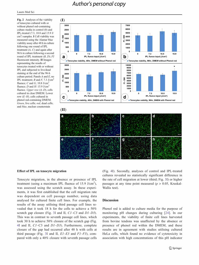
Analyses revealed that tenocytes cultured in clear DMEMand irradiated with increasing IPL fluencies (7.3, 10.8 and15.9 J/cm2) showed no significant difference in viability(p > 0.05, one-way ANOVA) compared with the controlgroup (i.e. no treatment) following the first and secondrounds of IPL treatment (Fig. 2IA, B). Similarly, therewas no significant effect of IPL treatment on the viabilityof tenocytes cultured in phenol red containing-DMEMfollowing one round of IPL treatment (Fig. 2IC). In con-trast, tenocytes cultured in phenol red-containing DMEMand subjected to two IPL treatments over the 96-h cultureperiod (Fig. 2ID) showed a significant difference in via-bility between treatment groups (p = 0.01, one-wayANOVA); furthermore, post hoc analyses revealed thattenocytes treated with a fluence of 15.9 J/cm2 showed asignificant increase in viability when compared with thecontrol group and 7.3 and 10.8 J/cm2 IPL treatmentgroups (p = 0.037, 0.002, 0.037 respectively, TukeyHSD).
The viability of tenocytes treated with or without IPLwas further assessed at the end of the 96-h culture periodusing live/dead staining (Fig. 2II), and these data wereconsistent with the results of the Alamar blue assay(Fig. 2I). Analyses revealed the presence of confluent,healthy cells in control cultures, with no evidence of celldeath in the absence (Fig. 2IA) or presence (Fig. 2IE) ofphenol red-containing culture media. Furthermore, datademonstrated that cell viability was not affected by IPLtreatment at any energy level, in the absence (Fig. 2IB–D)or presence (Fig. 2IF–H) of phenol red.
Lasers Med Sci
Author's personal copy
Effect of IPL on tenocyte migration
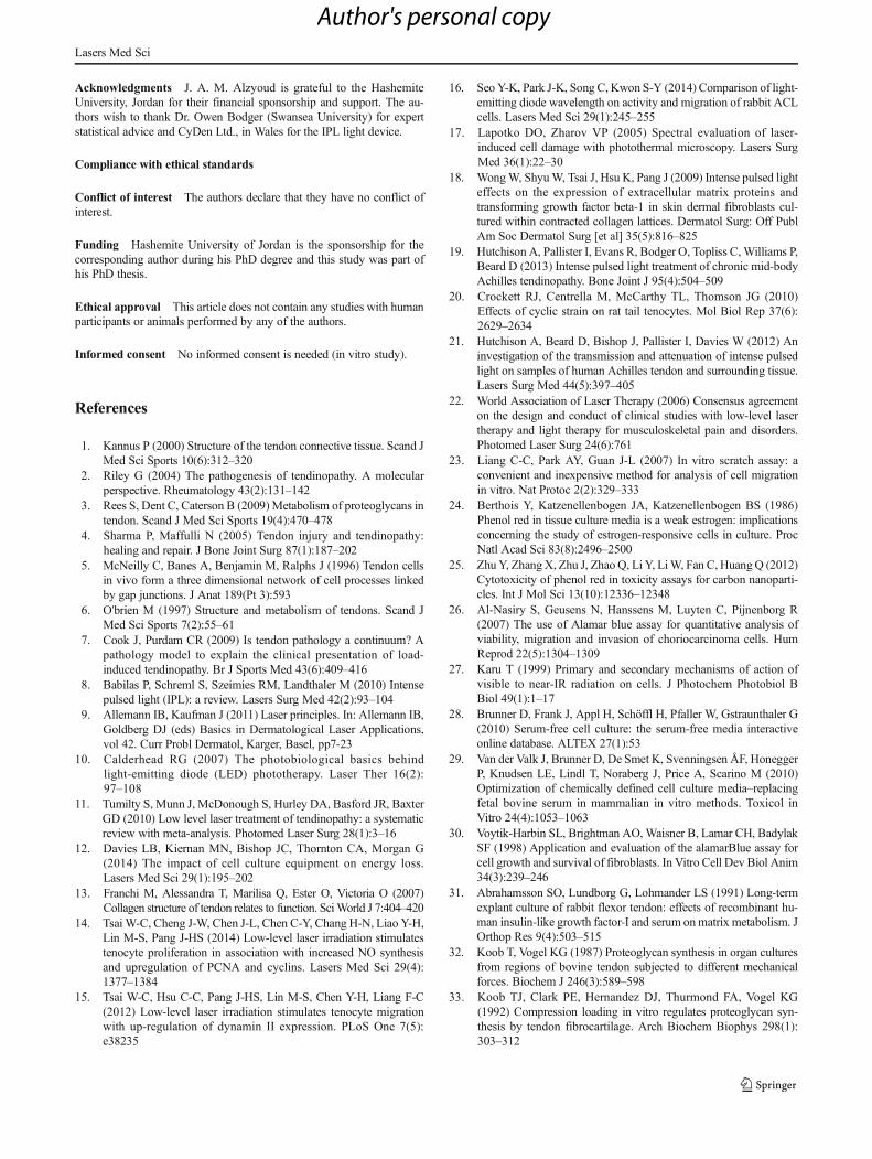
Tenocyte migration, in the absence or presence of IPLtreatment (using a maximum IPL fluence of 15.9 J/cm2),was assessed using the scratch assay. In these experi-ments, it was first established that the cell migration ratewas dependent on cell passage number, using dataanalysed for cultured finite cell lines. For example, theresults of the assay utilising third passage cell lines re-vealed that it took 18 h for the cells to achieve a 50%scratch gap closure (Fig. 3I and II, C1–C3 and D1–D3).This was in contrast to seventh passage cell lines, whichtook 30 h to achieve 50% closure of the scratch gap (Fig.4I and II, C1–C3 and D1–D3). Furthermore, completeclosure of the gap had occurred after 48 h with cells atthird passage (Fig. 3I and II, E1–E3 and F1–F3), com-pared with only a 40% closure with seventh passage cells
(Fig. 4I). Secondly, analyses of control and IPL-treatedcultures revealed no statistically significant difference inthe rate of cell migration at lower (third; Fig. 3I) or higherpassages at any time point measured (p > 0.05, Kruskal-Wallis test).
Discussion
Phenol red is added to culture media for the purpose ofmonitoring pH changes during culturing [24]. In ourexperiments, the viability of finite cell lines harvestedfrom bovine tendons was unaffected by the absence orpresence of phenol red within the DMEM, and theseresults are in agreement with studies utilising culturedHeLa cells, which found no evidence of cytotoxicity inassociation with high concentrations of this pH indicator
(I)
(II)
Fig. 2 Analyses of the viabilityof tenocytes cultured with orwithout phenol red-containingculture media in control (0) andIPL-treated (7.3, 10.8 and 15.9 J/cm2) samples. I Cell viability wasmeasured using the Alamar blueviability assay after 48 h in culturefollowing one round of IPLtreatment (A, C) and again after96 h in culture following a secondround of IPL treatment (B, D); FIfluorescent intensity. II Imagesrepresenting the results oftenocytes treated with or withoutIPL and subjected to live/deadstaining at the end of the 96-hculture period. Panels A and E, noIPL treatment; B and F, 7.3 J/cm2
fluence; C and G, 10.8 J/cm2
fluence; D and H: 15.9 J/cm2
fluence. Upper row (A–D), cellscultured in clear DMEM. Lowerrow (E–H), cells cultured inphenol red-containing DMEM.Green, live cells; red, dead cells;and blue, nuclear counterstain
Lasers Med Sci
Author's personal copy

(I)
(II)
Lasers Med Sci
Author's personal copy

[25]. Other studies have also shown that the presence ofphenol red-containing culture media did not interferewith Alamar blue viability assay fluorescence intensityreadings [26], which is consistent with our findings(Fig. 1). Interestingly, our data showed that cell viabilityincreased in the presence of phenol red when treated atthe highest IPL energy (Fig. 2I). This may be due tothe fact that phenol red increases energy absorption andheating of the culture media, which results in aphotobiostimulation effect through thermal reaction[27], as the phenol red molecules are in close proximityto the cell monolayer. Another possible explanation maybe that there is increased reflection of IPL light back tocells, which further enhances viability. Interestingly, instudies examining the impact of cell culture equipmenton energy loss, the use of phenol red-free media wasrecommended, as media containing phenol red wasfound to have a distinctive absorption peak at 560 nm[12]. However, our results show that this absorbancepeak may have a positive impact on cell viability andthat the optical window for photobiomodulation is stillvalid.
Serum provides various essential components, such asgrowth factors and other proteins necessary for the sur-vival of cultured cells [28, 29]. Indeed, our studies dem-onstrate that cultures of primary tenocytes required sup-plementation with FBS in order to survive and prolifer-ate, with increased FBS levels associated with high vi-ability (Fig. 1). Our results are in agreement with pre-vious studies which report a dose-dependent effect ofFBS (0–20% v/v) on fibroblast proliferation rate [30].Furthermore, ex vivo tendon tissue cultured usingserum-rich medium was found to enhance cell prolifer-ation in the epitenon layer [31]. Whilst adding FBS toculture media was shown to be important for tendonproteoglycan (PG) synthesis [32], conversely a low rateof PG synthesis was reported when tendon explantswere cultured in low-serum media [33, 34]. Therefore,serum-rich media (5–10%) is required for the culturingof bovine tendon [3, 28, 35].
In our experiments, tenocyte cultures were irradiatedevery 48 h, consistent with WALT (2012) recommendeddosages, as well as other published literature incorporat-ing laser or LED photobiomodulation treatment regimens.
Consequently, the actual maximum IPL dose received bythe tenocytes was in line with WALT recommendations(i.e. 12 J/cm2) [22, 36, 37]. Importantly, our results showthat IPL irradiation did not have a detrimental effect oncell viability (i.e. there was no decrease compared withuntreated cultures); moreover, an increased cell prolifera-tion was seen when higher levels of energy were deliveredto the cells, with a significant increase in cell viability atthe highest fluency, thus confirming the safety/validity ofthe devise in our in vitro system (Fig. 2). This may beexplained by the fact that higher energies enable deeperpenetration and absorption of light into tissue targets, es-pecially at higher wavelengths (600–900 nm) [38]. In ad-dition, high fluency IPL treatments have been shown to besafer than higher energy laser treatments, which supportsthe use of the former in the clinical situation. Our findingsare also consistent with a number of laboratory and clin-ical studies investigating the effects of IPL treatment onskin using high fluencies (up to 75 J/cm2), which reportincreased cell proliferation and viability with no evidenceof cytotoxicity, as well as improved clinical assessmentscores with few side effects [18, 39, 40]. Proposed mech-anisms for IPL enhancement of cell viability include thereversal of cell senescence via reduced reactive oxygenspecies production and enhanced telomerase activity, aswell as activation of cytochrome-c oxidase and releaseof nitrous oxide, leading to activation of cell signallingpathways [8, 38, 41, 42].
In our studies, the cell migratory rate, as well as the popu-lation doubling time, slowed as the cell passage number in-creased (third versus seventh passage) with or without IPLtreatment (Figs. 3 and 4). This may be explained by the pro-gressive ageing of the tenocytes in culture (senescence), astheir proliferative ability, motility and metabolic rate de-creases, leading to a decline in their regenerative ability[43–45], and is in keeping with previous studies. However,in contrast to our data, low-level laser irradiation has beenfound to stimulate tenocyte migration using an in vitro model[14, 15]. The lack of detection of any effect of IPL on cellmigration in our system might be due to a rapid proliferationof cells over a short period, or the lack of cumulative effect ofmultiple treatments over a longer period [14]. Future studiescould therefore be aimed at investigating a longer dosingregimen.
Conclusion
This study investigates for the first time the effect of IPL on abovine tendon model system; IPL fluencies of up to 15.9 J/cm2 were proven harmless to the tenocyte cultures, and thiswas the case using culture media with or without phenol red.Furthermore, there was a significant increase in cell viability
�Fig. 3 Analyses of tenocyte migration in the absence or presence of IPLtreatment, using third passage primary tenocytes. I Graph showingpercentage of scratch gap relative to time zero in control and IPL-treated cultures. Black arrow denotes the 50% closure of the scratcharea (18 h). II Images representing the scratch assay for tenocytestreated with or without IPL (n = 3). Panels A1–A3, C1–C3, and E1–E3,control cultures at 0, 18 and 48 h, respectively; B1–B3, D1–D3 and F1–F3, IPL-treated cultures at 0, 18 and 48 h, respectively. Scalebar = 250 μm
Lasers Med Sci
Author's personal copy

following irradiation with the highest energy under certainculture conditions. Future translational studies should be
carried out to elucidate the effects of IPL application in theclinical situation.
(I)
(II)
Fig. 4 Analyses of tenocytemigration in the absence orpresence of IPL treatment, usingseventh passage primarytenocytes. I Graph showingpercentage of scratch gap relativeto time zero in control and IPL-treated cultures. Black arrowdenotes the 50% closure of thescratch area (30 h). II Imagesrepresenting the scratch assay fortenocytes treated with or withoutIPL (n = 3). Panels A1–A3 andC1–C3, control cultures at 0 and30 h, respectively; B1–B3 andD1–D3, IPL-treated cultures at 0and 30 h, respectively. Scalebar = 250 μm
Lasers Med Sci
Author's personal copy
Acknowledgments J. A. M. Alzyoud is grateful to the HashemiteUniversity, Jordan for their financial sponsorship and support. The au-thors wish to thank Dr. Owen Bodger (Swansea University) for expertstatistical advice and CyDen Ltd., in Wales for the IPL light device.
Compliance with ethical standards
Conflict of interest The authors declare that they have no conflict ofinterest.
Funding Hashemite University of Jordan is the sponsorship for thecorresponding author during his PhD degree and this study was part ofhis PhD thesis.
Ethical approval This article does not contain any studies with humanparticipants or animals performed by any of the authors.
Informed consent No informed consent is needed (in vitro study).
References
1. Kannus P (2000) Structure of the tendon connective tissue. Scand JMed Sci Sports 10(6):312–320
2. Riley G (2004) The pathogenesis of tendinopathy. A molecularperspective. Rheumatology 43(2):131–142
3. Rees S, Dent C, Caterson B (2009) Metabolism of proteoglycans intendon. Scand J Med Sci Sports 19(4):470–478
4. Sharma P, Maffulli N (2005) Tendon injury and tendinopathy:healing and repair. J Bone Joint Surg 87(1):187–202
5. McNeilly C, Banes A, Benjamin M, Ralphs J (1996) Tendon cellsin vivo form a three dimensional network of cell processes linkedby gap junctions. J Anat 189(Pt 3):593
6. O'brien M (1997) Structure and metabolism of tendons. Scand JMed Sci Sports 7(2):55–61
7. Cook J, Purdam CR (2009) Is tendon pathology a continuum? Apathology model to explain the clinical presentation of load-induced tendinopathy. Br J Sports Med 43(6):409–416
8. Babilas P, Schreml S, Szeimies RM, Landthaler M (2010) Intensepulsed light (IPL): a review. Lasers Surg Med 42(2):93–104
9. Allemann IB, Kaufman J (2011) Laser principles. In: Allemann IB,Goldberg DJ (eds) Basics in Dermatological Laser Applications,vol 42. Curr Probl Dermatol, Karger, Basel, pp7-23
10. Calderhead RG (2007) The photobiological basics behindlight-emitting diode (LED) phototherapy. Laser Ther 16(2):97–108
11. Tumilty S, Munn J, McDonough S, Hurley DA, Basford JR, BaxterGD (2010) Low level laser treatment of tendinopathy: a systematicreview with meta-analysis. Photomed Laser Surg 28(1):3–16
12. Davies LB, Kiernan MN, Bishop JC, Thornton CA, Morgan G(2014) The impact of cell culture equipment on energy loss.Lasers Med Sci 29(1):195–202
13. Franchi M, Alessandra T, Marilisa Q, Ester O, Victoria O (2007)Collagen structure of tendon relates to function. SciWorld J 7:404–420
14. Tsai W-C, Cheng J-W, Chen J-L, Chen C-Y, Chang H-N, Liao Y-H,Lin M-S, Pang J-HS (2014) Low-level laser irradiation stimulatestenocyte proliferation in association with increased NO synthesisand upregulation of PCNA and cyclins. Lasers Med Sci 29(4):1377–1384
15. Tsai W-C, Hsu C-C, Pang J-HS, Lin M-S, Chen Y-H, Liang F-C(2012) Low-level laser irradiation stimulates tenocyte migrationwith up-regulation of dynamin II expression. PLoS One 7(5):e38235
16. Seo Y-K, Park J-K, Song C,Kwon S-Y (2014) Comparison of light-emitting diode wavelength on activity and migration of rabbit ACLcells. Lasers Med Sci 29(1):245–255
17. Lapotko DO, Zharov VP (2005) Spectral evaluation of laser-induced cell damage with photothermal microscopy. Lasers SurgMed 36(1):22–30
18. WongW, ShyuW, Tsai J, Hsu K, Pang J (2009) Intense pulsed lighteffects on the expression of extracellular matrix proteins andtransforming growth factor beta-1 in skin dermal fibroblasts cul-tured within contracted collagen lattices. Dermatol Surg: Off PublAm Soc Dermatol Surg [et al] 35(5):816–825
19. Hutchison A, Pallister I, Evans R, Bodger O, Topliss C,Williams P,Beard D (2013) Intense pulsed light treatment of chronic mid-bodyAchilles tendinopathy. Bone Joint J 95(4):504–509
20. Crockett RJ, Centrella M, McCarthy TL, Thomson JG (2010)Effects of cyclic strain on rat tail tenocytes. Mol Biol Rep 37(6):2629–2634
21. Hutchison A, Beard D, Bishop J, Pallister I, Davies W (2012) Aninvestigation of the transmission and attenuation of intense pulsedlight on samples of human Achilles tendon and surrounding tissue.Lasers Surg Med 44(5):397–405
22. World Association of Laser Therapy (2006) Consensus agreementon the design and conduct of clinical studies with low-level lasertherapy and light therapy for musculoskeletal pain and disorders.Photomed Laser Surg 24(6):761
23. Liang C-C, Park AY, Guan J-L (2007) In vitro scratch assay: aconvenient and inexpensive method for analysis of cell migrationin vitro. Nat Protoc 2(2):329–333
24. Berthois Y, Katzenellenbogen JA, Katzenellenbogen BS (1986)Phenol red in tissue culture media is a weak estrogen: implicationsconcerning the study of estrogen-responsive cells in culture. ProcNatl Acad Sci 83(8):2496–2500
25. ZhuY, ZhangX, Zhu J, Zhao Q, Li Y, LiW, Fan C,HuangQ (2012)Cytotoxicity of phenol red in toxicity assays for carbon nanoparti-cles. Int J Mol Sci 13(10):12336–12348
26. Al-Nasiry S, Geusens N, Hanssens M, Luyten C, Pijnenborg R(2007) The use of Alamar blue assay for quantitative analysis ofviability, migration and invasion of choriocarcinoma cells. HumReprod 22(5):1304–1309
27. Karu T (1999) Primary and secondary mechanisms of action ofvisible to near-IR radiation on cells. J Photochem Photobiol BBiol 49(1):1–17
28. Brunner D, Frank J, Appl H, Schöffl H, Pfaller W, Gstraunthaler G(2010) Serum-free cell culture: the serum-free media interactiveonline database. ALTEX 27(1):53
29. Van der Valk J, Brunner D, De Smet K, Svenningsen ÅF, HoneggerP, Knudsen LE, Lindl T, Noraberg J, Price A, Scarino M (2010)Optimization of chemically defined cell culture media–replacingfetal bovine serum in mammalian in vitro methods. Toxicol inVitro 24(4):1053–1063
30. Voytik-Harbin SL, Brightman AO,Waisner B, Lamar CH, BadylakSF (1998) Application and evaluation of the alamarBlue assay forcell growth and survival of fibroblasts. In Vitro Cell Dev Biol Anim34(3):239–246
31. Abrahamsson SO, Lundborg G, Lohmander LS (1991) Long-termexplant culture of rabbit flexor tendon: effects of recombinant hu-man insulin-like growth factor-I and serum onmatrix metabolism. JOrthop Res 9(4):503–515
32. Koob T, Vogel KG (1987) Proteoglycan synthesis in organ culturesfrom regions of bovine tendon subjected to different mechanicalforces. Biochem J 246(3):589–598
33. Koob TJ, Clark PE, Hernandez DJ, Thurmond FA, Vogel KG(1992) Compression loading in vitro regulates proteoglycan syn-thesis by tendon fibrocartilage. Arch Biochem Biophys 298(1):303–312
Lasers Med Sci
Author's personal copy
34. Vogel K, Hernandez D (1992) The effects of transforming growthfactor-beta and serum on proteoglycan synthesis by tendonfibrocartilage. Eur J Cell Biol 59(2):304–313
35. Rees S, Flannery C, Little C, Hughes C, Caterson B, Dent C (2000)Catabolism of aggrecan, decorin and biglycan in tendon. Biochem J350:181–188
36. Bastos J, Lizarelli R, Parizotto N (2009) Comparative study of laserand LED systems of low intensity applied to tendon healing. LaserPhys 19(9):1925–1931
37. Bjordal JM (2012) Low level laser therapy (LLLT) and worldAssociation for Laser Therapy (WALT) dosage recommendations.Photomed Laser Surg 30(2):61–62
38. Hamblin MR, Demidova TN (2006) Mechanisms of low level lighttherapy. Proc Spie 6140(61001):1-12.
39. Bahmer F, Drosner M, Hohenleutner U, Kaufmann R, Kautz G,Kimmig W, Landthaler M, Neumann R, Raulin C, Seeber N(2007) Recommendation for laser and intense pulsed light (IPL)therapy in dermatology. JDDG: J Dtsch Dermatol Ges 5(11):1036–1042
40. Bedewi AE, Khalafawy GE (2013) The use of synchrotron infraredmicrospectroscopy to demonstrate the effect of intense pulsed lighton dermal fibroblasts. J Cosmet Laser Ther 15(6):305–309
41. Raulin C, Greve B, Grema H (2003) IPL technology: a review.Lasers Surg Med 32(2):78–87
42. Karu T (2013) Is it time to consider photobiomodulation as a drugequivalent? Photomed Laser Surg 31(5):189–191
43. Zhou Z, Akinbiyi T, Xu L, Ramcharan M, Leong DJ, Ros SJ,Colvin AC, Schaffler MB, Majeska RJ, Flatow EL (2010)Tendon-derived stem/progenitor cell aging: defective self-renewaland altered fate. Aging Cell 9(5):911–915
44. López-Otín C, Blasco MA, Partridge L, Serrano M, Kroemer G(2013) The hallmarks of aging. Cell 153(6):1194–1217
45. Kohler J, Popov C, Klotz B, Alberton P, Prall WC, Haasters F,Müller-Deubert S, Ebert R, Klein-Hitpass L, Jakob F (2013)Uncovering the cellular and molecular changes in tendonstem/progenitor cells attributed to tendon aging and degeneration.Aging Cell 12(6):988–999
Lasers Med Sci
Author's personal copy