in vitro efficacy of some plant aqueous extracts against
TRANSCRIPT

Available online at http://www.ifgdg.org
Int. J. Biol. Chem. Sci. 14(8): 2699-2712, October 2020
ISSN 1997-342X (Online), ISSN 1991-8631 (Print)
© 2020 International Formulae Group. All rights reserved. 8633-IJBCS
DOI : https://dx.doi.org/10.4314/ijbcs.v14i8.4
Original Paper http://ajol.info/index.php/ijbcs http://indexmedicus.afro.who.int
In vitro efficacy of some plant aqueous extracts against two species of
Lasiodiplodia associated to mango decline in Burkina Faso
Oumarou Z. DIANDA1*, Issa WONNI1, Fernandez DIANA2, Oumarou TRAORÉ3,
Cyrille Tinlé ZOMBRÉ1 and Fousseni BORRO1
1Institut de l’Environnement et de Recherches Agricoles (INERA)/Centre National de Recherche Scientifique et
Technologique (CNRST), Station de Farako-Ba, 01 BP 910 Bobo Dioulasso 01, Burkina-Faso. 2Institut de Recherche pour le Développement (IRD) UMR 186 IIRD, Cirad, Univ. Montpellier "Interactions
Plante-Microorganismes Environnement" (IPME). 3Institut de Recherche en Sciences Appliquées et Technologiques (IRSAT)/ Centre National de Recherche
Scientifique et Technologique (CNRST); 01BP 2393 Bobo-Dioulasso 01 ; Burkina Faso. *Auteur correspondant ; E-mail : [email protected]; Tel. (00226) 75333401/73690211
ACKNOWLEDGEMENTS
The authors wish to sincerely thank the International Foundation for Science (IFS) through the grant
application No. 1I3_C_039006_REV and the National Research Fund project of Burkina Faso (FONRID) for
funding this research.
ABSTRACT
Mango decline is a serious disease in production areas in Burkina Faso. The aim of this study was to
contribute to the management of the disease through the use of plant aqueous extracts. Antifungal activities of
Azadirachta indica, Calotropis procera, Gmelina arborea, Jatropha curcas, Eucalyptus camaldulensis and the
synthetic fungicide (Mancozeb) were tested against Lasiodiplodia theobromae and Lasiodiplodia
pseudotheobromae associated to mango decline in Burkina Faso. Three different concentrations of leaf extracts
which 25%, 50%, 75% and 500 ppm of Mancozeb were tested for their antifungal activity in vitro. The results
showed that leaf extracts have an inhibitor effect on the growth of the two Lasiodiplodia species. The aqueous
extract of G. arborea was the most effective with average inhibition rates of L. theobromae of 42.62%, 73.84%
and 74.23% respectively with the concentrations of 25 g/l, 50 g/l and 75 g/l. The aqueous extract of A. indica
against L. pseudotheobromae showed maximum percentage inhibition with 50 g/l of 63.10% and with 75 g/l of
72.02%. Mancozeb completely inhibits the mycelial growth of both species of fungi. Ours findings showed that
aqueous extracts from plants could be tried for the eco-friendly management of mango decline pathogens.
© 2020 International Formulae Group. All rights reserved.
Keywords: Antifungal, plants extract, Lasiodiplodia spp., mango decline, Burkina Faso.
INTRODUCTION
Mango (Mangifera indica L.) is a tree
species producing delicious dessert fruits in the
Anacardiaceae family. Mango stands at the
first position in fruit production in Burkina
Faso and is therefore a crop of considerable
economic importance for the country with a
high potential for international (APROMAB,

O. Z. DIANDA et al. / Int. J. Biol. Chem. Sci. 14(8): 2699-2712, 2020
2700
2018). The mango sector employs many
producers and many other actors such as
consumers, processors, exporters, traders and
transporters who live from production and its
by-products. In 2018, fresh mango production
has increased from around 90 000 tons in 2017
to 200 000 tons (APROMAB, 2018). In terms
of resources, marketing of fresh and dried
mangoes generated more than 21 617 643.11
USD. M. indica is susceptible to a number of
diseases at all stages of its development
(Alemu et al., 2014). Fungal pathogens are the
most common agents causing diseases on
mango, including dieback, a form of
progressive death (Ismail et al., 2012). The
symptoms of dieback are commonly associated
with drying and withering of twigs from top to
downwards, followed by discoloration, drying
and eventual dropping of leaves (Saeed et al.,
2017 ; Dianda et al., 2018; Dianda, 2019).
Typically, a complete wilting and death of the
affected mango trees may occur within weeks
or few months after first symptoms occurred
(Saeed et al., 2017). Dieback is considered to
be the most destructive disease of the mango
tree, leading to significant yield loss and low
fruit quality (Saeed et al., 2017). Studies have
identified fungi of several Botryosphaeriaceae
species, such as L. theobromae (Pat.) Griffon
and Maubl. (Sutton, 1980) or L.
pseudotheobromae as the causal agents of
mango dieback disease in different areas of the
world, including Burkina Faso (Khanzada et
al., 2004; de Oliveira Costa et al., 2010; Ismail
et al., 2012; Ablormeti, 2016; Rodríguez-
Gálvez et al., 2017; Saeed et al., 2017; Dianda,
2019). Lasiodiplodia species have been
associated with several others disease
symptoms on mango plants including fruit rot,
stem-end rot, panicle brown rot, canker
(Sakalidis et al., 2011; Ismail et al., 2012).
The unbalanced use of fertilizer,
intercropping in the tree grooves, zero pruning
and certain abiotic constraints such as drought,
high temperatures, sunshine, water stress,
salinity and nutritional deficiencies can
contribute to the development and progression
of the decline caused by L. theobromae (Naqvi
and Perven, 2015). The common strategy for
controlling mango decline is using chemical
fungicides with high efficiency. Such fungicide
including Carbendazim, Fuguran, Mancozeb
are generally used to control fungal pathogens
by spraying copiously the infected plants
(Ablormeti, 2016; Tedihou et al., 2017).
However, due to growing concerns about the
potential risk that fungicides pose for human
health, environmental contamination, and the
development of fungicide resistance by
pathogens (El Ghaouth et al., 2003; Lema et al.,
2014) a control strategy based on antagonistic
microorganisms, or biocontrol, has become an
attractive alternative approach. Plants and their
derivatives have been extensively studied for
the control of pathogenic fungi. Indeed, the
extracts of acalypha (Acalypha hispida), siam
weed (Chromolaena odorata), aidan
(Tetrapleura tetraptera), and neem (A. indica)
were reported to significantly inhibit L.
theobromae isolates of cashew inflorescence
blight in Nigeria (Dele and Abiodun, 2015). In
another study, the extracts of J. curcas had
inhibitory effects against this fungal,
pathogenic on Rhizophora racemosa (Ukoima
et al., 2013). In Pakistan, in vitro evaluation of
the effectiveness of four plants extracts species
PEs) against the causal agent of mango dieback
revealed that safeda (E. camaldulensis) and
Neem extracts were the most effective while
garlic (Allium sativum) and onion (Allium
cepa) extracts were comparatively and
statistically less effective in inhibiting the
vegetative growth of the fungus causal agent of
quick decline of mango (Sahi et al., 2012).
Moreover, D. stramonium and E.
camaldulensis extracts reduced the growth of
L. theobromae causing stem end rot of mango

O. Z. DIANDA et al. / Int. J. Biol. Chem. Sci. 14(8): 2699-2712, 2020
2701
in Pakistan more efficiently than some Aloe-
vera extract (Ullah et al., 2017).
Given the threat of decline on mango
production and the harmful effects linked to the
use of pesticides in Burkina Faso, this study
aimed at assessing the antifungal activity of
extracts of certain plant species against L.
theobromae and L. pseudotheobromae isolated
from diseased mango trees in Burkina Faso.
MATERIALS AND METHODS
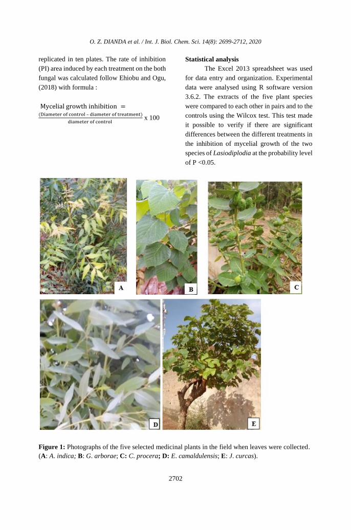
Source of plant materials
The plants used in this study were
collected at Farako-Bâ site located at 10 km
from Bobo Dioulasso. They were selected
based on their medicinal properties reported by
several authors (Sahi et al., 2012; Dele and
Abiodun, 2015) and availability from farm
lands within Farako-Bâ near Bobo-Dioulasso.
These plants include A. indica, C. procera, J.
curcas, G. arborae and E. camalulensis)
(Figure 1). Fresh leaves were manually plucked
and immediately transported in black
polyethylene bags to the laboratory for the
aqueous extraction.
Fungal isolates
One strain of L. theobromae (K11B)
and one strain of L. pseudotheobromae (H6B)
stored in the mycology laboratory of Institute
for the Environment and Agricultural Research
(INERA) of Bobo-Dioulasso were used for this
study. They were previously isolated from
mango trees with dieback symptoms. L.
theobromae and L. pseudotheobromae are
originated from Koloko
(N11°0571.14’W001°078.39’) and Peni
(N11°046.080°W004°366.6’) in Hauts Bassins
region, one of major area of mango production
in Burkina Faso. The fungal isolates were
identified based on DNA sequences of the
internal transcribed spacer region (ITS),
translation elongation factor (Tef1-alpha) and
beta-tubulin (Bt) (White et al., 1990; Alves et
al., 2008; Donaldson and GLass, 1995).
Pathogenicity of these strains was confirmed
by the detached leaves method described by
Ismail, (2011) and Munirah et al. (2017).
Preparation of aqueous extracts
The fresh leaves were first washed with
running tap water, then sterilized with 1%
sodium hypochloride and finally rinsed with
sterile distilled water.
They were then dried at laboratory
temperature on blotting paper for two weeks.
The dried leaves were crushed with a pestle in
a sterilized mortar and suspended in sterile
water at a ration 1:1 (w / v) according to the
method described by Rahee et al. (2018). The
aqueous extracts were filtered through two (02)
folds muslin clothes and centrifuged at 4000
rpm for 15 mn. The supernatant collected,
considered to be 100% concentration was
stored at 4 °C until subsequent use.
In vitro antifungal activity assay
Three different concentrations (C1:
25%; C2: 50% and C3: 75%) were prepared
from the stock solution (C0: 100%) to test their
ability to inhibit the mycelial growth of the two
species of Lasidioplodia. Each concentration
(v) of aqueous extract was inoculated into PDA
medium maintained in a 55 °C water bath. The
mixture was then poured into Petri dishes. PDA
medium without aqueous extract and PDA
inoculated with Mancozeb at 500 ppm were
used as negative and positive controls,
respectively. A 5 mm mycelial disc was cut
from the actively growing regions (peripheral
mycelium) of 7-days old culture of the two
species of Lasiodiplodia and deposited on the
medium inoculated with aqueous plant
extracts. After inoculation, the plates were
incubated in the inverted position at 28 ± 2 °C.
The efficacy of aqueous extract was assessed
when the fungal completely cover the medium
used as a negative test. Each treatment was
O. Z. DIANDA et al. / Int. J. Biol. Chem. Sci. 14(8): 2699-2712, 2020
2702
replicated in ten plates. The rate of inhibition
(PI) area induced by each treatment on the both
fungal was calculated follow Ehiobu and Ogu,
(2018) with formula :
Mycelial growth inhibition =(Diameter of control – diameter of treatment)
diameter of control x 100
Statistical analysis
The Excel 2013 spreadsheet was used
for data entry and organization. Experimental
data were analysed using R software version
3.6.2. The extracts of the five plant species
were compared to each other in pairs and to the
controls using the Wilcox test. This test made
it possible to verify if there are significant
differences between the different treatments in
the inhibition of mycelial growth of the two
species of Lasiodiplodia at the probability level
of P <0.05.
Figure 1: Photographs of the five selected medicinal plants in the field when leaves were collected.
(A: A. indica; B: G. arborae; C: C. procera; D: E. camaldulensis; E: J. curcas).

O. Z. DIANDA et al. / Int. J. Biol. Chem. Sci. 14(8): 2699-2712, 2020
2703
RESULTS
The two Lasiodiplodia strains recorded
a maximum mycelial growth in all negative
control plates 72 hours after incubation (hai).
However, their growth varied according to the
extracts from five plant species used and the
three concentrations tested (Figure 2 and Table
1).
Effects of plant extracts on the mycelial
growth of the L. theobromae and L.
pseudotheobromae.
Fungal growth observation at 72 h after
incubation showed that the aqueous extracts of
A. indica, J. curcas, C. procera, G. arborea and
E. camelendis haved antifungal properties
against the two Lasiodiplodia species tested at
for all the levels of concentrations tested 72h
after incubation (Table 1). Highest growth
inhibition was recorded at the concentration of
75% for all extracts. Except for Eucalyptus,
extracts from the other four plant species
generated mycelial growth inhibition rates
greater than 50%. At the dose 75%, G. arborea
aqueous extract inhibits the growth of L.
theobromae with mean rate of 74.23%. On the
other hand, C. procera recorded 72.20% of
inhibition rate on the mycelial growth of L.
pseudotheobromae. At 50% concentration,
aqueous extracts of G. arborea and A. indica
induced respectively inhibition rates of 73.87%
and 63.10% of L. theobromae and L.
pseudotheobromae. The lowest inhibition rates
of the both fungal species were observed with
the all aqueous plants extracts at 25%
concentration. Mancozeb used as a positive
control inhibits totally the mycelium growth of
the two species of Lasiodiplodia screened.
Efficacy of extracts from five plant species
on L. theobromae
Table 2 shows the p-values obtained
from the two-by-two comparisons of the plant
extracts on L. theobromae using Wilcox-test.
At the concentration of 75%, a significant
difference was observed between the inhibition
rates of the five plant extracts and the negative
control. Among the aqueous extracts, that of G.
arborea was found to be the most effective
against L. theobromae. It showed significant
differences with those of four other plant
species. Statistically, the effect was not
significantly different (P <0.05) between the
extracts of A. indica and C. procera.
Efficacy of extracts from five plant species
on L. pseudotheobromae
Results of Table 3 and Figure 3 showed
significant difference in per cent inhibition of
L. pseudotheobromae by extracts of five plants
species with the control. Among extracts of the
five plants species tested, maximum
percentage inhibition was recorded with C.
procera. It didn't show significant difference
with A. indica and J. curcas. Moreover, there
is no significant difference between J. curcas
and G. arborea.

O. Z. DIANDA et al. / Int. J. Biol. Chem. Sci. 14(8): 2699-2712, 2020
2704
Plant extracts Fungal
L. theobromae L. pseudotheobromae
G. arborea
A. indica
C. procera
J. curcas
E. camalendis
Figure 2: Effect of extracts of five plant species and Mancozeb (synthesis fungicide) on the mycelial
growth of the two Lasiodiplodia species 72 hours after incubation (dose used: A: 0 (control); B: 25%;
C: 50%; D: 75%; E: 500 ppm (Macozeb)).
A
B
C
D
E A
B
C
D
E
A
B
C
D
E A
B C
D
E
A E
B
A
C B C D
E
B
A
C D
E
B C D
A E
B
C D
A E
B
A
C D
E
O. Z. DIANDA et al. / Int. J. Biol. Chem. Sci. 14(8): 2699-2712, 2020
2705
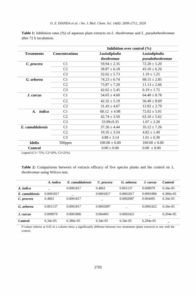
Table 1: Inhibition rates (%) of aqueous plant extracts on L. theobromae and L. pseudotheobromae
after 72 h incubation.
Inhibition over control (%)
Treatments Concentrations
Lasiodiplodia
theobromae
Lasiodiplodia
pseudotheobromae
C. procera C1 59.94 ± 2.35 72.20 ± 5.20
C2 38.87 ± 6.18 43.10 ± 6.26
C3 32.62 ± 5.73 1.19 ± 1.25
G. arborea C1 74.23 ± 6.74 68.15 ± 2.85
C2 73.87 ± 7.20 11.13 ± 2.66
C3 42.62 ± 5.45 6.19 ± 1.72
J. curcas C1 54.05 ± 4.60 64.40 ± 8.78
C2 42.32 ± 5.19 56.49 ± 8.69
C3 31.43 ± 4.67 13.92 ± 2.79
A. indica C1 60.12 ± 4.98 72.02 ± 5.01
C2 42.74 ± 3.50 63.10 ± 5.62
C3 33.99±9.35 1.07 ± 2.28
E. camaldulensis C1 37.26 ± 4.44 35.12 ± 7.26
C2 19.35 ± 3.54 4.82 ± 1.49
C3 4.88 ± 3.14 1.01 ± 0.38
Idefix 500ppm 100.00 ± 0.00 100.00 ± 0.00
Control 0.00 ± 0.00 0.00 ± 0.00
Legend (C1= 75%, C2=50%, C3=25%).
Table 2: Comparisons between of extracts efficacy of five species plants and the control on L.
theobromae using Wilcox-test.
A. indica E. camaldulensis C. procera G. arborea J. curcas Control
A. indica _ 0.0001817 0.4863 0.001137 0.008979 6.34e-05
E. camaldensis 0.0001817 _ 0.0001817 0.0001817 0.0001806 6.386e-05
C. procera 0.4863 0.0001817 _ 0.0002087 0.004495 6.34e-05
G. arborea 0.001137 0.0001817 0.0002087 _ 0.0002422 6.34e-05
J. curcas 0.008979 0.0001806 0.004495 0.0002422 _ 6.294e-05
Control 6.34e-05 6.386e-05 6.34e-05 6.34e-05 6.294e-05 _
P-values inferior at 0.05 in a column show a significantly different between two treatments (plant extracts) or one with the
control.

O. Z. DIANDA et al. / Int. J. Biol. Chem. Sci. 14(8): 2699-2712, 2020
2706
Table 3: Comparison of extracts efficacy of five species plants on L. pseudotheobromae using
Wilcox-test.
A. indica E. camaldensis C. procera G. arborea J. curcas control
A. indica _ 0.0001817 0.8198 0.02825 0.09593 6.386e-05
E. camaldensis 0.0001817 _ 0.0001776 0.0001806 0.0001817 6.203e-05
C. procera 0.8198 0.0001776 _ 0.02065 0.1197 6.203e-05
G. arborea 0.02825 0.0001806 0.02065 _ 0.2406 6.34e-05
J. curcas 0.09593 0.0001817 0.1197 0.2406 _ 6.386e-05
Control 6.386e-05 6.203e-05 6.203e-05 6.34e-05 6.386e-05 _
P-values inferior at 0.05 in a column show a significantly different between two treatments (plant extracts) or one with the
control.
Figure 3: Antifungal activity of five plant extracts and Mancozeb on the mycelial growth of L.
pseudothebromae in ten Petri dishes. (A: Mancozeb, B: A. indica; C: C. procera; D: G. arborae; E: J. curcas; F: E.
camaldulensis; CN: Petri dish control).

O. Z. DIANDA et al. / Int. J. Biol. Chem. Sci. 14(8): 2699-2712, 2020
2707
DISCUSSION
Efficacy of extracts from the five plant
species tested
The crude aqueous extracts from all five
plant species showed antifungal activities
against the two Lasiodiplodia species. Their
antifungal activities were lower than those of
the reference antifungal drug used, Mancozeb
which showed 100% inhibition. Chemical
fungicides are quite expensive and leave
drastic implications for human health and the
environment; so we have alternatively chosen
the botanical extracts under in vitro conditions
to find out the control of L. theobromae and L.
pseudotheobromae, pathogens responsibles of
the disease. Findings from this study were in
agreement with previous researchers, who
reported that most the mycelial growth of most
isolates of L. theobromae and L.
pseudotheobromae from mango and other
crops were significantly inhibited by leaf
extracts from various medicinal herbs and
spices (Rahee et al., 2018). About, acalypha
(Acalypha hispida), siam weed (Chromolaena
odorata), aidan (Tetrapleura tetraptera), and
neem (Azadirachta indica) were reported to
significantly inhibit L. theobromae isolates of
cashew inflorescence blight in Nigeria (Dele
and Abiodun, 2015). In the same country, in
vitro tests showed antimicrobial activity of
crude extracts against Botryodiplodia, one of
fungal pathogens of yam. The cold water
extracts of J. curcas had inhibitory effects
against this pathogen with range of inhibition
colony diameter of : 6.3 - 16.0 mm (Opara and
Nwokocha, 2015). In Pakistan, in vitro
evaluation of the effectiveness of the extract of
four plants species against the mycelial growth
of B. theobromae revealed that safeda (E.
camaldulensis) and Neem extracts were the
most effective while garlic (Allium sativum)
and onion (Allium cepa) extracts were
comparatively and statistically less effective
(Sahi et al., 2012). Moreover, comparative
analysis showed that D. stramonium and E.
camaldulensis extracts most efficiently
reduced the growth of L. theobromae causing
stem end rot of mango in Pakistan, in
comparison to Aloe-vera extract (Ullah et al.,
2017).
In contrast, extracts of E. camaldulensis
in our study showed the low inhibitory effect
(< 38% inhibition) on both target fungi (L.
theobromae and L. pseudotheobromae),
suggesting a possible variation in effectiveness
of the extract depending to the geographical
origine of the plant species. The effectiveness
of extracts from these plant species have also
been reported on other fungus species
indicating that they may have a large spectrum
of antifungal activity. Indeed, Ahmad et al.
(2016) revealed the efficacy of Eucalyptus
globulus, C. procera, Melia azedarach, Datura
stramonium et Acalypha indica in reducing the
mycelial growth of Alternaria alternata, seed-
borne fungal isolated from barley. Following
an in vitro study, crude extracts of leaves of six
different plants which A. indica (A.) and E.
camaldulensis showed antifungal activity
against five pathogenic fungi (Aspergillus
flavus, A. niger, Fusarium solani,
Macrophomina phaseolina and Rhizoctonia
solani) (Hussain et al., 2015). Pharmacological
research reviewed that G. arborea possess
various medicinal properties and biological
activities including antidiuretic, antidiarrhoeal,
antipyretic, antianalgesic, antioxidant,
antidiabetic, antihelmintic, antibacterial,
antifungal, cardiopotective, insecticidal,
antiulcer, gastro-protective, anticancer,
antihyperlipidemic and immunomodulatory
activity (Arora and Tamrakar, 2017).
Efficacy of concentrations of extracts and
phytochemical constituents
Growth reduction in L. theobromae and
L. pseudotheobromae was depending to the
type of plant extract, the concentration of
extract used and as well as the duration of
incubation. Findings of this study revealed that

O. Z. DIANDA et al. / Int. J. Biol. Chem. Sci. 14(8): 2699-2712, 2020
2708
the effectiveness of extracts increased as their
dose increased. This is in agreement with
earlier report (Ehiobu and Ogu, 2018). They
reported that M. esculenta leaf extracts at
concentrations (25 g/l and 50 g/l) generally
demonstrated least antifungal activities against
B. theobromae and A. niger, their activities
being significant at 75 g/l.
According to Banso and Adeyemo,
(2007) the actions of the antifungal substances
present in the plant extracts were fungistatic at
lower concentrations but became fungicidal at
higher concentrations. Antifungal activities
expressed by the five botanicals against the two
Lasiodiplodia species in the present study
suggest that they possess phytochemical
constituents against these fungal pathogens.
According to Alessandra et al. (2004), plant
secondary metabolites have great potential as a
source of effective antifungal agents. About, A.
indica, contains at least 35 biologically active
principals of which triterpenoides eg, nimbin
nimbidine and azadirachtin reported are the
most active insecticidal or antifungal
ingradients (Mordue et al., 2000 ; Brahmachari,
2004). A phytochemical analysis of aqueous
extracts of leaves of C. procera revealed the
presence of alkaloids, flavonoids, tannins,
steroids, triterpenoids, saponins, and saponins
glycosides (Hassan et al., 2006). Similary study
realised on aqueous extracts of leaves of J.
curcas by Opara and Nwokocha (2015)
allowed to detect bioactive compound such as
alkaloid, saponin, flavonoid, phenol, tannin,
phytate and HCN. Some constituents from
extract of G. arborea like 7’O-ethyl arboreol,
paulownin, gmelinol, epieudesmin and B-
sitosterol have been reported to exhibit
antifungal activity against Trametes versicolor
and Fomitopsis palustris, Aspergillus niger,
Penicillium notatum and Candida albican
(Arora and Tamrakar, 2017). The crude leaf
and stem bark extracts of G. arborea contains
also bioactive compounds such as alkaloids,
saponins, carbohydrates, phenolics,
anthraquinone and tannins recorded in the
others plants species tested in this study. The
secondary metabolites screening of E.
camaldulensis leaf extracts from Nigeria
confirmed presence of tannin, saponins, and
cardiac glycosides (Ayepola and Adeniyi,
2008). The chemical constituents such as
Tannins, saponins, terpenoids, anthraquinone,
glycoside, alkaloids, flavonoids, steroids and
reducing sugars are secondary metabolites of
plant that have been reported to serve as
defense mechanisms against predation by
many microorganisms, insects and herbivores
(Adeshina et al., 2009; Ogutu et al., 2012).
The presence of cardiac glycosides and
steroids have been documented to inhibit the
many microbes and found to possess
antioxidant potentials (Mujeeb et al., 2014).
The differences in the toxicity of different
extracts of the five plants species studied could
be attributed to the presence of the active
principles that are extracted by different
solvents, which may be influenced by several
factors such plant species, method of extraction
and type of extracting solvent (Onzo et al.,
2016). Although mancozeb a synthetic
fungicide consistently induced 100% inhibition
on the two Lasiodiplodia species irrespective
of the concentration used, it is advice that its
use can only be recommended when other
methods prove ineffectivess due to its toxic
effect on the environment (Markson et al.,
2012). Thus, the extracts of the five plants
species have to be tested in vivo on mango
seedling inoculated with the two Lasiodiplodia
species following fields trials. The extracts
with high could be used more efficacy could be
use as alternative to chemicals in the
management of these fungal pathogens
associated with mango decline.
Conclusion
The present study was carried out under
in vitro condition to find out the most effective
and eco-friendly approach to manage the

O. Z. DIANDA et al. / Int. J. Biol. Chem. Sci. 14(8): 2699-2712, 2020
2709
mango decline caused L. theobromae and L.
pseudotheobromae. The result proved that
plants may contain fungitoxic principles
against Lasiodiplodia species. G. arborea, C.
procera, A. indica and J. curcas were more
effective on L. theobromae and L.
pseudotheobromae compared to E.
camaldensis plants extracts at various
concentrations. These plants could therefore be
formulated and used as alternative to chemicals
in the management of fungal pathogens of
mango decline since they have less adverse
environmental effects, are easily available and
less difficult to prepare compared to the use of
synthetic fungicides which are very costly and
harmful to human and environment. However,
efficacy trials of these extracts must be carried
out on mango plants inoculated in a greenhouse
followed by field trials to detect the most
effective.
COMPENTING INTERESTS
The authors declare that they have no
competing interests.
AUTHORS’ CONTRIBUTIONS
DZO, WI and DF made a substantial
contribution to the design of the work. They
contribute to the acquisition, analysis or
interpretation of data. All authors brought a
contribution to the elaboration of the
manuscript and revised it critically for
important intellectual content, and approved to
be published.
ACKNOWLEDGEMENTS
We are also grateful to Institute of
Research for Development (IRD) and
University Agency of the Francophonie (AUF)
through the project PARFAO and the
International Mixed Laboratory (LMI-
PathoBios IRD-INERA) for their precious
cooperation.
REFERENCES
Ablormeti F. 2016. Aetiology, economic
importance and control of mango
(Mangifera indica l.) tree decline disease
in northern region. PhD Thesis,
University of Ghana, Ghana, p.127.
Adeshina G, Okeke C, Onwuegbuchulam N,
Ehinmidu J. 2010. Preliminary studies on
antimicrobial activities of ethanolic
extracts of Ficus sycomorus Linn. and
Ficus platyphylla Del. (Moraceae). Int. J.
Biol. Chem. Sci., 3: 1013-1020. DOI :
https://doi.org/10.4314/ijbcs.v3i5.51080.
Ahmad L, Pathak N, Zaidi RK. 2016.
Antifungal Potential of Plant Extracts
against Seed-borne Fungi Isolated from
Barley Seeds (Hordeum vulgare L.). J.
Plant Pathol. Microbiol., 07: 5-8. DOI :
https://doi.org/10.4172/2157-
7471.1000350.
Alemu K, Ayalew A, Woldetsadic K. 2014.
Effect of aqueous extracts of some
medicinal plants in controlling
anthracnose disease and improving
postharvest quality of mango fruit.
Persian Gulf Crop Prot., 3 : 84-92.
Alessandra DRA, Braga de Oliveira J, Dias de
Souza FJ, Antônio LFCB. 2004.
Antifungal constituents of Clytostoma
ramentaceum and Mansoa hirsuta.
Phytother Res., 6: 463-467. DOI :
https://doi.org/10.1002/ptr.1452.
Alves A, Crous PW, Correia A, Phillips JLA.
2008. Morphological and molecular data
reveal cryptic speciation in Lasiodiplodia
theobromae. Fungal Divers, 28: 1-13.
Apromab. 2018. Lancement de la campagne
mangue 2018 sous le thème: Réduire la
pression parasitaire pour la campagne
mangue 2018 au Burkina Faso. Apromab,
Burkina Faso.
Arora C, Tamrakar V. 2017. Gmelina arborea
: chemical constituents, pharmacological
activities and applications. Int. J.

O. Z. DIANDA et al. / Int. J. Biol. Chem. Sci. 14(8): 2699-2712, 2020
2710
Phytomedicine, 9: 528. DOI :
https://doi.org/10.5138/09750185.2149.
Ayepola O, Adeniyi BA. 2008. The
antibacterial activity of leaf extracts of
Eucalyptus camaldulensis (Myrtaceae). J.
Appl. Sci. Res., 11: 1410-1413. DOI :
https://doi.org/http://eprints.covenantuni
versity.edu.ng/id/eprint/368.
Banso A, Adeyemo S. 2007. Evalution of
Antibacterial Properties of tannins
isolated dichrostachys cineral. African J.
Biotechnol., 6: 1785-1787. DOI :
https://doi.org/10.5897/AJB2007.000-
2262.
Brahmachari G. 2004. Neem-An Omnipotent
Plant : A Retrospection. Chem. Bio.
Chem., 5: 408-421. DOI:
https://doi.org/10.1002/cbic.200300749.
de Oliveira Costa VS, Michereff SJ, Martins
RB, Gava CAT, Mizubuti ESG, Câmara
MPS. 2010. Species of
Botryosphaeriaceae associated on mango
in Brazil. Eur. J. Plant Pathol., 127 : 509-
519. DOI :
https://doi.org/10.1007/s10658-010-
9616-y.
Dele OA, Abiodun J. 2015. In vitro evaluation
of plant extracts against Lasiodiplodia
theobromae causing cashew inflorescent
blight. African J. Biotechnol., 14: 1139–
1142. DOI:
https://doi.org/10.5897/ajb2014.14336.
Dianda ZO. 2019. Caractérisation des agents
pathogènes associés au dessèchement du
manguier et essai de méthode de lutte
contre la maladie au Burkina Faso. Thèse
de doctorat Unique, Université Joseph
KI-ZERBO, Ouagadougou, 246 p.
Dianda ZO, Wonni I, Zombré C, Traoré O,
Sérémé D, Boro F, Ouédraogo I,
Ouédraogo SL, Sankara P. 2018.
Prévalence du dessèchement du manguier
et evaluation de la fréquence des
champignons associés à la maladie au
Burkina Faso. J. Appl. Biosci., 126:
12686. DOI:
https://doi.org/10.4314/jab.v126i1.6.
Donaldson GC, GLass NL. 1995. Development
of primer sets designed for use with the
PCR to amplify conserved genes from
filamentous asco- mycetes. Appl.
Environ. Microbiology, 61: 1323-1330.
DOI:
https://doi.org/10.1128/AEM.61.4.1323-
1330.1995.
Ehiobu JM, Ogu GI. 2018. Phytochemical
Content and In Vitro Antimycelial
Efficacy of Colocasia esculenta (L),
Manihot esculenta (Crantz ) and
Dioscorea rotundata (Poir) Leaf Extracts
on Aspergillus niger and Botryodiplodia
theobromae. J. Hortic. Plant Res., 1: 9-
18. DOI:
https://doi.org/10.18052/www.scipress.c
om/JHPR.1.9.
El Ghaouth A, Wilson C, Wisniewski M. 2003.
Control of postharvest decay of apple
fruit with Candida saitoana and induction
of defense responses. Phytopathology.,
93: 344-348. DOI:
https://doi.org/10.1094/PHYTO.2003.93.
3.344.
Hassan S, Bilbis F, Ladan M, Umar R,
Dangoggo S, Saidu Y, Abubakar M,
Faruk U. 2006. Evaluation of Antifungal
Activity and Phytochemical Analysis of
Leaves, Roots and Stem Barks Extracts of
Calotropis procera (Asclepiadaceae).
Pakistan J. Biol. Sci., 14: 2624–2629.
Hussain F, Abid M, Shaukat SS, Farzana
Akbar M. 2015. Anti-fungal activity of
some medicinal plants on different
pathogenic fungi. Pakistan J. Bot., 47:
2009–2013.
Ismail AM. 2011. Studies on the fungal disease
of mango with particular reference to
deseases caused by botryosphaeria
species. PhD Thesis, University of

O. Z. DIANDA et al. / Int. J. Biol. Chem. Sci. 14(8): 2699-2712, 2020
2711
Catania, Catania, 161 p.
Ismail AM, Cirvilleri G, Polizzi G, Crous PW,
Groenewald JZ, Lombard L. 2012.
Lasiodiplodia species associated with
dieback disease of mango (Mangifera
indica) in Egypt. Australas Plant Pathol.,
41: 649–660. DOI:
https://doi.org/10.1007/s13313-012-
0163-1.
Khanzada A, Lodhi M, Shahzad S. 2004.
Pathogenicity of Lasiodiplodia
theobromae and Fusarium solani on
mango. Pak. J. Bot., 36 : 181-189.
Lema E, Machunda R, Njau K. 2014.
Agrochemicals use in horticulture
industry in Tanzania and their potential
impact to water resources. Int. J. Biol.
Chem. Sci., 8: 831. DOI:
https://doi.org/10.4314/ijbcs.v8i2.38.
Markson AA, Amadioha A, Omosun G,
Madunagu B, Udo SE, Umana E. 2012.
Control of Bryodiplodia theobromae
causing tissue Rot of White yam
(Dioscorea rotundata Poir). Sch. J. Agric.
Sci., 1 : 1-7.
Mordue (Luntz) A, Jennifer N, Alasdai J. 2000.
Azadirachtin from the neem tree
Azadirachta indica : its action against
insects. An. da Soc. Entomológica do
Bras., 29: 615-632. DOI:
https://doi.org/10.1590/s0301-
80592000000400001.
Mujeeb F, Bajpai P, Pathak N. 2014.
Determination of Bioactive Components
from Leaves of Aegle marmelos. BioMed.
Res. Int. green-blue, 497606: 11p. DOI :
https://doi.org/10.1155/2014/497606.
Munirah M, Azmi A, Yong S, Nur Izzati M.
2017. Characterization of Lasiodiplodia
theobromae and L . pseudotheobromae
causing fruit rot on pre-harvest mango in
Malaysia. Plant Pathol. Quar., 7: 202-
213. DOI:
https://doi.org/10.5943/ppq/7/2/14.
Naqvi AHS, Perven R. 2015. Mango quick
decline manifestation on various cultivars
at plants of particular age in the Vicinity
of district Multan. Pak. J. Phytopathol.,
27 : 31-39.
Ogutu A, Lilechi D, Mutai C, Bii C. 2012.
Phytochemical analysis and antimicrobial
activity of Phytolacca dodecandra,
Cucumis aculeatus and Erythrina
excelsa. Int. J. Biol. Chem. Sci., 6: 692-
704. DOI:
https://doi.org/10.4314/ijbcs.v6i2.13;
Onzo CF, Azokpota P, Dah-Nouvlessounon D,
Lehmane TH, Adjatin A, Baba-Moussa L.
2016. Évaluation de l’activité
antimicrobienne de quatre feuilles
utilisées comme emballages dans
l’artisanat agroalimentaire au Bénin. J.
Appl. Biosci., 95: 9015. DOI:
https://doi.org/10.4314/jab.v95i1.11.
Opara EU, Nwokocha NJ. 2015. Antimicrobial
Activities of some Local Medicinal Plants
Against Post Harvest Yam Rot Pathogens
in Humid South Eastern Nigeria. J.
Microbiol. Res. Rev., 3 : 1-9.
Rahee B, Bireswar S, Sobita PD, Salam R,
Dinesh K, Chakma T. 2018. In vitro
Studies on Efficacy of Some Plant
Extracts and Biocontrol Agents against
Lasiodiplodia theobromae and
Lasiodiplodia pseudotheobromae. Int. J.
Curr. Microbiol. App. Sci., 7: 448-457.
DOI:
https://doi.org/10.20546/ijcmas.2018.707
.054.
Rodríguez-Gálvez E, Guerrero P, Barradas C,
Crous PW, Alves A. 2017. Phylogeny and
pathogenicity of Lasiodiplodia species
associated with dieback of mango in Peru.
Fungal Biol., 121: 452–465. DOI:
https://doi.org/10.1016/j.funbio.2016.06.
004.
Saeed EE, Sham A, AbuZarqa A, Khawla
AAS, Tahra SAN, Rabah I, Khaled ET,

O. Z. DIANDA et al. / Int. J. Biol. Chem. Sci. 14(8): 2699-2712, 2020
2712
AbuQamar Synan F. 2017. Detection and
Management of Mango Dieback Disease
in the United Arab Emirates. Int. J. Mol.
Sci., 18: 2086. DOI:
https://doi.org/10.3390/ijms18102086.
Sahi ST, Habib A, Ghazanfar MU, Badar A.
2012. In vitro Evaluation of Different
Fungicides and Plant Extracts Against
Botryodiplodia theobromae, the Causal
Agent of Quick Decline of Mango. J.
Phytopathol., 24 : 137-142.
Sakalidis ML, Ray JD, Lanoiselet V, Hardy
GES, Burgess TI. 2011. Pathogenic
Botryosphaeriaceae associated with
Mangifera indica in the Kimberley
Region of Western Australia. Eur. J.
Plant Pathol., 130: 379-391. DOI:
https://doi.org/10.1007/s10658-011-
9760-z.
Sutton BC. 1980. The Coelomycetes, Fungi
Imperfecti with Pycnidia, Acervuli and
Stromata. Commonwealth Mycological
Institute: UK.
Tedihou E, Kpemoua K, Tounou A. 2017.
Dépérissement des manguiers et citrus
dans la région centrale du Togo et
méthodes de lutte par des fongicides. J.
Appl. Biosci., 119: 11829-11838. DOI:
https://doi.org/10.4314/jab.v119i1.1.
Ukoima H, Ikata M, Pepple GA. 2013. Control
of Lasiodiplodia theobromae (PAT) on
Rhizophora racemosa using plants
extracts. Am. J. Biotechnol. Mol. Sci., 3:
1-7. DOI:
https://doi.org/10.5251/ajbms.2013.3.1.1
.7.
Ullah SF, Hussain Y, Iram S. 2017. Pathogenic
characterization of Lasiodiplodia causing
stem end rot of mango and its control
using botanicals. Pakistan J. Bot., 49:
1605-1613.
White TJ, Bruns T, Lee S, Taylor J. 1990.
Amplification and direct sequencing of
fungal ribosomal RNA genes for
phylogenetics. In PCR Protocols: A
Guide to Methods and Applications, Innis
MA, Gelfand DH, Sninsky JJ, White TJ
(eds.). Academic Press: New York; 315-
322. DOI : https://doi.org/10.1016/b978-
0-12-372180-8.50042-1.