in vitro breast cancer models as useful tools in therapeutics?
TRANSCRIPT

2
In Vitro Breast Cancer Models as Useful Tools in Therapeutics?
Emilie Bana and Denyse Bagrel Université Paul Verlaine – Metz
Laboratoire d’Ingénierie Moléculaire et Biochimie Pharmacologique France
1. Introduction
The increased use of animals in fundamental and applied research due to the remarkable
drug development in the 20th century has been an important matter of concern for people at
large, but also for the scientific community. This led Russel and Burch to examine the
decisions which could meliorate this situation, and they proposed, in 1959, the principle of
the 3Rs (Reduce, Refine, and Replace) nowadays largely admitted as an ethical and
incontrovertible principle (Russell & Bursch 1959). Alternatives to animal experiments
(Scheme 1) then knew a fantastic boom with the permanent objective of a high scientific
quality in order to prevent, treat and cure human illness.
Reaching the equilibrium between in vitro and in vivo models, observing the 3Rs rules, is
very difficult. Effectively, in vitro systems allow an excellent control of all parameters of the
experiments, and then, good quantifications. More the models are simple, more they are
easy to handle, but more they also are dedifferentiated and keep away from the in vivo
situation.
Scheme 1. In vitro systems as alternatives to the use of animals.
Within the framework of this book, the question becomes now: how the 3Rs could be the
best way to phase out animal experiments when considering breast cancer? We try to bring
some response elements in this chapter, emphasising the in vitro models the most useful and
the most frequently used. But we also show that no model is perfect and sufficient by itself,
and that pure in vitro models also need assistance of in vivo ones.
www.intechopen.com

Breast Cancer – Focusing Tumor Microenvironment, Stem Cells and Metastasis
22
2. Models for investigation on breast cancer
2.1 Established breast cancer cell lines
2.1.1 The different cell lines and their main properties
Significant amounts of data on breast cancer have been collected over the past 40 years,
thanks to the use of established cell lines. The first breast cancer cell lines (BCCL) have been
established in the sixties-seventies and very few new cell lines have been developed since.
Only a hundred of BCCL are currently available and three of them have been extensively
studied and represent now nearly 80% of the 35 000 publications mentioning breast cancer
cell lines (Lacroix & Leclercq 2004).
Most of the cell lines were created from cells derived from metastasis or from pleural
effusion. Pleural effusions contain large amounts of well isolated tumour cells and few
contaminating cells such as fibroblasts, thus making their recovery and growing easier than
those of cells directly derived from primary tumours or metastasis. Moreover, metastatic
cells are highly dedifferentiated cells, which allow their cultivation more successfully than
the primary tumour cells.
The three more used BCCL (MCF-7, MDA-MB-231 and T47D) are issued from pleural
effusion of an invasive ductal carcinoma (Soule et al. 1973; Cailleau et al. 1974; Keydar et al.
1979), and they mainly differ by their oestrogen receptor (ER) and progesterone receptor
(PgR) status: MCF-7 and T47D are ER+ PgR+ while MDA-MB-231 is ER- PgR-. Among these
three cell lines, MCF-7 was the most often used during the last ten years: it has been cited in
53% of all the scientific papers mentioning BCCL, while MDA-MB-231 and T47D were
respectively cited in about 18% and 7% of these articles (calculation made on the basis of a
Medline-based survey in March 2011).
The use of these lines has many technical advantages.
The complete control of environmental conditions and standardised culture conditions
ensures the reproducibility of results between experiments and laboratories.
Maintaining cells in culture is much less costly than working on animal models. Besides
the fact that some animal models are expensive by themselves, the care of animals and
the staff necessary to a good work in an animal house are the main drain of resources.
Conversely, the medium and the staff time required to growth cells are cheaper, thus
allowing the widespread use of BCCL.
Cryopreservation enables long-term conservation of the same strain and can
theoretically permit the use of these cell lines indefinitely.
These advantages have allowed to gather essential data for the study of breast cancer in the
last 40 years, making these cell lines reference models in the field with the establishment of a
complete genetic and proteinic profile.
2.1.2 The main drawbacks of these models
- Stability/instability In practice, these strains, although cryopreserved, undergo dedifferentiation resulting from
multiple subcultures, and leading to the lost of special characteristics. Moreover, differences
in the culturing practices (medium composition, time between subcultures, subculturing
technique, etc.), can explain some divergences observed for a same strain in different
laboratories.
www.intechopen.com

In Vitro Breast Cancer Models as Useful Tools in Therapeutics?
23
- Simplicity The relevance of cellular models is controversial since their over-simplicity implies difficulties in extrapolating results from the cell line to the tumour in humans and thus raises the question of their representativeness. Indeed, cell lines are homogeneous, theoretically consisting only of a single cell type (pure and clonal) due to the way they are established:
- The dislocation of tumours is followed by isolation of cells, in order to obtain the most stable culture during subcultures.
- The culture conditions eliminate some types of cells present in the original tumour, unable to grow on a synthetic surface, or whose rate of development is much lower than the one of the surviving cells.
- Cells in culture do not undergo the influence of nervous and hormonal regulatory systems active in vivo.
These particularities reduce the similarity with the primary tumour. - Limited representativeness The hundred of available cell lines do not cover all of the tumour features found in patients. Furthermore, the proportions of some characteristics are sometimes reversed, such as the ER and PgR status which is very different in cell lines, compared to that found in the patient population (Lacroix & Leclercq 2004). These dissimilarities can be explained by the fact that most lines are derived from pleural effusion and metastases containing cells which are already different from the original tumour and thus, more or less representative of this tumour. Indeed, the ER/PgR status sometimes differs between the metastasis and the original tumour from the same patient. Based on these observations, several teams have worked on the development of cell lines derived from a primary tumour (Amadori et al. 1993; Gazdar et al. 1998; Shen et al. 2009), which are much more representative of the in vivo cancerous tissues that lines derived from metastases, but which suffer from the same problems related to their relative homogeneity and instability in a long term use. Moreover, the establishment of cell lines from primary tumours remains a difficult achievement, failures mainly being the result of contamination by the stroma surrounding the tumour. - Confusion with some cell lines Besides these previous drawbacks, many criticisms have been made against BCCL because some of them have been proven not being from breast cancer origin. Indeed, some lines were contaminated by other cell types during their first years of use, then spread to other laboratories, and used on a large scale without further verifications of their true origin. Several cell lines were denounced as false, whereas it was not the case (Fogh et al. 1977; Nelson-Rees & Flandermeyer 1977). These contaminations have been subjects of controversial for a long time. However, studies have shown with certitude that two cell lines were not from their supposed origin. The MCF-7-ADRr cell line was developed in 1986 by Batist. It is derived from the lineage of human mammary adenocarcinoma MCF-7 and was rendered resistant to adriamycin treatment after exposition to increased concentrations of this drug. The obtained resistant cell line was also resistant to other agents such as actinomycin D, vinblastine and vincristine. However in 1998, the lineage between MCF-7 and MCF-7-ADRr became controversial, as shown by DNA fingerprinting studies and genetic comparison, so that the true origin of the cell line was undetermined and the cell line was renamed NCI/ADR-RES. Liscovitch and Ravid, in 2007, have collected data showing that NCI/ADR-RES were carcinoma ovarian
www.intechopen.com

Breast Cancer – Focusing Tumor Microenvironment, Stem Cells and Metastasis
24
cells (Liscovitch & Ravid 2007), and experiments of Affymetrix SNP array analysis at the Sanger Institute (Cancer Genome Project) and of karyotyping, helped to put in evidence an indisputable resemblance of NCI/ADR-RES with the OVCAR-8 human ovarian carcinoma cell line. The most likely scenario is that the stock of MCF-7 cells from the National Cancer Institute used in 1986 for the development of the lineage, was contaminated with OVCAR-8 cells before the first generation of MCF-7-ADR-r. OVCAR-8 cells are naturally resistant to adriamycin, and the in vitro selection probably eliminated the MCF-7 cells and allowed the survival of OVCAR-8 cells (Liscovitch & Ravid 2007). It can be noted that MCF-7-ADRr are no longer distributed by the international cell bank ATCC. The second misidentification concerns the MDA-MB-435 cell line established by Cailleau
and colleagues in 1978. This cell line has been controversial in 2000, further to the results of
DNA microarray analysis which suggested that these cells might be of melanocyte origin
(Ross & Perou 2001). Some other results, obtained by microsatellite comparison analysis,
karyotyping and comparative genomic hybridisation experiments (Rae et al. 2007),
confirmed that MDA-MB-435 cells are in fact M14 melanoma cells.
However, these two cell lines, MCF-7-ADRr and MDA-MB-235, are still used as breast
cancer cell lines for some studies and are used for publications in international journals,
while it has been proven that they are not from breast cancer origin (Lacroix 2008). The
verification of the origin of a cell line is essential, and a way of ensuring that the cell lines
are really from a well-defined origin is to make a short tandem repeat (STR) profiling.
This method is used to confirm the identity of a cell line by comparison to a known profile
and a periodic re-authentication of cell lines is advisable. Moreover, banks of cell lines
such as ATCC guarantee the exact origin of their cells. Several authors suggested to prove
the authenticity of the cell lines used for each publications (Burdall et al. 2003; Lacroix
2008).
2.1.3 Non cancerous immortalised cells as controls
It should be noticed that the study of mammary tumours also involves the use of non
cancerous cells which were immortalised. These cell lines were derived from healthy breast
tissue, but only few models, obtained by different methods, are available.
The immortalisation could be the consequence of a particular composition of the
growth medium. This is the case for the non-tumourigenic epithelial cell lines MCF-10A
(adherent cells) and MCF-10F (floating cells) which were established from the same
sample in the nineties (Soule et al. 1990). These cell lines were produced by a long-term
culture in a special medium containing a low concentration of Ca2+ and no serum
addition, which resulted in the apparition of immortalised cells with normal features of
mammalian epithelial cells.
Two other cell lines were derived from a mammoplastic surgery. These cells named
MCF-12A and MCF-12F became spontaneously immortal after unexpected exposition to
high temperatures (45°C during 72 hours, Pauley et al. 1991).
Another cell line, hTERT-HME1 was obtained from the HME1 cells (Human
Mammary Epithelial) which were immortalised by infection with the retrovirus
pBabepuro+hTER. The immortality feature results from the exogenous expression of
the telomerase gene coming from the viral infection (Van der Haegen & Shay 1993;
Gollahon & Shay 1996).
www.intechopen.com

In Vitro Breast Cancer Models as Useful Tools in Therapeutics?
25
Under chemical pressure, normal cells in culture can also be immortalised. This is the case for some cell lines as 184A1 and 184B5 which were obtained by exposition to benzo[a]pyrène, a chemical carcinogen, leading to clonal events which are the origin of these immortal cell lines (Stampfer 1989).
The use of these “non cancerous” cell lines is important to give a comparison point to results obtained with cancerous cell lines. However, there are drawbacks and controversy to their use, the major one concerning the way they were obtained. Indeed, if they are still non-tumourigenic, they suffer of genetic modifications which lead them to become immortal. They are looking like normal cells, but they are not.
2.1.4 Breast cell lines and metabolism of therapeutic drugs
- Drug metabolism The metabolic equipment of a cell can explain its sensitivity/resistance to drugs. Indeed,
any xenobiotic molecule (therapeutic drugs included) undergoes the same metabolic fate in
the cells. Briefly, enzymes of Phase I (essentially cytochromes P450 (CYP) dependent
enzymes) ensure a bioactivation of the molecules while enzymes of Phase II conjugate the
metabolites issued from Phase I to endogenous molecules (glucuronic acid, glutathione,
sulfates…) in order to make them more water-soluble and to facilitate their elimination.
Finally, transporters of Phase III are responsible for exporting these last products out of the
cells. Each human organ is equipped with these enzymes, but their expression pattern
differs quantitatively and qualitatively. The liver is the most efficient organ in metabolising
processes, even if we know that some enzymes are more specifically expressed in non
hepatic tissues.
When considering the usefulness of breast cell lines as in vitro tools to predict sensitivity or resistance to a molecule, it is easy to perform, in first line, simple cytotoxicity tests. However, in order to explain the reasons of these cells behavior, or to predict the metabolism of a new compound, the knowledge of the metabolic equipment of the cells is necessary. As it is impossible, and not very interesting, to decline the results of the literature concerning breast cell lines and assays with the numerous chemical molecules which have been, precisely or not precisely, tested, we chose two examples of therapeutic drugs, used in breast cancer, that need to be bioactivated by CYP before exerting their deleterious effects in the cells: oxazaphosphorines and ellipticine. - Metabolism of oxazaphosphorines The oxazaphosphorines generally used in pharmacology (i.e. cyclophosphamide (CPA),
ifosfamide (IFO), and trofosfamide) represent an important group of chemotherapeutic
agents. However, their use is limited by severe toxic side effects. New oxazaphosphorines
derivatives have been developed in order to improve selectivity and to reduce toxicity but
they won’t be studied here, due to their bioactivation process which is different from that of
previous molecules (Zhang et al. 2005).
Both CPA and IFO, the most widely used as alkylating agents, are prodrugs whose metabolism involves different cytochromes P450 (CYPs) catalysing 4-hydroxylations leading to acrolein and nitrogen mustards capable of reacting with DNA molecules leading to cell apoptosis and/or necrosis. Another pathway consists in an N-dealkylation whose last product is the toxic chloroacetaldehyde (Figure 1) (Rooseboom et al. 2004; Zhang et al. 2005). All these metabolites are highly reactive metabolites responsible for urotoxicity, neurotoxicity and nephrotoxicity. As all the mechanisms underlying these toxicities are not
www.intechopen.com

Breast Cancer – Focusing Tumor Microenvironment, Stem Cells and Metastasis
26
elucidated, Mesna (Sodium 2-mercaptoethanesulfonate) is often used to limit these side effects (Giraud et al. 2010).
Fig. 1. First phase of metabolism of the oxazaphosphorines by CYPs: hydroxylation leads to oxazaphosphorine mustards, and N-dealkylation results in chloracetaldehyde formation. From Rooseboom et al. 2004 with permission from ASPET.
As already mentioned, several CYPs are involved in these drug metabolism: CYP2B6 (Wang & Tompkins 2008; Mo et al. 2009; Bray et al. 2010), CYP3A4 (Kivisto et al. 1995), but also CYP2A6 (Di et al. 2009), CYP2C9, CYP2C19, CYP3A5 (Bray et al. 2010) and probably others. Figure 2 below, extracted from Wang & Tompkins 2008, shows the expression of the different human hepatic CYP and their contribution to metabolize clinically-used drugs. No analog study was performed in breast tissue, and a fortiori in breast cancer cell lines. However, the literature reports the presence of CYP3A4 (the CYP enzyme the most involved in drug metabolism) in MCF-7, T47D and MDA-MB-231 (Nagaoka et al. 2006; Chen et al. 2009; Mitra et al. 2011), of CYP2B6 in MCF-7 and T47D (Lo et al. 2010) whereas this information is not available for MDA-MB-231. While CYP2D6 and splicing variants similar to those found in breast cancer tissues were shown expressed in MCF-7 (Huang et al. 1997), no information about this CYP, to our knowledge, was related for T47D and MDA-MB-231.
www.intechopen.com

In Vitro Breast Cancer Models as Useful Tools in Therapeutics?
27
Fig. 2. Hepatic CYP expression (A) and their contribution to metabolism of clinically-used drugs (B). From Wang & Tompkins 2008, permission granted by Bentham Science Publishers Ltd.
- Metabolism of ellipticine Another example is given by ellipticine. This alkaloid compound found in several plants (Ochrosia, Aspidoserma subincanum, Bleekeria vitiensis) is a topoisomerase poison often used in ovarian and breast cancer treatment. It is also a prodrug whose efficiency depends on CYP activation. 13-hydroxy- and 12-hydroxy-ellipticine, responsible for the formation of DNA adducts, are generated by CYP1A1/2, CYP3A4 and CYP2C9.
Fig. 3. Main pathways of ellipticine metabolism. Reprinted from Stiborova et al. 2011, ©2011, with permission from Elsevier.
Members of the CYP1 family are usually expressed in extrahepatic tissues and it is not strange to find CYP1A1 in MCF-7 (Androutsopoulos et al. 2009; Stiborova et al. 2011), in MDA-MB-231 and T47D (Macpherson & Matthews 2010). We already mentioned the presence of CYP3A4 in the three cell lines, but no precise information is available for CYP2C9. This slight overview shows that the three main breast cancer cell lines are able to give interesting information about drugs that have to be bioactivated before exerting their deleterious effects in cancer cells. However, we must keep in mind that polymorphic variants of the genes coding these enzymes, or splicing variants, may influence the
www.intechopen.com

Breast Cancer – Focusing Tumor Microenvironment, Stem Cells and Metastasis
28
pharmacology of any drugs. Very few information about that are available in patients, but no study was performed in breast cancer cells. BCCL have been created to study tumour development and related mechanisms and to test molecules potentially active. They are inevitable models for many studies. However, their extensive use in all areas of research on breast cancer remains sometimes controversial due to the over simplicity of the model, the instability of the strain, the existence of “false cell lines” and the failures of representativeness of the tumour. Thus, it clearly appears that these models are not sufficient to answer all the questions on breast cancer, and it is essential to turn to complementary models. Consequently, new models were introduced in the late 70s. They were used to a lesser extent than cell lines for a long time, but they tend to be more used now.
2.2 Improving representativeness of the model: Direct culture of tumour fragment
There are several methods to circumvent the problem of representativeness of BCCL, e.g.
the direct culture of tumour fragments. The first attempts in this direction were made in the
late 60s from tumours of 1mm3 volume (Matoska & Stricker 1967). However, these cultures
were proven difficult due to the high thickness of the samples, preventing the diffusion of
nutrients and oxygen to the center of the sample, and thus, avoiding a long-term cultivation
in vitro. This method has been modified over time, and with the use of microtome, problems
associated with diffusion of nutrients have been resolved. The samples are now constituted
of extremely thin slices of about 150 to 200 µM thick (Nissen et al. 1983).
This type of model was used to study the different inter-tumoural cell interactions and also
to test the sensitivity to drugs (Milani et al. 2010). The slice tumour model associated with
the development of microscopic analysis methods, such as the triple-fluorescence viability
assay developed by Van Der Kuip, allowed the study of the cytotoxic effect of Taxol on this
breast cancer model (Van Der Kuip et al. 2006).
Another example of drug study is the evaluation of the action of cytokines and cytotoxic
drugs on animal (MMTV-Neu mice) breast cancer slices, especially the monitoring of
apoptosis increase and DNA damage after treatment with interferon-gamma or doxorubicin
(Parajuli & Doppler 2009).
The last noticeable example is the use of a tropism-modified oncolytic adenovirus, and a
wild-type adenovirus on these slices to treat breast cancer. The results showed that the
modified oncolytic adenovirus can infect and replicate in breast cancer tissue slices,
suggesting the great potential of this model for evaluating the potential of oncolytic
adenovirus constructs (Pennington et al. 2010).
This list is not exhaustive and the literature shows that a lot of results were obtained by the
slice culturing method, more particularly on the study of drugs effects like tamoxifen or
paclitaxel (Conde et al. 2008; Sonnenberg et al. 2008; Rajendran et al. 2011).
Although used since the late 70's, the slice technique evolved over time and was adapted to
technological innovations. We may especially underline the use of silicon sensor chips
wearing electrodes and sensors as a carrier of culture slice. The samples are deposited on the
chip and data concerning the tumour-slice are analysed continuously during its cultivation
and during its contact with drugs; measurements are made in real-time by the readout of
ionic-sensitive field effect transistors and an oxygen electrode. This model was used to study
the effects of Taxol on 200 slices of breast cancer, which revealed a dose-dependent decrease
www.intechopen.com
In Vitro Breast Cancer Models as Useful Tools in Therapeutics?
29
of the metabolic activity showed by the measurement of a decrease in the acidification of the
medium (Mestres et al. 2006).
This technique has advantages and drawbacks. The direct culture of tumour fragment has the major advantage of preserving tissue architecture and all the cell populations constituting the human tumour. This method is thus a valuable technique which permits to take into account the whole tumour environment in vivo, allowing the investigation of the role of 3-dimensional structures and stromal interactions in tumour. It also allows to study the response of a particular tumour type to environmental stimulations, drugs, and cytokines under well-defined and reproducible conditions. However, the culture of tumour samples presents limitations that do not allow its
widespread use. Obtaining tumour samples is submitted to ethical constraints relative to the
use of patient samples for research. In addition, it must be performed under ideal
conditions. Thus, the samples have to be prepared very quickly after their excision, which
means that the research laboratory should have particular facilities to have a direct access to
fresh tissues. Moreover, the samples excised by the surgeon are becoming smaller and
smaller, due to early diagnoses, and the major part of the samples is kept for diagnosis.
Then, if some sample is still available for research, priority is given to research on
biomarkers of the tumour in order to give personalised therapies, and, only after, it is
disposable for fundamental research. Additionally to the availability restrictions, the same
sample cannot be used for many tests because of the limitations of growth of this tissue in
vitro. Repetition of assays and comparative measurements are thus more difficult with this
model.
The use of samples from animal models with mammary tumour partially resolves the
problem of availability of samples, but it also raises questions on the representativeness of
the samples with human breast tumours. High improvements for providing human tissues
of good quality will be brought by the emergence of biobanks.
2.3 Circumventing the lack of diversity: Co-culturing of cell lines
The co-culturing represents another way to circumvent the lack of cell diversity found in cell
lines and to allow understanding of the tumoural proliferation mechanisms and inter-
cellular interactions within a tumour. It is an indispensable tool to elucidate the regulation
of the tumour by epithelial and stromal components surrounding it.
This model can be used by different ways: co-culturing of two cell types with a direct
contact or co-culturing with a separating porous membrane between both cell types. The
first method implicates to be able to differentiate the two cell types by microscopy. For that
the use of fluorescent markers is a valuable tool (see Figure 4 for an example of co-culture of
MDA-MB-231 with hASCs (adipose stem cells) respectively stained by the lipophilic tracers
DiI (dialkylindocarbocyanines) and DiO (dialkyloxacarbocyanines), Pinilla et al. 2009).
The second method allows a relative isolation of the two cell types, the porosity of the
membrane separating them allowing the exchange of substances. The two techniques give
complementary information on the behavior of cells studied, especially the crucial role of
the inter-cellular communication (Cappelletti et al. 1991).
In example, we could cite the co-culture of MDA-MB-231 and MCF-7, which has highlighted the importance of the heterogeneity of tumours for their growth and the role of oestrogen receptors. In this study, the co-culture of MCF-7 and MDA-MB-231 (respectively ER+ and
www.intechopen.com

Breast Cancer – Focusing Tumor Microenvironment, Stem Cells and Metastasis
30
ER-) in a membrane separation system, was characterised by an increase of the MCF-7 cells growth rate in comparison with monocultures. This suggests that complex interactions between heterogenous cells population in tumour could explain the variability in tumour progression between different patients and the failure in response to endocrine treatment for some patients with ER+ tumours.
Fig. 4. Human stem cells derived from adipose tissue (hASCs) and breast cancer cells (MDA-MB-231) cultured in a monolayer co-culture system. (a) Direct microscopic observation of the co-culture of MDA-MB-231 and hASCs cells. (b) Overlay of DiO (hASCs), DiI (MDA-MB-231) and DAPI (nucleus) stainings. (c) DiO staining of hASCs derived stem cells (green). (d) DiI staining of MDA-MB-231 breast cancer cells (red). Reprinted from Pinilla et al. 2009, ©2009, with permission from Elsevier.
Another example concerns the direct co-culturing of MCF-10A, a non-cancerous breast
cell line, with the cancerous one MCF-7. An exposure to hormonal treatment with 17┚-
estradiol was able to inhibit the proliferation of MCF-7 cells in this co-culture, whereas
this phenomenon was not observed in a monoculture of MCF-7. This highlighted the
complex interactions between ER+ MCF-7 and ER- MCF-10A cells which may reflect
physiologically relevant mechanisms of the paracrine regulation of cell proliferation
(Spink et al. 2006).
The co-culture of MCF-7 with fibroblasts derived from normal biopsies or from cancer biopsies also allowed to highlight the crucial role of fibroblasts in breast tumours. The results of two studies, one in direct co-culturing (Samoszuk et al. 2005) the second in membrane separated system (Dong-Le Bourhis et al. 1997), showed that MCF-7 growth rate was inhibited by fibroblasts issued from non cancerous tissues, but not by fibroblasts issued from tumourous tissues or serum-activated fibroblasts which enhanced MCF-7 growth rate. This suggests that fibroblasts could release some tumour growth inhibiting or activating factors. The role of tumour-associated macrophages in the proliferation of tumour cells was also studied by co-culturing macrophages with MCF-7 cells in a membrane separated system. This co-culture lead to a significant increase of MCF-7 invasiveness in vitro (Hagemann et al. 2004).
www.intechopen.com

In Vitro Breast Cancer Models as Useful Tools in Therapeutics?
31
These repeatable techniques have permitted to highlight the regulation of mammary tumours by the surrounding stroma and the complex interactions between the cell subtypes of the tumour.
2.4 A model with a tumour-like structure and cell diversity: 3-D culture
Another particular model allows cells to grow in 3-dimensions, generally with a matrix support (Yuhas et al. 1978). This type of culture permits an in vitro depiction of tumour tissue more accurate than classical 2-dimensional cultures in monolayers, as this last model does not correctly imitate the architecture and cellular gradients of oxygen and nutrients that are found in poorly vascularised regions of the tumour. Only few cell lines are spontaneously able to establish spheroid architectures under certain
culture conditions, but most of the systems require the use of synthetic or non-synthetic
matrix. Systems are most often made of agar matrices or collagen support (Kim et al. 2004b).
The Figure 5 show the growth of a MCF-7 spheroid growth in a hydrogel agarose matrix
system (Fritsch et al. 2010).
Fig. 5. Growth of a MCF-7 tumour spheroid in agarose hydrogel. The pictures represent the spheroid at 2 days old (a), 11 days old (b) and 27 days old (c) (the scale bar represent 50 μm). Reprinted by permission from Macmillan Publishers Ltd: Nature Physics, Fritsch et al. 2010, ©2010. http://www.nature.com/nphys.
Co-culturing of multiple cell types on these 3-dimensional systems is often used to study the
relationship between cells, while simulating the tumour architecture with the most fidelity.
These systems generally implicate the cultivation of tumour cells with other cell types like
stromal, endothelial, fibroblasts and immune-competent cells. Moreover, this type of model,
structurally like-looking the tumour, can be used quite indefinitely because it relies on the
use of immortalised cells lines. This allows circumventing the problem of the lack of
samples which is the major drawback of the tumour fragment culturing.
More advanced systems have been derived from this principle; one can cite the microfluidic-
based 3-dimensional culturing (Bauer et al. 2010) that allows to grow multicellular tumour
spheroid on a microchannel support, in order to analyze complex and heterotypic cellular
interactions between breast cancer cells and fibroblast from the surrounding stroma. It has
many advantages compared to the standard 3D culture: the culture volume and the number
of needed cells are smaller than in standard support, the molecules are only distributed by
diffusion mechanisms and the model is adapted to high throughput screenings.
www.intechopen.com
Breast Cancer – Focusing Tumor Microenvironment, Stem Cells and Metastasis
32
2.5 Xenografts: An intermediary model between cell lines and in vivo models
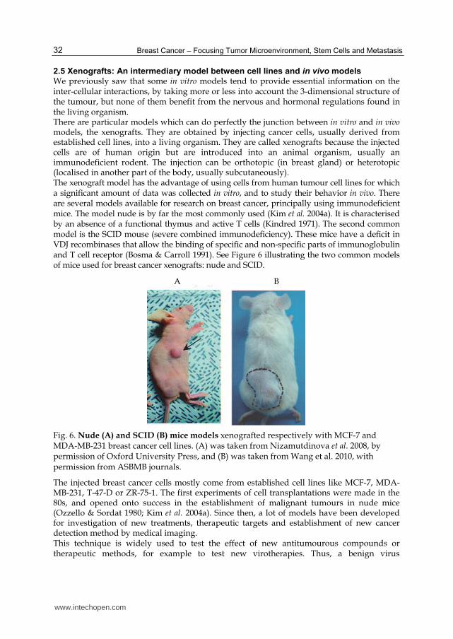
We previously saw that some in vitro models tend to provide essential information on the inter-cellular interactions, by taking more or less into account the 3-dimensional structure of the tumour, but none of them benefit from the nervous and hormonal regulations found in the living organism. There are particular models which can do perfectly the junction between in vitro and in vivo models, the xenografts. They are obtained by injecting cancer cells, usually derived from established cell lines, into a living organism. They are called xenografts because the injected cells are of human origin but are introduced into an animal organism, usually an immunodeficient rodent. The injection can be orthotopic (in breast gland) or heterotopic (localised in another part of the body, usually subcutaneously). The xenograft model has the advantage of using cells from human tumour cell lines for which a significant amount of data was collected in vitro, and to study their behavior in vivo. There are several models available for research on breast cancer, principally using immunodeficient mice. The model nude is by far the most commonly used (Kim et al. 2004a). It is characterised by an absence of a functional thymus and active T cells (Kindred 1971). The second common model is the SCID mouse (severe combined immunodeficiency). These mice have a deficit in VDJ recombinases that allow the binding of specific and non-specific parts of immunoglobulin and T cell receptor (Bosma & Carroll 1991). See Figure 6 illustrating the two common models of mice used for breast cancer xenografts: nude and SCID.
A B
Fig. 6. Nude (A) and SCID (B) mice models xenografted respectively with MCF-7 and MDA-MB-231 breast cancer cell lines. (A) was taken from Nizamutdinova et al. 2008, by permission of Oxford University Press, and (B) was taken from Wang et al. 2010, with permission from ASBMB journals.
The injected breast cancer cells mostly come from established cell lines like MCF-7, MDA-MB-231, T-47-D or ZR-75-1. The first experiments of cell transplantations were made in the 80s, and opened onto success in the establishment of malignant tumours in nude mice (Ozzello & Sordat 1980; Kim et al. 2004a). Since then, a lot of models have been developed for investigation of new treatments, therapeutic targets and establishment of new cancer detection method by medical imaging. This technique is widely used to test the effect of new antitumourous compounds or therapeutic methods, for example to test new virotherapies. Thus, a benign virus
www.intechopen.com

In Vitro Breast Cancer Models as Useful Tools in Therapeutics?
33
Coxsackievirus 21 (CVA21) was intravenously injected in SCID mice xenografted with MDA-MB-231 breast cancer cells. CVA21 virus targets the receptors ICAM-1 and DAF that are overexpressed in breast cancer cells. In this experiment a rapid lysis focused on cancer cells was observed in all mice, making this virus a good candidate for use in systemic therapy (Skelding et al. 2009). See Figure 7 illustrating the effect of the virus on xenografted mice, visualised by bioluminescent analysis.
Fig. 7. Observation of the oncolytic activity of CVA21 virus in SCID mouse xenografted with MDA-MB-231-luc. The breast cancer cells were xenografted into the mammary fat pad, mice were then treated with PBS or CVA21. Metastases were detected 3 weeks post-cell injection. The mice on the pictures are representative for bioluminescent observation at day 42 post treatment. From Skelding et al. 2009, with kind permission from Springer Science and Business Media B.V.
In the investigation of new treatments, the vitamin D3 receptors constitute good targets as they are present in over 80% of mammary tumours and they are negative growth regulator of both oestrogen-dependent and independent breast cancer cells in vitro. In a study published in 1998 it was shown that EB1089, a vitamin D3 analog, was able to highly reduce the growth of tumour in nude mice xenografted with MCF-7 cells (tumours were 4-fold smaller than those in untreated mice). This reduction was resulting from an enhancement of apoptosis and reducing proliferation of tumour epithelial cells, suggesting the great potential of vitamin D3 analogs such as EB1089 against human breast cancer (VanWeelden et al. 1998). This model can also be used to explore new potential targets for anticancer therapies. A good example is the targeting of receptor ER┚. In an experiment, standard T47D ER┙+ ER┚- and modified T47D ER┙+ ER┚+ (T47D stably transfected with a plasmid allowing the expression of the receptor ER┚), were xenografted in SCID mice. 17┚-estradiol was then injected into mice. The treatment triggered an acceleration of tumour growth in mice xenografted with the native T47D strain, and conversely a regression of tumours T47D ER┚+. These results emphasize the antagonistic role of ER receptors that appear to play an antitumourigenic role, and offered prospects for the development of ER-selective inhibitors. (Hartman et al. 2006). The targets cited above are non exhaustive. Many other therapeutic targets are tested with xenografts models, as it is the case of the VEGF pathway implicated in tumour angiogenesis (Le et al. 2008), or of cell cycle regulating proteins such as CDK kinases (Fry et al. 2004). The use of established cell lines for producing xenografts raises several questions about their relevance. The murine model presents considerable differences with the human body, concerning the biochemical and physiological regulation. Moreover, the stroma that will grow surround the tumour will be of murine origin and it will result in a chimeric tumour
www.intechopen.com
Breast Cancer – Focusing Tumor Microenvironment, Stem Cells and Metastasis
34
which biology may significantly differ from human one (Kim et al. 2004a). Furthermore, in humans, the immune system plays an important role in the fight against tumour, whereas in xenografts models the immune system is totally absent. The xenograft model has some limitations but is the most accomplished of all models because it takes into account the complexity of the organism. Besides the xenografts, there are also murine models which can develop tumours spontaneously or under the influence of inducing compounds (Russo & Russo 1996). Although the achievement of these models is easy, their use is largely debated because of their relevance to the clinical situation. Indeed, murine breast cancers are most often caused by viral infections and are not hormone dependent, whereas a considerable proportion of human cancers are oestrogen dependent. To date there is no evidence suggesting a viral induction of breast cancer in humans. The biology of spontaneous rodent tumours differs from the human ones. The size, the oncogenic targets or the degree of maturation and differentiation of cells differ between the two species, making them hardly comparable.
3. Conclusion
In this chapter, we described the main models used in breast cancer research in order to obtain results of high scientific quality. In summary, we can say that BCCL models allow repeatable experiments with simple material and methods. They are inevitable models for basic studies and mechanistic explorations, but their use is still controversial owing to their approximate representativeness of breast tumours in human and to the existence of misidentified cell lines. Cultures of cancerous tissues preserve the tumour architecture and the cell diversity of a tumour but this model suffers of limited reproducibility and cannot be easily maintained for a long time. Co-culture systems offer an alternative with reproducible long term culture systems, and offer the possibility to study the relations between different types of cells in tumour, but this model suffers from the same controversies as BCCL as it mainly relies on their use. 3-dimensional systems allow the mimicking of the tumour architecture and microenvironment, but very few cell lines are able to form spheroids under specific conditions. Considering the advantages and drawbacks of these models, the xenografts appear to be good alternative models as they enable to take into account the tumour structure, its microenvironment, the role of the metabolism and they preserve the cell diversity of the tumour. But as other models, they also have drawbacks principally due to the metabolic and physiological differences existing between human and rodents, and to the fact that the role of the immune system against tumour is not taken into account with the immunodeficient rodent models used for xenografts. Application of the 3Rs principle leaded to the development of all these models, but we showed that none of them is sufficient by itself and able to perfectly mimic breast cancer in human. However it clearly appears that all these models are essential to accumulate data and information to fight breast cancer.
4. Acknowledgements
The ‘‘Ligue contre le Cancer” (54, 55, 57 and 88 Departmental committees) is acknowledged for its financial support. E.B is recipient of an AFR grant of the National Research Fund, Luxembourg.
www.intechopen.com

In Vitro Breast Cancer Models as Useful Tools in Therapeutics?
35
5. References
Amadori, D., Bertoni, L., Flamigni, A., Savini, S., De Giovanni, C., Casanova, S., De Paola, F., Amadori, A., Giulotto, E. and Zoli, W. (1993). Establishment and characterization of a new cell line from primary human breast carcinoma. Breast Cancer Res Treat, Vol. 28, No.3: 251-260.
Androutsopoulos, V. P., Li, N. and Arroo, R. R. (2009). The methoxylated flavones eupatorin and cirsiliol induce CYP1 enzyme expression in MCF7 cells. J Nat Prod, Vol. 72, No.8: 1390-1394.
Bauer, M., Su, G., Beebe, D. J. and Friedl, A. (2010). 3D microchannel co-culture: method and biological validation. Integr Biol (Camb), Vol. 2, No.7-8: 371-378.
Bosma, M. J. and Carroll, A. M. (1991). The SCID mouse mutant: definition, characterization, and potential uses. Annu Rev Immunol, Vol. 9: 323-350.
Bray, J., Sludden, J., Griffin, M. J., Cole, M., Verrill, M., Jamieson, D. and Boddy, A. V. (2010). Influence of pharmacogenetics on response and toxicity in breast cancer patients treated with doxorubicin and cyclophosphamide. Br J Cancer, Vol. 102, No.6: 1003-1009.
Burdall, S. E., Hanby, A. M., Lansdown, M. R. and Speirs, V. (2003). Breast cancer cell lines: friend or foe? Breast Cancer Research, Vol. 5, No.2: 89-95.
Cailleau, R., Young, R., Olive, M. and Reeves, W. J., Jr. (1974). Breast tumor cell lines from pleural effusions. J Natl Cancer Inst, Vol. 53, No.3: 661-674.
Cappelletti, V., Ruedl, C., Granata, G., Coradini, D., Del Bino, G. and Di Fronzo, G. (1991). Interaction between hormone-dependent and hormone-independent human breast cancer cells. Eur J Cancer, Vol. 27, No.9: 1154-1157.
Chen, Y., Tang, Y., Chen, S. and Nie, D. (2009). Regulation of drug resistance by human pregnane X receptor in breast cancer. Cancer Biol Ther, Vol. 8, No.13: 1265-1272.
Conde, S. J., Luvizotto, R. A., Sibio, M. T., Katayama, M. L., Brentani, M. M. and Nogueira, C. R. (2008). Tamoxifen inhibits transforming growth factor-alpha gene expression in human breast carcinoma samples treated with triiodothyronine. J Endocrinol Invest, Vol. 31, No.12: 1047-1051.
Di, Y. M., Chow, V. D., Yang, L. P. and Zhou, S. F. (2009). Structure, function, regulation and polymorphism of human cytochrome P450 2A6. Curr Drug Metab, Vol. 10, No.7: 754-780.
Dong-Le Bourhis, X., Berthois, Y., Millot, G., Degeorges, A., Sylvi, M., Martin, P. M. and Calvo, F. (1997). Effect of stromal and epithelial cells derived from normal and tumorous breast tissue on the proliferation of human breast cancer cell lines in co-culture. Int J Cancer, Vol. 71, No.1: 42-48.
Fogh, J., Wright, W. C. and Loveless, J. D. (1977). Absence of HeLa cell contamination in 169 cell lines derived from human tumors. J Natl Cancer Inst, Vol. 58, No.2: 209-214.
Fritsch, A., Höckel, M., Kiessling, T., Nnetu, K. D., Wetzel, F., Zink, M. and Käs, J. A. (2010). Are biomechanical changes necessary for tumour progression? Nature Physics, Vol. 6: 730–732.
Fry, D. W., Harvey, P. J., Keller, P. R., Elliott, W. L., Meade, M., Trachet, E., Albassam, M., Zheng, X., Leopold, W. R., Pryer, N. K. and Toogood, P. L. (2004). Specific inhibition of cyclin-dependent kinase 4/6 by PD 0332991 and associated antitumor activity in human tumor xenografts. Mol Cancer Ther, Vol. 3, No.11: 1427-1438.
Gazdar, A. F., Kurvari, V., Virmani, A., Gollahon, L., Sakaguchi, M., Westerfield, M., Kodagoda, D., Stasny, V., Cunningham, H. T., Wistuba, II, Tomlinson, G., Tonk, V., Ashfaq, R., Leitch, A. M., Minna, J. D. and Shay, J. W. (1998). Characterization of paired tumor and non-tumor cell lines established from patients with breast cancer. Int J Cancer, Vol. 78, No.6: 766-774.
www.intechopen.com

Breast Cancer – Focusing Tumor Microenvironment, Stem Cells and Metastasis
36
Giraud, B., Hebert, G., Deroussent, A., Veal, G. J., Vassal, G. and Paci, A. (2010). Oxazaphosphorines: new therapeutic strategies for an old class of drugs. Expert Opin Drug Metab Toxicol, Vol. 6, No.8: 919-938.
Gollahon, L. S. and Shay, J. W. (1996). Immortalization of human mammary epithelial cells transfected with mutant p53 (273his). Oncogene, Vol. 12, No.4: 715-725.
Hagemann, T., Robinson, S. C., Schulz, M., Trumper, L., Balkwill, F. R. and Binder, C. (2004). Enhanced invasiveness of breast cancer cell lines upon co-cultivation with macrophages is due to TNF-alpha dependent up-regulation of matrix metalloproteases. Carcinogenesis, Vol. 25, No.8: 1543-1549.
Hartman, J., Lindberg, K., Morani, A., Inzunza, J., Strom, A. and Gustafsson, J. A. (2006). Estrogen receptor beta inhibits angiogenesis and growth of T47D breast cancer xenografts. Cancer Res, Vol. 66, No.23: 11207-11213.
Huang, Z., Fasco, M. J. and Kaminsky, L. S. (1997). Alternative splicing of CYP2D mRNA in human breast tissue. Arch Biochem Biophys, Vol. 343, No.1: 101-108.
Keydar, I., Chen, L., Karby, S., Weiss, F. R., Delarea, J., Radu, M., Chaitcik, S. and Brenner, H. J. (1979). Establishment and characterization of a cell line of human breast carcinoma origin. Eur J Cancer, Vol. 15, No.5: 659-670.
Kim, J. B., O'Hare, M. J. and Stein, R. (2004a). Models of breast cancer: is merging human and animal models the future? Breast Cancer Res, Vol. 6, No.1: 22-30.
Kim, J. B., Stein, R. and O'Hare, M. J. (2004b). Three-dimensional in vitro tissue culture models of breast cancer-- a review. Breast Cancer Res Treat, Vol. 85, No.3: 281-291.
Kindred, B. (1971). Antibody response in genetically thymus-less nude mice injected with normal thymus cells. J Immunol, Vol. 107, No.5: 1291-1295.
Kivisto, K. T., Kroemer, H. K. and Eichelbaum, M. (1995). The role of human cytochrome P450 enzymes in the metabolism of anticancer agents: implications for drug interactions. Br J Clin Pharmacol, Vol. 40, No.6: 523-530.
Lacroix, M. (2008). Persistent use of "false" cell lines. Int J Cancer, Vol. 122, No.1: 1-4. Lacroix, M. and Leclercq, G. (2004). Relevance of breast cancer cell lines as models for breast
tumours: an update. Breast Cancer Res Treat, Vol. 83, No.3: 249-289. Le, X. F., Mao, W., Lu, C., Thornton, A., Heymach, J. V., Sood, A. K. and Bast, R. C., Jr.
(2008). Specific blockade of VEGF and HER2 pathways results in greater growth inhibition of breast cancer xenografts that overexpress HER2. Cell Cycle, Vol. 7, No.23: 3747-3758.
Liscovitch, M. and Ravid, D. (2007). A case study in misidentification of cancer cell lines: MCF-7/AdrR cells (re-designated NCI/ADR-RES) are derived from OVCAR-8 human ovarian carcinoma cells. Cancer Lett, Vol. 245, No.1-2: 350-352.
Lo, R., Burgoon, L., Macpherson, L., Ahmed, S. and Matthews, J. (2010). Estrogen receptor-dependent regulation of CYP2B6 in human breast cancer cells. Biochim Biophys Acta, Vol. 1799, No.5-6: 469-479.
Macpherson, L. and Matthews, J. (2010). Inhibition of aryl hydrocarbon receptor-dependent transcription by resveratrol or kaempferol is independent of estrogen receptor alpha expression in human breast cancer cells. Cancer Lett, Vol. 299, No.2: 119-129.
Matoska, J. and Stricker, F. (1967). Following human tumours in primary organ culture. Neoplasma, Vol. 14, No.5: 507-519.
Mestres, P., Morguet, A., Schmidt, W., Kob, A. and Thedinga, E. (2006). A new method to assess drug sensitivity on breast tumor acute slices preparation. Ann N Y Acad Sci, Vol. 1091: 460-469.
Milani, C., Welsh, J., Katayama, M. L., Lyra, E. C., Maciel, M. S., Brentani, M. M. and Folgueira, M. A. (2010). Human breast tumor slices: a model for identification of
www.intechopen.com
In Vitro Breast Cancer Models as Useful Tools in Therapeutics?
37
vitamin D regulated genes in the tumor microenvironment. J Steroid Biochem Mol Biol, Vol. 121, No.1-2: 151-155.
Mitra, R., Guo, Z., Milani, M., Mesaros, C., Rodriguez, M., Nguyen, J., Luo, X., Clarke, D., Lamba, J., Schuetz, E., Donner, D. B., Puli, N., Falck, J. R., Capdevila, J., Gupta, K., Blair, I. A. and Potter, D. A. (2011). CYP3A4 mediates growth of ER+ breast cancer cells, in part, by nuclear translocation of phospho-Stat3 through biosynthesis of ({+/-})-14,15-EET. J Biol Chem, Vol.:
Mo, S. L., Liu, Y. H., Duan, W., Wei, M. Q., Kanwar, J. R. and Zhou, S. F. (2009). Substrate specificity, regulation, and polymorphism of human cytochrome P450 2B6. Curr Drug Metab, Vol. 10, No.7: 730-753.
Nagaoka, R., Iwasaki, T., Rokutanda, N., Takeshita, A., Koibuchi, Y., Horiguchi, J., Shimokawa, N., Iino, Y., Morishita, Y. and Koibuchi, N. (2006). Tamoxifen activates CYP3A4 and MDR1 genes through steroid and xenobiotic receptor in breast cancer cells. Endocrine, Vol. 30, No.3: 261-268.
Nelson-Rees, W. A. and Flandermeyer, R. R. (1977). Inter- and intraspecies contamination of human breast tumor cell lines HBC and BrCa5 and other cell cultures. Science, Vol. 195, No.4284: 1343-1344.
Nissen, E., Tanneberger, S., Weiss, H. and Bender, E. (1983). [In vitro cultivation of vital tissue slices: a new variation of organ culture technics]. Biomed Biochim Acta, Vol. 42, No.7-8: 907-916.
Nizamutdinova, I. T., Lee, G. W., Lee, J. S., Cho, M. K., Son, K. H., Jeon, S. J., Kang, S. S., Kim, Y. S., Lee, J. H., Seo, H. G., Chang, K. C. and Kim, H. J. (2008). Tanshinone I suppresses growth and invasion of human breast cancer cells, MDA-MB-231, through regulation of adhesion molecules. Carcinogenesis, Vol. 29, No.10: 1885-1892.
Ozzello, L. and Sordat, M. (1980). Behavior of tumors produced by transplantation of human mammary cell lines in athymic nude mice. Eur J Cancer, Vol. 16, No.4: 553-559.
Parajuli, N. and Doppler, W. (2009). Precision-cut slice cultures of tumors from MMTV-neu mice for the study of the ex vivo response to cytokines and cytotoxic drugs. In Vitro Cell Dev Biol Anim, Vol. 45, No.8: 442-450.
Pauley, R. J., Paine, T. J., Herbert, D. and Soule, D. N. (1991). Immortal Human Mammary Epithelial Cell Sublines. United States Patent, Patent n°. 5206165,
Pennington, K., Chu, Q. D., Curiel, D. T., Li, B. D. and Mathis, J. M. (2010). The utility of a tissue slice model system to determine breast cancer infectivity by oncolytic adenoviruses. J Surg Res, Vol. 163, No.2: 270-275.
Pinilla, S., Alt, E., Abdul Khalek, F. J., Jotzu, C., Muehlberg, F., Beckmann, C. and Song, Y. H. (2009). Tissue resident stem cells produce CCL5 under the influence of cancer cells and thereby promote breast cancer cell invasion. Cancer Lett, Vol. 284, No.1: 80-85.
Rae, J. M., Creighton, C. J., Meck, J. M., Haddad, B. R. and Johnson, M. D. (2007). MDA-MB-435 cells are derived from M14 melanoma cells--a loss for breast cancer, but a boon for melanoma research. Breast Cancer Res Treat, Vol. 104, No.1: 13-19.
Rajendran, S., O'Hanlon, D., Morrissey, D., O'Donovan, T., O'Sullivan, G. C. and Tangney, M. (2011). Preclinical evaluation of gene delivery methods for the treatment of loco-regional disease in breast cancer. Exp Biol Med (Maywood), Vol. 236, No.4: 423-434.
Rooseboom, M., Commandeur, J. N. and Vermeulen, N. P. (2004). Enzyme-catalyzed activation of anticancer prodrugs. Pharmacol Rev, Vol. 56, No.1: 53-102.
Ross, D. T. and Perou, C. M. (2001). A comparison of gene expression signatures from breast tumors and breast tissue derived cell lines. Dis Markers, Vol. 17, No.2: 99-109.
Russell, W. M. S. and Bursch, R. L. (1959). The Principles of Humane Experimental Technique. London, Methuen, 978-0900767784.
www.intechopen.com
Breast Cancer – Focusing Tumor Microenvironment, Stem Cells and Metastasis
38
Russo, I. H. and Russo, J. (1996). Mammary gland neoplasia in long-term rodent studies. Environ Health Perspect, Vol. 104, No.9: 938-967.
Shen, C., Gu, M., Liang, D., Miao, L., Hu, L., Zheng, C. and Chen, J. (2009). Establishment and characterization of three new human breast cancer cell lines derived from Chinese breast cancer tissues. Cancer Cell Int, Vol. 9: 2.
Skelding, K. A., Barry, R. D. and Shafren, D. R. (2009). Systemic targeting of metastatic human breast tumor xenografts by Coxsackievirus A21. Breast Cancer Res Treat, Vol. 113, No.1: 21-30.
Sonnenberg, M., van der Kuip, H., Haubeis, S., Fritz, P., Schroth, W., Friedel, G., Simon, W., Murdter, T. E. and Aulitzky, W. E. (2008). Highly variable response to cytotoxic chemotherapy in carcinoma-associated fibroblasts (CAFs) from lung and breast. BMC Cancer, Vol. 8: 364.
Soule, H. D., Maloney, T. M., Wolman, S. R., Peterson, W. D., Jr., Brenz, R., McGrath, C. M., Russo, J., Pauley, R. J., Jones, R. F. and Brooks, S. C. (1990). Isolation and characterization of a spontaneously immortalized human breast epithelial cell line, MCF-10. Cancer Res, Vol. 50, No.18: 6075-6086.
Soule, H. D., Vazguez, J., Long, A., Albert, S. and Brennan, M. (1973). A human cell line from a pleural effusion derived from a breast carcinoma. J Natl Cancer Inst, Vol. 51, No.5: 1409-1416.
Spink, B. C., Cole, R. W., Katz, B. H., Gierthy, J. F., Bradley, L. M. and Spink, D. C. (2006). Inhibition of MCF-7 breast cancer cell proliferation by MCF-10A breast epithelial cells in coculture. Cell Biol Int, Vol. 30, No.3: 227-238.
Stampfer, M. R. (1989). Continuous human cell lines and method of making same. United States Patent, Patent n°. 4808532,
Stiborova, M., Rupertova, M. and Frei, E. (2011). Cytochrome P450- and peroxidase-mediated oxidation of anticancer alkaloid ellipticine dictates its anti-tumor efficiency. Biochim Biophys Acta, Vol. 1814, No.1: 175-185.
Van der Haegen, B. A. and Shay, J. W. (1993). Immortalization of human mammary epithelial cells by SV40 large T-antigen involves a two step mechanism. In Vitro Cell Dev Biol, Vol. 29A, No.3 Pt 1: 180-182.
Van Der Kuip, H., Murdter, T. E., Sonnenberg, M., McClellan, M., Gutzeit, S., Gerteis, A., Simon, W., Fritz, P. and Aulitzky, W. E. (2006). Short term culture of breast cancer tissues to study the activity of the anticancer drug taxol in an intact tumor environment. BMC Cancer, Vol. 6: 86.
VanWeelden, K., Flanagan, L., Binderup, L., Tenniswood, M. and Welsh, J. (1998). Apoptotic regression of MCF-7 xenografts in nude mice treated with the vitamin D3 analog, EB1089. Endocrinology, Vol. 139, No.4: 2102-2110.
Wang, H. and Tompkins, L. M. (2008). CYP2B6: new insights into a historically overlooked cytochrome P450 isozyme. Curr Drug Metab, Vol. 9, No.7: 598-610.
Wang, Z., Bryan, J., Franz, C., Havlioglu, N. and Sandell, L. J. (2010). Type IIB procollagen NH(2)-propeptide induces death of tumor cells via interaction with integrins alpha(V)beta(3) and alpha(V)beta(5). J Biol Chem, Vol. 285, No.27: 20806-20817.
Yuhas, J. M., Tarleton, A. E. and Molzen, K. B. (1978). Multicellular tumor spheroid formation by breast cancer cells isolated from different sites. Cancer Res, Vol. 38, No.8: 2486-2491.
Zhang, J., Tian, Q., Yung Chan, S., Chuen Li, S., Zhou, S., Duan, W. and Zhu, Y. Z. (2005). Metabolism and transport of oxazaphosphorines and the clinical implications. Drug Metab Rev, Vol. 37, No.4: 611-703.
www.intechopen.com
Breast Cancer - Focusing Tumor Microenvironment, Stem cells andMetastasisEdited by Prof. Mehmet Gunduz
ISBN 978-953-307-766-6Hard cover, 584 pagesPublisher InTechPublished online 14, December, 2011Published in print edition December, 2011
InTech EuropeUniversity Campus STeP Ri Slavka Krautzeka 83/A 51000 Rijeka, Croatia Phone: +385 (51) 770 447 Fax: +385 (51) 686 166www.intechopen.com
InTech ChinaUnit 405, Office Block, Hotel Equatorial Shanghai No.65, Yan An Road (West), Shanghai, 200040, China
Phone: +86-21-62489820 Fax: +86-21-62489821
Cancer is the leading cause of death in most countries and its consequences result in huge economic, socialand psychological burden. Breast cancer is the most frequently diagnosed cancer type and the leading causeof cancer death among females. In this book, we discussed characteristics of breast cancer cell, role ofmicroenvironment, stem cells and metastasis for this deadly cancer. We hope that this book will contribute tothe development of novel diagnostic as well as therapeutic approaches.
How to referenceIn order to correctly reference this scholarly work, feel free to copy and paste the following:
Emilie Bana and Denyse Bagrel (2011). In Vitro Breast Cancer Models as Useful Tools in Therapeutics?,Breast Cancer - Focusing Tumor Microenvironment, Stem cells and Metastasis, Prof. Mehmet Gunduz (Ed.),ISBN: 978-953-307-766-6, InTech, Available from: http://www.intechopen.com/books/breast-cancer-focusing-tumor-microenvironment-stem-cells-and-metastasis/in-vitro-breast-cancer-models-as-useful-tools-in-therapeutics-

© 2011 The Author(s). Licensee IntechOpen. This is an open access articledistributed under the terms of the Creative Commons Attribution 3.0License, which permits unrestricted use, distribution, and reproduction inany medium, provided the original work is properly cited.