hahlbohm.spirometry in the primary care setting · -provides clear document ation of lung function...
TRANSCRIPT

2/5/2019
1
SPIROMETRY
Dewey Hahlbohm, PA-C, AE-C
• To understand the uses and importance of spirometry testing
• To perform spirometry testing including reversibility testing
• To identify normal and abnormal patterns and classify asthma severity
• To review the definitions of lung volumes and capacities
Objectives
• To understand the value of spirometry for asthma diagnosis and management in the primary care setting
• To feel comfortable in the interpretation of PFTs and be able to use them as an aid in the diagnosis of obstructive and restrictive pulmonary disease
• To have a basic understanding of how to properly administer a PFT
Objectives

2/5/2019
2
• Spirometry is a poor test of little benefit• Equipment is expensive, works poorly• Spirometry is hard to do right• Numbers are difficult to interpret
Myths
Objective Testing
• Spirometry is a powerful diagnostic and assessment tool
- Provides clear, objective documentation of lung function
- Reliable tool to obtain pulmonology vital signs
- easy to use and accurate- carried out in primary care setting
Spirometry in primary care…
• Improves clinical outcomes through better diagnosis and staging
• Supports motivation and lifestyle• Promotes more appropriate referrals to
specialists• Generates revenue

2/5/2019
3
Spirometry in the Managementof Asthma and COPD
• Spirometry permits an objective measurement of the degree of airway obstruction (impairment and risk)– Patients’ perceptions of obstruction are notoriously inaccurate– Significant obstruction can be present even when the chest is clear
on physical examination– Clinical symptoms alone will underestimate severity up to 30% of
the time in primary care– PEF (peak expiratory flow) testing alone is highly variable, is not a
very sensitive measure of obstruction, and is no longer recommended for diagnosis
– 1. Stout, JW, et al., Archives of Pediatrics and Adolescent Medicine Medicine 2006; 160:844-850– 2. Cowen, M. et al., Journal of Asthma 2007; 44:711-715– 3. Fuhlbrigge AL, et al., J Allergy Clin Immunol 2001; 107:61-67
Barriers to PerformingSpirometry in PrimaryCare
– Lack of training for support staff and providers
– Lack of a spirometer (or its use)– Lack of time (problems with work flow
and lack of planned visits)– Lack of interest or enthusiasm in
incorporating a new device and procedure– Lack of incentives
Desktop Electronic Spirometers
• Portable• Easy to calibrate• Immediate feedback• Billable

2/5/2019
4
When to Utilize Spirometry
• Symptoms Signs:- chronic cough - hyperinflation- frequent colds - expiratory
slowing- dyspnea - cyanosis- wheezing - chest deformity- orthopnea - chest pain
Indications for Spirometry
• Used to determine:– Presence and severity of disease– Response to treatment– Etiology of disease– Reversibility– Surgery risk evaluation– Disability– Progression and variability of disease
When should spirometry be performed and how often?
• 1-6 month intervals:– At time of initial assessment– After treatment is initiated and symptoms and
PEF have stabilized– During periods of progressive decline or extended
loss of asthma control
At least every 1-2 years

2/5/2019
5
What is Spirometry?
• Spirometry is a method of assessing lung function by measuring the volume of air the patient can expel from the lungs after a maximal inspiration.
Benefits of Spirometry
• Spirometry results can help confirm a diagnosis of asthma
• Spirometry shows severity of airways obstruction- peak flow shows only a moment in time- spirometry looks at the breathing process over time
• Spirometry and the bronchodilatator test-allows patient to see benefit of medication- allows physician to better assess patient response to medication and adjust treatment regimen as appropriate
Spirometry
• Quantifies patients ability to exhale• Measures basic lung function –
spirometry values- Total exhaled volume: forced vital
capacity (FVC)- Forced expiratory volume exhaled in
first second (FEV1)- Ratio of volume exhaled in first
second to total (FEV1/FVC)

2/5/2019
6
Interpreting Results
• Spirometry allows comparison of patient’s lung function to reference values
• Helps to define disease class: obstructive, restrictive or mixed type
Inter-individual variability
• Age• Sex• Race
– Race correction factor• NHANES for those 8 and older• Wang et al for children < 8 years old
• Height (measure with shoes off)• Room temperature
Predicted normal lung values
• Based on large population surveys• Predicted values are the mean values
obtained from the survey• No surveys have been done in elderly
populations
2/5/2019
7
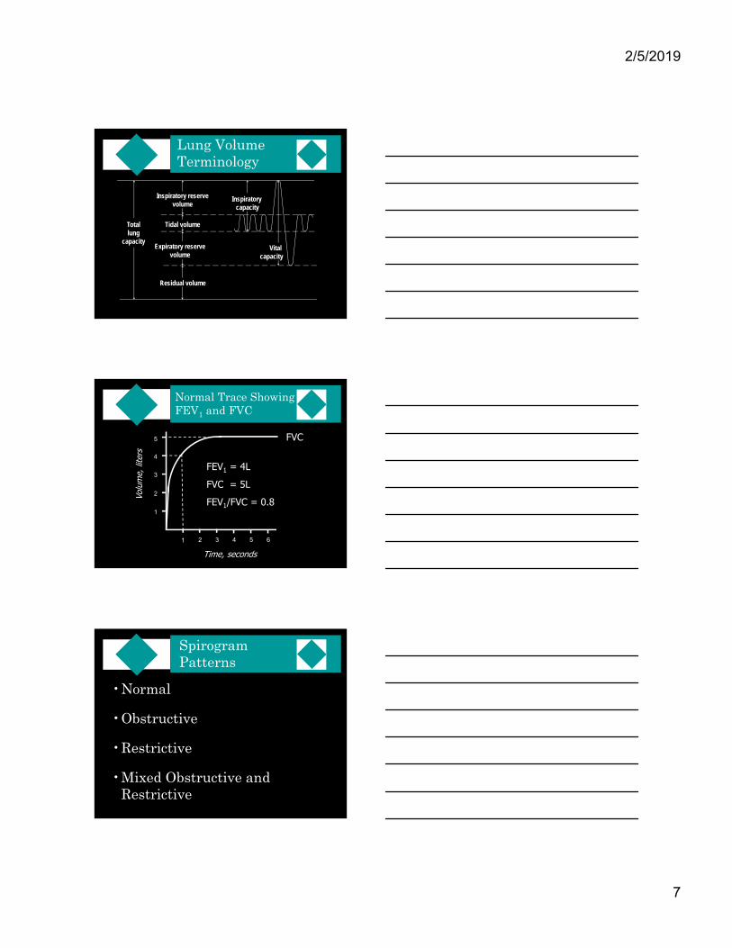
Totallung
capacity
Tidal volume
Inspiratory reservevolume
Expiratory reservevolume
Residual volume
Inspiratory capacity
Vital capacity
Lung Volume Terminology
Normal Trace Showing FEV1 and FVC
1 2 3 4 5 6
1
2
3
4
Volu
me,
lite
rs
Time, seconds
FVC5
1
FEV1 = 4LFVC = 5LFEV1/FVC = 0.8
Spirogram Patterns
•Normal
•Obstructive
•Restrictive
•Mixed Obstructive and Restrictive

2/5/2019
8
Volu
me,
lite
rs
Time, seconds
54
3
2
1
1 2 3 4 5 6
FEV1 = 1.8LFVC = 3.2LFEV1/FVC = 0.56
Normal
Obstructive
Spirometry: Obstructive Disease
CRJ1
Diseases Associated w/ Airflow Obstruction
• Obstruction: Characterized by a limitation of expiratory airflow so that airways cannot empty as rapidly compared to normal (such as through narrowed airways from bronchospasm, inflammation, etc.)
• COPD
• Asthma
• Bronchiectasis
• Cystic fibrosis
• Post-tuberculosis
• Lung cancer (greater risk in COPD)
• Obliterative bronchiolitis
Diseases Associated w/ Airflow Restriction
• Restriction: Characterized by reduced lung volumes/decreased lung compliance
• Interstitial fibrosis
• Scoliosis
• Obesity
• Lung resection
• Neuromuscular diseases
• Cystic fibrosis

Slide 22
CRJ1 Sue i have inserted a bracket and shifted the obstructive label. The FVC in this slide is about 3.4 by eyeball - shoudl be moved down to 3.2 or the numbers should be changed Christine Jenkins, 4/14/2008

2/5/2019
9
Bronchodilator Reversibility Testing in Asthma
Results
•An increase in FEV1 that is both greater than 200 ml and 12% above the pre-bronchodilator FEV1 (baseline value) is considered significant
•It is usually helpful to report the absolute change (in ml) as well as the % change from baseline to set the improvement in a clinical context
CRJ2
Flow Volume Loop
• Standard on most desk-top spirometers• Adds more information than volume time
curve• Less understood but not too difficult to
interpret• Better at demonstrating mild airflow
obstruction
Flow Volume LoopExpiration is the area above the “waterline” Indicates lung disease
Expiratory flow rateL/sec
Volume (L)
FVC
Maximum expiratory flow (PEF)
Inspiratory flow rate
L/sec
RVTLC

Slide 25
CRJ2 need to delete Figure reference. Christine Jenkins, 4/14/2008

2/5/2019
10
Flow Volume Loop Inspiration is the area below the “waterline” Indicates extrathoracic area
Expiratory flow rateL/sec
Volume (L)
FVC
Maximum expiratory flow (PEF)
Inspiratory flow rate
L/sec
RVTLC
Flow Volume Curve Patterns
Obstructive Severe obstructive Restrictive
Volume (L)
Expi
rato
ry f
low
rate
Expi
rato
ry f
low
rate
Expi
rato
ry f
low
rate
Volume (L) Volume (L)
Steeple pattern, reduced peak flow,
rapid fall off
Normal shape, normal peak flow, reduced volume
Reduced peak flow, scooped out mid-
curve
Volu
me,
lite
rs
Time, seconds
54
3
2
1
1 2 3 4 5 6
FEV1 = 1.8LFVC = 3.2LFEV1/FVC = 0.56
Normal
Obstructive
Spirometry: Volume Time Curve
CRJ4

Slide 30
CRJ4 Sue i have inserted a bracket and shifted the obstructive label. The FVC in this slide is about 3.4 by eyeball - shoudl be moved down to 3.2 or the numbers should be changed Christine Jenkins, 4/14/2008

2/5/2019
11
Obstructive Restrictive Mixed
Time Time Time
Volu
me
Volu
me
Volu
me
Spirometry: Abnormal Patterns
Slow rise, reduced volume expired;
prolonged time to full expiration
Fast rise to plateau at reduced
maximum volume
Slow rise to reduced maximum volume; measure static lung
volumes and full PFT’s to confirm
Ensuring Accuracy: Best Effort
• Best effort- inhale as deeply as possible- exhale as fast and as long as possible- exhale for at least six seconds
• Reproducibility- two “best efforts” out of a minimum of three exhalations, no more than 6-8 attempts
- Largest and second largest FVC and FEV1 within 0.15 L of each other
No spirometry effort should be rejected due to poor reproducibility, just document
UnacceptableEfforts
• Lack of full inspiration• Lack of maximum effort• Effort too short• Presence of cough in first second• Obstructed mouthpiece• Unsatisfactory start, hesitation• Excessive variability between efforts

2/5/2019
12
Preparing the patient..
Patients are asked:• Avoid smoking within 2 hrs of test• Avoid drinking alcohol within 4 hours• Avoid vigorous exercise within 30 minutes• Avoid restrictive clothing• Avoid eating substantial meal within 2 hours• Avoid SABA within 4-6 hours • Avoid LABA within 12 hours
Preparing and coaching
• Patient should sit (feet on floor) or stand with chair behind patient in case of dizziness (document if done standing, repeat with future testing)
• Loosen any restrictive clothing• Reassure patient; help them feel relaxed• Explain in simple terms what the test measures• Explain the technique in simple terms and then
demonstrate how it is done• Make sure the mouthpiece is placed between the
teeth and that the tongue and teeth do not occlude the mouthpiece.
Special considerations in pediatric patients
• Be creative• Use incentives• Even with the best of environments and
coaching, a child may not be able to perform spirometry
• Patients need a calm, relaxed environment and good coaching. Patience is key
• Ability to perform spirometry dependent on developmental age of child, personality, and interest

2/5/2019
13
Coach the patient!!!!
• BLAST IT OUT!!!
• BLOW!! BLOW!! BLOW!!
• SQUEEZE! SQUEEZE!! SQUEEZE!!!!
• PUSH! PUSH!! PUSH!!! PUSH!!!!
• KEEP GOING! KEEP GOING!•
Troubleshooting
Examples - Unacceptable Traces
Unacceptable Trace:Poor Effort
Volu
me,
lite
rs
Time, seconds
May be accompanied by a slow start
Inadequate sustaining of effort
Variable expiratory effortNormal

2/5/2019
14
Volu
me,
lite
rs
Time, seconds
Unacceptable Trace:Stop Early
Normal
Volu
me,
lite
rs
Time, seconds
Unacceptable Trace:Slow Start
Normal
Volu
me,
lite
rs
Time, seconds
Unacceptable Trace Coughing 1ST Second
Normal

2/5/2019
15
Volu
me,
lite
rs
Time, seconds
Unacceptable TraceExtra Breath
Normal
Spirometry vs. Peak flow meter
• Peak Flow Meter is used for monitoring only
• Measures only large airway function• No graphic display or printout• No regular calibration
Spirometryreimbursement
• Cost of spirometry: • 94010 Spirometry test – FVC: $70.00• 94060 Pre-Post Bronchodilator
Spirometry test: $145.00

2/5/2019
16
CPT Codes
Summary
Spirometry is a powerful diagnostic and assessment tool-provides clear documentation of lung function
• Spirometry is easy to use and accurate- can be carried out in the primary care setting- offers test results to include in patient’s chart
• Spirometry measures lung airflow- helps detect obstructive and restrictive lung
disease- objectively measures and illustrates the
severity of lung disease
Questions?
• MT Asthma Control Program• Jessie Fernandes, Program Manager
– (406) 444-9155, [email protected]
• Dewey Hahlbohm, Medical Consultant- (406) 442-6934, [email protected]