in reactivity to stress individual differences distribute or post,...large individual differences in...
TRANSCRIPT

203
10Individual Differences in Reactivity to Stress
We have all seen how differently people respond to stress. Not just in their initial reactions but also in their ability to cope, and even
in their physiological responses. We might say that the subject of individual
Chapter Objectives
1. Develop an appreciation for stress reactivity as an individual difference variable that can have normative levels, exaggerated levels, or dimin-ished levels.
2. Consider the possibility that departures from normal in either the exaggerated or diminished direction may signal systems dysregulation.
3. Individual differences in stress reactivity may derive from responsivity at three levels in the system: frontal-limbic interactions, hypothalamic and brainstem processes, and peripheral organs and tissues.
4. Exaggerated reactivity provides evidence that the system is operating out of the normal range and therefore is altered by underlying disease or disease risk.
Copyright ©2016 by SAGE Publications, Inc. This work may not be reproduced or distributed in any form or by any means without express written permission of the publisher.
Do not
copy
, pos
t, or d
istrib
ute

204——Stress and Health
differences is what makes the study of stress so interesting. Stress reactivity has been described as “an individual’s propensity to experience cardiovas-cular reactions of greater or lesser magnitude, in relation to those of other persons, when encountering behavioral stimuli experienced as engaging, challenging, or aversive” (Manuck, 1994). Other studies show that stress reactivity is a stable characteristic of the individual, such that a tendency to have large or small stress responses will characterize the system over a long period of time, perhaps for a lifetime. In this chapter, we will discuss individual differences in stress reactivity with two goals: First, we will consider a theoretical distribution of stress reactivity falling on a continu-ous distribution, with a normative range of responses toward the center of the distribution and high and low extremes at the tails. Second, we will refer to material in Chapters 5, 6, and 7 to consider ways that persons may come to differ in how they react to stress. Using the model in Chapter 3 showing that physiological functioning has a layered hierarchy of controls, this chapter describes three major sources of individual differences in reac-tivity to psychological stressors: At Level I, persons may differ because of their cognitive and emotional characteristics, reflecting the operation of frontal-limbic processes on patterns of response. At Level II, persons may have exaggerated autonomic and endocrine responses because of differen-tial activation of hypothalamic and brainstem outputs. At Level III, per-sons may have altered peripheral responses that reflect changes in the tissues themselves. Finally, in Chapter 11, we will consider implications of these sources of individual differences for health and disease.
A Proposed Classification of Individual Differences in Reactivity
If we believe that individual differences in stress reactivity have implications for health, a first question is how to separate persons with “good” or “healthy” reactivity profiles from “bad” or “unhealthy” ones. Traditional approaches to stress reactivity in psychosomatic medicine argued that per-sons prone to psychosomatic diseases had fixed patterns of autonomic reac-tivity, known as autonomic response stereotypy, which indicated a systemic weakness, a lack of flexibility in response patterns, and an elevated risk for disease (Lacey & Lacey, 1958; Wenger, Clemens, Coleman, Cullen, & Engel, 1961). More recent formulations of stress reactivity and disease risk have assumed that large or exaggerated responses indicate risk for disease and, by exclusion, that smaller responses are better. Based on this assumption, most
Copyright ©2016 by SAGE Publications, Inc. This work may not be reproduced or distributed in any form or by any means without express written permission of the publisher.
Do not
copy
, pos
t, or d
istrib
ute

CHAPTER 10: Individual Differences in Reactivity to Stress——205
stress and health studies focus on persons in the upper percentiles of the response distribution; highly reactive persons are suspected of having poor health prognoses, and everyone else is seen as being at low risk. However, the theme of this chapter and Chapter 11 is that reactivity deviations sig-nificantly above and below normal should be candidates for further study of health outcomes.
Figure 10.1 illustrates this notion. If a large number of persons are tested on some standard stressor and measured on some stress response, such as cortisol, blood pressure, or heart rate, then we can produce a statistical distribution of responses that might approximate a normal curve. In such cases, most responses fall into a normative midrange of reactivity. In statistical terms, “normal” responses are frequently defined as ones that fall within a 95% confidence interval (± 1.96 standard deviations from the mean), such that 95% of the observed cases fall within this range and the remaining 5% fall outside this range (2.5% on each tail of the distribution). Confidence intervals are often applied to the interpretation of medical lab tests. Once a normal range for a test is established, if a given patient’s value falls outside the normal range, the patient is presumed to have an abnormal reading, and additional testing or treatment is called for. A similar logic can be applied to stress responses; extremely large and extremely small responses may be consid-ered outside the normal range and indicating an “abnormal” or “mal-adaptive” reactivity profile (Sinha, 2001).
An example of a stressor challenge to determine normal systems function-ing is the treadmill stress test. Norms exist for the length of time healthy persons of different ages are able to run on the treadmill, and deviations above or below this normative time window are considered to have clinical significance. In particular, falling below the norm for one’s age group can signal that possible cardiac disease is present, resulting in further tests. Performing significantly above one’s normative cutoffs usually indicates a high degree of cardiovascular fitness, an abnormally good result.
If we think of a stress response as a health indicator that tells us about systemic integrity, any response that deviates from normal may indicate some form of systems dysregulation. Exaggerated or diminished reactivity profiles might result from different forms of systems dysregulation that could indicate a preclinical disease state. This line of reasoning suggests a potential value in studying persons with unexpectedly large or small stress responses. The present chapter will present a way of thinking about normal and abnormal systems integration leading to stress responses outside the normal range.
Copyright ©2016 by SAGE Publications, Inc. This work may not be reproduced or distributed in any form or by any means without express written permission of the publisher.
Do not
copy
, pos
t, or d
istrib
ute

206——Stress and Health
Persons May Differ in Stress Reactivity Because of Inborn Factors or Experience
Before considering evidence of reactivity and disease in Chapter 11, it is use-ful to think first about how persons may come to differ in stress reactivity. What is it that changes in the person’s psychological makeup or physiological constitution to produce an abnormal blood pressure response or cortisol response to a stressor? As noted in Chapter 9, experience may have a lasting effect on responses to stressors. Michael Meaney’s work clearly demonstrates
Low Reactors
Z = −2 0 +2
High Reactors
Normative Reactors
Figure 10.1 Hypothetical stress response curve representing group data. In this hypothetical example, persons with presumably normative stress responses occupy the middle of the curve, and persons with reactivity levels presumed to be lower than normal fall two or more Z scores below the mean, while high reactors are two or more Z scores above the mean. Persons falling outside the 95% confidence interval, either above or below the mean, would be considered to have stress reactivity tendencies that depart from normality on statistical grounds alone and would therefore be of interest for studies of altered stress reactivity and health outcomes. This statistical definition of exaggerated and blunted reactivity is for illustration purposes and does not imply any agreed cut-offs in research practice.
Copyright ©2016 by SAGE Publications, Inc. This work may not be reproduced or distributed in any form or by any means without express written permission of the publisher.
Do not
copy
, pos
t, or d
istrib
ute

CHAPTER 10: Individual Differences in Reactivity to Stress——207
that maternal nurturing in rats can increase serotonin activity and reduce glucocorticoid responsiveness throughout life. On the other hand, Levine’s and Nemeroff’s work shows that repeated or severe stress early in life has deleterious effects on the animal’s responsiveness. In both cases, the reactivity of the central nervous system has been shifted by early life experience. In addition to the role of experience, we know that genetic factors can determine differences in psychological and physiological responses to stress. The clearest evidence of inborn differences in stress response comes from studies of the spontaneously hypertensive rat, an animal that is genetically hyperreactive both behaviorally and physiologically (Knardahl & Hendley, 1990). Similarly, monkeys from the same species raised in identical circumstances will show large individual differences in heart rate responses to behavioral threats as well as differences in aggressiveness and affiliative tendencies (Kaplan, Manuck, Clarkson, & Prichard, 1985).
The work summarized in Chapter 9 on genes, experience, and health outcomes alerts us to the ways that our genetic endowment and our life experience interact in determining our characteristic ways of reacting to challenges in daily life. To reduce this complexity, I have found it helpful in my own thinking about individual differences to consider the organization of our central nervous system and peripheral response systems at three somewhat separable levels. We will discuss these and present some data suggesting that the three-level approach is useful in interpreting studies of health outcomes.
Individual Differences in Stress Responses May be Conditioned by Functional Alterations at Three Levels in the System
There are potentially three levels at which persons may differ in how they react to threats. These levels are summarized in Figure 10.2, and they cor-respond to major components of the model of central nervous system func-tioning from Chapter 5.
First, at the top level of organization, persons may differ in frontal-limbic brain activity in relation to their primary appraisals of events or secondary appraisals of coping resources, based on personal experiences (Figure 5.7). These processes correspond to activities associated with cognitive evalua-tions in working memory and the affective biasing of these evaluations necessary for making decisions. These decision-making functions are associ-ated with the prefrontal cortex working in light of inputs from the limbic
Copyright ©2016 by SAGE Publications, Inc. This work may not be reproduced or distributed in any form or by any means without express written permission of the publisher.
Do not
copy
, pos
t, or d
istrib
ute

208——Stress and Health
Frontal CortexAppraisals
Coping ResponsesEvaluation of Coping
MemorySeptal RegionN. Accumbens
VTADopamine
LocusCeruleus
NEAutonomic and
EndocrineOutputs
Peripheral TissuesResponse
Characteristics
III
II
I
Amygdalaand
BNST
Brainstem
RapheSerotonin
HypothalamusHACER and PVN
Figure 10.2 Three levels of control over stress reactivity. Level I: Individual differences in reaction to threats from the environment may occur as the outcome of frontal-limbic processes. These bring together the actions of working memory, declarative memories, and Pavlovian conditioning, and they are shaped by inputs from the amygdala and the central feedback subsystem. This level of reactivity shaping appears clearly in subjects’ self-reports, subjective experiences, and psychological or temperament-based personal characteristics. Level II: Persons may differ in the reactive tendency of the hypothalamus, especially the paraventricular nucleus and the area identified as the HACER. At this same level of analysis, differences in outputs to frontal-limbic areas from the aminergic brainstem nuclei that constitute the central feedback subsystem will also shape individual differences in reactivity. They provide an affective bias on the frontal-limbic areas and they affect descending activity in autonomic pathways. Level III: Peripheral organs may function differently in different persons and therefore have different patterns and degrees of response for any given output from the hypothalamus and brainstem.
Copyright ©2016 by SAGE Publications, Inc. This work may not be reproduced or distributed in any form or by any means without express written permission of the publisher.
Do not
copy
, pos
t, or d
istrib
ute

CHAPTER 10: Individual Differences in Reactivity to Stress——209
system. These limbic inputs arise in particular from the amygdala and bed nucleus of the stria terminalis and their actions on subcortical nuclei that interact with the prefrontal cortex.
Second, evaluations and emotions shaped by these higher-level pro-cesses act downward on the hypothalamus and brainstem to influence physiological and behavioral outputs. Hypothalamic areas such as the HACER and paraventricular nucleus may consistently be more or less reactive to a given set of messages from higher areas in the brain. We might think of response tendencies at the level of the hypothalamus and brainstem as increasing or decreasing the strength of descending signals that result from prefrontal-limbic processes. These gain factors may deter-mine the responsiveness of the hypothalamus to descending activation, and this may constitute a consistent source of differences between persons in how they respond to stress. In similar fashion, the brainstem aminergic nuclei may contribute to interpersonal reactivity differences in two ways: (a) the aminergic nuclei may differ from person to person in the strength and patterning of the signals they return to the rest of the central nervous system; and (b) the descending outputs of these brainstem nuclei may similarly differ in the strength of their outputs to the peripheral organs via the brainstem and spinal cord.
Third, peripheral organs themselves may be more or less responsive to the signals they receive. They may have different complements of autonomic receptors and they may be genetically prone to disease, and both of these factors may determine that peripheral responsivity is altered from normal. Specific examples of reactivity differences arising at these levels in the sys-tem are presented in more detail elsewhere (Lovallo, 2005; Lovallo & Gerin, 2003).
Individual Differences in Evaluative and Emotional Processes—Level I
The top level of the diagram in Figure 10.2 concerns brain areas specialized for shaping our evaluations and associated emotional responses to events, pro-cesses we associate with primary and secondary appraisals. There are several lines of evidence that suggest there may be stable differences between persons in the formation of situational judgments and the development of the emotions that accompany them. Based on personal experience, we recognize the perva-siveness of declarative memories and Pavlovian-conditioned responses and how we use these memories to help us classify events and choose responses to them, especially if they are threats to our well-being. The second line of evidence comes from work on temperament differences, affective response differences,
Copyright ©2016 by SAGE Publications, Inc. This work may not be reproduced or distributed in any form or by any means without express written permission of the publisher.
Do not
copy
, pos
t, or d
istrib
ute
210——Stress and Health
and accompanying physiological differences in response to specific challenges (Jahn et al., 2010). In both cases, there are neurophysiological mechanisms that generate descending signals to the hypothalamus and brainstem and cause responses in the periphery.
Neurophysiological Evidence on Prefrontal-Limbic Connections and Emotions
In Lazarus and Folkman’s model of appraisals and coping (Figure 5.1), we think first of how cognitive evaluations occur in working memory, how these deliberations are informed by experiences maintained in declarative memory, and we consider that these functions depend on activities of the prefrontal cortex. Because we each differ in our life experience, we are likely to differ in how we interpret events and how we form emotions in relation to these interpretations. Interpretation of events and their evaluation based on experience occurs in working memory. Work by Goldman-Rakic (Ungerleider, 1995) shows that areas of the dorsolateral prefrontal cortex are differentially activated during tasks requiring working memory. These corti-cal areas are tied to the architecture of sensory systems and are accessible to declarative (long-term) memory. This allows the prefrontal cortex to have the benefit of prior experience while processing current inputs.
However, we know that declarative and working memory alone do not fully determine normal, adaptive decision making. Chapter 5 presented the famous case of Phineas Gage, who had lost the normal communica-tion between his limbic system and prefrontal cortex. We now know that the cognitive apparatus of the brain also needs inputs from areas we usu-ally associate with the emotions (Damasio, 1994). As Damasio has argued, making good decisions takes more than cold facts. We need gut feelings developed through experience to help us feel which alternative is best. This affective biasing depends on inputs from limbic structures, especially the amygdala and anterior insula, acting on frontal-limbic circuits, such as those referenced in Figure 5.7. These limbic inputs are shaped in turn by other aspects of experience, especially the motivational or affective components of our experience that we retain through Pavlovian condi-tioning. The cooperation of the prefrontal cortex and limbic system is the basis of our formulation of frontal-limbic processes in guiding behavior and shaping stress responses within a psychological stress framework. A clear example of emotion-driven physiological response patterning arising from conscious processes, and presumed prefrontal-limbic interactions, is the impact of asking subjects to recall an emotional scene or a specific emotional state, such as joy or sorrow, while recording physiological
Copyright ©2016 by SAGE Publications, Inc. This work may not be reproduced or distributed in any form or by any means without express written permission of the publisher.
Do not
copy
, pos
t, or d
istrib
ute

CHAPTER 10: Individual Differences in Reactivity to Stress——211
activity. In this case, relatively specific patterns of cardiovascular activity have been observed to voluntarily induced emotions (Sinha, Lovallo, & Parsons, 1992).
In a similar line of reasoning, LeDoux (1993) considers the hippocampus to be critical for the recall of specific events in our lives and the amygdala to be essential for recall of the contextual aspects of these events through its role in Pavlovian conditioning. We may therefore think of working memory as the highest venue for integrating our past with our present via declarative memories and Pavlovian associations. We can then invest our present experi-ence with the benefit of our emotional evaluations. These joint influences of experience on working memory suggest that stable, but not fixed, differences between persons may be formed by life experiences that in turn may alter the evaluation of current events and the bodily outputs associated with them. These processes fall into the province of our individual psychology, but they cannot be divorced from the brain systems involved, and in the context of our discussion of reactivity, we can say that frontal-limbic processes instanti-ate the ground for the action of our affective-experiential response biases. These frontal-limbic processes not only shape our responses, but they are accessible to consciousness , and they give rise to subjective experiences that we can communicate to others.
An influential approach to understanding frontal-limbic interactions, and their effect on physiology and behavior, is a model by Jeffrey Gray (Gray, 1987, 1991). In bringing together cognitive and emotional components of current experience as determinants of outputs to the body, Gray proposed two distinct frontal-limbic systems responsible for interpretation of incoming events and formulation of responses. One is the behavioral inhibition system, thought to play a central role in fear and anxiety. This system organizes auto-nomic and behavioral avoidance responses to aversive conditioned stimuli. In animals, the freezing response forms a complete behavioral complex, includ-ing immobility, heightened attention, and motor preparedness in the face of threat. It has been viewed as a behavioral pattern that forms a preparation for the active component of the fight-or-flight response. Gray also postulates a behavioral approach system that responds to appetitive events and is linked to positive emotions. Its activities serve to motivate exploration and appeti-tive approach behaviors. Gray’s system incorporates activities of the septal nuclei. To briefly review points raised in Chapter 5, the septal nuclei include the nucleus accumbens and related structures found at the base of the ventro-medial prefrontal cortex and at the rostral tip of the hypothalamus. These nuclei have inputs from the hippocampus and the amygdala along with fibers that ascend from brainstem aminergic nuclei. The aminergic nuclei them-selves have inputs from the amygdala and frontal cortex, and so they form
Copyright ©2016 by SAGE Publications, Inc. This work may not be reproduced or distributed in any form or by any means without express written permission of the publisher.
Do not
copy
, pos
t, or d
istrib
ute

212——Stress and Health
key players in the frontal-limbic loops essential for conscious evaluation of events in working memory, or through less-conscious processes via Pavlovian conditioning. Recall that these connections at the septal region were the ones specifically damaged in the case of Phineas Gage. In turn, Gage became less capable at evaluating his courses of action to guide his long-term behavior. He also became emotionally less stable.
Direct evidence of Level I neurophysiological processes in relation to stress reactivity was obtained in a functional magnetic resonance imaging study of healthy adults living in New York City during the World Trade Center Bombing on September 11, 2001. Volunteers were exposed to scenes of the World Trade Center on that emotionally significant day while func-tional scans were acquired and cortisol was measured before and after the scan. Subjects with the largest cortisol responses had the highest degrees of frontolimbic response to the pictures, including the amygdala, hippocampus, and ventromedial prefrontal cortex (Root et al., 2009). Similarly, compared to high–heart rate reactors, persons having smaller heart rate responses to mental arithmetic outside the scanner had reduced activity in the amygdala and less response in the anterior midcingulate cortex to an executive func-tion task in the scanner (Ginty, Gianaros, Derbyshire, Phillips, & Carroll, 2013). Both of these studies suggest frontolimbic activational differences to be a source of individual differences in cardiovascular activity to emotional or otherwise activational tasks.
A general lesson is that interactions between conscious processes served by the prefrontal cortex and affective responses formulated by the limbic system can support stable individual differences in stress reactivity. This research provides us with a neurophysiologically based view of how people differ in the shaping of their evaluative processes, emotional responses, and response choices.
Level I Processes Can Bias Physiological and Behavioral Reactivity to Stress: Research on Hostility
Level I processes involve conscious evaluations of the environment and emotional reactions to accompany those conscious processes. Not surpris-ingly, persons may have persistent biases in their cognitive-emotional habits, and these biases will produce consistent differences in physiological reactiv-ity and behavior. For example, in work on acute reactivity associated with Level I processes, persons who ruminate about prior stressors have pro-longed activation of cardiovascular function (Gerin, Davidson, Christenfeld, Goyal, & Schwartz, 2006; Gerin et al., 2012). Schwartz et al. (2003) argues that rumination about emotionally charged events may contribute to the
Copyright ©2016 by SAGE Publications, Inc. This work may not be reproduced or distributed in any form or by any means without express written permission of the publisher.
Do not
copy
, pos
t, or d
istrib
ute

CHAPTER 10: Individual Differences in Reactivity to Stress——213
total health burden of increased reactivity. To illustrate the impact of Level I cognitive-emotional biases on stress reactivity, we can also take examples from research comparing hostile and nonhostile individuals.
Suarez has shown that persons high in cynical hostility, as measured by self-reports on the Cook-Medley Hostility Scale, produce larger blood pres-sure responses to a task performed immediately after a social encounter with rude laboratory assistant (Suarez & Williams, 1989). Observations from a related study by Susan Everson illustrate how social cues can trigger specific cognitive evaluations in hostility-prone persons and how these can alter emotional and physiological responses (Everson, McKey, & Lovallo, 1995). Everson interviewed subjects to assess their potential for interpersonal hos-tility and invited the most- and least-hostile men back to the laboratory for a second, ostensibly unrelated, experiment to measure “how blood pressure was affected by mental challenges.” Each person worked on two identically difficult mental arithmetic tasks separated by a 20-minute rest. The first task was done under neutral conditions for all subjects. This provided a simple baseline of emotional and physiological reactivity to an activating task in the absence of emotional challenge. In the second task, one group of subjects again performed under neutral conditions to control for psychological and physiological responses during repetition of the first task. The remaining subjects were harassed to compare the reactions of these low- and high-hostile groups to a social provocation.
At the end of the rest interval between the tasks, a new female experi-menter entered the testing room and rudely announced that the original experimenter had forgotten a prior appointment and had departed from the lab, and she said that she would now have to finish the testing, conveying a sense of irritation at this inconvenience. She removed the subject’s reading material and started to describe the second task just as a phone outside the door rang. She then held a staged conversation, gossiping about friends’ hairdos and dating habits, at last remarking in a bored voice that she had to “finish up with this guy in here.” She then made several gratuitous harassing comments during the second task.
The subjects’ reports during debriefing give us insight into cognitive dif-ferences in how the high- and low-hostile men saw the interpersonal dynam-ics of this social situation. First, one high-hostile subject became irate during the task, removed his electrodes, and loudly announced that he would not continue to participate in a study conducted by such rude and uncaring people! (Naturally, he was debriefed and told about the purpose of the study, as were all subjects.) Second, high-hostile men in general said they felt that the experimenter was directing her callous attitude toward them personally, while low-hostile men often imagined that the new experimenter was merely
Copyright ©2016 by SAGE Publications, Inc. This work may not be reproduced or distributed in any form or by any means without express written permission of the publisher.
Do not
copy
, pos
t, or d
istrib
ute
214——Stress and Health
having a bad day. These reports indicate an important difference in the groups’ primary appraisals of the situation. Third, when questioned about possible suspicions during the study, low-hostile men were twice as likely to have doubts about the validity of the scenario. Highly hostile men therefore interpreted the scenario from a hostile personal perspective and were not likely to see through the pretense. We pushed their hostile buttons and they responded accordingly.
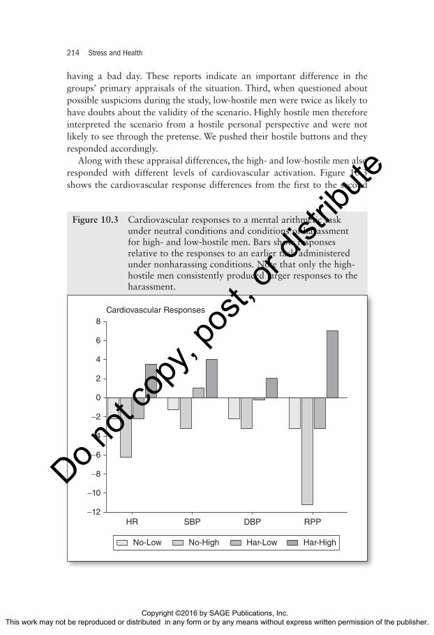
Along with these appraisal differences, the high- and low-hostile men also responded with different levels of cardiovascular activation. Figure 10.3 shows the cardiovascular response differences from the first to the second
−12
8
−8
−6
−10
2
4
6
Cardiovascular Responses
−2
0
HR SBP DBP RPP
−4
No-Low No-High Har-Low Har-High
Figure 10.3 Cardiovascular responses to a mental arithmetic task under neutral conditions and conditions of harassment for high- and low-hostile men. Bars show responses relative to the responses to an earlier task administered under nonharassing conditions. Note that only the high-hostile men consistently produced larger responses to the harassment.
Copyright ©2016 by SAGE Publications, Inc. This work may not be reproduced or distributed in any form or by any means without express written permission of the publisher.
Do not
copy
, pos
t, or d
istrib
ute

CHAPTER 10: Individual Differences in Reactivity to Stress——215
tasks in the control and harassment conditions. The high-hostile men had consistently greater responses to the second task in the harassment condi-tion. For example, one measure we used was the rate pressure product (heart rate multiplied by systolic blood pressure), which is a simple measure of oxygen demand by the heart muscle that indexes the workload of the heart. Harassment during the second task increased the rate pressure product responses of the hostile men. In contrast, the low-hostile group and the nonharassed controls had lower rate-pressure product values during the second task.
The high-hostile subjects apparently evaluated the situation within a hos-tile cognitive scheme, became offended and angry, and then produced high levels of sympathetically mediated cardiovascular activity. We speculate that these autonomic response differences were derived ultimately from emotion-related differences in frontal-limbic processing of the harassment during the second task. As proof, the high- and low-hostile subjects did not differ in cardiovascular responses to the first task under neutral conditions, indicat-ing that simple work on mental arithmetic did not differentiate the groups and suggesting no inherent physiological biases. These in turn may have resulted in greater hypothalamic activation and therefore responses of brain-stem cardiovascular control centers.
In this section, we focused on persons who manifest hostile traits and who were exposed to hostility provocations in the lab. The studies suggest that the high-hostile subjects were primed to draw hostile interpretations of social interactions with resulting differences in their emotional reactions. We believe these were accompanied by different frontal-limbic response patterns, resulting in disproportionate effects on the hypothalamus and brainstem outputs to autonomic and endocrine pathways. In the next sec-tion, we will consider whether there are individual differences in stress reactivity that do not depend on differences in situational appraisals or lack of coping resources.
Individual Differences in Hypothalamic and Brainstem Responses to Stress—Level II
Cognitive and emotional biases are not the only source of individual differ-ences in stress reactivity. We suspect that individual differences in reactivity may also occur due to differences in hypothalamic or brainstem reactivity. Research on cardiovascular reactivity provides us with examples of persons who have quite elevated cardiovascular and endocrine responses to various challenges, even when they report no differences in cognitive evaluations or
Copyright ©2016 by SAGE Publications, Inc. This work may not be reproduced or distributed in any form or by any means without express written permission of the publisher.
Do not
copy
, pos
t, or d
istrib
ute

216——Stress and Health
emotional reactions to the situation. As a result, there is no obvious basis for saying that cognitive-emotional interpretations caused these stress reactions to differ. We might therefore ask if it is reasonable to look at the hypothala-mus and brainstem as sources of these individual differences.
Stability of Cardiovascular Reactivity
There are considerable individual differences in the magnitude of heart rate change to mentally demanding tasks, leading to speculation that per-sons having consistently larger responses may be at higher risk of coronary artery disease and hypertension (Sherwood & Turner, 1992). A precondi-tion of suspecting that heart rate reactivity is a risk factor for disease is knowing whether it is a stable individual difference. Manuck and col-leagues found that persons who had large heart rate rises to a cognitively challenging task had similarly large responses to the same task, and to a different cognitive task when retested 13 months later (Manuck & Garland, 1980). Others have reported reasonably stable levels of reactivity over 10 years (Sherwood et al., 1997). Heart rate reactivity is also stable across different types of tasks, such as cold pressor and reaction time tests conducted 2 weeks to 13 months apart (Lovallo, Pincomb, & Wilson, 1986b) and across public speaking and mental arithmetic tasks 3 weeks apart (Sgoutas-Emch et al., 1994), again reinforcing its trait-like qualities. This stability over time and across situations provides a basis for us to consider reactivity tendencies as capable of affecting health. The idea that persistently large cardiovascular responses may themselves be a disease risk is known as the cardiovascular reactivity hypothesis.
Persons who tend to respond to stress with relatively large heart rate and blood pressure increases appear to have elevated levels of sympathetic out-flow from brainstem cardiovascular control centers. We discussed auto-nomic controls on cardiovascular function in Chapters 3 and 4. Cacioppo and colleagues provide an extensive account of measures reflecting sympa-thetic and parasympathetic influences on the heart (Berntson, Cacioppo, & Quigley, 1993, 1994; Cacioppo et al., 1994).
In considering the possible sources of individual differences in heart rate reactivity, we may refer to Figure 5.7. Persons who are more reactive could be more responsive at the level of the brainstem cardiovascular control nuclei, at the level of the paraventricular nucleus of the hypothalamus, or at the level of the HACER. We do not presently have strong evidence allowing us to separate these sources of differences in response magnitude. However, there are reasons to speculate that differences in heart rate reactivity arise especially at the level of the hypothalamus.
Copyright ©2016 by SAGE Publications, Inc. This work may not be reproduced or distributed in any form or by any means without express written permission of the publisher.
Do not
copy
, pos
t, or d
istrib
ute

CHAPTER 10: Individual Differences in Reactivity to Stress——217
Hypothalamus as a Source of Individual Differences in Stress Reactivity
In studies from our lab, we were able to make a reasonable interpretation that group reactivity differences can arise due to processes other than subjec-tive experience and presumed psychological processes. We interpret these results collectively as implicating hypothalamic mechanisms as the source of these reactivity differences.
In one study, my colleagues and I selected high– and low–heart rate reac-tivity subjects using their responses to a painful cold pressor test. We then tested each person on two reaction time tasks using threat of electric shock in one of them and monetary rewards in the other (Lovallo, Pincomb, & Wilson, 1986a; 1986b). Persons with large heart rate increases to the cold pressor test (> 19 beats per minute) also had the largest heart rate changes to both reaction time tasks, regardless of the nature of the incentive. Most important for our present discussion, the heart rate reactivity groups did not differ in their perceptions or evaluations of the tasks. In both studies, high– and low–heart rate reactors reported feeling activated and distressed to an equal degree (Lovallo, Pincomb, Brackett, & Wilson, 1990). Not surprisingly, everyone found shock avoidance more aversive than monetary rewards.
Because the heart rate response groups did not have different subjective experiences, we suspect that their cardiovascular response differences did not result from processes at the cognitive-emotional level in Figure 10.1. Instead, it appears that the response difference was based on activational differences lower in the system. This interpretation is consistent with find-ings in borderline hypertensives compared with controls at low risk of future hypertension (al’Absi et al., 1994). The borderlines had larger cortisol and cardiovascular responses to nonaversive mental stressors with no difference in reported feelings of activation or distress, suggesting greater hypothalamic activation as the source of the reactivity difference.
Integrated Cardiac and Endocrine Reactivity
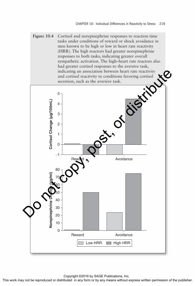
Chapters 5 and 6 noted that during aversive stressors, the hypothalamic paraventricular nucleus was capable of stimulating both cortisol secretion and cardiovascular responses. This dual role also allowed us to focus on the hypothalamus as one source of individual differences in reactivity. My col-leagues and I reexamined heart rate reactivity and cortisol responses from our studies on aversive and rewarded versions of the reaction time task (Lovallo et al., 1990). We expected high–heart rate reactors to have greater norepinephrine responses to both tasks because both called for heightened
Copyright ©2016 by SAGE Publications, Inc. This work may not be reproduced or distributed in any form or by any means without express written permission of the publisher.
Do not
copy
, pos
t, or d
istrib
ute

218——Stress and Health
attention and response preparedness, but we expected to see greater cortisol responses only to the aversive tasks. This prediction was based on a theory that cortisol is secreted preferentially during aversive situations, evoking negative emotions, and that norepinephrine, representing the sympathetic nervous system, could be elevated during any situation calling for arousal and response preparation (Lundberg, 1980; Lundberg & Frankenhaeuser, 1980; Smyth et al., 1998).
The data are displayed in Figure 10.4. First, the low–heart rate reactors showed little or no change in cortisol or norepinephrine to either of the tasks. Second, the high–heart rate reactors had larger norepinephrine responses to both tasks, indicating greater global sympathetic activation tendencies. We also note that the high–heart rate reactors produced significant cortisol rises to the aversive task but not to the rewarded task. This was expected because other work had shown that cortisol responses are especially sensitive to expo-sure to aversive or distressing incentives such as pain or threat of shock (Lundberg, 1980; Lundberg & Frankenhaeuser, 1980). A question then arose as to the nature of the cortisol response in the high–heart rate reactors. Was it due to their emotional responses (a Level I response) or was it a Level II response involving the hypothalamus? When we compared reactivity groups in their activation and distress scores to the rewarded or aversive reaction time tasks, we found that the groups had indistinguishable subjective response scores to both types of incentive. Accordingly, we concluded that the cortisol reactivity difference was not secondary to a difference in emotional reactivity in this case. Instead, we concluded that reactivity groups differed in heart rate response and cortisol response at a level lower in the system. Since sympathetic responses and cortisol responses are integrated at the hypotha-lamic paraventricular nucleus, we concluded that the high reactors were producing this response difference at the hypothalamus. In regard to the incentive task, neither group had a cortisol response. We argued that the sympathetic response to the incentive task was due to elevated alertness and cognitive effort, as reported by both reactivity groups. However, in this case, the expenditure of effort without the sense of distress did not engage the paraventricular nucleus in either group but instead engaged brainstem auto-nomic centers that stimulated the heart. Once again, this process was exag-gerated only in the high reactors. These results show that physiologically reactive persons may produce integrated patterns of sympathetic outflow and pituitary–adrenal activation in the absence of differences in cognitive or emo-tional activity. The fact that high–heart rate reactors differed in response to a cold pressor task, a rewarded reaction time task, and an aversive reaction time task suggests that the reactivity difference is a stable characteristic of these persons, making them worthwhile candidates for disease risk.
Copyright ©2016 by SAGE Publications, Inc. This work may not be reproduced or distributed in any form or by any means without express written permission of the publisher.
Do not
copy
, pos
t, or d
istrib
ute
CHAPTER 10: Individual Differences in Reactivity to Stress——219
−1
5
1
0
3
4
Reward
Co
rtis
ol C
han
ge
(µg
/10
0mL
)
Avoidance
2
0
80
70
20
10
50
60
Reward
No
rep
inep
hri
ne
Ch
ang
e (p
g/m
l)
Avoidance
30
40
Low HRR High HRR
Figure 10.4 Cortisol and norepinephrine responses to reaction time tasks under conditions of reward or shock avoidance in men known to be high or low in heart rate reactivity (HRR). The high reactors had greater norepinephrine responses to both tasks, indicating greater overall sympathetic activation. The high–heart rate reactors also had greater cortisol responses to the aversive task, indicating an association between heart rate reactivity and cortisol reactivity to conditions favoring cortisol secretion, such as the aversive task.
Copyright ©2016 by SAGE Publications, Inc. This work may not be reproduced or distributed in any form or by any means without express written permission of the publisher.
Do not
copy
, pos
t, or d
istrib
ute
220——Stress and Health
Central Nervous System Activity in Relation to Cardiovascular and Endocrine Reactivity
The studies reviewed above resulted in two major conclusions. First, high– and low–heart rate reactors, as opposed to high- and low-hostility subjects, appear to experience challenging events in the same way, suggesting that they are not differentially reactive because of differences in evaluations of the situa-tion or the resulting emotions. Second, the tendency for large heart rate responses to be accompanied by large cortisol responses in both high–heart rate reactors and in those at high risk for hypertension suggests that this individual difference in stress reactivity is organized at a level above the separate output pathways for autonomic and endocrine outflow. This leads us to focus on the hypothalamus as the structure most capable of producing this integrated response pattern. Whether the difference is at the level of the HACER or por-tions of the hypothalamus that communicate more directly with the pituitary and brainstem, such as the paraventricular nucleus, is not clear from these data; however, the pattern of results is consistent with a focus on the hypothalamus.
This line of reasoning leads to a picture of differences in heart rate reactiv-ity that result from individual differences in the hypothalamic amplification of equivalent signals arriving from evaluative and emotion-producing cen-ters of the brain. The presumed amplification difference may therefore result in consistent individual differences in integrated autonomic and endocrine outflow, accounting for the differences between subjects in both heart rate and cortisol responses.
Although the case for differences in hypothalamic amplification of descending signals is indirect, there is a plausible mechanism for such a sys-tem in limbic and hypothalamic outputs. As noted in Chapter 7, high levels of cortisol exposure can sensitize the amygdala and consequently exaggerate CRF neuron inputs to the PVN and HACER during times of stress (Schulkin et al., 1998; Shepard et al., 2000, 2003). More direct evidence has emerged from a study of humans who died with complications of hypertension. In the deceased hypertensive patients, the number of CRF neurons in the hypotha-lamic PVN was greater than in control autopsy tissue (Goncharuk, Van Heerikhuize, Swaab, & Buijs, 2002). In the researchers’ words, “Increased activity of CRH-producing neurons in the PVN of hypertensive patients is proposed not only to entail hyperactivity of the hypothalamo-pituitary-adrenal axis, but also of the sympathetic nervous system and, thus, to be involved in the pathogenesis of hypertension.” In relation to our present discussion, such persons would have larger cardiovascular and endocrine responses to a variety of stressors regardless of their individual cognitive-emotional reactions. In addition, the contributions of such reactivity differ-ences to reactivity and health may extend beyond hypertension.
Copyright ©2016 by SAGE Publications, Inc. This work may not be reproduced or distributed in any form or by any means without express written permission of the publisher.
Do not
copy
, pos
t, or d
istrib
ute
CHAPTER 10: Individual Differences in Reactivity to Stress——221
Individual Differences in Peripheral Responses to Stress—Level III
The preceding sections indicate that exaggerated or altered responses to stress may originate in altered prefrontal-limbic system interactions (Level I) or because of altered amplification of descending signals at the level of the hypothalamus or brainstem (Level II). This leaves a consideration of the peripheral organs themselves (Level III). We consider that responses to stress can be altered because peripheral organs themselves are disease prone or because they have been altered by existing disease. In endocrinol-ogy, for example, failure to rapidly remove glucose from the blood follow-ing glucose challenge may indicate impending diabetes. In cardiology, premature fatigue, shortness of breath, exaggerated blood pressure response, or abnormal cardiac rhythm during the stress of exercise may signal the presence of coronary artery disease, hypertension, or autonomic dysregulation, all of which are diseases of the peripheral organs. Similarly, physical alterations occur to the blood vessels during hypertension devel-opment, and these include thickening of the muscle layer of the blood ves-sel wall. This narrows the internal diameter of the vessel so that any increase in blood flow, as in exercise, results in an exaggerated blood pres-sure rise (Jackson, Squires, Grimes, & Beard, 1983; Wilson, Sung, Pincomb, & Lovallo, 1990). As a result, in prehypertensive persons, otherwise nor-mal autonomic and endocrine adjustments to exercise may still cause abnormal blood pressure responses. Such studies suggest that preclinical alterations in vascular resistance (Lovallo & al’Absi, 1998) can cause a disproportionate rise in blood pressure relative to an otherwise normal demand for blood flow. In such cases, the abnormal reactivity may be an indicator of underlying pathology, thus serving as a marker of disease but not necessarily acting as a cause.
While we often look to central nervous system determinants of differ-ences in reactivity, it is possible that in some instances persons are more reactive because of alterations in peripheral mechanisms. In this case, a person might have exaggerated responses to a stressor without any altera-tion in appraisals, emotions, or centrally induced activation of endocrine or autonomic outflow. For example, persons may cluster into groups on the basis of peripheral alpha- and beta-adrenoreceptor sensitivity, and hence may have different cardiovascular responses to otherwise similar degrees of central activation (Mills et al., 1994; Mills, Dimsdale, Ziegler, Berry, & Bain, 1990). Similarly, an exaggerated heart rate response to a mental arith-metic challenge may be associated with peripheral adrenoreceptor function (Mills et al., 1990).
Copyright ©2016 by SAGE Publications, Inc. This work may not be reproduced or distributed in any form or by any means without express written permission of the publisher.
Do not
copy
, pos
t, or d
istrib
ute

222——Stress and Health
Finally, persons developing hypertension may have altered vascular reac-tivity that is of peripheral origin, resulting in enhanced blood pressure responses to pharmacological challenges. One such challenge is caffeine, which potentiates the action of norepinephrine at the sympathetic nerve terminal and elevates peripheral vascular resistance. In studying blood pres-sure responses to caffeine, we saw progressively greater pressure rises in 185 subjects stratified into four risk groups, ranging from low-risk controls to medicated hypertensives (Hartley et al., 2000). The subjects in these separate studies reported no differences in subjective activation or distress to caffeine or the stressful tasks (Lovallo et al., 1991; Pincomb et al., 1996; Sung, Lovallo, Whitsett, & Wilson, 1995). In another study, caffeine given to bor-derline hypertensives induced a more prolonged increase in blood pressure over an hour of mental stress (al’Absi, Bongard, & Lovallo, 2000), again without differences in reported activation or distress (al’Absi, Everson, & Lovallo, 1995). These studies suggest that the high-risk groups were experi-encing caffeine and the stressors in the same way as the low-risk subjects. Caffeine raises blood pressure by actions at the blood vessel wall (Pincomb et al., 1996). The absence of self-report differences between the groups and the known peripheral actions of caffeine strongly suggest that the pressure differences between hypertension risk groups were peripheral in origin.
Discussion
The emergence of an integrated field of study incorporating neurophysiol-ogy, personality theory, autonomic and endocrine function, immune system activity, and health provides us with a very powerful set of tools to take a multilevel approach to our understanding of health and disease. We have attempted to systematize disparate lines of research using our model of cen-trally determined stress responses presented in Chapter 5 into the three-level model in Figure 10.2.
This chapter has presented evidence that individual differences in stress response can arise out of differences in situational appraisals and resulting emotional responses, pointing to activity in prefrontal and limbic system structures (Level I). Other evidence has been presented that response biases can also be determined by activational differences at the hypothalamus and brainstem (Level II) or by differences in peripheral tissues and organs (Level III). The following chapter presents evidence that both psychologically and physiologically based reactivity differences could result in impaired health. This allows a top-to-bottom integration of how the system could be altered by what we think and feel, and in turn these tendencies to think and feel in
Copyright ©2016 by SAGE Publications, Inc. This work may not be reproduced or distributed in any form or by any means without express written permission of the publisher.
Do not
copy
, pos
t, or d
istrib
ute

CHAPTER 10: Individual Differences in Reactivity to Stress——223
certain ways may have a strong biological and experiential basis. Ultimately, our physiological behaviors accompanying our evaluations and emotions become major influences on our bodies.
The ultimate value of attempting to integrate information on evaluative processes, emotions, and autonomic-endocrine outflow into our models of disease etiology is twofold. First, we will be able to view health and disease as the outcome of psychophysiological processes encompassing the psychology, behavior, and physiology of the individual. Second, the individual-differences approach described here helps us understand more clearly the mechanisms relating behavior to health, and it provides us with ways to identify persons at greatest risk of disease.
We started this chapter with a conceptual presentation of the notion of stress reactivity falling along a normal distribution. This statistical model implies that abnormal reactivity is not confined to the upper reaches of the distribution but includes the lower end as well. This chapter has presented evidence on untoward effects of exaggerated reactivity in keeping with the dominant thinking in research on stress and health. The next chapter will concern itself with the low end of the distribution and ask the question of whether this tail also presages deleterious health outcomes.
If we return to our basic question concerning how ideas can come to have power over our bodies, we can now see that there are meaningful relation-ships between evaluations, mood states, and central nervous system function in relation to emotion tendencies. These in turn determine autonomic and endocrine outflow. The evidence reviewed here suggests that there are indeed systematic differences between persons and their reactions to psychological stressors. These differences translate into consistent differences in endocrine impacts on the immune system and autonomic alterations on cardiovascular structures. But our list of targets for such peripheral changes is surely not complete. The response systems we have discussed so far are intended to represent how reactivity differences might arise and how these alterations in function might connect with disease processes. But we have no reason to believe that individual differences in psychological stress response and health consequences are limited to the influences of cardiovascular reactions and adrenocortical responses. There are likely to be other mechanistic relation-ships between differences in the activity of the central nervous system and health. We should also recognize that the examples discussed in this chapter all represent ways that persons can become hyperreactive to stress, repre-senting the right tail of the distribution in Figure 10.1. In Chapter 11, we will focus on the left tail of the distribution and consider ways that persons may become stress hyporeactive and whether this diminished reactivity is benign or a risk factor for disease.
Copyright ©2016 by SAGE Publications, Inc. This work may not be reproduced or distributed in any form or by any means without express written permission of the publisher.
Do not
copy
, pos
t, or d
istrib
ute

224——Stress and Health
Summary
In this chapter, we have reviewed evidence that persons may have different ways of responding to potential threats. Persons may differ systematically in how they perceive and evaluate situations they face. These differences may determine consistent differences in emotional responses. For example, persons having strongly negative emotions to a variety of situations are likely to have greater autonomically mediated cardiovascular reactions, even ones inappropriate to the situation at hand. Persons may also differ in their hypothalamic and brainstem activational tendencies, even when they do not differ in their perceptions and emotional reactions. Research on persons with greater heart rate responses shows that there is considerable stability of such responses across situations and over time, pointing to a physiological basis for this reactivity. Studies in groups at high risk for hypertension show increased cortisol reactivity in the absence of affective or evaluative differences. Finally, other studies point to peripheral sources of reactivity differences between groups. Whether the source of exaggerated physiological activation is an emotional response bias, such as trait hostil-ity; an autonomic response bias; or a peripheral difference, the exaggerated reactivity has the same impact on the periphery. These individual differences in stress reactivity may predict differences in health outcomes. More gener-ally, these differences in reactivity to mental stressors indicate that the ways in which ideas can influence the body can vary systematically from person to person and become major predictors of health and disease.
FURTHER READING
Contrada, R. J., & Baum, A. (Eds.). (2011). The handbook of stress science. New York, NY: Springer.
Section IV of this edited volume has a discussion of stress mechanisms in relation to a wide range of health outcomes.
Light, K. C. (2001). Hypertension and the reactivity hypothesis: The next generation. Psychosomatic Medicine, 63, 744–746.
A commentary on refinement of contemporary reactivity hypotheses in test-ing relationships to disease risk.
Turner, J. R. (1994). Cardiovascular reactivity and stress: Patterns of physiological response. New York, NY: Plenum Press.
This volume is a very thorough coverage of cardiovascular reactivity research in relation to individual differences.
Copyright ©2016 by SAGE Publications, Inc. This work may not be reproduced or distributed in any form or by any means without express written permission of the publisher.
Do not
copy
, pos
t, or d
istrib
ute
CHAPTER 10: Individual Differences in Reactivity to Stress——225
Turner, J. R., Sherwood, A., & Light, K. C. (Eds.) (1992). Individual differences in cardiovascular response to stress. New York, NY: Plenum Press.
This is an extended discussion of individual differences in stress reactivity, including factors not dealt with here, such as reactivity differences due to gender and race.
Treiber, F. A., Kamarck, T., Schneiderman, N., Sheffield, D., Kapuku, G., & Taylor, T. (2003). Cardiovascular reactivity and development of preclinical and clinical disease states. Psychosomatic Medicine, 65, 46–62.
A thorough review of evidence linking reactivity to health and disease, with a discussion of modifier variables and assessment of future research needs.
DISCUSSION POINTS
1. Using statistical principles, why should we be concerned with diminished stress reactivity when exaggerated reactivity is considered an indicator of health risk?
2. Relate the three-level model of reactivity in Figure 10.2 to the diagram of responses to psychological stress described in Chapter 5.
3. Relate both Figure 10.2 and Figure 5.7 to Figure 5.1, integrating concepts of psychological stress, central determinants of stress reactivity, and levels in the system determining individual differences in reactivity.
4. If exaggerated reactivity is an indicator of health risk, how might diminished reactivity also be an indicator of altered health?
Copyright ©2016 by SAGE Publications, Inc. This work may not be reproduced or distributed in any form or by any means without express written permission of the publisher.
Do not
copy
, pos
t, or d
istrib
ute