infl ammatory myofi broblastic tumor of thigh: a case …
TRANSCRIPT

261 International Journal of Scientifi c Study | November 2014 | Vol 2 | Issue 8
Infl ammatory Myofi broblastic Tumor of Thigh: A Case Report and Review of LiteratureTapan Kumar Sahoo1, Ipsita Dhal2, Chandra Prava Mishra2, Saroj Kumar Das Majumdar3, Dillip Kumar Parida4
1Senior Resident, Department of Radiation Oncology, All India Institute of Medical Sciences, Bhubaneswar, Odisha, India, 2Postgraduate, Department of Pathology, Shrirama Chandra Bhanj Medical College, Cuttack, Odisha, India, 3Assistant Professor, Department of Radiation Oncology, All India Institute of Medical Sciences, Bhubaneswar, Odisha, India, 4Professor and Head, Department of Radiation Oncology,All India Institute of Medical Sciences, Bhubaneswar, Odisha, India
patient with soft tissue IMT of left thigh who underwent surgery and post-operative radiotherapy.
CASE REPORT
A 56-year-old man initially presenting with growth over left thigh since 12 months which gradually increase in size to the present size (3 cm × 3 cm), non-tender, hard in consistency, there is no superfi cial venous engorgement and no local rise of temperature. Fine-needle aspiration cytology from the lesion revealed benign spindle cell lesion. Further investigation ruled out possibility of distant metastasis. Wide local excision was done under general anesthesia. The histopathological examination showed possibility of IMT or spindle cell sarcoma. Immunohistochemical examination showed CD34, S-100, bcl2, and ALK1 negative, also smooth muscle actin positive focally and KI67 positive focally (30%)favoring IMT. Post-operative computed tomography (CT) scan showed mild soft tissue thickening with a small collection. He was received external beam radiotherapy of 60 Gy in 30 fractions to left thigh with two opposing portals
INTRODUCTION
Inflammatory myofibroblastic tumor (IMT)is a rare neoplasm that consists of spindle cell proliferation with a distinctive fi broinfl ammatory and even pseudosarcomatous appearance.1 IMT affects both sexes equally and is more prevalent in children and young adults.2 These tumors most frequently occur in the lungs.1 Only a few extra-pulmonary lesions such as liver, genitor-urinary tract, mesentery, omentum, extremities, head and neck, orbit, nasal sinuses, liver, spleen, pancreas, bowel, kidney, urinary bladder, testis, heart, and lymphatic systems have been reported.1 Microscopic examination showed presence of histiocytes, fi broblasts, infl ammatory cells, collagen bundles and plasma cells (Figures 1-3). We describe a
Case Report
Abstract
Infl ammatory myofi broblastic tumor (IMT)is a rarely reported tumor of unknown etiology and pathogenesis. Neoplastic growths of myofi broblast, on a background of plasma cell and lymphocytic proliferation, have been designated as IMT. It occurs primarily in the lungs but has occurred in other extra-pulmonary sites, also. Abdomen is the most common extra-pulmonary site. The biological behavior is still uncertain. We report a case of 56-year-old man presenting with a lump over left thigh for 6 months. Post-excision histopathological examination showed possibility of IMT or spindle cell sarcoma. Immunohistochemical examination showed CD34, S-100, bcl2, and ALK1 negative, also smooth muscle actin positive focally and KI67 positive focally 30% favoring IMT.
Keywords: Immunohistochemistry, Infl ammatory myofi broblastic tumor, Neoplastic growth, Thigh
Corresponding Author:Dr. Tapan Kumar Sahoo, Senior Resident, Department of Radiation Oncology, All India Institute of Medical Sciences, Bhubaneswar - 751 019, Odisha, India. Phone: +91-9437219525. E-mail: [email protected]
Access this article online
Website: www.ijss-sn.com
Sahoo, et al.: Inflammatory Myofibroblastic Tumor
262International Journal of Scientifi c Study | November 2014 | Vol 2 | Issue 8
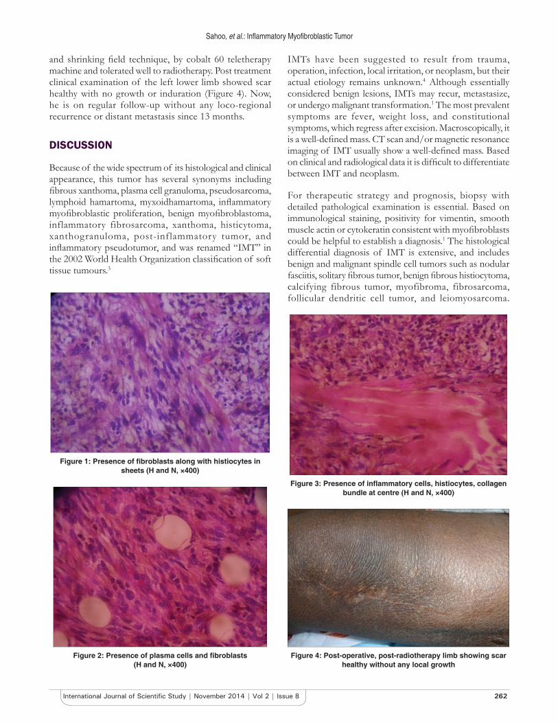
and shrinking fi eld technique, by cobalt 60 teletherapy machine and tolerated well to radiotherapy. Post treatment clinical examination of the left lower limb showed scar healthy with no growth or induration (Figure 4). Now, he is on regular follow-up without any loco-regional recurrence or distant metastasis since 13 months.
DISCUSSION
Because of the wide spectrum of its histological and clinical appearance, this tumor has several synonyms including fi brous xanthoma, plasma cell granuloma, pseudosarcoma, lymphoid hamartoma, myxoidhamartoma, infl ammatory myofi broblastic proliferation, benign myofi broblastoma, inflammatory fibrosarcoma, xanthoma, histicytoma, xanthogranuloma, post-inflammatory tumor, and infl ammatory pseudotumor, and was renamed “IMT” in the 2002 World Health Organization classifi cation of soft tissue tumours.3
IMTs have been suggested to result from trauma, operation, infection, local irritation, or neoplasm, but their actual etiology remains unknown.4 Although essentially considered benign lesions, IMTs may recur, metastasize, or undergo malignant transformation.1 The most prevalent symptoms are fever, weight loss, and constitutional symptoms, which regress after excision. Macroscopically, it is a well-defi ned mass. CT scan and/or magnetic resonance imaging of IMT usually show a well-defi ned mass. Based on clinical and radiological data it is diffi cult to differentiate between IMT and neoplasm.
For therapeutic strategy and prognosis, biopsy with detailed pathological examination is essential. Based on immunological staining, positivity for vimentin, smooth muscle actin or cytokeratin consistent with myofi broblasts could be helpful to establish a diagnosis.1 The histological differential diagnosis of IMT is extensive, and includes benign and malignant spindle cell tumors such as nodular fasciitis, solitary fi brous tumor, benign fi brous histiocytoma, calcifying fibrous tumor, myofibroma, fibrosarcoma, follicular dendritic cell tumor, and leiomyosarcoma.
Figure 1: Presence of fi broblasts along with histiocytes in sheets (H and N, ×400)
Figure 2: Presence of plasma cells and fi broblasts (H and N, ×400)
Figure 3: Presence of infl ammatory cells, histiocytes, collagen bundle at centre (H and N, ×400)
Figure 4: Post-operative, post-radiotherapy limb showing scar healthy without any local growth

Sahoo, et al.: Inflammatory Myofibroblastic Tumor
263 International Journal of Scientifi c Study | November 2014 | Vol 2 | Issue 8
Histologically, IMTs contain much more prominent infl ammatory infi ltrate than nodular fasciitis.5 In addition, they lack the “c” shaped fascicles, and mucin-rich stroma that is responsible for the characteristic “tissue culture-like or feathery” appearance in nodular fasciitis.6 Solitary fi brous tumor was excluded due to lack of hemangiopericytoma-like areas and strong CD34 immunoreactivity.7 The diagnosis of benign fi brous histiocytoma was not favored because of the lack of characteristic storiform pattern.8 Calcifying fi brous tumor, a rare benign neoplasm, is uniformly hypocellular and contains scattered dystrophic calcification.2 The diagnosis of myofi broma was excluded due to lack of biphasic growth pattern with hemangiopericytoma-like blood vessels.9 Fibrosarcoma was excluded due to lack of malignant features, collagenous areas herringbone pattern that characterize it. Additionally, it typically lacks a signifi cant infl ammatory infi ltrate.9 Follicular dendritic cell tumor is differentiated from IMTs by its characteristic distribution of infl ammatory infi ltrate admixed with dendritic spindle cells. It is easily distinguished by immunohistochemical staining for CD21, CD23 and/or CD35.5 If there was a predominant lymphocytic and/or plasmacytic component, a plasma cell neoplasm or lymphoma should be excluded. In our case, the immunohistochemical analysis revealed CD34, S-100, bcl2, and ALK1 negative, also smooth muscle actin positive focally and KI67 positive focally 30% favoring IMT. According to the histological and immunohistochemical features of the present case, a fi nal diagnosis IMT was made.
IMTs are tumors with unpredictable clinical behavior, requiring complete surgical excision and continuous monitoring of clinical consequences. According to the World Health Organization IMTs are classifi ed as tumors of intermediate biological potential due to a tendency of local recurrence and small risk of distant metastasis.5 Surgical resection remains the recommended treatment for IMT. Prognosis is excellent when the tumor is completely removed. Recurrence has been reported in 15-37% of abdominal IMT.1 Radiotherapy, chemotherapy, or steroid treatment has been reported to successfully treat some
patients, but benefi ts have not been proven in a large series of patients.10
CONCLUSIONS
IMT is a rare spindle cell tumor with an uncertain malignant potential. Thus, early recognition and complete surgical resection are necessary to avoid recurrences and prevent the spread of locally aggressive tumors. Soft tissue IMT in thigh is a rare disease and represents a diagnostic dilemma for surgeons. Due to nonspecifi c radiological and clinical presentations, diagnosis of IMT is rarely made before excision. The diagnosis is dependent on histological and immunohistochemical examination. Surgical resection remains the recommended treatment for IMT. Close follow-up is recommended to prevent recurrence.
REFERENCES
1. Coffi n CM, Watterson J, Priest JR, Dehner LP. Extrapulmonary infl ammatory myofi broblastic tumor (infl ammatory pseudotumor). A clinicopathologic and immunohistochemical study of 84 cases. Am J Surg Pathol 1995;19:859-72.
2. Roberts G, Farrell M, Allcutt D. Spinal infl ammatory pseudotumours. Br J Neurosurg 2001;15:197-8.
3. Aizawa T, Sato T, Tanaka Y, Kishimoto K, Watanabe M, Kokubun S. Intramedullary plasma cell granuloma in the cervicothoracic spine. Case report. J Neurosurg 2002;97:235-8.
4. Allanore Y, Pham XV, Clerc DA, Menkès CJ, Kahan A. Sacral infl ammatory pseudotumor revealed by paraneoplastic syndrome. Rheumatol Int 2004;24:166-8.
5. Gleason BC, Hornick JL. Infl ammatory myofi broblastic tumours: where are we now? J Clin Pathol 2008;61:428-37.
6. Dayan D, Nasrallah V, Vered M. Clinico-pathologic correlations of myofi broblastic tumors of the oral cavity: 1. Nodular fasciitis. J Oral Pathol Med 2005;34:426-35.
7. Shimoyama T, Horie N, Ide F. Solitary fi brous tumor of the palate: a case report and review of the literature. J Oral Maxillofac Surg 2004;62:895-7.
8. Alves FA, Vargas PA, Coelho Siqueira SA, Coletta RD, de Almeida OP. Benign fi brous histiocytoma of the buccal mucosa: Case report with immunohistochemical features. J Oral Maxillofac Surg 2003;61:269-71.
9. Neville B, Damm D, Allen C, Bouquot J. Oral and Maxillofacial Pathology. 3rd ed. Philadelphia: W.B. Saunders; 2008.
10. Cho YS, Kim SM, Chung WH, Hong SH. Infl ammatory pseudotumour involving the skull base and cervical spine. J Laryngol Otol 2001;115:580-4.
How to cite this article: Sahoo TK, Dhal I, Mishra CP, Majumdar SK, Parida DK. Infl ammatory myofi broblastic tumour of thigh: A case report and review of literature. Int J Sci Stud 2014;2(8):261-263.
Source of Support: Nil, Confl ict of Interest: None declared.