imunodeficiência comum variável em adultos e crianças ... · estudo retrospectivo dos processos...
TRANSCRIPT

171R E V I S T A P O R T U G U E S A D E I M U N O A L E R G O L O G I A
ARTIGO ORIGINAL
RESUMO
Fundamentos: A imunodeficiência comum variável (IDCV) é uma doença heterogénea caracterizada por infec-ções recorrentes e deficiência de anticorpos. Têm sido descritas várias classificações que correlacionam diferentes perfis imunológicos das células B e T com distintos fenótipos clínicos. Objectivos: Conhecer as características clínicas e sua correlação com as subpopulações linfocitárias de adultos e crianças com IDCV. Material e métodos: Dos doentes com IDP observados na consulta de Imunodeficiência Primária dos HUC e HPC, entre 1 de Janeiro de 2000 e 31 de Dezembro de 2010, seleccionámos aqueles que cumpriam o diagnóstico de IDCV e procedeu -se ao estudo retrospectivo dos processos clínicos. Efectuou -se o estudo de imunofenotipagem dos linfócitos B e T através de citometria de fluxo e os resultados foram comparados com 12 controlos saudáveis. Resultados: Foram avalia-dos 14 doentes com IDCV, 12 adultos e 2 crianças. Os adultos, com uma relação feminino/masculino de 2:1, tinham uma média de idade de 48,5 anos e uma média de idade na altura do diagnóstico de 36 anos. Nestes doentes, as infecções respiratórias baixas recorrentes e as gastrintestinais estavam presentes em 75% e 33% dos casos, respec-tivamente. As complicações associadas foram bronquiectasias e doenças autoimunes (50% dos casos), doenças lin-
Imunodeficiência comum variável em adultos e crianças: Correlação entre fenótipos clínicos e imunológicos
Correlation between the clinical and immunological phenotypes in adults and children with common variable immunodeficiency
Eugénia Almeida1, Emília Faria1,2, Nuno Sousa1, Tiago Carvalheiro3, Sónia Lemos2, Artur Paiva3, António Segorbe Luís1
1 Serviço de Imunoalergologia. Hospitais da Universidade de Coimbra (HUC)2 Consulta de Imunodeficiência Primária. Departamento de Pediatria de Centro Hospitalar dos Covões (HPC)3 Laboratório de Citometria. Centro de Histocompatibilidade do Centro
R e v P o r t I m u n o a l e r g o l o g i a 2 0 1 1 ; 1 9 ( 3 ) : 1 7 1 - 1 8 1
Data de recepção / Received in: 11/03/2011
Data de aceitação / Accepted for publication in: 25/07/2011
Imuno (19) 3 - Miolo 2ª PROVA PT.indd Sec2:171Imuno (19) 3 - Miolo 2ª PROVA PT.indd Sec2:171 19-10-2011 10:26:3719-10-2011 10:26:37

172R E V I S T A P O R T U G U E S A D E I M U N O A L E R G O L O G I A
foproliferativas (25%), esplenomegalia e enteropatia crónica (17%). As crianças, ambas de sexo feminino, com 8 e 12 anos, para além da clínica de infecções respiratórias recorrentes, apresentavam bronquiectasias. Da análise das sub-populações linfocitárias nos adultos, salienta -se a diminuição de células T reguladoras em 2 doentes, diminuição in-ferior a 1% das células CD19 em 2 doentes e diminuição das células B memória switched em 10 doentes. Dos 4 doentes com diminuição de CD4 naive, 2 apresentavam doenças autoimunes e os restantes complicações linfopro-liferativas. Nas crianças, o estudo das subpopulações B e T revelou -se normal. Conclusões: A análise efectuada das subpopulações de células B e T demonstrou que a principal alteração encontrada nos doentes com complicações linfoproliferativas, autoimunes e esplenomegalia foi a acentuada diminuição das células B memória. Ao contrário, os doentes com IDCV sem complicações associadas não apresentam alterações das subpopulações T e B, o que pode-rá documentar um prognóstico mais favorável.
Palavras -chave: Bronquiectasias, fenótipos clínicos, imunodeficiência comum variável, imunoglobulinas, subpopulações linfocitárias B e T.
ABSTRACT
Background: Common variable immunodeficiency (CVID) is a heterogeneous disorder characterised by recurrent infections and antibody deficiency. Various classification schemes for distinct clinical phenotypes have been developed and correlated with different B cell and T cell phenotypes. Aim: To understand the clinical characteristics and their correlation with the B and T cell phenotypes in adult and children CVID patients. Material and methods: We selected the CVID patients followed -up in HUC and HPC Primary Immunodeficiency Outpatient Clinics from 1st January 2000 to 31st December 2010 and performed a retrospective study of their clinical files. Different T and B lymphocyte immunophenotyping was then performed using the flow cytometer and the results were compared with 12 healthy controls. Results: Fourteen CVID patients (12 adults/2children) were evaluated. The adults had a mean age of 48.5 years and mean age at diagnosis of 36 years, F/M ratio 2:1. Recurrent lower respiratory tract infections and gastrointestinal infections were found in 75% and 33% of cases, respectively. The asso-ciated complications were bronchiectasis and autoimmune diseases (50%), lymphoproliferative disease (25%), splenomegaly and chronic enteropathy (17%). The children, both female, aged 8 and 12, had recurrent respiratory tract infections and bron-chiectasis. In the adult patients, T and B lymphocyte subpopulations showed decrease of Treg cells in 2 patients, CD19 less than 1% in 2 patients and decreased switched memory B cells in 10 patients. Two of the 4 patients with reduction of naive CD4 cells had autoimmune diseases and the others lymphoproliferative diseases. In the children, the T and B lymphocyte subpopula-tions were normal. Conclusions: The B and T cell subset analysis revealed that the major abnormality was the more significant decrease of switched memory B cells in the patients with additional lymphoproliferative and autoimmune diseases and spleno-megaly. In contrast, CVID patients without associated complications have no changes in T and B subpopulations, which may mean a more favourable prognosis.
Key-words: Bronchiectasis, clinical phenotypes, common variable immunodeficiency, immunoglobulins, B and T lymphocyte subsets
Eugénia Almeida, Emília Faria, Nuno Sousa, Tiago Carvalheiro, Sónia Lemos, Artur Paiva, António Segorbe Luís
Imuno (19) 3 - Miolo 2ª PROVA PT.indd Sec2:172Imuno (19) 3 - Miolo 2ª PROVA PT.indd Sec2:172 19-10-2011 10:26:4019-10-2011 10:26:40
173R E V I S T A P O R T U G U E S A D E I M U N O A L E R G O L O G I A
INTRODUÇÃO
A imunodefi ciência comum variável (IDCV), descri-ta pela primeira vez em 1953, é uma imunodefi -ciência primária (IDP) caracterizada por altera-
ções na função terminal das células B com compromisso na produção de anticorpos e, clinicamente, por infecções recorrentes. Segundo a classifi cação da European Society for Immunodefi ciencies (ESID) e Pan -American Group for Immu-nodefi ciency (PAGID), são critérios de IDCV a diminuição do nível de IgG, IgM e/ou IgA (pelo menos dois desvios--padrão abaixo do valor médio para o grupo etário), início dos sintomas depois dos 2 anos, ausência de isoemagluti-ninas e/ou defi ciente resposta à vacinação, após a exclusão de outras causas de hipogamaglobulinemia1. Mais recente-mente, o European Initiative for Primary Immunodefi ciencies (EUROPID) sugere a alteração do critério idade para um mínimo de 4 anos2.
A IDCV é resultante de um defeito primário de imu-nidade celular B com falha na diferenciação das células B maduras em plasmócitos, estando também descritas algu-mas alterações na função e expressão do receptor da célula T e na produção e expressão de citocinas. Foram recentemente encontradas alterações genéticas em doen-tes com IDCV, nomeadamente mutações em genes codi-ficadores das moléculas envolvidas na sobrevivênvia da célula B e no switch de isotipo (CD19, TACI, ICOS)2,3,4.
Na IDCV, os sintomas podem ocorrer em qualquer idade, com dois picos de incidência, entre 5 e 10 anos e entre os 20 e os 30 anos5. Considerada a IDP sintomática mais frequente no adulto, esta doença afecta de igual modo ambos os sexos e a sua incidência está estimada em 1 para 10 000 -50 000 casos6.
A IDCV é uma doença heterogénea com uma varieda-de ampla de manifestações clínicas de gravidade variável. As infecções sinopulmonares recorrentes são as mais fre-quentes, embora as doenças gastrintestinais e autoimunes tenham uma prevalência elevada e possam ser a manifes-tação inicial de IDCV. As bronquiectasias, frequentemente presentes na altura do diagnóstico, são o resultado de
infecções graves e repetidas do aparelho respiratório in-ferior e não parecem estar relacionadas com o nível de IgG2. As manifestações gastrintestinais de natureza infec-ciosa, neoplásica, inflamatória ou autoimune podem mimeti-zar doenças gastrintestinais bem conhecidas, como doença inflamatória intestinal ou doença celíaca e ainda síndromes de malabsorção, ou coexistir com estas doenças. O me-canismo fisiopatológico subjacente à maioria destas mani-festações, para além de deficiências da célula B, parece também estar relacionado com disfunção da célula T 7,8. Cerca de 20% dos doentes com IDCV desenvolvem com-plicações autoimunes, sendo as mais frequentes a púrpura trombocitopénica imune e a anemia hemolítica. Tal como a doença granulomatosa sistémica, são situações de prog-nóstico menos favorável. Permanece por esclarecer a pre-sença desta inesperada autorreactividade em doentes que ao mesmo tempo apresentam deficiência de anticorpos9,10. Os doentes com IDCV têm também risco aumentado de carcinoma gástrico e linfoma, particularmente do tipo B, associado ao vírus Epstein -Barr. No sexo feminino, o lin-foma apresenta uma maior incidência na quinta ou sexta décadas de vida11.
Recentemente, os estudos realizados na criança mos-tram uma apresentação clínica da IDCV semelhante ao adulto no que se refere a patologias auto -imunes e a pa-tologias infecciosas. Contudo, demonstram uma presença mais elevada de asma e a associação com deficiência da hormona de crescimento12,13. Os casos clínicos descritos de IDCV de início precoce são menos frequentes, não sendo o diagnóstico em geral efectuado antes dos 4 ou 6 anos de idade. Na criança, o diagnóstico diferencial estabe-lece -se com outras IDP, nomeadamente hipogamaglobu-linemia transitória da infância, agamaglobulinemia ligada ao X, deficiência de subclasses de IgG, deficiência de IgA e síndrome de Hiper -IgM. Perante uma criança com imu-nidade celular T normal mas com hipogamaglobulinemia, definida como redução dos níveis de um ou mais isotipos de imunoglobulinas (pelo menos dois desvios -padrão abai-xo do valor médio para o grupo etário), há que conside-rar poder tratar -se de imunodeficiência transitória, adqui-
IMUNODEFICIÊNCIA COMUM VARIÁVEL EM ADULTOS E CRIANÇAS: CORRELAÇÃO ENTRE FENÓTIPOS CLÍNICOS E IMUNOLÓGICOS / ARTIGO ORIGINAL
Imuno (19) 3 - Miolo 2ª PROVA PT.indd Sec2:173Imuno (19) 3 - Miolo 2ª PROVA PT.indd Sec2:173 19-10-2011 10:26:4019-10-2011 10:26:40

174R E V I S T A P O R T U G U E S A D E I M U N O A L E R G O L O G I A
rida ou fisiológica, que pode persistir após os 6 meses de idade14.
Nos últimos 10 anos, assistiu -se a um número cres-cente de publicações de IDCV na idade adulta, o que permitiu o conhecimento mais aprofundado desta doen-ça. É actualmente considerada uma síndrome com múlti-plos fenótipos clínicos, correlacionada com diferentes perfis imunológicos. Chapel et al, num estudo recente, verifica que 83% dos 334 doentes com IDCV estudados apresentava um dos cinco seguintes fenótipos clínicos: sem complicações, autoimunidade, infiltração linfocítica policlonal, enteropatia e neoplasia linfóide, que se reflec-tem em diferentes taxas de sobrevivência4. Foi encontra-do, como factor clínico preditivo, a associação de infiltra-ção linfocítica policlonal com risco aumentado de neoplasia linfóide, que se correlaciona com o nível sérico superior de IgM. A doença autoimune correlaciona -se com diminuição de células CD8 circulantes. Outros autores sugerem a definição de subgrupos com base no fenótipo da célula B, baseado na análise por citometria de fluxo das populações de células B periféricas. Em 2002, o estu-do de Warnatz et al foi um dos primeiros a classificar os doentes com IDCV, baseando -se na quantificação das cé-lulas B memória switched (CD27+IgM -IgD -) e correlacionando -a com fenótipos clínicos15. Posterior-mente, o estudo multicentro EUROclass, envolvendo 303 doentes com IDCV, confirma uma redução significativa de células de memória switched na maioria dos doentes com elevado risco de esplenomegalia e doença granulomatosa, adiciona o conceito de expansão de células B CD21low em doentes com esplenomegalia e expansão de células B transitional (grupo SmB -Trhi) associada a linfadenopatia (no grupo de doentes com uma percentagem de células de memória switched inferior ou igual a 2 %)16.
Mais recentemente, o estudo prospectivo francês DEFI concluiu que a redução das células de memória switched e das células CD4 e CD8 naive, encontradas nos doentes com IDCV que apenas desenvolvem infecções, é mais pro-nunciada nos doentes que apresentam doenças linfopro-liferativas, citopenias autoimunes ou enteropatia crónica.
Este estudo demonstrou ainda que os doentes com com-plicações linfoproliferativas e citopenias autoimunes apre-sentam diminuição das células T reguladoras17.
À semelhança do observado no adulto, Yong et al de-tectaram, recentemente, associação entre diferentes fenó-tipos clínicos e alterações imunológicas, nomeadamente diminuição do número de células B de memória switched em 45 crianças com IDCV. Estas crianças apresentavam níveis séricos de IgG e IgM mais baixos que se correlacio-navam com maior número de infecções, doença autoimune, bronquiectasias, doença granulomatosa pulmonar e neopla-sias hematológicas18.
A terapêutica de reposição regular com imunoglobu-lina G é a base do tratamento desta entidade, para além da instituição precoce de antibioterapia durante as infec-ções agudas, sendo discutível a utilidade de antibiótico profiláctico. A realização de cinesiterapia respiratória re-gular é fundamental.
Vários autores demonstraram a diminuição da frequên-cia e gravidade das infecções e a melhoria do prognóstico desta IDP em doentes com terapêutica adequada com imunoglobulina2,19. A terapêutica imunossupressora pode ser usada no tratamento de doença autoimune. A preco-cidade do diagnóstico e a optimização da terapêutica tem reduzido consideravelmente a mortalidade e aumentado a qualidade da vida destes doentes nas últimas décadas.
MATERIAL E MÉTODOS
Foi realizado o estudo retrospectivo dos processos clínicos de adultos e crianças com o diagnóstico de IDP observadas nas consultas de imunodeficiência primária dos Hospitais da Universidade de Coimbra (HUC) e no Hos-pital Pediátrico de Coimbra (HPC), entre 1 de Janeiro de 2000 e 31 de Dezembro de 2009. Procedeu -se à caracte-rização das IDP com base na classificação do Comité da União Internacional da Sociedade de IDP20. Foram selec-cionados os casos de adultos e crianças que, em Dezembro de 2009, cumpriram os critérios de IDCV e foi analisada
Eugénia Almeida, Emília Faria, Nuno Sousa, Tiago Carvalheiro, Sónia Lemos, Artur Paiva, António Segorbe Luís
Imuno (19) 3 - Miolo 2ª PROVA PT.indd Sec2:174Imuno (19) 3 - Miolo 2ª PROVA PT.indd Sec2:174 19-10-2011 10:26:4019-10-2011 10:26:40
175R E V I S T A P O R T U G U E S A D E I M U N O A L E R G O L O G I A
a clínica, terapêutica efectuada, tomografia computarizada (TC) torácica, ecografia abdominal e espirometria. Procedeu -se também à quantificação das subpopulações linfocitárias nos doentes e em doze controlos saudáveis. Para marcação das células T CD4 e CD8 naive, memória e efectoras, utilizaram -se os anticorpos anti -CD 4 pacific B, anti -CD3 FITC, anti -CCR7 ficoeritrina, anti -CD8 PerCP, anti -CD45RA APC; para as células B naive e memória, os anticorpos anti -CD20 pacific B, anti -CD45 pacific orange, anti -IgG FITC, anti -IgE PE, anti -IgD PE, anti -CD27 PC5, anti -CD19 PC7, anti -IgM APC e anti -CD38 APC -H7 e, para células T reguladoras, os anticorpos anti -CD25 FITC, anti--CD127 PE e anti -CD4 PerCPCy5.5, através da expressão combinada para CD4, CD25 de expressão forte e expres-são débil a negativa para CD127. A aquisição de resultados foi realizada em duas fases consecutivas no citómetro de fluxo FACSCanto IITM com recurso ao software FACSDiva e, no caso particular das células T reguladoras, no citóme-tro de fluxo FACSCalibur com recurso ao software Cell-Quest Pro. A análise de resultados foi realizada usando o sofware de análise Infinicyt.
RESULTADOS
Foram seleccionados 12 doentes adultos com o diag-nóstico de IDCV, 14,5% do total de doentes adultos com IDP seguidos na respectiva consulta dos HUC, e 2 crianças do HPC com o mesmo diagnóstico as quais representam 2,4% dos doentes da consulta de IDP deste hospital.
Na população de doentes adultos com IDCV, encon-trámos uma relação sexo feminino/sexo masculino de 2:1, com média de idade de 48,5 anos, tendo o início dos sin-tomas ocorrido, em média, aos 24 anos para as mulheres e aos 12 anos para os homens. A média das idades na al-tura do diagnóstico foi de 36 anos. O tempo médio entre o início dos sintomas e o diagnóstico foi de 16 anos. Apenas um doente tinha um filho com deficiência de IgA e IgG2.
Na série de doentes adultos, as infecções respiratórias baixas recorrentes estavam presentes em 75% dos doentes
e as gastrintestinais em 33%. Um dos doentes apresentava apenas clínica de infecções urinárias e outro sofreu um episódio de encefalite a enterovírus na infância. Metade dos doentes apresentava bronquiectasias na altura do diag-nóstico. Na última avaliação, 3 doentes (25%) apresentavam síndrome obstrutivo (D2, D7, D8) e 2 (17%) síndrome restritivo no estudo funcional ventilatório. As complicações mais frequentes foram as doenças autoimunes, a par das bronquiectasias (50%), seguidas das doenças linfoprolife-rativas (25%) e, por último, esplenomegalia e enteropatia crónica (ambas 17%). Um dos doentes apresentava síndrome de malabsorção grave, tendo sido excluída doença celíaca e linfoma intestinal (D1). O exame parasitológico das fezes e/ou biópsia por endoscopia digestiva alta detectou giar-díase em 50% dos casos. Em três doentes (25%) foi detec-tada hiperplasia nodular linfóide e dois desenvolveram neo-plasias malignas (carcinoma papilar da tiróide e adenoma do antro gástrico) (Quadro 1).
Nos doentes com IDCV em idade adulta, os valores das imunoglobulinas na altura do diagnóstico, a contagem total de leucócitos, células B, células T, subpopulações de células T e células de memória switched são apresentados no Qua-dro 2. Todos os doentes apresentavam diminuição das três classes de imunoglobulinas. Os linfócitos T CD4 encontra-vam -se diminuídos em 9 casos e os linfócitos CD8 estavam aumentados noutros 9. Apenas dois doentes apresentavam uma diminuição de linfócitos CD19 inferior a 1% (D2 e D10). Encontramos inversão da relação CD4/CD8 em 50%. So-mente dois doentes apresentavam diminuição das células T reguladoras em relação ao mesmo grupo. Dez doentes apre-sentavam diminuição das células B memória switched, sendo que, em 7 deles, o valor é inferior ou igual a 2%. Os dois doentes que apresentavam apenas clínica de infecções não tinham alterações no número de células B memória. Na contagem diferencial das células CD4 e CD8 verificamos diminuição das células CD4 naive e das CD8 naive em 4 e 6 doentes, respectivamente, na comparação com o grupo--controlo. Na análise das subpopulações linfocitárias do gru-po de doentes que desenvolveram complicações clínicas, dos 3 três que desenvolveram complicações linfoproliferativas,
IMUNODEFICIÊNCIA COMUM VARIÁVEL EM ADULTOS E CRIANÇAS: CORRELAÇÃO ENTRE FENÓTIPOS CLÍNICOS E IMUNOLÓGICOS / ARTIGO ORIGINAL
Imuno (19) 3 - Miolo 2ª PROVA PT.indd Sec2:175Imuno (19) 3 - Miolo 2ª PROVA PT.indd Sec2:175 19-10-2011 10:26:4019-10-2011 10:26:40

176R E V I S T A P O R T U G U E S A D E I M U N O A L E R G O L O G I A
dois apresentavam simultaneamente diminuição de células T reguladoras e diminuição grave das células B memória (0%). O mesmo número apresentava diminuição na percentagem de células CD4 naive. No grupo de doentes que desenvol-veram complicações autoimunes, 2/3 apresentavam diminui-ção das células B memória, CD4 e CD8 naive; 1/3 diminuição de células CD4 naive e apenas um deles apresentava as cé-lulas T reguladoras diminuídas. Os doentes que tinham so-mente complicações infecciosas apresentavam valores nor-mais nas subpopulações linfocitárias B e T analisadas.
As crianças, de 8 e 12 anos, do sexo feminino, apre sen-tavam -se com clínica de infecções do aparelho respiratório superior e inferior, necessitando de internamento em mé-dia uma vez por ano e, uma delas, também com infecções envolvendo a pele e o aparelho urinário. Em ambos os casos, foi identificada colonização das vias respiratórias por Haemophylus influenza e bronquiectasias na altura do diagnóstico. Ambas apresentavam asma brônquica. Um dos casos revelou uma repercussão negativa no desenvolvi-mento estatoponderal (Quadro 3). O estudo das imuno-
Eugénia Almeida, Emília Faria, Nuno Sousa, Tiago Carvalheiro, Sónia Lemos, Artur Paiva, António Segorbe Luís
Quadro 1. Características clínicas e terapêutica diária dos doentes com IDCV em idade adulta
Doentes SexoIdade (anos) Infecções
RepetiçãoFEV1
%BQ Enteropatia
crónica/HNL Giardia Espleno-megalia
Doençaautoimune
Doenças linfoproliferativas
TerapêuticadiáriaActual Início Diagnóstico
1 M 45 6 30 RA+RB+GI 97 SimSim
(SMA)Sim Sim Não
Carcinomatiróide
LepicortinoleLevotiroxinaCloreto de potássio
2 F 36 10 27 RA+RB+GI 79 SimSimHNL
Sim Não Não NãoDefl azacortTinidazolCloreto de potássio
3 M 37 2 30 RB 93 Não Não Sim Sim NãoGranulomatosesistémica
Não
4 F 41 31 31 RA+GI 88 NãoNãoHNL Sim Não Poliartrite Não
SalazopirinaNaproxenoTinidazol
5 M 48 30 40 RA+RB+C 93 Não Não Sim Não TCAI Não Não
6 F 57 47 52 RA+RB 89 Sim Não Sim Não TCAI? Não LevotiroxinaMucolítico
7 F 61 26 41 RB 68 Não Não Não Não GCA Não CI+BDLevotiroxina
8 F 57 33 46 RA+RB 57 Sim NãoHNL Não Não Não Adenoma
antro gástricoCI+BDOxigenoterapia
9 F 57 6 32 RB+GI 108 Sim Não Não Não GCA Não Captopril
10 F 66 18 40 RB 136 Sim Não Não Não GCA Não Omeprazol
11 M 21 11 28 RAEncefalite 88 Não Não Não Não Não Não Mucolítico
Anticonvulsionante
12 F 56 20 37 U 130 Não Não Não Não Não Não Não
HNL – Hiperplasia nodular linfóide; RA – Infecções respiratórias altas; RB – Infecções respiratórias baixas; GI – Infecções gastrintestinais; C – Candidíase oral; U – Infecções urinárias BQ – Bronquiectasias; GCA – Gastrite crónica atrófica; TAI – Tiroidite autoimune; TCAI – Trombocitopenia autoimune; CI – Corticóide inalado; BD – Broncodilatador inalado; SMA – Síndrome de malabsorção
Imuno (19) 3 - Miolo 2ª PROVA PT.indd Sec2:176Imuno (19) 3 - Miolo 2ª PROVA PT.indd Sec2:176 19-10-2011 10:26:4019-10-2011 10:26:40
177R E V I S T A P O R T U G U E S A D E I M U N O A L E R G O L O G I A
IMUNODEFICIÊNCIA COMUM VARIÁVEL EM ADULTOS E CRIANÇAS: CORRELAÇÃO ENTRE FENÓTIPOS CLÍNICOS E IMUNOLÓGICOS / ARTIGO ORIGINAL
Quadro 2. Imunidade humoral e celular dos doentes com IDCV em idade adulta
DoentesLeucócitos
totais(cels/μL)
CD19 IgG(mg/dL)
IgM(mg/dL)
IgA(mg/dL)
CD4 CD8 RazãoCD4//CD8
TregCél. B memória
switched(CD27+ IgM - IgD -)
céls/μL % céls/μL % céls/μL % céls/μL % céls/μL %
1 12000 240 2 <3 23 <7 840 28 2010 67 0,42 5 0,2 0 0
2 7900 24 0,3 260 17,8 23 583 41 782 55 0,75 9 0,6 0,48 2
3 5400 162 3 542 4 <6 522 46 408 36 1,27 4 0,4 0 0
4 6100 122 2 321 30 <6,6 869 57 655 43 1,32 6 0,4 1,22 1
5 5700 228 4 391 18 52 520 57 337 37 1,54 7 0,7 75 33
6 5400 108 2 287 64 <6 441 48 459 50 0,96 6 0,7 0 0
7 5600 112 2 497 72 <7 521 30 1180 68 0,44 7 0,4 4,48 4
8 4800 96 2 225 9 6,6 206 43 240 50 0,86 7 1,5 1,92 2
9 7300 365 5 444 14 <7 906 43 494 52 0,83 5 0,5 14,6 4
10 4600 5 0,1 396 <4 <7 290 15 1623 84 0,18 4 0,2 0 0
11 4000 120 3 37 4 <6 640 50 550 43 1,16 7 0,5 6 5
12 5600 185 3 212 250 22 497 65 238 31 2,09 9 1,2 18,96 14
C(n=12)
6383±1377
283±220
4,17±2,55 NE NE NE 973
±48563,3±8,6
454±196
30,5±7,5
2,22±0,77
6,25±2,09
0,52±0,29
44,3±43,7
20,9±16,6
Valoresreferência 700 -1600 40 -230 70 -160 6,5 -29,2
Quadro 3. Características clínicas, exames complementares de diagnóstico e terapêutica dos doentes com IDCV em idade pediátrica
Doente 13 14
Sexo F F
Idade
Actual 8 anos 12 anos
Início sintomas 4 meses 2 anos
No diagnóstico 7 anos 12 anos
Infecções de repetição RB RA+RB+U
FEV1 (%) 102,6 60
Bronquiectasias Sim Sim
Outras complicações(Esplenomegalia, giardíase,
doença autoimune, doenças malignas) Não Não
Terapêutica diáriaIGSC (desde 7A)
CI+BD Amoxicilina+ácido clavulânico(dose profi láctica)
IGSC (desde 13A)CI
Cinesiterapia respiratória regular
RA – Infecções respiratórias altas; RB – Infecções respiratórias baixas; U – Infecções urinárias; CI – corticóide inalado;BD – Broncodilatador inalado; IGSC – Imunoglobulina subcutânea
Imuno (19) 3 - Miolo 2ª PROVA PT.indd Sec2:177Imuno (19) 3 - Miolo 2ª PROVA PT.indd Sec2:177 19-10-2011 10:26:4019-10-2011 10:26:40

178R E V I S T A P O R T U G U E S A D E I M U N O A L E R G O L O G I A
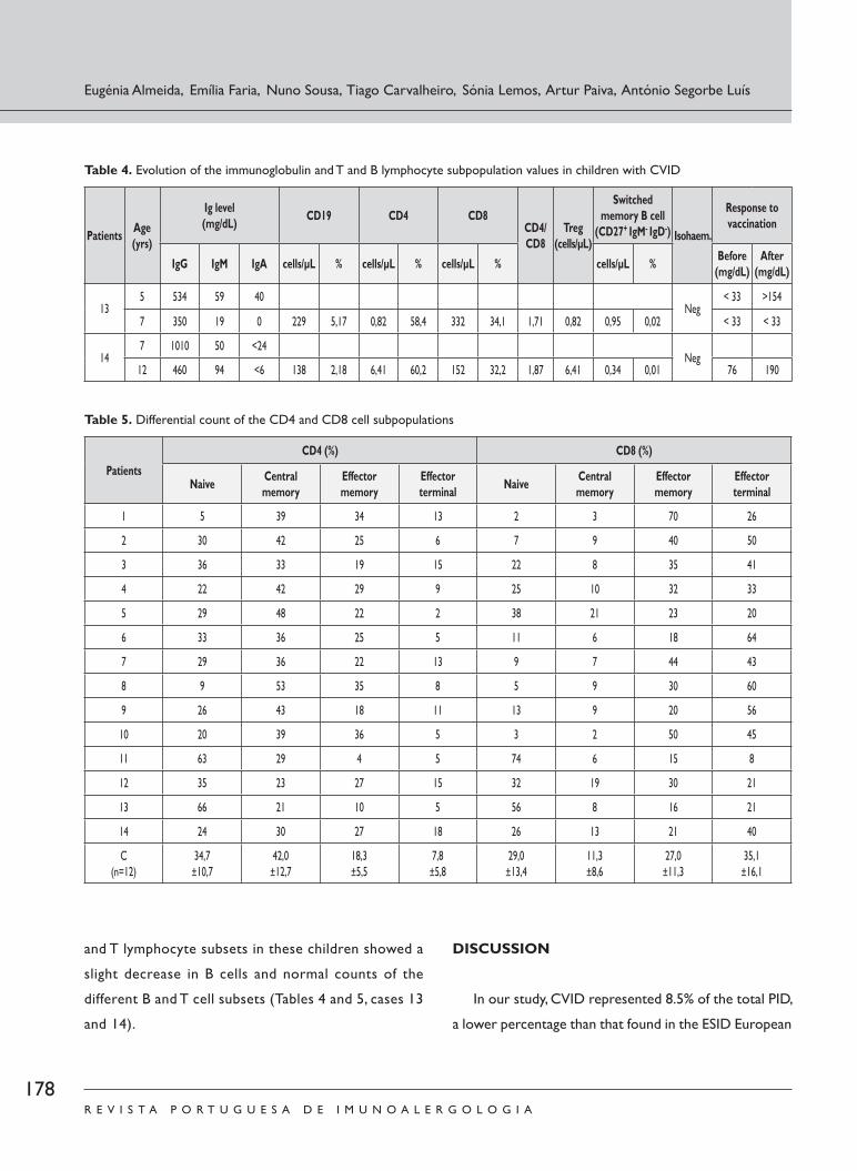
globulinas e subpopulações linfocitárias B e T destas crian-ças apresentava ligeira diminuição das células B e valores normais das diferentes subpopulações de células B e T (Quadros 4 e 5, casos 13 e 14).
DISCUSSÃO
No presente estudo, a IDCV representa 8,5 % do total de IDP, percentagem inferior ao encontrado na base euro-
Eugénia Almeida, Emília Faria, Nuno Sousa, Tiago Carvalheiro, Sónia Lemos, Artur Paiva, António Segorbe Luís
Quadro 4. Evolução dos valores de imunoglobulinas e subpopulações linfocitárias T e B das crianças com IDCV
Doentes Idade(anos)
Nível de Ig(mg/dL) CD19 CD4 CD8
CD4//CD8
Treg(céls/μL)
Cel. B memória switched
(CD27+ IgM - IgD -) TítuloIsoem.
Resposta à vacinação
IgG IgM IgA céls/μL % céls/μL % céls/μL % céls/μL % Antes(mg/dL)
Depois(mg/dL)
135 534 59 40
Neg< 33 >154
7 350 19 0 229 5,17 0,82 58,4 332 34,1 1,71 0,82 0,95 0,02 < 33 < 33
147 1010 50 <24
Neg12 460 94 <6 138 2,18 6,41 60,2 152 32,2 1,87 6,41 0,34 0,01 76 190
Quadro 5. Contagem diferencial das subpopulações de células CD4 e CD8
Doentes
CD4 (%) CD8 (%)
Naive Memóriacentral
Memóriaefectora
Terminalefectora Naive Memória
centralMemóriaefectora
Terminalefectora
1 5 39 34 13 2 3 70 26
2 30 42 25 6 7 9 40 50
3 36 33 19 15 22 8 35 41
4 22 42 29 9 25 10 32 33
5 29 48 22 2 38 21 23 20
6 33 36 25 5 11 6 18 64
7 29 36 22 13 9 7 44 43
8 9 53 35 8 5 9 30 60
9 26 43 18 11 13 9 20 56
10 20 39 36 5 3 2 50 45
11 63 29 4 5 74 6 15 8
12 35 23 27 15 32 19 30 21
13 66 21 10 5 56 8 16 21
14 24 30 27 18 26 13 21 40
C(n=12)
34,7±10,7
42,0±12,7
18,3±5,5
7,8±5,8
29,0±13,4
11,3±8,6
27,0±11,3
35,1±16,1
Imuno (19) 3 - Miolo 2ª PROVA PT.indd Sec2:178Imuno (19) 3 - Miolo 2ª PROVA PT.indd Sec2:178 19-10-2011 10:26:4119-10-2011 10:26:41
179R E V I S T A P O R T U G U E S A D E I M U N O A L E R G O L O G I A
peia da ESID entre os anos 2006 -2008 (20,7%). O atraso no diagnóstico foi de 16 anos, em média, sobreponível ao encontrado por outros autores. Este eventual subdiagnós-tico poderá reflectir o facto de esta doença ainda não ser reconhecida por alguns profissionais de saúde. O período de tempo que decorreu entre o início da sintomatologia e o diagnóstico não parece estar relacionado com o desen-volvimento de complicações estruturais, já que 50% dos doentes apresentavam bronquiectasias na altura do diag-nóstico, independente do tempo de evolução. Por outro lado, as complicações autoimunes, imunoproliferativas e inflamatórias também não parece estarem relacionadas com o tempo de evolução da doença. Encontraram -se síndromes restritivo e obstrutivo, apesar da função pulmonar estar normal na maioria dos casos. A prevalência de doença gas-trintestinal, na forma de diarreia crónica (variável entre as séries, de 21 a 56,6%), foi de 14,3% na presente série, infe-rior ao esperado, ao contrário da hiperplasia nodular lin-fóide, descrita em 8% dos casos, e, neste estudo, presente em 25% dos doentes7. Relativamente às complicações de natureza linfoproliferativa, salienta -se o facto de nenhum doente ter desenvolvido linfoma, patologia de elevada mor-talidade, com risco aumentado nos doentes com IDCV.
Nos 6 doentes que desenvolveram complicações au-toimunes (42,8%), a maioria apresenta diminuição da per-centagem de células B memória switched (metade destes inferior a 2%), assim como das células CD8 naive, e 1/3
redução das células CD4 naive, tal como é observado no estudo prospectivo DEFI17. Em contrapartida, apenas um doente apresenta diminuição das células T reguladoras. De modo semelhante, nos 3 doentes com doenças linfoproli-ferativas (21,4%), as células B memória switched estão di-minuídas, a par duma diminuição das células CD 4 naive em dois casos. O número absoluto de células T regulado-ras apenas se encontra diminuído em um dos doentes.
Os dois doentes com enteropatia crónica apresentavam diminuição de células B memória, num inferior a 2%. Num dos casos verificou -se ainda diminuição das células CD4 naive. Estas observações estão de acordo com o observa-do no estudo DEFI17. Um destes apresenta ainda diminuição
das células T reguladoras. Nos dois casos com esplenome-galia só se verifica diminuição acentuada das células B me-mória, semelhante ao encontrado noutras séries.16,17
Os quatro doentes (28%) que apresentavam apenas in-fecções respiratórias de repetição (dois deles crianças) tinham valores normais de células CD4 naive, CD 8 naive e B me-mória switched, o que contrasta com o encontrado no estu-do já referido, onde esses valores estavam diminuídos17.
A evolução clínica observada nas crianças incluídas nes-te estudo ilustra as dificuldades no diagnóstico definitivo desta doença em idade pediátrica. As duas crianças apresen-tam as primeiras manifestações de doença nos primeiros 2 anos de vida, com os estudos imunológicos iniciais normais aos 4 anos. Entre os 6 e 8 anos, assiste -se a uma diminuição progressiva dos valores das imunoglobulinas e resposta ine-ficaz na produção de anticorpos anti -pneumococcus após vacinação, associada ao aparecimento de rápida degradação da função pulmonar e de bronquiectasias. À semelhança do descrito, estas crianças com IDCV apresentavam doença pulmonar obstrutiva associada21. O tempo médio entre o início dos sintomas e o estabelecimento do diagnóstico, com consequente introdução de terapêutica de substituição, foi de 7 anos, semelhante ao que se encontra descrito12.
Apesar dos estudos na idade pediátrica sugerirem pre-sença de fenótipos de IDCV diferentes do adulto, poucos são os que avaliaram as subpopulações de células B e ainda não existem estudos que correlacionem as células B e T
com determinadas características clínicas. Os estudos que incluíram doentes em idade pediátrica sugerem que a dimi-nuição no número de células B memória switched se associa a um risco mais elevado de complicações e a um prognós-tico menos favorável18. Nas duas crianças, a caracterização imunológica das subpopulações de células B e T revelou -se normal, designadamente as células B memória, o que pode justificar a ausência de complicações, excepto bronquiec-tasias. O prognóstico destes doentes poderá ser favorável, pois, segundo Yong et al., o mesmo parece estar dependen-te da quantificação das células B memória na criança18.
Dez doentes efectuam terapêutica de substituição ade-quada com imunoglobulina IgG por via endovenosa e qua-
IMUNODEFICIÊNCIA COMUM VARIÁVEL EM ADULTOS E CRIANÇAS: CORRELAÇÃO ENTRE FENÓTIPOS CLÍNICOS E IMUNOLÓGICOS / ARTIGO ORIGINAL
Imuno (19) 3 - Miolo 2ª PROVA PT.indd Sec2:179Imuno (19) 3 - Miolo 2ª PROVA PT.indd Sec2:179 19-10-2011 10:26:4119-10-2011 10:26:41

180R E V I S T A P O R T U G U E S A D E I M U N O A L E R G O L O G I A
tro por via subcutânea. Estudos revelam que a instituição precoce de IgG policlonal de substituição não parece di-minuir o risco de desenvolvimento de bronquiectasias, apesar da manutenção dum nível de IgG pré -infusão ade-quado (superior a 500 mg/dl)22.
O prognóstico dos doentes com IDCV depende das alterações estruturais na altura do diagnóstico e do apare-cimento de complicações, nomeadamente de doença au-toimune, neoplásica ou doença granulomatosa sistémica.
CONCLUSÕES
Este estudo encontrou distintas características imu-nológicas nos diferentes fenótipos clínicos. Nesta série, a análise efectuada das subpopulações de células B e T demonstrou que, nos doentes com complicações linfo-proliferativas, autoimunes e esplenomegalia, a principal alteração encontrada foi a acentuada diminuição das cé-lulas B memória. Nos doentes apenas com complicações infecciosas não encontrámos alterações nas subpopula-ções linfocitárias. Salientamos o facto de as bronquiecta-sias estarem presentes em metade dos doentes na altura do diagnóstico e de o desenvolvimento de complicações não estar relacionado com o tempo de evolução da doen-ça. Atendendo à natureza sistémica e à gravidade das complicações associadas a esta doença, o seguimento regular destes doentes é decisivo para a sua identificação precoce e consequente intervenção no sentido de um melhor prognóstico.
Financiamento: NenhumDeclaração de conflitos de interesse: Nenhum a declarar
Contacto:Prof. António Segorbe LuísServiço de ImunoalergologiaHospitais da Universidade de CoimbraPraceta Prof. Mota PintoCoimbra
REFERÊNCIAS
1. European Society of Immunodeficiencies (ESID).2008. Available from:
http://www.esid.org.
2. Chapel H, Cunningham -Rundles C. Update in understanding com-
mon variable immunodeficiency disorders (CVIDs) and the manage-
ment of patients with this conditions. Br J Haematol 2009; 145:
709 -27.
3. Ahn S, Cunningham -Rundles C. Role of B cells in common variable
immunodeficiency. Expert Rev Clin Immunol 2009; 5(5):557 -64.
4. Chapel H, Lucas M, Lee M, Bjorkander J,Webster D, Grimbacher
B, et al. Common variable immunodeficiency disorders: division
into distinct clinical phenotypes. Blood 2008; 112(2):277 -86.
5. Glocker E, Ehl S, Grimbacher B. Common variable immunodefi-
ciency in children. Curr Opin Pediatr 2007; 19(6):685 -92.
6. Touw CML, Van de Ven AA, De Jong PA, Terheggen -Lagro S, Beek E,
Sanders EAM, et al. Detection of pulmonary complications in com-
mon variable immunodeficiency. Pediatr Allergy Immunol 2010; 21:
793–805.
7. Agarwal S, Mayer L. Pathogenesis and treatment of gastrointestinal
disease in antibody deficiency syndromes. J Allergy Clin Immunol
2009; 124(4): 658 -64.
8. Kalha I, Sellin JH. Common variable immunodeficiency and the gas-
trointestinal tract. Curr Gastroenterol Rep 2004; 6:377 -83.
9. Cunningham -Rundles C. Autoimmune manifestations in common
variable immunodeficiency. J Clin Immunol 2008; 28(Suppl 1):
S42 -5.
10. Silva SL, Ferreira MB, Rizzo LV. Imunodeficiência comum variável
e doença autoimune. Rev Port Imunoalergologia 2008; 16 (3):
231 -40.
11. Gompels MM, Hodges E, Lock RJ, Angus B, Whites H, Larkin A,
et al. Lymphoproliferative disease in antibody deficiency: a multi-
-centre study. Clin Exp Immunol 2003; 134:314 -20.
12. Urschel S, Kayikci L, Wintergerst U, Notheis G, Jansson A, Be-
lohradsky B. Common Variable Immunodeficiency Disorders in
Children: Delayed Diagnosis Despite Typical Clinical Presentation.
J Pediatr 2009; 154:888 -94.
13. Ogershok PR, Hogan MB, Welch JE, Corder WT, Wilson NH. Spectrum of illness in pediatric common variable immunodefi-
ciency. Ann Allergy Asthma Immunol 2006; 97:653 -56.
14. Keles S, Artac H, Kara R, Gokturk B, Ozen A, Reisli I. Transient
hypogammaglobulinemia and unclassified hypogammaglobulinemia:
‘Similarities and differences’. Pediatr Allergy Immunol 2010;
21:843 -51.
15. Warnatz K, Denz A, Dräger R, Braun M, Groth C, Wolff -Vorbeck
G et al. Severe deficiency of switched memory B cells (CD27(+)
IgM( -)IgD( -)) in subgroups of patients with common variable im-
munodeficiency: a new approach to classify a heterogeneous
disease. Blood 2002; 99(5):1544 -51.
Eugénia Almeida, Emília Faria, Nuno Sousa, Tiago Carvalheiro, Sónia Lemos, Artur Paiva, António Segorbe Luís
Imuno (19) 3 - Miolo 2ª PROVA PT.indd Sec2:180Imuno (19) 3 - Miolo 2ª PROVA PT.indd Sec2:180 19-10-2011 10:26:4119-10-2011 10:26:41

181R E V I S T A P O R T U G U E S A D E I M U N O A L E R G O L O G I A
16. Wehr C, Kivioja T, Schmitt C, Ferry B, Witte T, Eren E, et al. The
EUROclass trial: defining subgroups in common variable immuno-
deficiency. Blood 2008; 111(1):77 -85.
17. Mouillot G, Carmagnat M, Gérard L, Garnier JL, Fieschi C, Vince N, et
al. B -cell and T -cell phenotypes in CVID patients correlate with the
clinical phenotype of the disease. J Clin Immunol 2010; 30:746 -55
18. Yong PL, Orange JS, Sullivan KE. Pediatric common variable immu-
nodeficiency: Immunologic and phenotypic associations with switched
memory B cells. Pediatr Allergy Immunol 2010; 21: 852 -8.
19. Faria E, Silva S, Español T. Tratamento das imunodeficiências
primárias por deficiência predominante de anticorpos. Rev Port
Imunoalergologia 2004; 12(3): 251 -60.
20. Geha RS, Notarangelo LD, Casanova J -L, Chapel H, Conley ME,
Fischer A, et al. Primary immunodeficiency diseases: An update from
the International Union of Immunological Societies Primary Im-
munodeficiency Diseases Classification Committee. J Allergy Clin
Immunol 2007; 120(4): 776 -94.
21. Llobet MP, Bertrán JM, Español T. Immunodeficiencia común variable
en la edad pediátrica. Allergol et Immunopathol 2002; 30(1): 42 -6.
22. Kainulainen L, Varpula M, Liippo K, Svedström E, Nikoskelainen J,
Ruuskanen O. Pulmonary abdormalities in patients with primary
hypogammaglobulinemia. JACI 1999; 104(5):1031 -6.
IMUNODEFICIÊNCIA COMUM VARIÁVEL EM ADULTOS E CRIANÇAS: CORRELAÇÃO ENTRE FENÓTIPOS CLÍNICOS E IMUNOLÓGICOS / ARTIGO ORIGINAL
Imuno (19) 3 - Miolo 2ª PROVA PT.indd Sec2:181Imuno (19) 3 - Miolo 2ª PROVA PT.indd Sec2:181 19-10-2011 10:26:4119-10-2011 10:26:41
171R E V I S T A P O R T U G U E S A D E I M U N O A L E R G O L O G I A
ORIGINAL ARTICLE
ABSTRACT
Background: Common Variable Immunodeficiency (CVID) is a heterogeneous disorder characterised by recur-
rent infections and antibody deficiency. Various classification schemes for distinct clinical phenotypes have been de-
veloped and correlated with different B cell and T cell phenotypes. Aim: To understand the clinical characteristics
and their correlation with the B and T cell phenotypes in adult and children CVID patients. Material and Methods:
We selected the CVID patients followed -up in HUC and HPC Primary Immunodeficiency Outpatient Clinics from 1st
January 2000 to 31st December 2010 and performed a retrospective study of their clinical files. Different T and B
lymphocyte immunophenotyping was then performed using the flow cytometer and the results were compared with
12 healthy controls. Results: Fourteen CVID patients (12 adults/2children) were evaluated. The adults had a mean
age of 48.5 years and mean age at diagnosis of 36 years, F/M ratio 2:1. Recurrent lower respiratory tract infections
and gastrointestinal infections were found in 75% and 33% of cases, respectively. The associated complications were
bronchiectasis and autoimmune diseases (50%), lymphoproliferative disease (25%), splenomegaly and chronic en-
Correlation between the clinical and immunological phenotypes in adults and children with common variable immunodeficiency
Imunodeficiência comum variável em adultos e crianças: Correlação entre fenótipos clínicos e imunológicos
Eugénia Almeida1, Emília Faria1,2, Nuno Sousa1, Tiago Carvalheiro3, Sónia Lemos2, Artur Paiva3, António Segorbe Luís1
1 Allergology and Clinical Immunology Department. Hospitais da Universidade de Coimbra. (HUC)2 Primary Immunodeficiency Outpatient Clinic. Paediatrics Unit, Centro Hospitalar dos Covões. (HPC)3 Cytometry Laboratory. Centro de Histocompatibilidade do Centro.
R e v P o r t I m u n o a l e r g o l o g i a 2 0 1 1 ; 1 9 ( 3 ) : 1 7 1 - 1 8 1
Data de recepção / Received in: 11/03/2011
Data de aceitação / Accepted for publication in: 25/07/2011

172R E V I S T A P O R T U G U E S A D E I M U N O A L E R G O L O G I A
teropathy (17%). The children, both female, aged 8 and 12, had recurrent respiratory tract infections and bronchiecta-
sis. In the adult patients, T and B lymphocyte subpopulations showed decrease of Treg cells in 2 patients, CD19 less
than 1% in 2 patients and decreased switched memory B cells in 10 patients. Two of the 4 patients with reduction of
naive CD4 cells had autoimmune diseases and the other lymphoproliferative diseases. In the children, the T and B
lymphocyte subpopulations were normal. Conclusions: The B and T cell subset analysis revealed that the major
abnormality was the more significant decrease of switched memory B cells in the patients with additional lymphop-
roliferative and autoimmune diseases and splenomegaly. In contrast, CVID patients without associated complications
have no changes in T and B subpopulations, which may mean a more favourable prognosis.
Key-words: Bronchiectasis, clinical phenotypes, common variable immunodeficiency, immunoglobulins, B and T lym-phocyte subsets
RESUMO
Fundamentos: A imunodeficiência comum variável (IDCV) é uma doença heterogénea caracterizada por infecções re-correntes e deficiência de anticorpos. Têm sido descritas várias classificações que correlacionam diferentes perfis imunológicos das células B e T com distintos fenótipos clínicos. Objectivos: Conhecer as características clínicas e sua correlação com as subpopulações linfocitárias de adultos e crianças com IDCV. Material e métodos: Dos doentes com IDP observados na consulta de Imunodeficiência Primária dos HUC e HPC, entre 1 de Janeiro de 2000 e 31 de Dezembro de 2010, seleccio-námos aqueles que cumpriam o diagnóstico de IDCV e procedeu -se ao estudo retrospectivo dos processos clínicos. Efectuou--se o estudo de imunofenotipagem dos linfócitos B e T através de citometria de fluxo e os resultados foram comparados com 12 controlos saudáveis. Resultados: Foram avaliados 14 doentes com IDCV, 12 adultos e 2 crianças. Os adultos, com uma relação feminino/masculino de 2:1, tinham uma média de idade de 48,5 anos e uma média de idade na altura do diagnós-tico de 36 anos. Nestes doentes, as infecções respiratórias baixas recorrentes e as gastrintestinais estavam presentes em 75% e 33% dos casos, respectivamente. As complicações associadas foram bronquiectasias e doenças autoimunes (50% dos casos), doenças linfoproliferativas (25%), esplenomegalia e enteropatia crónica (17%). As crianças, ambas de sexo feminino, com 8 e 12 anos, para além da clínica de infecções respiratórias recorrentes, apresentavam bronquiectasias. Da análise das subpopulações linfocitárias nos adultos, salienta -se a diminuição de células T reguladoras em 2 doentes, diminuição inferior a 1% das células CD19 em 2 doentes e diminuição das células B memória switched em 10 doentes. Dos 4 doentes com diminuição de CD4 naive, 2 apresentavam doenças autoimunes e os restantes complicações linfoproliferativas. Nas crianças, o estudo das subpopulações B e T revelou -se normal. Conclusões: A análise efectuada das subpopulações de células B e T demonstrou que a principal alteração encontrada nos doentes com complicações linfoproliferativas, autoimunes e espleno-megalia foi a acentuada diminuição das células B memória. Ao contrário, os doentes com IDCV sem complicações associadas não apresentam alterações das subpopulações T e B, o que poderá documentar um prognóstico mais favorável.
Palavras -chave: Bronquiectasias, fenótipos clínicos, imunodeficiência comum variável, imunoglobulinas, subpopulações linfocitárias B e T.
Eugénia Almeida, Emília Faria, Nuno Sousa, Tiago Carvalheiro, Sónia Lemos, Artur Paiva, António Segorbe Luís

173R E V I S T A P O R T U G U E S A D E I M U N O A L E R G O L O G I A
INTRODUCTION
Common Variable Immunodeficiency (CVID) was
first described in 1953. It is a primary immuno-
deficiency (PID) characterised by abnormalities
in the terminal function of B cells with compromised anti-
body production and clinically by recurrent infections. The
European Society for Immunodeficiencies (ESID) and the
Pan -American Group for Immunodeficiency (PAGID) clas-
sify as CVID criteria a decreased IgG, IgM and/or IgA levels
(at least 2 standard -deviations below the mean for the age
group), onset of symptoms after 2 years of age, lack of
isohaemagglutinins and/or deficient response to vaccina-
tion, after the exclusion of other causes of hypogamma-
globulinaemia1. More recently, the European Initiative for
Primary Immunodeficiencies (EUROPID) has suggested a
change in the criterion of age to a a minimum of 4 years2.
CVID is the result of a primary defect of B cell immu-
nity with lack of differentiation of mature B cells into plasma
cells, and some abnormalities in the function and expression
of T cell receptor and in cytokine production and expression
have also been described. Genetic abnormalities have re-
cently been seen in CVID patients, particularly mutations in
the coding genes of the molecules involved in the survival of
the B cell and the isotype switch (CD19, TACI, ICOS)2,3,4.
Symptoms in CVID can onset at any age, with two
peaks of incidence; 5 -10 and 20 -30 years of age5. Consid-
ered the most frequent symptomatic PID in adults, this
disease affects both sexes equally and its incidence is es-
timated at 1 in 10,000 -50,000 cases6.
CVID is a heterogeneous disease with a wide variety of
clinical manifestations which vary in severity. Recurrent
sinopulmonary infections are the most frequent, while gas-
trointestinal and autoimmune diseases are common and can
represent an initial manifestation of CVID. Bronchiectasies,
usually seen when diagnosis is made, stem from severe re-
current infections of the lower respiratory tract and do not
seem to be related to IgG level2. Infectious gastrointestinal
infections, neoplastic, inflammatory or autoimmune dis-
eases can mimic well -known gastrointestinal diseases, such
as inflammatory intestinal disease, coeliac disease, or mal-
absorption syndromes, or coexist with these diseases. The
physiopathological mechanism underlying the majority of
these manifestations, in addition to B cell deficiency, seems
also to be associated with T cell dysfunction7,8. Around 20%
of CVID patients develop autoimmune complications, with
the most frequent being immune thrombocytopaenic pur-
pura and haemolytic anaemia. This and systemic granuloma-
tous disease have a less favourable associated prognosis. This
unexpected autorreactivity in patients who have antibody
deficiency at the same time remains to be elucidated9,10.
Patients with CVID also run a higher risk of gastric carci-
noma and lymphoma, particularly type B, associated with
Epstein -Barr virus. Lymphoma in females presents more
frequently in their fifth or sixth decade of life11.
Recent studies conducted with children have shown a
clinical presentation of CVID similar to that seen in adults
in terms of autoimmune and infectious diseases. They show
a higher rate of asthma and an association with growth
hormone deficiency12,13. There are less frequent early -onset
clinical cases of CVID described, and the diagnosis is not
usually made before the child is 4 -6 years old. The differen-
tial diagnosis in the child is made with other PID, namely
transitory hypogammaglobulinaemia of infancy, X -linked
agammaglobulinaemia, IgE subclass deficiency, IgA deficien-
cy and Hyper -IgA syndrome. When a child has normal T
cellular immunity but hypogammaglobulinaemia, defined as
a decrease in levels of one or more imunoglobulin isotypes
(at least 2 standard -deviations below the mean for the age
group), the hypothesis of an acquired or physiological tran-
CORRELATION BETWEEN THE CLINICAL AND IMMUNOLOGICAL PHENOTYPES IN ADULTS AND CHILDREN WITH COMMON VARIABLE IMMUNODEFICIENCY / ORIGINAL ARTICLE

174R E V I S T A P O R T U G U E S A D E I M U N O A L E R G O L O G I A
sitory immunodeficiency which might persist beyond 6
months of age should be considered14.
The last 10 years have seen an increasing number of pub-
lications on adult -age CVID, allowing a deeper understanding
of this disease. It is currently considered a syndrome with
multiple clinical phenotypes, correlated with different immu-
nological profiles. In a recent study, Chapel et al. found 83%
of the 334 CVID patients studied had one of the 5 following
clinical phenotypes: no complication, autoimmunity, polyclonal
lymphocyte infiltration, enteropathy and lymphoid neoplasia,
reflected in the different survival rates4. The association of
polyclonal lymphocyte infiltration with increased risk of lym-
phoid neoplasia was found as a predictive clinical factor, which
correlates with an increased serum IgA level. Autoimmune
disease correlated with decreased circulating CD8 cells.
Other authors have suggested the definition of subgroups
with B cell phenotype base, based on flow cytometry analysis
of the populations of peripheral B cells. The 2002 Warnatz et
al. study was one of the first to classify CVID patients based
on the count of switched memory B cells (CD27+IgM -IgD)
and correlate this with clinical phenotypes15. After that, the
multicentre EUROclass study of 303 CVID patients confirmed
a significant decrease in switched memory cells in the major-
ity of patients at higher risk of splenomegaly and granuloma-
tous disease, added to the concept of expansion of B CD21low
cells in patients with splenomegaly and expansion of Transi-
tional B cells (group SmB -Trhi) associated to lymphadenopa-
thy (in the group of patients with a percentage of switched
memory cells below or equal to 2%)16.
More recently, the prospective French DEFI study con-
cluded that a decrease in switched memory cells and naive
CD4 and CD8 cells found in CVID patients which only
develops infections is more pronounced in patients with
lymphoproliterative diseases, autoimmune cytopaenias or
chronic enteropathy. This study also showed that patients
with lymphoproliterative complications and autoimmune
cytopaenias have decreased Treg cells17.
Similar to that seen in adults, Yong et al. recently found
an association between different clinical phenotypes and
immunological abormalities, namely decreased number of
switched memory B cells in 45 children with CVID. These
children had lower serum IgG and IgM levels, which cor-
related with a greater number of infections, autoimmune
disease, bronchiectasias, pulmonary granulomatous disease
and haematological neoplasia18.
Regular replacement with immunoglobulin G is the
basis of treatment of this condition, in addition to the
early institution of antibiotics during acute infections, with
opinion divided over the usefulness of prophylactic anti-
biotics. Regular respiratory kinesitherapy is fundamental.
Several authors have shown the decreased rate and
severity of infections and the improved prognosis of this
PID in patients with adequate treatment with immuno-
globulin2,19. Immunosuppression therapy can be used in
the treatment of autoimmune disease. The speed of diag-
nosis and optimisation of treatment has considerably re-
duced mortality and increased the quality of life of these
patients in the last few decades.
MATERIAL AND METHODS
We performed a retrospective study into the clinical files
of adults and children diagnosed with PID followed -up at Pri-
mary Immunodeficiency outpatient appointments at the Hos-
pitais da Universidade de Coimbra (HUC) and the Hospital
Pediátrico de Coimbra (HPC) January 1 2000 -December 31
2009. We characterised PID based on the classification of the
International Union of Immunological Societies Primary Im-
munodeficiency Diseases Classification Committee20. We se-
Eugénia Almeida, Emília Faria, Nuno Sousa, Tiago Carvalheiro, Sónia Lemos, Artur Paiva, António Segorbe Luís

175R E V I S T A P O R T U G U E S A D E I M U N O A L E R G O L O G I A
lected adults and children who in December 2009 met the
criteria for CVID, and analysed the clinical aspects, treatment
administered, computerised thoracic tomography (CT), ab-
dominal scan and spirometry. We also made a count of the
lymphocyte subpopulations in the patients and in 12 healthy
controls. To stain the naive, memory and effector CD4 and CD8
T cells, we used Pacific B anti -CD4 antibodies, anti -CD3 FITC,
anti -CCR7 phycoerythrin, anti -CD8 PerCP and anti -CD45RA
APC; for naive and memory B cells, Pacific B anti -CD4 antibod-
ies, Pacific Orange anti -CD45, anti -IgG FITC, anti -IgE PE, anti -IgD
PE, anti -CD27 PC5, anti -CD19 PC7, anti -IgM APC and anti-
-CD38 APC -H7 and, for Treg cells, anti -CD25 FITC antibodies,
anti -CD127 PE and anti -CD4 PerCPCy5.5, using combined
expression for CD4, strong expression for CD25 and weak to
negative expression for CD127. Acquisition of results was per-
formed in two consecutive stages in a FACSCanto IITM flow
cytometer using FACSDiva software and for Treg cells, FACS-
Calibur flow cytometer using CellQuest Pro software. Analysis
of results was performed using Infinicyt analysis software.
RESULTS
We selected 12 adult patients diagnosed with CVID,
14.5% of the total adult patients with PID followed -up at
HUC, and 2 children from HPC with the same diagnosis
who represented 2.4% of the patients having PID appoint-
ments at this hospital.
In the population of adult patients with CVID, there
was a 2:1 female/male ratio, mean age 48.5 years, with
symptoms beginning at mean 24 years of age in women
and 12 years of age for men. The mean age at time of di-
agnosis was 36 years. Mean time between onset of symp-
toms and diagnosis was 16 years. Only one patient had a
child with IgA and IgG2 deficiency.
In the adult series of patients, recurring lower respiratory
tract infections were found in 75% of patients and gastroin-
testinal infections in 33%. One patient had only a clinical picture
of urinary infections and another experienced an episode of
childhood encephalitis and enterovirus. Half of the patients
had bronchiectasias at the time of diagnosis. In the last evalu-
ation, lung function study found 3 patients (25%) had obstruc-
tive syndrome (D2, D7, D8) and 2 (17%) restrictive syndrome.
The most frequent complications were autoimmune diseases
such as bronchiectasias (50%), followed by lymphoproliferative
disease (25%) and finally splenomegaly and chronic enteropa-
thy (both 17%). One patient had severe malabsorbtion syn-
drome, with celiac disease and intestinal lymphoma excluded
(D1). The faecal paracytology exam and/or upper digestive
tract endoscopic biopsy detected giardiasis in 50% of cases.
Three patients (25%) had nodular lymphoid hyperplasia and
2 developed malignant neoplasias (papillary carcinoma of the
thyroid and adenoma of the gastric antrum) (Table 1).
Table 2 shows the immunoglobulin levels, total leukocyte
count, B cells, T cells, T cell subsets and switched memory cells
at the time of diagnosis in adult age CVID patients. All patients
had decrease in the three immunoglobulin classes. T CD4
lymphocytes were decreased in 9 cases and CD8 lymphocytes
were increased in another 9. Only 2 patients had CD19 lym-
phocytes decreased below 1% (D2 and D10). We found an
inverse CD4/CD8 relationship in 50%. Only 2 patients had
decreased Treg cells in relation to the same group. Ten patients
had decreased switched memory B cells with the count in 7
below or equal to 2%. The 2 patients who had only a clinical
picture of infections had no change in the number of memo-
ry B cells. The differential count of CD4 and CD8 cells showed
a decrease in naive CD4 cells and naive CD8 cells in 4 and 6
patients in turn, in comparison with the control group. In an
analysis of lymphocyte subpopulations in the group of patients
with clinical complications, 2 of the 3 who developed lym-
CORRELATION BETWEEN THE CLINICAL AND IMMUNOLOGICAL PHENOTYPES IN ADULTS AND CHILDREN WITH COMMON VARIABLE IMMUNODEFICIENCY / ORIGINAL ARTICLE

176R E V I S T A P O R T U G U E S A D E I M U N O A L E R G O L O G I A
phoproliferative complications had simultaneous decrease of
Treg cells and severe decrease in memory B cells (0%). The
same number had a decreased percentage of naive CD4 cells.
Two -thirds of the group of patients who developed autoim-
mune complications had decreased memory B cells and CD4
and naive CD8 cells. One -third had decreased naive CD4 cells
and only one of them had decreased Treg cells. The patients
who had only infectious complications had normal counts in
the B and T lymphocyte subpopulations analysed.
The children aged 8 -12 years old, female, had a
clinical picture of upper and lower respiratory tract
infections needing hospital admission, mean once a
year. One of them also had skin and urinary tract infec-
tions. In both cases colonisation of the respiratory
tract by Haemophylus influenza and bronchiectasias was
identified at diagnosis. Both had bronchial asthma. One
case had a negative impact on height and weight de-
velopment (Table 3). Study of immunoglobulins and B
Eugénia Almeida, Emília Faria, Nuno Sousa, Tiago Carvalheiro, Sónia Lemos, Artur Paiva, António Segorbe Luís
Table 1. Daily clinical and treatment characteristics of adults with CVID
Patients SexAge (yrs) Repeat
InfectionsFEV1
%BQ
Chronic Enteropathy
/HNLGiardiasis Spleno-
megalyAutoimmune
DiseaseLymphoproliferative
Disease Daily TreatmentCurrent Start Diagnosis
1 M 45 6 30 RA+RB+GI 97 YesYes
(SMA)Yes Yes No Thyroid Carcinoma
LepicortinoloLevothyroxinePotassium chloride
2 F 36 10 27 RA+RB+GI 79 YesYes
HNLYes No No No
Defl azacortTinidazolPotassium chloride
3 M 37 2 30 RB 93 No No Yes Yes NoSystemic granulomatous
Não
4 F 41 31 31 RA+GI 88 NoNo
HNL Yes No Polyarthritis NoSalazopyrinNaproxenTinidazol
5 M 48 30 40 RA+RB+C 93 No No Yes No TCAI No Não
6 F 57 47 52 RA+RB 89 Yes No Yes No TCAI? No LevothyroxineMucolytic
7 F 61 26 41 RB 68 No No No No GCA No CI+BDLevothyroxine
8 F 57 33 46 RA+RB 57 Yes NoHNL No No No Adenoma
Gastric antrumCI+BDOxygentherapy
9 F 57 6 32 RB+GI 108 Yes No No No GCA No Captopril
10 F 66 18 40 RB 136 Yes No No No GCA No Omeprazol
11 M 21 11 28 RAEncephalitis 88 No No No No No No Mucolytic
Anticonvulsant
12 F 56 20 37 U 130 No No No No No No Não
HNL – Lymphoid nodular hyperplasia; RA – Upper respiratory infections; RB – Lower respiratory infections; GI – Gastrointestinal infections; C – oral candidiasis; U – Urinary infections; BQ – Bronchiectasias; GCA – Chronic atrophic gastritis; TAI – Autoimmune thyroiditis; TCAI – Autoimmune thrombocytopaenia; CI – Inhaled corticosteroids; BD – Inhaled bronchodilatr; SMA – Malabsorption syndrome

177R E V I S T A P O R T U G U E S A D E I M U N O A L E R G O L O G I A
CORRELATION BETWEEN THE CLINICAL AND IMMUNOLOGICAL PHENOTYPES IN ADULTS AND CHILDREN WITH COMMON VARIABLE IMMUNODEFICIENCY / ORIGINAL ARTICLE
Table 2. Humoral and cellular immunity in adults with CVID
PatientsTotal
Leukocytes(cels/μL)
CD19 IgG(mg/dL)
IgM(mg/dL)
IgA(mg/dL)
CD4 CD8 CD4/CD8Ratio
TregSwitched
memory B cell(CD27+ IgM - IgD -)
cells/μL % cells/μL % cells/μL % cells/μL % cells/μL %
1 12000 240 2 <3 23 <7 840 28 2010 67 0,42 5 0,2 0 0
2 7900 24 0,3 260 17,8 23 583 41 782 55 0,75 9 0,6 0,48 2
3 5400 162 3 542 4 <6 522 46 408 36 1,27 4 0,4 0 0
4 6100 122 2 321 30 <6,6 869 57 655 43 1,32 6 0,4 1,22 1
5 5700 228 4 391 18 52 520 57 337 37 1,54 7 0,7 75 33
6 5400 108 2 287 64 <6 441 48 459 50 0,96 6 0,7 0 0
7 5600 112 2 497 72 <7 521 30 1180 68 0,44 7 0,4 4,48 4
8 4800 96 2 225 9 6,6 206 43 240 50 0,86 7 1,5 1,92 2
9 7300 365 5 444 14 <7 906 43 494 52 0,83 5 0,5 14,6 4
10 4600 5 0,1 396 <4 <7 290 15 1623 84 0,18 4 0,2 0 0
11 4000 120 3 37 4 <6 640 50 550 43 1,16 7 0,5 6 5
12 5600 185 3 212 250 22 497 65 238 31 2,09 9 1,2 18,96 14
C(n=12)
6383±1377
283±220
4,17±2,55 NE NE NE 973
±48563,3±8,6
454±196
30,5±7,5
2,22±0,77
6,25±2,09
0,52±0,29
44,3±43,7
20,9±16,6
Reference values 700 -1600 40 -230 70 -160 6,5 -29,2
Table 3. Clinical characteristics, ancillary diagnostic exams and treatment in children with CVID
Patient 13 14
Sex F F
Age
Current 8 yrs 12 yrs
At start of symptoms 4 mths 2 yrs
At diagnosis 7 yrs 12 yrs
Recurrent infections RB RA+RB+U
FEV1 (%) 102,6 60
Bronchiectasias Yes Yes
Other complications(Splenomegaly, Giardiasis,
Autoimmune Disease, Malignant Disease) No No
Daily treatmentIGSC (since 7 yrs)
CI+BD Amoxicillin + clavulanic acid(prophylactic dose)
IGSC (since 13 yrs)CI
Regular respiratory kinesiotherapy
RA – Upper respiratory infections; RB – Lower respiratory infections; U – Urinary infections; CI – Inhaled corticosteroid; BD – Inhaled bronchodilator; IGSC – Subcutaneous immunoglobulin
178R E V I S T A P O R T U G U E S A D E I M U N O A L E R G O L O G I A
and T lymphocyte subsets in these children showed a
slight decrease in B cells and normal counts of the
different B and T cell subsets (Tables 4 and 5, cases 13
and 14).
DISCUSSION
In our study, CVID represented 8.5% of the total PID,
a lower percentage than that found in the ESID European
Eugénia Almeida, Emília Faria, Nuno Sousa, Tiago Carvalheiro, Sónia Lemos, Artur Paiva, António Segorbe Luís
Table 4. Evolution of the immunoglobulin and T and B lymphocyte subpopulation values in children with CVID
Patients Age(yrs)
Ig level(mg/dL) CD19 CD4 CD8
CD4/CD8
Treg(cells/μL)
Switched memory B cell
(CD27+ IgM - IgD -) Isohaem.
Response to vaccination
IgG IgM IgA cells/μL % cells/μL % cells/μL % cells/μL % Before(mg/dL)
After(mg/dL)
135 534 59 40
Neg< 33 >154
7 350 19 0 229 5,17 0,82 58,4 332 34,1 1,71 0,82 0,95 0,02 < 33 < 33
147 1010 50 <24
Neg12 460 94 <6 138 2,18 6,41 60,2 152 32,2 1,87 6,41 0,34 0,01 76 190
Table 5. Differential count of the CD4 and CD8 cell subpopulations
PatientsCD4 (%) CD8 (%)
Naive Central memory
Effector memory
Effectorterminal Naive Central
memory Effector memory
Effectorterminal
1 5 39 34 13 2 3 70 26
2 30 42 25 6 7 9 40 50
3 36 33 19 15 22 8 35 41
4 22 42 29 9 25 10 32 33
5 29 48 22 2 38 21 23 20
6 33 36 25 5 11 6 18 64
7 29 36 22 13 9 7 44 43
8 9 53 35 8 5 9 30 60
9 26 43 18 11 13 9 20 56
10 20 39 36 5 3 2 50 45
11 63 29 4 5 74 6 15 8
12 35 23 27 15 32 19 30 21
13 66 21 10 5 56 8 16 21
14 24 30 27 18 26 13 21 40
C(n=12)
34,7±10,7
42,0±12,7
18,3±5,5
7,8±5,8
29,0±13,4
11,3±8,6
27,0±11,3
35,1±16,1

179R E V I S T A P O R T U G U E S A D E I M U N O A L E R G O L O G I A
database for 2006 -2008 (20.7%). The time to diagnosis
was a mean 16 years, similar to that found by other au-
thors. This possible underdiagnosis could be a reflection
of this disease not yet being recognised by some health-
care professionals. The timelag between onset of symp-
toms and diagnosis does not seem to be related to the
development of structural complications, as 50% of pa-
tients had bronchiectasias at diagnosis, independently of
how long they had had the disease. Equally so, autoim-
mune, immunoproliferative and inflammatory complica-
tions also do not seem to be related to how long the
patient has had the disease. Restrictive and obstructive
syndromes were found, despite lung function being normal
in the majority of cases. The rate of gastrointestinal disease
in the form of chronic diarrhoea ranges in various series
from 21 -56.6% and was 14.3% in our series. This was
lower than expected, unlike nodular lymphoid hyperplasia,
which was seen in 8% of cases and in our study found in
25% of patients7. In terms of lymphoproliferative compli-
cations, we highlight that no patient had development of
lymphoma, a pathology with high mortality and of which
CVID patients run high risk.
The majority of the 6 patients who developed autoim-
mune complications (42.8%) had a decreased switched
memory B cell count (half of them below 2%), along with
naive CD8 cells and one -third reduction of naive CD4
cells, as was seen in the prospective DEFI study17. In
counterpoint, only 1 patient had decreased Treg cells.
Similarly, in the 3 patients with lymphoproliferative dis-
eases (21.4%), the switched memory B cells were low-
ered, as were the naive CD4 cells in 2 cases. The absolute
number of Treg cells was only found decreased in one of
the patients.
The 2 patients with chronic enteropathy had lowered
memory B cell count, in one below 2%. One had in addition
lowered naive CD4 cell count. These findings are in line
with those seen in the DEFI study17. One of these also had
decreased Treg cells. Marked decrease in memory B cells
was only seen in the two cases with splenomegaly, similar
to what was found in other series16,17.
The 4 patients (28%) who only had recurrent respira-
tory infections (two of them children) had normal naive
CD4 cell, naive CD8 and switched B memory cell counts,
unlike that seen in the abovementioned study in which
these counts were lowered17.
The clinical evolution seen in the children in this study
illustrates the difficulties in making a definite diagnosis in
this disease in children. Two children presented the first
manifestations of disease within the first 2 years of life,
with the initial immunology studies normal at 4 years.
Between the ages of 6 -8 there was a progressive decrease
in immunoglobulin levels and inefficient response in the
production of anti -pneumococcus antibodies after vaccina-
tion, associated to the appearance of rapid degradation
of lung function and bronchiectasias. Similar to that de-
scribed, these children with CVID had associated obstruc-
tive pulmonary disease21. Mean time between onset of
symptoms and establishment of diagnosis, with conse-
quent introduction of replacement therapy, was 7 years,
similar to that described12.
Despite studies into children suggesting the presence
of different CVID phenotypes to those found in adults,
there are only a handful of studies which evaluate the B
cell subpopulations and there are as yet no studies which
correlate the B and T cells with determined clinical char-
acteristics. Studies which include children suggest that the
decreased switched memory B cell count is associated
with a higher risk of complications and a less favourable
prognosis18. In the two children, the immunological char-
acterisation of the B and T cell subpopulations was normal,
CORRELATION BETWEEN THE CLINICAL AND IMMUNOLOGICAL PHENOTYPES IN ADULTS AND CHILDREN WITH COMMON VARIABLE IMMUNODEFICIENCY / ORIGINAL ARTICLE

180R E V I S T A P O R T U G U E S A D E I M U N O A L E R G O L O G I A
particularly memory B cells, which might explain the lack
of complications, except bronchiectasias. The prognosis
of these patients might be favourable as, according to Yong
et al., it seems to depend on the memory B cell count in
the child18.
Ten patients underwent adequate replacement therapy
with intravenous IgG and 4 with subcutaneous IgE. Studies
show that the early institution of polyclonocal replacement
IgE does not seem to lower the risk of developing bron-
chiectasias, despite the maintaining of an adequate pre-
-infusion IgE level (over 500 mg/dl)22.
The prognosis of CVID patients depends on the struc-
tural abnormalities at the time of diagnosis and any com-
plications, namely autoimmune, neoplasia or systemic
granulomatous disease.
CONCLUSIONS
This study found distinct immunological characteristics
in the different clinical phenotypes. In this series the anal-
ysis carried out in the B and T cell subsets showed that
the main abnormality found in the patients with lymphop-
roliferative, autoimmune and splenomegaly complications
was a marked decrease in memory B cells. In the patients
who only had infectious complications, we found no ab-
normalities in the lymphocyte subpopulations. We highlight
that half the patients had bronchiectasias at diagnosis and
development of complications was not related to the dis-
ease’s length of evolution. Given the systemic nature and
the severity of the complications associated with this dis-
ease, the regular follow -up of patients is vital for an early
identification and consequent intervention for a better
prognosis.
Funding: None
Conflict of interest disclosure: None
REFERENCES
1. European Society of Immunodeficiencies (ESID).2008. Available from:
http://www.esid.org.
2. Chapel H, Cunningham -Rundles C. Update in understanding com-
mon variable immunodeficiency disorders (CVIDs) and the manage-
ment of patients with this conditions. Br J Haematol 2009; 145:
709 -27.
3. Ahn S, Cunningham -Rundles C. Role of B cells in common variable
immunodeficiency. Expert Rev Clin Immunol 2009; 5(5):557 -64.
4. Chapel H, Lucas M, Lee M, Bjorkander J,Webster D, Grimbacher
B, et al. Common variable immunodeficiency disorders: division
into distinct clinical phenotypes. Blood 2008; 112(2):277 -86.
5. Glocker E, Ehl S, Grimbacher B. Common variable immunodefi-
ciency in children. Curr Opin Pediatr 2007; 19(6):685 -92.
6. Touw CML, Van de Ven AA, De Jong PA, Terheggen -Lagro S, Beek E,
Sanders EAM, et al. Detection of pulmonary complications in com-
mon variable immunodeficiency. Pediatr Allergy Immunol 2010; 21:
793–805.
7. Agarwal S, Mayer L. Pathogenesis and treatment of gastrointestinal
disease in antibody deficiency syndromes. J Allergy Clin Immunol
2009; 124(4): 658 -64.
8. Kalha I, Sellin JH. Common variable immunodeficiency and the gas-
trointestinal tract. Curr Gastroenterol Rep 2004; 6:377 -83.
9. Cunningham -Rundles C. Autoimmune manifestations in common
variable immunodeficiency. J Clin Immunol 2008; 28(Suppl 1):
S42 -5.
10. Silva SL, Ferreira MB, Rizzo LV. Imunodeficiência comum variável
e doença autoimune. Rev Port Imunoalergologia 2008; 16 (3):
231 -40.
11. Gompels MM, Hodges E, Lock RJ, Angus B, Whites H, Larkin A,
et al. Lymphoproliferative disease in antibody deficiency: a multi-
-centre study. Clin Exp Immunol 2003; 134:314 -20.
12. Urschel S, Kayikci L, Wintergerst U, Notheis G, Jansson A, Be-
lohradsky B. Common Variable Immunodeficiency Disorders in
Children: Delayed Diagnosis Despite Typical Clinical Presentation.
J Pediatr 2009; 154:888 -94.
13. Ogershok PR, Hogan MB, Welch JE, Corder WT, Wilson NH. Spectrum of illness in pediatric common variable immunodefi-
ciency. Ann Allergy Asthma Immunol 2006; 97:653 -56.
14. Keles S, Artac H, Kara R, Gokturk B, Ozen A, Reisli I. Transient
hypogammaglobulinemia and unclassified hypogammaglobulinemia:
‘Similarities and differences’. Pediatr Allergy Immunol 2010;
21:843 -51.
15. Warnatz K, Denz A, Dräger R, Braun M, Groth C, Wolff -Vorbeck
G et al. Severe deficiency of switched memory B cells (CD27(+)
IgM( -)IgD( -)) in subgroups of patients with common variable im-
munodeficiency: a new approach to classify a heterogeneous
disease. Blood 2002; 99(5):1544 -51.
Eugénia Almeida, Emília Faria, Nuno Sousa, Tiago Carvalheiro, Sónia Lemos, Artur Paiva, António Segorbe Luís

181R E V I S T A P O R T U G U E S A D E I M U N O A L E R G O L O G I A
16. Wehr C, Kivioja T, Schmitt C, Ferry B, Witte T, Eren E, et al. The
EUROclass trial: defining subgroups in common variable immuno-
deficiency. Blood 2008; 111(1):77 -85.
17. Mouillot G, Carmagnat M, Gérard L, Garnier JL, Fieschi C, Vince N, et
al. B -cell and T -cell phenotypes in CVID patients correlate with the
clinical phenotype of the disease. J Clin Immunol 2010; 30:746 -55
18. Yong PL, Orange JS, Sullivan KE. Pediatric common variable immu-
nodeficiency: Immunologic and phenotypic associations with switched
memory B cells. Pediatr Allergy Immunol 2010; 21: 852 -8.
19. Faria E, Silva S, Español T. Tratamento das imunodeficiências
primárias por deficiência predominante de anticorpos. Rev Port
Imunoalergologia 2004; 12(3): 251 -60.
20. Geha RS, Notarangelo LD, Casanova J -L, Chapel H, Conley ME,
Fischer A, et al. Primary immunodeficiency diseases: An update from
the International Union of Immunological Societies Primary Im-
munodeficiency Diseases Classification Committee. J Allergy Clin
Immunol 2007; 120(4): 776 -94.
21. Llobet MP, Bertrán JM, Español T. Immunodeficiencia común variable
en la edad pediátrica. Allergol et Immunopathol 2002; 30(1): 42 -6.
22. Kainulainen L, Varpula M, Liippo K, Svedström E, Nikoskelainen J,
Ruuskanen O. Pulmonary abdormalities in patients with primary
hypogammaglobulinemia. JACI 1999; 104(5):1031 -6.
CORRELATION BETWEEN THE CLINICAL AND IMMUNOLOGICAL PHENOTYPES IN ADULTS AND CHILDREN WITH COMMON VARIABLE IMMUNODEFICIENCY / ORIGINAL ARTICLE