imr in stable...
TRANSCRIPT

IMR in Stable Patients
William F. Fearon, MD
Associate Professor of Medicine
Director, Interventional Cardiology
Stanford University Medical Center

Indications for IMR in Stable
Patients
To evaluate the etiology of chest
pain/abnormal stress test in a patient with
angiographically appearing normal coronaries
To assess for the likelihood of peri-PCI
myocardial infarction
Research purposes

Clinical Application of IMR65 year old man with HTN, Chol, and chest pain
with anterior ischemia on ETT-Echo

IMR = 77 x 0.12 = 9
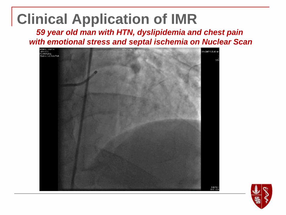
Clinical Application of IMR59 year old man with HTN, dyslipidemia and chest pain
with emotional stress and septal ischemia on Nuclear Scan

IMR = 76 x 0.70 = 53

Clinical Application of IMR68 year old man HTN and tobacco use with negative stress echo
4 months ago, but increasingly severe classic exertional angina
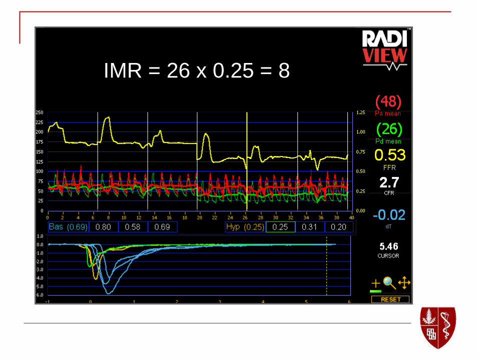
IMR = 26 x 0.25 = 8

Slow Pullback in LAD
Distal LAD Proximal LAD

IVUS of LAD

Chest Pain and “Normal Coronaries”
139 patients referred for coronary
angiography because of symptoms and/or
abnormal stress test and found to have
“normal” appearing coronaries
FFR, IMR, CFR, IVUS and acetylcholine
challenge were performed down the LAD
Lee, Tremmel, et al. Unpublished data

Chest Pain and “Normal Coronaries”
Lee, Tremmel, et al. Unpublished data
Patient Characteristic n=139
Age (years) 54 ±11
Female 77%
Hypertension 53%
Diabetes 23%
Dyslipidemia 63%
Tobacco Use 8%
Typical Angina 32%
Positive Stress Test 42%

Chest Pain and “Normal Coronaries”
The mean IMR was 19.6 ±9.1
Microvascular dysfunction was present in
21% (defined as IMR ≥ 25)
Typical angina was more frequent (52% vs 26%,
p=0.01) in patients with microvascular dysfunction
Positive stress tests were more common (65% vs.
41%, p=0.04)
Predictors of microvascular dysfunction were
age, BMI, and typical angina
Lee, Tremmel, et al. Unpublished data

Chest Pain and “Normal Coronaries”
4% of patients had an FFR of the LAD ≤ 0.80
44% had epicardial endothelial dysfunction
44% had a myocardial bridge
42% had nonischemic FFR, normal IMR, no
significant endothelial dysfunction
34% had nonischemic FFR, normal IMR, no
endothelial dysfunction and no “bridge”
Lee, Tremmel, et al. Unpublished data

Indications for IMR in Stable
Patients
To evaluate the etiology of chest
pain/abnormal stress test in a patient with
angiographically appearing normal coronaries
To assess for the likelihood of peri-PCI
myocardial infarction
Research purposes

IMR after PCI in Stable Patients
50 patients randomized to conventional stenting with predilatation versus direct stenting
IMR measured after PCI and correlated with troponin release
In the 10 patients with elevated Tn post PCI, IMR was 24.7 ±13.3 vs. 16.9 ±10.2, p=0.04.
Cuisset, et al. J Am Coll Cardiol;2008:51:1060
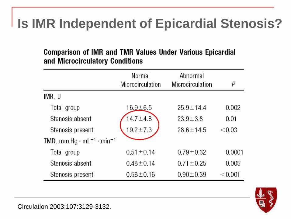
Is IMR Independent of Epicardial Stenosis?
Circulation 2003;107:3129-3132.

Importance of Collaterals when Measuring IMR
Resistance = Pressure / Qmyo
Qmyo = Qcor + Qcoll
Simplified IMR = Pd x Tmn
But Tmn is inversely proportional to coronary flow
Catheter Cardiovasc Interv 2004;62:56-63.

Importance of Collaterals when Measuring IMR
Qcor Qcoll Pd Rmyo
Catheter Cardiovasc Interv 2004;62:56-63.

Importance of Collaterals when Measuring IMR
Qcor Qcoll Pd IMRapp
To measure true IMR, must measure coronary
wedge pressure to incorporate collateral flow
Catheter Cardiovasc Interv 2004;62:56-63.
Flow ’s more than it should, Tmn ’s and IMRapp = Pd x Tmn ’s
IMR = Pd x Tmn x (FFRcor / FFRmyo)

IMR is not affected by epicardial stenosis severity:
Circulation 2004;109:2269-2272
Animal Validation

IMR is not affected by epicardial stenosis severity:
Aarnoudse, et al. Circulation 2004;110:2137-42
stenosis
After
stenting
10% AS
50% AS
75% AS
FFR = 0.53± 0.19
FFR = 0.90 ± 0.12
FFR = 0.84 ± 0.08
IMR = 22 ± 15
FFR = 0.69 ± 0.09
IMR = 23 ± 14
FFR = 0.52 ± 0.11
IMR = 23 ± 14
stenosis
After
stenting
10% AS
50% AS
75% AS
FFR = 0.53± 0.19
FFR = 0.90 ± 0.12
FFR = 0.84 ± 0.08
IMR = 22 ± 15
FFR = 0.69 ± 0.09
IMR = 23 ± 14
FFR = 0.52 ± 0.11
IMR = 23 ± 14
stenosis
After
stenting
10% AS
50% AS
75% AS
FFR = 0.53± 0.19
FFR = 0.90 ± 0.12
FFR = 0.84 ± 0.08
IMR = 22 ± 15
FFR = 0.69 ± 0.09
IMR = 23 ± 14
FFR = 0.52 ± 0.11
IMR = 23 ± 14
Human Validation

IMR is not affected by epicardial stenosis severity:
Aarnoudse, et al. Circulation 2004;110:2137-42
Human Validation
0.40.50.60.70.80.91.0
15
20
25
30
35
40
45
FFR
IMR
IMRapp
IMR

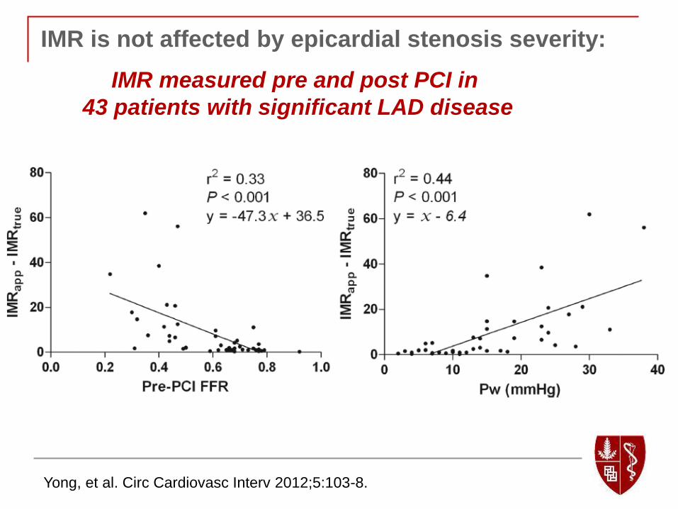
IMR is not affected by epicardial stenosis severity:
Yong, et al. Circ Cardiovasc Interv 2012;5:103-8.
IMR measured pre and post PCI in
43 patients with significant LAD disease

IMR is not affected by epicardial stenosis severity:
Yong, et al. Circ Cardiovasc Interv 2012;5:103-8.
IMR measured pre and post PCI in
43 patients with significant LAD disease
IMR is not affected by epicardial stenosis severity:
Yong, et al. Circ Cardiovasc Interv 2012;5:103-8.
IMR measured pre and post PCI in
43 patients with significant LAD disease

IMR Before PCI in Stable Patients
IMR measured before PCI in 50 stable patients undergoing LAD PCI
Ng, et al. Circ Cardiovasc Interv 2012;5:515-22.

IMR Before PCI in Stable Patients
IMR measured before LAD PCI in 50 stable patients
Multivariable Regression Analysis
Ng, et al. Circ Cardiovasc Interv 2012;5:515-22.

IMR Before PCI in Stable Patients
IMR measured before PCI in 54 stable patients
Multivariable Regression Analysis
Layland, et al. Heart 2012;98:1492-7.

Estimating True IMR without Wedge
IMR = Pd x Tmn x (FFRcor / FFRmyo)
IMR = Pd x Tmn x ((Pd-Pw)/(Pa-Pw) / (Pd/Pa))
If there is a relationship between FFRcor and
FFRmyo, perhaps we can estimate FFRcor
without having to measure the coronary
wedge pressure.
Yong, et al. J Am Coll Cardiol Intv 2013;6:53-8.

Estimating True IMR without Wedge
In a derivation cohort of 50 patients, a strong linear relationship
was found between FFRcor and FFRmyo.
Yong, et al. J Am Coll Cardiol Intv 2013;6:53-8.

Estimating True IMR without Wedge
In a validation cohort of 72 patients, there was no significant
difference in IMR with estimate FFRcor or measured FFRcor.
Yong, et al. J Am Coll Cardiol Intv 2013;6:53-8.

Estimating True IMR without Wedge
In a validation cohort of 72 patients, there was no significant
difference in IMR with estimate FFRcor or measured FFRcor.
Yong, et al. J Am Coll Cardiol Intv 2013;6:53-8.

Indications for IMR in Stable
Patients
To evaluate the etiology of chest
pain/abnormal stress test in a patient with
angiographically appearing normal coronaries
To assess for the likelihood of peri-PCI
myocardial infarction
Research purposes

IMR post Statin Therapy
Fujii, et al. J Am Coll Cardiol Intv 2011; 4:513-20.
IMR measured after PCI in 80 patients randomized to
either 1 month pretreatment with pravastatin or placebo

IMR post ACE Inhibitor Therapy
Mangiacapra, et al. J Am Coll Cardiol 2013; 61:615-21.
40 patients randomized to IC enalaprilat or placebo prior to PCI

IMR post ACE Inhibitor Therapy
Mangiacapra, et al. J Am Coll Cardiol 2013; 61:615-21.
40 patients randomized to IC enalaprilat or placebo prior to PCI

IMR post Stem Cell Therapy
Tayyareci, et al. Angiology 2008;59:145
IMR measured in 15 patients with ischemic cardiomyopathy
before and 6 months after intracoronary stem cell delivery

Conclusions:
Measurement of FFR and IMR can help to
diagnose the etiology of chest pain/abnormal
stress test in patient with angiographically
normal appearing coronaries.
IMR measured at the time of PCI can predict
peri-procedural myocardial infarction.
IMR is a useful research tool for evaluating
the efficacy of various therapies.