improved phylogenetic resolution within siphonophora ... · rudja kovia sp bargmannia lata...
TRANSCRIPT

Improved phylogenetic resolution within Siphonophora (Cnidaria)with implications for trait evolution
Catriona Munro1*,**, Stefan Siebert1,2*, Felipe Zapata3, Mark Howison4,5, Alejandro Damian Serrano1,10,Samuel H. Church1,6, Freya E. Goetz1,7, Philip R. Pugh8, Steven H.D. Haddock9, Casey W. Dunn10
1 Department of Ecology and Evolutionary Biology, Brown University, Providence, RI 02912, USA2 Current address: Department of Molecular & Cellular Biology, University of California Davis, Davis, CA95616, USA3 Department of Ecology and Evolutionary Biology, University of California Los Angeles, Los Angeles, CA90095, USA4 Brown Data Science Practice, Brown University, Brown University, Providence, RI 02912, USA5 Current address: Watson Institute for International and Public Affairs, Brown University, Providence, RI02912, USA6 Current address: Department of Organismic and Evolutionary Biology, Harvard University, Cambridge,MA 02138, USA7 Current address: Smithsonian Institution, National Museum of Natural History, Washington, DC 20560,USA8 National Oceanography Centre, Southampton, SO14 3ZH, UK9 Monterey Bay Aquarium Research Institute, Moss Landing, CA 95039, USA10 Department of Ecology and Evolutionary Biology, Yale University, New Haven, CT 06520, USA
* Authors contributed equally
** Corresponding author, [email protected]
Abstract
Siphonophores are a diverse group of hydrozoans (Cnidaria) that are found at all depths of the ocean - fromthe surface, like the familiar Portuguese man of war, to the deep sea. Siphonophores play an important rolein ocean ecosystems, and are among the most abundant gelatinous predators. A previous phylogenetic studybased on two ribosomal RNA genes provided insight into the internal relationships between major siphonophoregroups, however there was little support for many deep relationships within the clade Codonophora. Here, wepresent a new siphonophore phylogeny based on new transcriptome data from 30 siphonophore species analyzedin combination with 13 publicly available genomic and transcriptomic datasets. We use this new phylogenyto reconstruct several traits that are central to siphonophore biology, including sexual system (monoecyvs. dioecy), gain and loss of zooid types, life history traits, and habitat. The phylogenetic relationships in thisstudy are largely consistent with the previous phylogeny, but we find strong support for new clades withinCodonophora that were previously unresolved. These results have important implications for trait evolutionwithin Siphonophora, including favoring the hypothesis that monoecy arose twice.
1. Introduction
Siphonophores (Fig. 1 and 2) are among the most abundant gelatinous predators in the open ocean, andhave a large impact on ocean ecosystems (Choy et al., 2017; Pagès et al., 2001; Pugh, 1984; Pugh et al.,1997; Purcell, 1981; Williams and Conway, 1981). Siphonophores, which belong to Hydrozoa (Cnidaria), arefound at all depths in the ocean. The most familiar species is the Portuguese man of war Physalia physalis,which floats at the surface and can wash up conspicuously onto beaches (Totton, 1960). Most species are
1
All rights reserved. No reuse allowed without permission. (which was not peer-reviewed) is the author/funder, who has granted bioRxiv a license to display the preprint in perpetuity.
The copyright holder for this preprint. http://dx.doi.org/10.1101/251116doi: bioRxiv preprint first posted online Jan. 20, 2018;

planktonic, living in the water column, where some grow to be more than 30 meters in length (Mackie et al.,1987). There is also a small clade of benthic siphonophores, Rhodaliidae (Pugh, 1983), that are tethered tothe bottom for part of their lives. There are currently 187 valid described siphonophore species.
Siphonophores remain poorly known, in large part because they are fragile and difficult to collect. Theyhave, however, been of great interest for more than 150 years due to their unique structure and development(Mackie et al., 1987; Mapstone, 2014). Like many other cnidarians, they are colonial: they grow by incompleteasexual reproduction. Each colony arises from a single embryo that forms the protozooid, the first body. Oneor two growth zones (Fig. 2) then arise that asexually produce other genetically identical zooids that remainattached (Carré and Carré, 1995, 1991; Carré, 1969, 1967). These zooids are each homologous to a solitaryanimal, but are physiologically integrated (Dunn and Wagner, 2006; Mackie et al., 1987; Totton, 1965).Siphonophores differ significantly from other colonial animals in terms of colony structure and development –their zooids are highly functionally specialized and arranged in precise, repeating, species-specific patterns(Beklemishev, 1969; Cartwright and Nawrocki, 2010). The functions that zooids are specialized for includefeeding, reproducing, or swimming (Fig. 2) (Dunn and Wagner, 2006).
Understanding the unique ecology, morphology, and development of siphonophores requires a well-resolvedphylogeny of the group. The relationship of siphonophores to other hydrozoans has been difficult to resolve(Cartwright et al., 2008; Cartwright and Nawrocki, 2010; Zapata et al., 2015), but there has been progresson their internal relationships. A phylogeny (Dunn et al., 2005) based on two genes (16S, 18S) from52 siphonophore taxa addressed several long standing questions about siphonophore biology. Includingthe relationships of the three historically recognised groups, Cystonectae, Physonectae, and Calycophorae.Cystonectae was found to be sister to all other siphonophores, while Calycophorae were nested within“Physonectae”. The name Codonophora was given to this clade of “Physonectae” and Calycophorae (Dunn etal., 2005).
Major questions remained after this early work, though. There was, in particular, little support for importantdeep relationships within Codonophora. Understanding these relationships is key to resolving the evolutionof several traits of importance, including sexual systems (monoecy versus dioecy) and the gain and loss ofparticular zooids, such as palpons (Fig. 2). Here we present a broadly sampled phylogenetic analysis ofSiphonophora that considers transcriptomic data from 33 siphonophore species and 10 outgroup species (2outgroups were subsequently excluded due to poor sampling). Using 1,071 genes, we find strong supportfor many relationships found in the earlier phylogeny (Dunn et al., 2005), and also provide new resolutionfor key relationships that were unresolved in that previous study. Using this phylogeny, we reconstruct theevolutionary history of characters central to the unique biology of siphonophores, including zooid type, lifehistory traits, and habitat.
2. Material and methods
All scripts for the analyses are available in a git repository at https://github.com/caseywdunn/siphonophore_phylogeny_2017. The most recent commit at the time of the analysis presented here was7c33159bed8a993b09bb1908cf9bd317c04ef3d2.
2.1 Collecting
Specimens were collected in the north-eastern Pacific Ocean, Mediterranean, and the Gulf of California.Collection data on all examined specimens, a description of the tissue that was sampled from the colony,collection mode, sample processing details, mRNA extraction methods, sequencing library preparationmethods, and sequencing details are summarized in the file Supplementary data 1 (also found in the gitrepository). Monterey Bay and Gulf of California specimens were collected by remotely operated underwatervehicle (ROV) or during blue-water SCUBA dives. Chelophyes appendiculata and Hippopodius hippopus (Fig.1C) specimens were collected in the bay of Villefranche-sur-Mer, France, during a plankton trawl on 13 April2011. Available physical vouchers have been deposited at the Museum of Comparative Zoology (HarvardUniversity), Cambridge, MA, the Peabody Museum of Natural History (Yale University), New Haven, CT, or
2
All rights reserved. No reuse allowed without permission. (which was not peer-reviewed) is the author/funder, who has granted bioRxiv a license to display the preprint in perpetuity.
The copyright holder for this preprint. http://dx.doi.org/10.1101/251116doi: bioRxiv preprint first posted online Jan. 20, 2018;
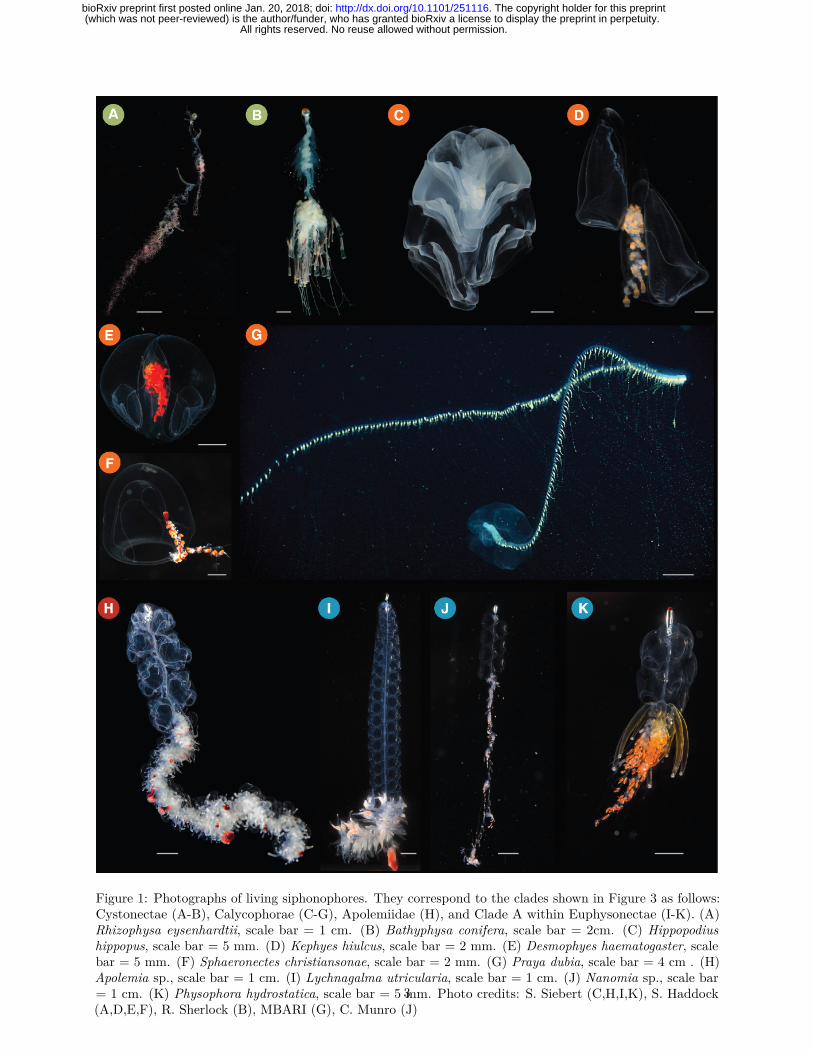
Figure 1: Photographs of living siphonophores. They correspond to the clades shown in Figure 3 as follows:Cystonectae (A-B), Calycophorae (C-G), Apolemiidae (H), and Clade A within Euphysonectae (I-K). (A)Rhizophysa eysenhardtii, scale bar = 1 cm. (B) Bathyphysa conifera, scale bar = 2cm. (C) Hippopodiushippopus, scale bar = 5 mm. (D) Kephyes hiulcus, scale bar = 2 mm. (E) Desmophyes haematogaster, scalebar = 5 mm. (F) Sphaeronectes christiansonae, scale bar = 2 mm. (G) Praya dubia, scale bar = 4 cm . (H)Apolemia sp., scale bar = 1 cm. (I) Lychnagalma utricularia, scale bar = 1 cm. (J) Nanomia sp., scale bar= 1 cm. (K) Physophora hydrostatica, scale bar = 5 mm. Photo credits: S. Siebert (C,H,I,K), S. Haddock(A,D,E,F), R. Sherlock (B), MBARI (G), C. Munro (J)
3
All rights reserved. No reuse allowed without permission. (which was not peer-reviewed) is the author/funder, who has granted bioRxiv a license to display the preprint in perpetuity.
The copyright holder for this preprint. http://dx.doi.org/10.1101/251116doi: bioRxiv preprint first posted online Jan. 20, 2018;

8
Siph
osom
eN
ecto
som
e
Nectophore
Pneumatophore
Gastrozooid
Palpon
Tentacle
Tentilla
Gonophore (male)
Gonophore (female)
Nectophore
AB
C Bract
B
C
Cormidium
Figure 2: Schematic of the siphonophore Nanomia bijuga, orientated with the anterior of the colony at thetop of the page, and the ventral side to the left. Adapted from http://commons.wikimedia.org/wiki/File:Nanomia_bijuga_whole_animal_and_growth_zones.svg, drawn by Freya Goetz. (A) Overview of thewhole mature colony. (B) Inset of the pneumatophore and nectosomal growth zone. A series of buds giverise to nectophores. (C) Inset of the siphosomal growth zone. Probuds subdivide to give rise to zooids inrepeating-units (cormidia). The gastrozooid (specialized feeding polyp) is the posterior-most zooid withineach cormidium.
4
All rights reserved. No reuse allowed without permission. (which was not peer-reviewed) is the author/funder, who has granted bioRxiv a license to display the preprint in perpetuity.
The copyright holder for this preprint. http://dx.doi.org/10.1101/251116doi: bioRxiv preprint first posted online Jan. 20, 2018;

had been previously deposited at the United States National Museum (Smithsonian Institution), Washington,DC. Accession numbers are given in Supplementary data 1. In cases where physical vouchers were unavailablewe provide photographs to document species identity (see git repository).
2.2 Sequencing
When possible, specimens were starved overnight in filtered seawater at temperatures close to ambient watertemperatures at the time of specimen collection. mRNA was extracted directly from tissue using a variety ofmethods (Supplementary data 1): Magnetic mRNA Isolation Kit (NEB, #S1550S), Invitrogen DynabeadsmRNA Direct Kit (Ambion, #61011), Zymo Quick RNA MicroPrep (Zymo #R1050), or from total RNAafter Trizol (Ambion, #15596026) extraction and through purification using Dynabeads mRNA PurificationKit (Ambion, #61006). In case of anticipated very small total RNA quantities, only a single round of beadpurification was performed. Extractions were performed according to the manufacturer’s instruction. Allsamples were DNase treated (TURBO DNA-free, Invitrogen #AM1907; or on column DNase treatmentwith Zymo Quick RNA MicroPrep). Libraries were prepared for sequencing using the Illumina TruSeq RNASample Prep Kit (Illumina, #FC-122-1001, #FC-122-1002), the Illumina TruSeq Stranded Library PrepKit (Illumina, #RS-122-2101) or the NEBNext RNA Sample Prep Master Mix Set (NEB, #E6110S). Wecollected long read paired end Illumina data for de novo transcriptome assembly. In the case of large tissueinputs, libraries were sequenced separately for each tissue, subsequently subsampled and pooled in silico.Libraries were sequenced on the HiSeq 2000, 2500, and 3000 sequencing platforms. Summary statistics foreach library are given in the file Supplementary data 2. All sequence data have been deposited in the NCBIsequence read archive (SRA) with Bioproject accession number PRJNA255132.
2.3 Analysis
New data were analysed in conjunction with 13 publicly available datasets, with a total number of 43 species.Sequence assembly, annotation, homology evaluation, gene tree construction, parsing of genes trees to isolateorthologous sequences, and supermatrix construction were conducted with Agalma v. 1.0.0 (Dunn et al.,2013; Guang et al., 2017). This workflow integrates a variety of existing tools (Altschul et al., 1990; Enrightet al., 2002; Grabherr et al., 2011; Katoh and Standley, 2013; Langmead and Salzberg, 2012; Li and Dewey,2011; Li et al., 2009; Sukumaran and Holder, 2010; Talavera and Castresana, 2007) and new methods.Maximum likelihood analyses of the supermatrix were conducted with RAxML v 8.2.0 (Stamatakis, 2006)and implemented via Agalma. Bayesian Inference (BI) analyses of the supermatrix were conducted usingPhylobayes v. 1.7a-mpi (Lartillot et al., 2009). Sequence alignments, sampled and consensus trees, andvoucher information are available in the git repository. Tree figures were rendered with ggtree (Yu et al.,2016).
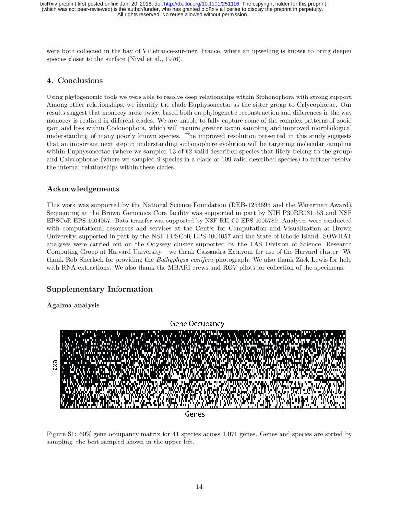
Two outgroup species, Atolla vanhoeffeni and Aegina citrea, were removed from the final supermatrix due tolow gene occupancy (gene sampling of 20.8% and 14.5% respectively in a 50% occupancy matrix with 2,203genes). The final analyses presented here consider 33 siphonophore species and 8 outgroup species. Thisincludes new data for 30 species. In the final analyses, we sampled 1,071 genes to generate a supermatrixwith 60% occupancy and a length of 378,468 amino acids (Fig. S1).
ML analyses were conducted on the unpartitioned supermatrix using the WAG+Γ model of amino acidsubstitution, and bootstrap values were estimated using 1000 replicates. BI was conducted using two differentCAT models, CAT-Poisson and CAT-GTR (Lartillot and Philippe, 2004). Two independent MCMC chainswere run under the CAT-GTR model, and four independent MCMC chains were run under the CAT-Poissonmodel. The BI analyses did not converge (CAT-Poisson: maxdiff=1, meandiff=0.0148; CAT-GTR: maxdiff=1,meandiff=0.0127). Only the results from the CAT-Poisson model are presented here. Visual inspection of thetraces indicated that a burn in of 400 trees was sufficient for all CAT-Poisson runs. This left 15847 trees inthe posterior.
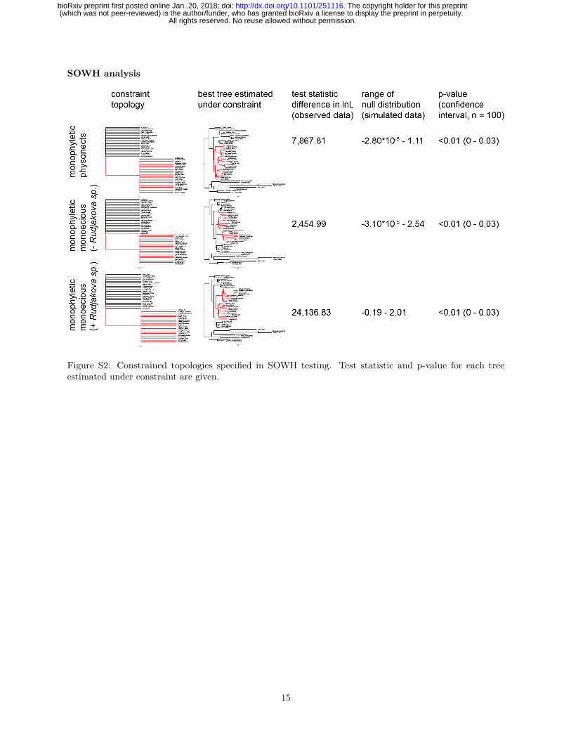
We used the Swofford-Olsen-Waddell-Hillis (SOWH) test (Swofford et al., 1996) to evaluate two hypotheses(Fig. 3C, S2): (i) “Physonectae” is monophyletic (Totton, 1965); (ii) monoecious species are monophyletic(Dunn et al., 2005). As the sexual system of Rudjakovia sp. is unclear, we carried out two tests of the
5
All rights reserved. No reuse allowed without permission. (which was not peer-reviewed) is the author/funder, who has granted bioRxiv a license to display the preprint in perpetuity.
The copyright holder for this preprint. http://dx.doi.org/10.1101/251116doi: bioRxiv preprint first posted online Jan. 20, 2018;

monophyly of monoecy, one with Rudjakovia sp. included as a monoecious species, and one without (Fig. 3C,S2). We used SOWHAT (Samuel H. Church et al., 2015) dev. version 0.39 (commit fd68ef57) to carry outthe SOWH tests in parallel with the default options and an initial sample size of 100 (analysis code can befound in the git repository). For each hypothesis we defined a topology with a single constrained node thatwas inconsistent with the most likely topology (Fig. 3). We used a threshold for significance of 0.05 andfollowing the initial 100 samples, we evaluated the confidence interval around the p-value to determine ifmore samples were necessary.
Morphological character data used in trait mapping were obtained from the literature or direct observationof available voucher material. Depth distribution data was queried from the MBARI VARS database (http://www.mbari.org/products/research-software/video-annotation-and-reference-system-vars/) (Schliningand Stout, 2006). We used stochastic character mapping to infer the probable evolution of traits on thetree in R using the phytools package (Huelsenbeck et al., 2003; Revell, 2012). Subsequent analyses wereconducted in R and integrated into this manuscript with the knitr package. See Supplementary Informationfor R package version numbers.
3. Results and Discussion
3.1 Species phylogeny and hypothesis testing
Most relationships received strong support across analysis methods (Fig. 3A), with a couple of localizedexceptions (Fig. 3B). The phylogenetic relationships recovered in this study are largely consistent withthose found in a previous study based on two genes (16S and 18S ribosomal RNA) (Dunn et al., 2005).Relationships that receive strong support in both studies include the placement of Cystonectae as sister toCodonophora (the clade that includes all other siphonophores), the placement of Apolemiidae as sister to allother codonophorans, and the placement of Calycophorae within the paraphyletic “Physonectae”. Multiplenodes that were not resolved in the previous two-gene analysis receive strong support in the present 1,071-genetranscriptome analyses. There is strong support for Pyrostephidae as sister to all other non-apolemiidcodonophorans. Within the clade that is sister to Pyrostephidae, we find two main clades, Calycophorae anda clade we here name Euphysonectae (Fig. 3A). It includes the remaining non-apolemiid, non-pyrostephid“Physonectae”. We define Euphysonectae as the clade consisting of Agalma elegans and all taxa that are moreclosely related to it than to Diphyes dispar.
Euphysonectae consists of two reciprocally monophyletic groups that we here provisionally refer to as CladeA and Clade B (Fig. 3A). The presence of an involucrum, a fold around the base of the cnidoband (Totton,1965), is a potential synapomorphy for Clade A. Species of Clade A also have a descending mantle canalwithin the nectophores (Fig. S6), a structure that is also present in some calycophorans. Members of CladeA are also monoecious (Fig. 5). There is not a clear synapomorphy for Clade B. Within Clade B there is lowsupport for the placement of Erenna richardi, which is placed as sister to Clade A in some analyses (Fig. 3B).More taxon sampling will be required to determine the relationship of species within this clade.
Within Clade A, Physophora gilmeri along with Lychnagalma utricularia (Fig. 1I) (both not included in theprevious phylogeny) are sister to Agalmatidae, a clade restricted to Agalma, Athorybia, Melophysa, Halistemmaand Nanomia (Dunn et al., 2005; Pugh, 2006). In the rDNA study, P. hydrostatica (the presumed sisterspecies to P. gilmeri) was sister to Forskaliidae with low support. The position of Cordagalma cordiforme (=Cordagalma ordinatum) (Pugh, 2016) was previously unresolved, while in this analysis Cordagalma sp. isin a clade with Forskalia asymmetrica, falling outside of Agalmatidae. Placement of Cordagalma outsideAgalmatidae is consistent with previous analyses of morphological and molecular data (Dunn et al., 2005;Pugh, 2006).
Within Calycophorae, taxon sampling is shallower here than in the previous study. The calycophoran relation-ships that can be investigated, however, are in broad agreement with the previous analysis. Calycophoranshave in the past been split into two groups, prayomorphs and diphyomorphs, based on morphology afterMackie et al. (1987). As in the previous study, the results presented here indicate that the prayomorphsare paraphyletic with respect to the diphyomorphs. Craseoa lathetica and Desmophyes sp. are sister to
6
All rights reserved. No reuse allowed without permission. (which was not peer-reviewed) is the author/funder, who has granted bioRxiv a license to display the preprint in perpetuity.
The copyright holder for this preprint. http://dx.doi.org/10.1101/251116doi: bioRxiv preprint first posted online Jan. 20, 2018;

New data Species SRA Number Depth (m) Lat LonAgalma elegans 3–20 35.56 N 122.55 W
Y Bargmannia elongata SRR1548343;SRR1548344;SRR1548345;SRR1548346;SRR1548347 412/805/636/818 36.12 N 122.67 WY Frillagalma vityazi SRR1548362;SRR1548363;SRR1548364 407 36.69 N 122.05 WY&N Nanomia bijuga SRR1548376;SRR1548377;SRR871527 414/387 36.60 N 122.15 W
Physalia physalis SRR871528Abylopsis tetragona SRR871525Aegina citrea SRS893439Aiptasia pallida SRR6967; SRR6967; SRR6967Alatina alata SRR1952741
Y Apolemia rubriversa SRR1548342 767 36.70 N 122.05 WAtolla vanhoeffeni SRR1952729
Y Chelophyes appendiculata SRR1548354 3–20Y Chuniphyes multidentata SRR1548355 327 36.79 N 122.00 W
Clytia hemisphaericaY Cordagalma sp SRR1548356 252 36.70 N 122.06 W
Ectopleura larynx SRR923510Y Erenna richardi SRR1548360 1044 36.61 N 122.38 WY Forskalia asymmetrica SRR1548361 253 36.80 N 122.00 WY Hippopodius hippopus SRR1548371 3–20 43.69 N 7.315 E
Hydra magnipapillataHydractinia symbiolongicarpus SRX474878
Y Kephyes ovata SRR1548372 452 36.36 N 122.81 WY Lilyopsis fluoracantha SRR1548373 320 36.69 N 122.04 WY Lychnagalma utricularia SRR1548374 431 36.69 N 122.04 WY Marrus claudanielis SRR1548375 1427 36.07 N 122.29 W
Nematostella vectensisY Undescribed Sp. L SRR1548381 1463 36.70 N 122.57 WY Podocoryna carnea SRR1266262Y Desmophyes sp. SRR1548358 1363 35.48 N 123.64 W
Craseoa lathetica SRR871529Y Resomia ornicephala SRR1548382 322 35.48 N 123.86 WY Rhizophysa filiformis SRR1548383 10 27.23 N 110.46 WY Stephalia dilata SRR1548384 3074 35.62 N 122.67 WY Apolemia lanosa 1073 36.70 N 122.08 WY Apolemia sp 461 36.60 N 122.15 WY Bargmannia amoena 1251 36.70 N 122.08 WY Bargmannia lata 1158 36.067 N 122.30 WY Rudjakovia sp 334 36.00 N 122.42 WY Stephalia sp 3255 36.39 N 122.67 WY Physophora gilmeri 242 36.36 N 122.40 WY Halistemma rubrum 313 24.68 N 109.90WY Athorybia rosacea 3–20 22.92 N 108.36 WY Diphyes dispar 3–20 35.93 N 122.93 W
Table 1: A complete list of specimens collected for this work. New data indicated by Y, blank fields indicatethat data were already published.
Hippopodius hippopus in this study, while in the previous study, the relationship between C. lathetica and theclade including H. hippopus was unresolved.
Using the Swofford-Olsen-Waddell-Hillis (SOWH) test (Swofford et al., 1996), we evaluated the followingthree alternative phylogenetic hypotheses against the most likely tree topology (Fig. 3C): (i) monophyleticPhysonectae, (ii) and (iii) monophyletic monoecious siphonophores, with and without Rudjakovia sp. respec-tively. In all three tests the alternative hypothesis was rejected (p-value <0.01, confidence interval: <0.001 -0.03, Fig. S2).
The broad sampling approach of this phylogeny provides new evidence for the relationships between majorsiphonophore clades within Codonophora, specifically between Pyrostephidae, Calycophorae, and the newlynamed Euphysonectae. This opens up new questions about key relationships within both Calycophorae andEuphysonectae – where future transcriptome sampling efforts should be focused. Within Euphysonectae,two clades (Clade A and Clade B) are hypothesized, however there is weaker support for Clade B (Fig. 3A,3B). Expanding sampling of species that probably fall in Clade B, including other Erenna species, rhodaliids,and relatives of Undescribed sp L, will greatly expand our understanding of these two groups and perhapsprovide evidence of Clade B synapomorphies. Similarly, within Calycophorae, increased taxon sampling is
7
All rights reserved. No reuse allowed without permission. (which was not peer-reviewed) is the author/funder, who has granted bioRxiv a license to display the preprint in perpetuity.
The copyright holder for this preprint. http://dx.doi.org/10.1101/251116doi: bioRxiv preprint first posted online Jan. 20, 2018;

Aiptasia pallida
Clytia hemisphaerica
Podocoryna carneaHydractinia symbiolongicarpus
Physalia physalisRhizophysa filiformis
Rudjakovia spBargmannia lataBargmannia elongataBargmannia amoena
Desmophyes spCraseoa lathetica
Hippopodius hippopus
Lilyopsis fluoracantha
Chelophyes appendiculataDiphyes disparAbylopsis tetragona
Chuniphyes multidentataKephyes ovata
Stephalia spStephalia dilata
Marrus claudanielisUndescribed sp L
Erenna richardi
Frillagalma vityaziResomia ornicephala
Cordagalma spForskalia asymmetrica
Physophora gilmeriLychnagalma utricularia
Nanomia bijuga
Athorybia rosaceaAgalma elegans
Halistemma rubrum
Apolemia spApolemia lanosaApolemia rubriversa
Ectopleura larynxHydra magnipapillata
Alatina alataNematostella vectensis
69/100
Support values (%)
Bootstrap / Posterior
Unlabeled nodes are >99 / >99
100/77
90/100
100/47
Calycophorae(109)
Clade A(37)
Clade B(25)
Pyrostephidae(6)
Apolemiidae(5)
Cystonectae(5)
Outgroups
Codonophora
Euphysonectae
Siphonophora
HydrozoaMedusozoaCnidaria
Outgroups
C SOWH ConstraintsB Observed Conflicting Topologies
Phys
onec
tae
PyrostephidaeClade BClade ACalycophorae
CystonectaeApolemiidae
Outgroups
mon
oeci
ous
PyrostephidaeClade B Clade ACalycophorae
CystonectaeApolemiidae
A Maximum Likelihood Phylogram
Nanomia bijugaAthorybia rosaceaAgalma elegans
Halistemma rubrum
0/53 Clade A0/23Erenna richardiClade B (remainder)
Topologies from posterior distribution that conflict with ML phylogram Both rejected
Figure 3: (A) Maximum likelihood (ML) phylogram with bipartition frequencies from the ML bootstraps andthe Bayesian posterior distribution of trees. Unlabeled nodes have support >0.99 for both bootstraps andposteriors. The numbers of valid described species estimated to be based in each clade based on taxonomyare shown below each clade name on the right. (B) The topologies found in the posterior distribution of treesthat conflict with the ML tree. (C) The topologies evaluated by the SOWH tests. For more details on theSOWH topologies refer to Fig. S2.
8
All rights reserved. No reuse allowed without permission. (which was not peer-reviewed) is the author/funder, who has granted bioRxiv a license to display the preprint in perpetuity.
The copyright holder for this preprint. http://dx.doi.org/10.1101/251116doi: bioRxiv preprint first posted online Jan. 20, 2018;

needed. This study, and the previous phylogenetic study (Dunn et al., 2005), suggest that the prayomorphsare paraphyletic, but for slightly different reasons given the different sampling of the analyses. In Dunn et al.(2005), a clade of prayomorphs including Praya dubia (Fig. 1G), Nectadamas diomedeae, and Nectopyramisnatans (not included in this study) were found to be sister to all other calycophorans, while in this study,the prayomorph Lilyopsis fluoracantha (not included in the previous study) is found in a clade includingdiphyomorph calycophorans that is sister to all other prayomorphs. Expanded taxon sampling, particularly P.dubia or a nectopyramid, but also extensive sampling across the major prayomorph and diphyomorph groups,will expand our understanding of relationships within Calycophorae. This will be especially important forunderstanding trait evolution within Calycophorae, for example, the release of eudoxids (Fig. 4), or thearrangement of male and female zooids along the stem (see section 3.2 below).
3.2 Character Evolution
3.3 Evolution of Monoecy
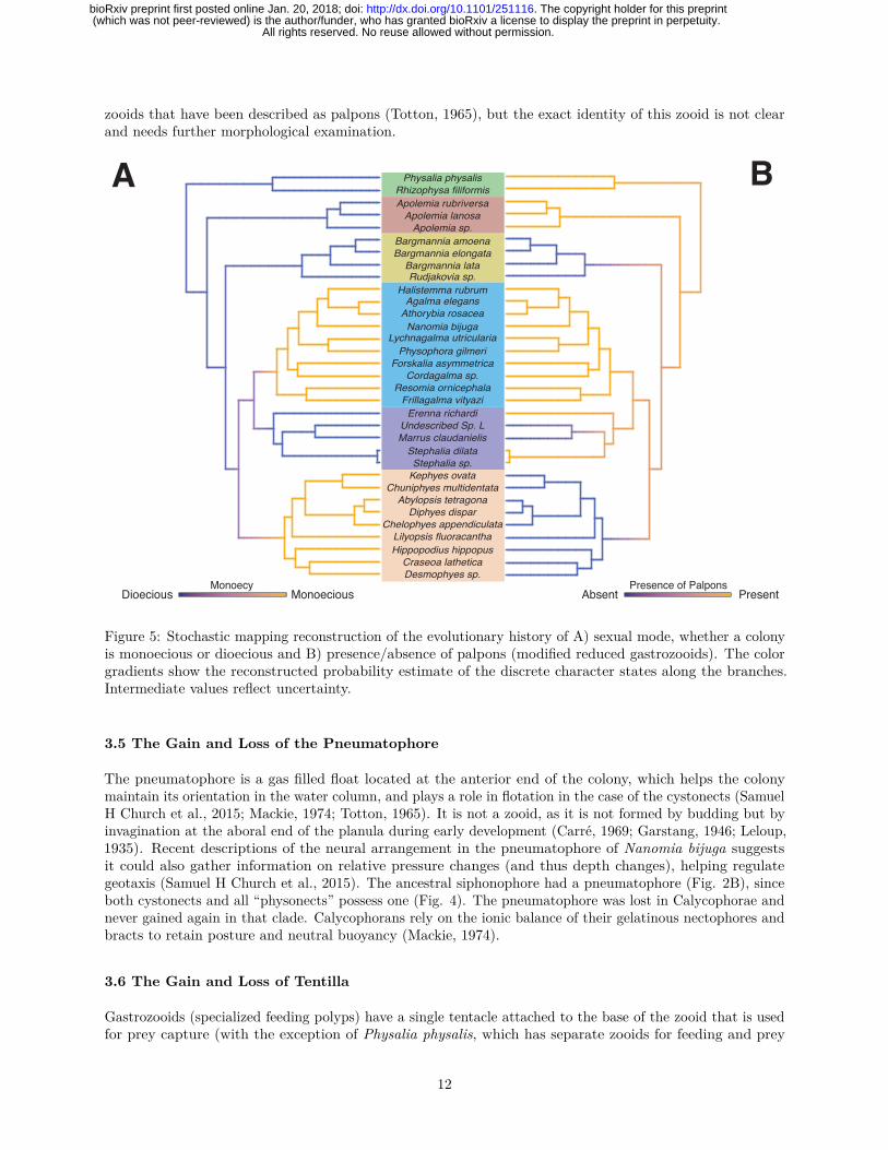
In all siphonophores, each gonophore (sexual medusa that produces gametes) is either male or female. Withineach siphonophore species, colonies are either monoecious (male and female gonophores are on the samecolony) or dioecious (male and female gonophores are on different colonies). Previous analyses suggestedthat the common ancestor of siphonophores was dioecious, and was consistent with a single gain of monoecywithin Codonophora and no secondary losses (Dunn et al., 2005). The better-resolved tree in the currentanalyses indicates that the evolution of monoecy is more complicated than this. The two clades of monoecioussiphonophores, Calycophorae and Clade A (Fig. 3A), do not form a monophyletic group. This is becauseClade B, which contains dioecious species, is also descended from their most recent common ancestor. SOWHtests strongly reject the placement of the monecious clades Calycophorae and Clade A as a group thatexcludes Clade B (Figs. 3C and S2). The positions of the only two taxa from Clade B in the previousanalysis (Dunn et al., 2005), Erenna and Stephalia, were unresolved in that previous study. This difference inconclusions regarding trait evolution, therefore, does not reflect a contradiction between alternative stronglysupported results, but the resolution of earlier polytomies in a way that indicates there has been homoplasyin the evolution of monoecy.
The distribution of monoecy is consistent with two scenarios (Fig. 4). In the first, there is a single shift fromdioecy to monoecy along the branch that give rise to the most recent common ancestor of Calycophorae andEuphysonectae, followed by a shift back to dioecy along the branch that gave rise to Clade B. In the second,monoecy arose twice – once along the branch that gave rise to Clade A and again along the branch that gaverise to Calycophorae.
Ancestral character state reconstructions favor the hypothesis that monoecy arose twice (Fig. 5A), once inCalycophorae and once in Clade A. This is consistent with differences in the arrangements of male and femalegonophores in the two clades. In Clade A, male and female zooids are found within the same cormidium (asingle reiterated sequence of zooids along the stem, see Fig. 2). In these species, the male and female zooidsare placed at very different but well defined locations within the cormidium. Meanwhile in calycophorans,each cormidium bears either male or female gonophores. In this form of monoecy, the male and femalecormidia can either be in an alternating pattern, or there can be several male or female cormidia in a row. Ineither case, male and female zooids are found at the same corresponding locations within the cormidia. Insum, homoplasy in sexual system evolution along with variation in the spatial arrangement of gonophoreswithin a colony suggest that siphonophores have evolved different ways to be monoecious. Both Calycophoraeand Clade A have a large proportion of shallow water species (see section 3.7), suggesting that there may bean association between habitat depth and sexual mode. Similar independent transitions from gonochorism(separate sex) to hermaphroditism (both sexes in the same individual) have been identified in shallow-waterscleractinian corals (Anthozoa, Cnidaria) (Kerr et al., 2011). To test this hypothesis, a more extensive taxonsampling of the Calycophorae is needed.
Within Calycophorae there are some variations in sexual mode – in Sulculeolaria (not included in thisphylogeny) colonies appear to consist of only one sex, however they are monoecious and protandrous, withfemale gonophores developing after the release of male gonophores (Carré, 1979). Environmental influences
9
All rights reserved. No reuse allowed without permission. (which was not peer-reviewed) is the author/funder, who has granted bioRxiv a license to display the preprint in perpetuity.
The copyright holder for this preprint. http://dx.doi.org/10.1101/251116doi: bioRxiv preprint first posted online Jan. 20, 2018;

may also play a role in determining the expressed sex. Colonies of the calycophoran Chelophyes appendiculatacollected in the field always bear both male and female gonophores whereas when kept in culture onlygonophores of one sex are maintained (Carré and Carré, 2000). This suggests a high plasticity of the sexualstate in some calycophoran taxa and underlines the need for caution when evaluating the state of thischaracter in rarely collected species.
3.4 The Evolution of Zooid Types
One of the most striking aspects of siphonophore biology is their diversity of unique zooid types (Beklemishev,1969; Cartwright and Nawrocki, 2010). For example, members of Forskalia have 5 basic zooid types(nectophore, gastrozooid, palpon, bract, and gonophore), and in some species, a total of 9 when consideringzooid subtypes (4 types of bract, male & female gonophores)(Pugh, 2003). Here we reconstruct the evolutionaryorigins of several zooid types on the present transcriptome tree (Fig. 4).
Nectophores (Fig. 2) are non-reproductive propulsive medusae. In Codonophora, the nectophores are localizedto a region known as the nectosome (Fig. 2B), which has its own growth zone, and are used for coordinatedcolony-level swimming. Planktonic cystonects like Bathyphysa sibogae and Rhizophysa filiformis (Fig. 1A)instead move through the water column using repeated contraction and relaxation of the stem, and usemodified flattened gastrozooids with wings (called ptera) to increase surface area and prevent colony sinking(Biggs and Harbison, 1976). Nectophores are also present within the gonodendra (reproductive structures) ofcystonects, and are thought to propel the gonodendra when they detach from the colony (Totton, 1960, 1965).It is not clear whether the nectophores found within the siphosome of the cystonects are homologous to thenectophores borne on the nectosome of codonophorans. Similarly, the homology of the special nectophoreassociated with gonophores of the calycophoran Stephanophyes superba is also unclear (Chun, 1891). As such,we only consider the evolution of the nectosome in this study, and not the presence/absence of nectophores.The present analyses, as well as the analyses of Dunn et al. (2005), are consistent with a single origin of thenectosome (Fig. S5).
Within the nectosome, the nectophores can be attached along the dorsal or ventral side of the stem, followingthe orientation framework of Haddock et al. (2005). Our ancestral reconstructions for this character (Fig.S7) suggest that ventral attachment of nectophores was the ancestral state in Codonophora, and that dorsalattachment has independently evolved twice – once along the stem of Agalmatidae and once along the stemof Pyrostephidae. The functional implication of dorsal vs. ventral attachment is not clear.
Bracts are highly reduced zooids unique to siphonophores, where they are only present in Codonophora(Fig. 4). Bracts are functional for protection of the delicate zooids and to help maintain neutral buoyancy(Jacobs, 1937). Some calycophorans are able to actively exclude sulphate ions in their bracts to adjusttheir buoyancy along the colony (Bidigare and Biggs, 1980). Bracts were lost in Hippopodiidae, someclausophyids, Physophora hydrostatica (Fig. 1K), and in Gymnopraia lapislazula. These patterns of lossare not captured in this study, as most of these species are not included in the present phylogeny. Inspecies without bracts, a diversity of zooids appear to fulfil the roles of neutral buoyancy and protection. InPhysophora hydrostatica, enlarged palpons surround all other siphosomal zooids and move in a coordinatedmanner to inflict a powerful sting (Totton, 1965). While Hippopodius hippopus have up to twelve nectophoresand can retract the stem within the nectophores – the nectophores play a role in maintaining neutral buoyancyand possibly also in defense, by bioluminescing and blanching in response to stimuli (Fig. 1C shows theblanching of nectophores)(Bassot et al., 1978).
Palpons are modified reduced gastrozooids used for digestion and circulation of the gastrovascular fluid(Mackie et al., 1987). We do not distinguish here between gonopalpons (palpons associated with gonodendra,without a tentacle, as in the cystonects) and palpons borne on the stem (typically with a reduced tentacle orpalpacle) (Totton, 1965). We reconstruct them as present in the common ancestor of siphonophores (Fig.5B), retained in most species, but lost three times independently in the branches leading to Pyrostephidae(represented here by the genera Bargmannia and Rudjakovia), in calycophorans, and in Marrus claudanielis.Within the calycophorans, one species Stephanophyes superba (not included in this phylogeny) has polyp-like
10
All rights reserved. No reuse allowed without permission. (which was not peer-reviewed) is the author/funder, who has granted bioRxiv a license to display the preprint in perpetuity.
The copyright holder for this preprint. http://dx.doi.org/10.1101/251116doi: bioRxiv preprint first posted online Jan. 20, 2018;

Desm
ophy
es s
p.
Cras
eoa
lath
etica
Hipp
opod
ius
hipp
opus
Lilyo
psis
fluor
acan
tha
Chel
ophy
es a
ppen
dicu
lata
Diph
yes
disp
ar
Abylo
psis
tetra
gona
Chun
iphy
es m
ultid
enta
ta
Keph
yes
ovat
a
Step
halia
sp.
Step
halia
dila
ta
Mar
rus
claud
anie
lis
Unde
scrib
ed S
p. L
Eren
na ri
char
di
Frilla
galm
a vit
yazi
Reso
mia
orn
iceph
ala
Cord
agal
ma
sp.
Fors
kalia
asy
mm
etric
a
Phys
opho
ra g
ilmer
i
Lych
naga
lma
utric
ular
ia
Nano
mia
biju
ga
Atho
rybi
a ro
sace
a
Agal
ma
eleg
ans
Halis
tem
ma
rubr
um
Rudj
akov
ia s
p.
Barg
man
nia
lata
Barg
man
nia
elon
gata
Barg
man
nia
amoe
na
Apol
emia
sp.
Apol
emia
lano
sa
Apol
emia
rubr
ivers
a
Phys
alia
phy
salis
Rhizo
phys
a fili
form
is
Tentilla
Eudoxia
Monoecy
Palpons
Bracts
Nectosome
Pneumatophore
AbsentPresent
Dep
th (m
)
Observed Blue Water Diving
0
1000
2000
3000
4000
Dorsal Nectosome
Descending Mantle Canal
?
Definition SensitiveUnknown?Non-Applicable
P. physalis floatingon the surface.
Figure 4: Siphonophore phylogeny showing the distribution of the main anatomical characters and thebathymetric distributions of the different species. Bottom: siphonophore phylogeny, colored by clade. Middlepanel: diagram showing the presence/absence of traits across Siphonophora, with the physical location of thetrait shown on a schematic of Nanomia bijuga (schematic by Freya Goetz). Top: Bathymetric distribution ofsiphonophore species. Physalia illustration by Noah Schlottman, taken from http://phylopic.org/
11
All rights reserved. No reuse allowed without permission. (which was not peer-reviewed) is the author/funder, who has granted bioRxiv a license to display the preprint in perpetuity.
The copyright holder for this preprint. http://dx.doi.org/10.1101/251116doi: bioRxiv preprint first posted online Jan. 20, 2018;
zooids that have been described as palpons (Totton, 1965), but the exact identity of this zooid is not clearand needs further morphological examination.
Absent PresentPresence of Palpons
Desmophyes sp.Craseoa lathetica
Hippopodius hippopusLilyopsis fluoracantha
Chelophyes appendiculataDiphyes dispar
Abylopsis tetragonaChuniphyes multidentata
Kephyes ovataStephalia sp.
Stephalia dilataMarrus claudanielisUndescribed Sp. L
Erenna richardiFrillagalma vityazi
Resomia ornicephalaCordagalma sp.
Forskalia asymmetricaPhysophora gilmeri
Lychnagalma utriculariaNanomia bijuga
Athorybia rosaceaAgalma elegans
Halistemma rubrumRudjakovia sp.
Bargmannia lataBargmannia elongataBargmannia amoena
Apolemia sp.Apolemia lanosa
Apolemia rubriversaRhizophysa filiformis
Physalia physalisA B
Dioecious MonoeciousMonoecy
Figure 5: Stochastic mapping reconstruction of the evolutionary history of A) sexual mode, whether a colonyis monoecious or dioecious and B) presence/absence of palpons (modified reduced gastrozooids). The colorgradients show the reconstructed probability estimate of the discrete character states along the branches.Intermediate values reflect uncertainty.
3.5 The Gain and Loss of the Pneumatophore
The pneumatophore is a gas filled float located at the anterior end of the colony, which helps the colonymaintain its orientation in the water column, and plays a role in flotation in the case of the cystonects (SamuelH Church et al., 2015; Mackie, 1974; Totton, 1965). It is not a zooid, as it is not formed by budding but byinvagination at the aboral end of the planula during early development (Carré, 1969; Garstang, 1946; Leloup,1935). Recent descriptions of the neural arrangement in the pneumatophore of Nanomia bijuga suggestsit could also gather information on relative pressure changes (and thus depth changes), helping regulategeotaxis (Samuel H Church et al., 2015). The ancestral siphonophore had a pneumatophore (Fig. 2B), sinceboth cystonects and all “physonects” possess one (Fig. 4). The pneumatophore was lost in Calycophorae andnever gained again in that clade. Calycophorans rely on the ionic balance of their gelatinous nectophores andbracts to retain posture and neutral buoyancy (Mackie, 1974).
3.6 The Gain and Loss of Tentilla
Gastrozooids (specialized feeding polyps) have a single tentacle attached to the base of the zooid that is usedfor prey capture (with the exception of Physalia physalis, which has separate zooids for feeding and prey
12
All rights reserved. No reuse allowed without permission. (which was not peer-reviewed) is the author/funder, who has granted bioRxiv a license to display the preprint in perpetuity.
The copyright holder for this preprint. http://dx.doi.org/10.1101/251116doi: bioRxiv preprint first posted online Jan. 20, 2018;

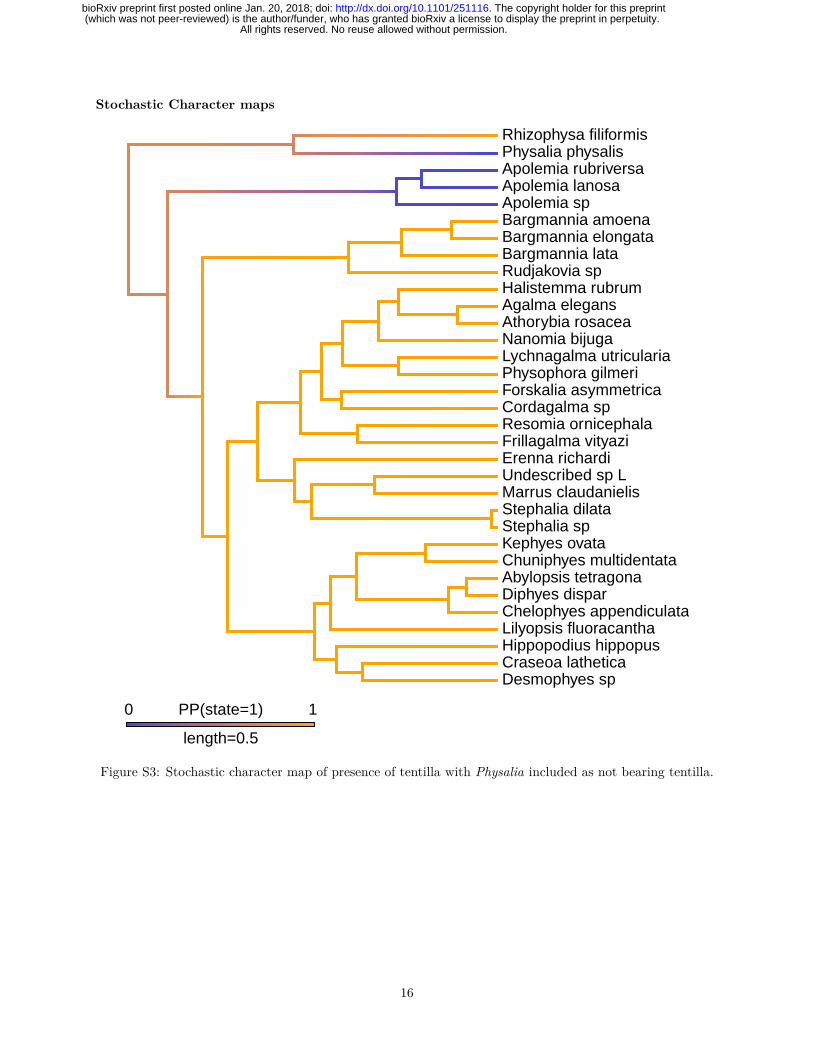
capture). As in other cnidarians, nematocyst stinging capsules, arranged in dense nematocyst batteries, playa critical role in prey capture. In many siphonophore species these batteries are found in side branches ofthe tentacle, termed tentilla (Fig. 2A). Outside of Siphonophora, most hydrozoans bear simple tentacleswithout side branches. It is still an open question whether the common ancestor of Siphonophora had tentilla.The only siphonophores species regarded as lacking tentilla are Physalia physalis, Apolemia spp. (Fig. 1H),and Bathyphysa conifera (Fig. 1B). Since B. conifera is the only member of the Rhizophysidae (and of theBathyphysa genus) lacking tentilla, we assume this is a case of secondary loss. When we reconstruct theevolution of this character on the current phylogeny, 70% of simulations support a common ancestor bearingtentilla, with two independent losses leading to Physalia and Apolemia (Fig. S3). However, this leaves a 30%support for a simple-tentacled common ancestor followed by 2 independent gains of tentilla in the branchesleading to Rhizophysidae and non-apolemiid codonophorans.
How we define absence of tentilla, especially for Physalia physalis, is also important. The tentacles of thisspecies, when uncoiled, show very prominent, evenly spaced, bulging buttons which contain in the ectodermall functional nematocytes (carrying mature nematocysts) used by the organism for prey capture (Hessingerand Ford, 1988; Totton, 1960). Siphonophore tentilla are complete diverticular branchings of the tentacleectoderm, mesoglea, and gastrovascular canal (lined by endoderm). Physalia’s buttons enclose individualfluid-filled chambers connected by narrow channels to the tentacular canal, lined by endoderm (Bardi andMarques, 2007). This suggests they are not just ectodermal swellings, but probably reduced tentilla. Whenwe define Physalia physalis as tentilla bearing, the results for the character reconstruction lead to a morerobust support for a tentilla-bearing common ancestor followed by independent losses of tentilla in the branchleading to Apolemiidae (Fig. S4), and in Bathyphysa conifera. The application of phylogenetic methods tothe evolution of tentillum morphology would be a crucial step towards understanding the evolution of thesestructures, and their relationship with the feeding ecology of siphonophores.
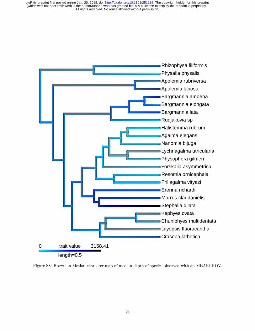
3.7 The Evolution of Vertical Habitat Use
Siphonophores are abundant predators in the pelagic realm, ranging from the surface (Physalia physalis)to bathypelagic depths (Figs. 4 and S8) (Mackie et al., 1987; Mapstone, 2014). The depth distribution ofsiphonophore populations is not always static, as some species are known to be vertical migrators, althoughthis is within a relatively narrow depth range (<100m) (Pugh, 1984). Some species such as Nanomia bijugaexhibit synchronous diel migration patterns (Barham, 1966). Using the present phylogeny, we reconstructedthe median depth changes along the phylogeny under a Brownian Motion model (Fig. S9), which had thestrongest AICc support (compared to non-phylogenetic distributions, and to Ohrnstein-Uhlenbeck). Thismodel indicates a mesopelagic most recent common ancestor, with several independent transition events toepipelagic and bathypelagic waters. There was only a single transition to benthic lifestyle on the branchof Rhodaliidae, and a single transition to a pleustonic lifestyle on the branch of Physalia physalis. Thereis evidence that habitat depth is conserved within some clades, with the exception of Calycophorae whichhave diversified across the water column (Figs. S8, S9). Depth appears to be phylogenetically conserved inEuphysonectae after the split between Clade A (shallow living species) and Clade B (deep dwelling species);however several shallow-living species that likely belong in Clade B were not included in this analysis. Thepresent sampling is also not sufficient to capture significant variation in depth distributions between closelyrelated species. Previous studies have shown that many species that are collected at the same locality arefound to occupy discrete, largely non-overlapping depth distributions, including between species that areclosely related (Pugh, 1974). This suggests that vertical habitat use is more labile than it appears and maybe an important mechanism in siphonophore ecology.
This reconstruction (Fig. S8) only included depths recorded using an ROV, thus it excludes many otherindependent colonizations of the epipelagic habitat. The ROV observations are reliable below 200m, and noquantitative measurements were made on SCUBA dives. Species such as Hippopodius hippopus, Athorybiarosacea, Diphyes dispar, and Chelophyes appendiculata are often encountered blue water diving less than 20mfrom the surface (Fig. 4). We also reconstructed the median depth changes along the phylogeny using mediandepths of 20m for all species collected by SCUBA diving or via a shallow trawl (Fig. S9), and still findsupport for a mesopelagic ancestor. It is important to note, however, that H. hippopus and C. appendiculata
13
All rights reserved. No reuse allowed without permission. (which was not peer-reviewed) is the author/funder, who has granted bioRxiv a license to display the preprint in perpetuity.
The copyright holder for this preprint. http://dx.doi.org/10.1101/251116doi: bioRxiv preprint first posted online Jan. 20, 2018;
were both collected in the bay of Villefrance-sur-mer, France, where an upwelling is known to bring deeperspecies closer to the surface (Nival et al., 1976).
4. Conclusions
Using phylogenomic tools we were able to resolve deep relationships within Siphonophora with strong support.Among other relationships, we identify the clade Euphysonectae as the sister group to Calycophorae. Ourresults suggest that monoecy arose twice, based both on phylogenetic reconstruction and differences in the waymonoecy is realized in different clades. We are unable to fully capture some of the complex patterns of zooidgain and loss within Codonophora, which will require greater taxon sampling and improved morphologicalunderstanding of many poorly known species. The improved resolution presented in this study suggeststhat an important next step in understanding siphonophore evolution will be targeting molecular samplingwithin Euphysonectae (where we sampled 13 of 62 valid described species that likely belong to the group)and Calycophorae (where we sampled 9 species in a clade of 109 valid described species) to further resolvethe internal relationships within these clades.
Acknowledgements
This work was supported by the National Science Foundation (DEB-1256695 and the Waterman Award).Sequencing at the Brown Genomics Core facility was supported in part by NIH P30RR031153 and NSFEPSCoR EPS-1004057. Data transfer was supported by NSF RII-C2 EPS-1005789. Analyses were conductedwith computational resources and services at the Center for Computation and Visualization at BrownUniversity, supported in part by the NSF EPSCoR EPS-1004057 and the State of Rhode Island. SOWHATanalyses were carried out on the Odyssey cluster supported by the FAS Division of Science, ResearchComputing Group at Harvard University – we thank Cassandra Extavour for use of the Harvard cluster. Wethank Rob Sherlock for providing the Bathyphysa conifera photograph. We also thank Zack Lewis for helpwith RNA extractions. We also thank the MBARI crews and ROV pilots for collection of the specimens.
Supplementary Information
Agalma analysis
Figure S1: 60% gene occupancy matrix for 41 species across 1,071 genes. Genes and species are sorted bysampling, the best sampled shown in the upper left.
14
All rights reserved. No reuse allowed without permission. (which was not peer-reviewed) is the author/funder, who has granted bioRxiv a license to display the preprint in perpetuity.
The copyright holder for this preprint. http://dx.doi.org/10.1101/251116doi: bioRxiv preprint first posted online Jan. 20, 2018;
SOWH analysis
Figure S2: Constrained topologies specified in SOWH testing. Test statistic and p-value for each treeestimated under constraint are given.
15
All rights reserved. No reuse allowed without permission. (which was not peer-reviewed) is the author/funder, who has granted bioRxiv a license to display the preprint in perpetuity.
The copyright holder for this preprint. http://dx.doi.org/10.1101/251116doi: bioRxiv preprint first posted online Jan. 20, 2018;
Stochastic Character maps
Desmophyes spCraseoa latheticaHippopodius hippopusLilyopsis fluoracanthaChelophyes appendiculataDiphyes disparAbylopsis tetragonaChuniphyes multidentataKephyes ovataStephalia spStephalia dilataMarrus claudanielisUndescribed sp LErenna richardiFrillagalma vityaziResomia ornicephalaCordagalma spForskalia asymmetricaPhysophora gilmeriLychnagalma utriculariaNanomia bijugaAthorybia rosaceaAgalma elegansHalistemma rubrumRudjakovia spBargmannia lataBargmannia elongataBargmannia amoenaApolemia spApolemia lanosaApolemia rubriversaPhysalia physalisRhizophysa filiformis
0 1PP(state=1)
length=0.5
Figure S3: Stochastic character map of presence of tentilla with Physalia included as not bearing tentilla.
16
All rights reserved. No reuse allowed without permission. (which was not peer-reviewed) is the author/funder, who has granted bioRxiv a license to display the preprint in perpetuity.
The copyright holder for this preprint. http://dx.doi.org/10.1101/251116doi: bioRxiv preprint first posted online Jan. 20, 2018;

Desmophyes spCraseoa latheticaHippopodius hippopusLilyopsis fluoracanthaChelophyes appendiculataDiphyes disparAbylopsis tetragonaChuniphyes multidentataKephyes ovataStephalia spStephalia dilataMarrus claudanielisUndescribed sp LErenna richardiFrillagalma vityaziResomia ornicephalaCordagalma spForskalia asymmetricaPhysophora gilmeriLychnagalma utriculariaNanomia bijugaAthorybia rosaceaAgalma elegansHalistemma rubrumRudjakovia spBargmannia lataBargmannia elongataBargmannia amoenaApolemia spApolemia lanosaApolemia rubriversaPhysalia physalisRhizophysa filiformis
0 1PP(state=1)
length=0.5
Figure S4: Stochastic character map of presence of tentilla with Physalia included as bearing tentilla.
17
All rights reserved. No reuse allowed without permission. (which was not peer-reviewed) is the author/funder, who has granted bioRxiv a license to display the preprint in perpetuity.
The copyright holder for this preprint. http://dx.doi.org/10.1101/251116doi: bioRxiv preprint first posted online Jan. 20, 2018;

Desmophyes spCraseoa latheticaHippopodius hippopusLilyopsis fluoracanthaChelophyes appendiculataDiphyes disparAbylopsis tetragonaChuniphyes multidentataKephyes ovataStephalia spStephalia dilataMarrus claudanielisUndescribed sp LErenna richardiFrillagalma vityaziResomia ornicephalaCordagalma spForskalia asymmetricaPhysophora gilmeriLychnagalma utriculariaNanomia bijugaAthorybia rosaceaAgalma elegansHalistemma rubrumRudjakovia spBargmannia lataBargmannia elongataBargmannia amoenaApolemia spApolemia lanosaApolemia rubriversaPhysalia physalisRhizophysa filiformis
0 1PP(state=1)
length=0.5
Figure S5: Stochastic character map of presence of nectosome.
18
All rights reserved. No reuse allowed without permission. (which was not peer-reviewed) is the author/funder, who has granted bioRxiv a license to display the preprint in perpetuity.
The copyright holder for this preprint. http://dx.doi.org/10.1101/251116doi: bioRxiv preprint first posted online Jan. 20, 2018;

Desmophyes spCraseoa latheticaHippopodius hippopusLilyopsis fluoracanthaChelophyes appendiculataDiphyes disparAbylopsis tetragonaChuniphyes multidentataKephyes ovataStephalia spStephalia dilataMarrus claudanielisUndescribed sp LErenna richardiFrillagalma vityaziResomia ornicephalaCordagalma spForskalia asymmetricaPhysophora gilmeriLychnagalma utriculariaNanomia bijugaAgalma elegansHalistemma rubrumRudjakovia spBargmannia lataBargmannia elongataBargmannia amoenaApolemia spApolemia lanosaApolemia rubriversa
0 1PP(state=1)
length=0.447
Figure S6: Stochastic character map of presence of a descending mantle canal in the nectophores. Cystonectsand Athorybia were excluded as they do not have a nectosome.
19
All rights reserved. No reuse allowed without permission. (which was not peer-reviewed) is the author/funder, who has granted bioRxiv a license to display the preprint in perpetuity.
The copyright holder for this preprint. http://dx.doi.org/10.1101/251116doi: bioRxiv preprint first posted online Jan. 20, 2018;

Desmophyes spCraseoa latheticaHippopodius hippopusLilyopsis fluoracanthaChelophyes appendiculataDiphyes disparAbylopsis tetragonaChuniphyes multidentataKephyes ovataStephalia spStephalia dilataMarrus claudanielisUndescribed sp LErenna richardiFrillagalma vityaziResomia ornicephalaCordagalma spForskalia asymmetricaPhysophora gilmeriLychnagalma utriculariaNanomia bijugaAgalma elegansHalistemma rubrumRudjakovia spBargmannia lataBargmannia elongataBargmannia amoenaApolemia spApolemia lanosaApolemia rubriversa
0 1PP(state=Ventral)
length=0.447
Figure S7: Stochastic character map for the evolution of the position of the nectosome. Cystonects wereexcluded as they do not have a nectosome.
20
All rights reserved. No reuse allowed without permission. (which was not peer-reviewed) is the author/funder, who has granted bioRxiv a license to display the preprint in perpetuity.
The copyright holder for this preprint. http://dx.doi.org/10.1101/251116doi: bioRxiv preprint first posted online Jan. 20, 2018;
Craseoa lathetica
Lilyopsis fluoracantha
Chuniphyes multidentata
Kephyes ovata
Stephalia dilata
Marrus claudanielis
Erenna richardi
Frillagalma vityazi
Resomia ornicephala
Forskalia asymmetrica
Physophora gilmeri
Lychnagalma utricularia
Nanomia bijuga
Agalma elegans
Halistemma rubrum
Rudjakovia sp
Bargmannia lata
Bargmannia elongata
Bargmannia amoena
Apolemia lanosa
Apolemia rubriversa
Physalia physalis
Rhizophysa filiformis
0 3158.41trait value
length=0.5
Figure S8: Brownian Motion character map of median depth of species observed with an MBARI ROV.
21
All rights reserved. No reuse allowed without permission. (which was not peer-reviewed) is the author/funder, who has granted bioRxiv a license to display the preprint in perpetuity.
The copyright holder for this preprint. http://dx.doi.org/10.1101/251116doi: bioRxiv preprint first posted online Jan. 20, 2018;

Desmophyes spCraseoa latheticaHippopodius hippopusLilyopsis fluoracanthaChelophyes appendiculataDiphyes disparAbylopsis tetragonaChuniphyes multidentataKephyes ovataStephalia spStephalia dilataMarrus claudanielisUndescribed sp LErenna richardiFrillagalma vityaziResomia ornicephalaCordagalma spForskalia asymmetricaPhysophora gilmeriLychnagalma utriculariaNanomia bijugaAthorybia rosaceaAgalma elegansHalistemma rubrumRudjakovia spBargmannia lataBargmannia elongataBargmannia amoenaApolemia spApolemia lanosaApolemia rubriversaPhysalia physalisRhizophysa filiformis
0 3255trait value
length=0.5
Figure S9: Brownian Motion character map of median depth of species including blue water diving observations.
22
All rights reserved. No reuse allowed without permission. (which was not peer-reviewed) is the author/funder, who has granted bioRxiv a license to display the preprint in perpetuity.
The copyright holder for this preprint. http://dx.doi.org/10.1101/251116doi: bioRxiv preprint first posted online Jan. 20, 2018;

Software versions
This manuscript was computed on Fri Jan 19 20:06:20 2018 with the following R package versions.
R version 3.4.1 (2017-06-30)Platform: x86_64-apple-darwin15.6.0 (64-bit)Running under: macOS Sierra 10.12.2
Matrix products: defaultBLAS: /Library/Frameworks/R.framework/Versions/3.4/Resources/lib/libRblas.0.dylibLAPACK: /Library/Frameworks/R.framework/Versions/3.4/Resources/lib/libRlapack.dylib
locale:[1] en_GB.UTF-8/en_GB.UTF-8/en_GB.UTF-8/C/en_GB.UTF-8/en_GB.UTF-8
attached base packages:[1] grid parallel stats graphics grDevices utils datasets[8] methods base
other attached packages:[1] bindrcpp_0.2 phylolm_2.5 geomorph_3.0.5 rgl_0.98.1[5] adephylo_1.1-10 ade4_1.7-8 phylobase_0.8.4 geiger_2.0.6[9] phangorn_2.2.0 phytools_0.6-20 picante_1.6-2 nlme_3.1-131
[13] vegan_2.4-4 lattice_0.20-35 permute_0.9-4 ape_4.1[17] hutan_0.5.0 FactoMineR_1.38 factoextra_1.0.5 gridExtra_2.3[21] seriation_1.2-2 fields_9.0 maps_3.2.0 spam_2.1-1[25] dotCall64_0.9-04 ggtree_1.8.2 treeio_1.0.2 cowplot_0.8.0[29] xtable_1.8-2 jsonlite_1.5 knitr_1.17 digest_0.6.12[33] magrittr_1.5 forcats_0.2.0 stringr_1.2.0 dplyr_0.7.4[37] purrr_0.2.3 readr_1.1.1 tidyr_0.7.1 tibble_1.3.4[41] ggplot2_2.2.1 tidyverse_1.1.1
loaded via a namespace (and not attached):[1] readxl_1.0.0 uuid_0.1-2[3] backports_1.1.1 fastmatch_1.1-0[5] plyr_1.8.4 igraph_1.1.2[7] lazyeval_0.2.0 sp_1.2-5[9] splines_3.4.1 rncl_0.8.2
[11] foreach_1.4.3 htmltools_0.3.6[13] viridis_0.4.0 gdata_2.18.0[15] cluster_2.0.6 gclus_1.3.1[17] modelr_0.1.1 gmodels_2.16.2[19] prettyunits_1.0.2 jpeg_0.1-8[21] colorspace_1.3-2 rvest_0.3.2[23] ggrepel_0.7.0 haven_1.1.0[25] bindr_0.1 survival_2.41-3[27] iterators_1.0.8 glue_1.1.1[29] registry_0.3 gtable_0.2.0[31] seqinr_3.4-5 kernlab_0.9-25[33] prabclus_2.2-6 DEoptimR_1.0-8[35] scales_0.5.0 mvtnorm_1.0-6[37] DBI_0.7 Rcpp_0.12.13[39] plotrix_3.6-6 viridisLite_0.2.0[41] progress_1.1.2 spdep_0.6-15
23
All rights reserved. No reuse allowed without permission. (which was not peer-reviewed) is the author/funder, who has granted bioRxiv a license to display the preprint in perpetuity.
The copyright holder for this preprint. http://dx.doi.org/10.1101/251116doi: bioRxiv preprint first posted online Jan. 20, 2018;

[43] flashClust_1.01-2 foreign_0.8-69[45] subplex_1.4-1 bold_0.5.0[47] mclust_5.3 deSolve_1.20[49] stats4_3.4.1 animation_2.5[51] htmlwidgets_0.9 httr_1.3.1[53] gplots_3.0.1 fpc_2.1-10[55] modeltools_0.2-21 pkgconfig_2.0.1[57] reshape_0.8.7 XML_3.98-1.9[59] flexmix_2.3-14 deldir_0.1-14[61] nnet_7.3-12 crul_0.4.0[63] tidyselect_0.2.0 labeling_0.3[65] rlang_0.1.2 reshape2_1.4.2[67] munsell_0.4.3 cellranger_1.1.0[69] tools_3.4.1 broom_0.4.2[71] evaluate_0.10.1 yaml_2.1.14[73] robustbase_0.92-7 caTools_1.17.1[75] dendextend_1.5.2 mime_0.5[77] whisker_0.3-2 taxize_0.9.0[79] adegenet_2.1.0 leaps_3.0[81] xml2_1.1.1 compiler_3.4.1[83] curl_2.8.1 clusterGeneration_1.3.4[85] RNeXML_2.0.7 stringi_1.1.5[87] highr_0.6 trimcluster_0.1-2[89] Matrix_1.2-11 psych_1.7.8[91] msm_1.6.4 LearnBayes_2.15[93] combinat_0.0-8 data.table_1.10.4[95] bitops_1.0-6 httpuv_1.3.5[97] R6_2.2.2 TSP_1.1-5[99] KernSmooth_2.23-15 codetools_0.2-15
[101] boot_1.3-20 MASS_7.3-47[103] gtools_3.5.0 assertthat_0.2.0[105] rprojroot_1.2 mnormt_1.5-5[107] diptest_0.75-7 mgcv_1.8-22[109] expm_0.999-2 hms_0.3[111] quadprog_1.5-5 coda_0.19-1[113] class_7.3-14 rmarkdown_1.6[115] rvcheck_0.0.9 shiny_1.0.5[117] numDeriv_2016.8-1 scatterplot3d_0.3-40[119] lubridate_1.6.0
References
Altschul, S.F., Gish, W., Miller, W., Myers, E.W., Lipman, D.J., 1990. Basic local alignment search tool. J.Mol. Biol. 215, 403–410.
Bardi, J., Marques, A., 2007. Taxonomic redescription of the Portuguese man-of-war, Physalia physalis(Cnidaria, Hydrozoa, Siphonophorae, Cystonectae) from Brazil. Iheringia. Ser. Zool. 97, 425–433.
Barham, E.G., 1966. Deep scattering layer migration and composition: observations from a diving saucer.Science 151, 1399–1403.
Bassot, J.-M., Bilbaut, A., Mackie, G., Passano, L., De Ceccatty, M.P., 1978. Bioluminescence and otherresponses spread by epithelial conduction in the siphonophore Hippopodius. Biol. Bull. 155, 473–498.
Beklemishev, W.N., 1969. Principles of Comparative Anatomy of Invertebrates. Volume I. Promorphology.
24
All rights reserved. No reuse allowed without permission. (which was not peer-reviewed) is the author/funder, who has granted bioRxiv a license to display the preprint in perpetuity.
The copyright holder for this preprint. http://dx.doi.org/10.1101/251116doi: bioRxiv preprint first posted online Jan. 20, 2018;

Oliver & Boyd, Edinburgh.
Bidigare, R.R., Biggs, D.C., 1980. The role of sulfate exclusion in buoyancy maintenance by siphonophoresand other oceanic gelatinous zooplankton. Comp. Biochem. Physiol. A Physiol. 66, 467–471.
Biggs, D., Harbison, G., 1976. The siphonophore Bathyphysa sibogae Lens and Van Riemsdijk, 1908, in theSargasso Sea, with notes on its natural history. Bull. Mar. Sci. 26, 14–18.
Carré, C., 1979. Sur le genre Sulculeolaria Blainville, 1834 (Siphonophora, Calycophorae, Diphyidae). Ann.Inst. Oceanogr. (Paris) 55, 27–48.
Carré, C., Carré, D., 1995. Ordre des siphonophores, in: Grassé, P.-P. (Ed.), Traité de Zoologie. Anatomie,Systématique, Biologie. Paris:Masson, pp. 523–596.
Carré, C., Carré, D., 1991. A complete life cycle of the calycophoran siphonophore Muggiaea kochi (Will) inthe laboratory, under different temperature conditions: ecological implications. Philos. Trans. R. Soc. Lond.,B, Biol. Sci. 334, 27–32.
Carré, D., 1969. Etude histologique du developpement de Nanomia bijuga (Chiaje, 1841), SiphonophorePhysonecte, Agalmidae. Cah. Biol. Mar. 10, 325–341.
Carré, D., Carré, C., 2000. Origin of germ cells, sex determination, and sex inversion in medusae of the genusClytia (Hydrozoa, Leptomedusae): The influence of temperature. J. Exp. Zool. 287, 233–242.
Carré, C, 1967. Le developpement larvaire d’Abylopsis tetragona. Cah. Biol. Mar. 8, 185–193.
Cartwright, P., Evans, N.M., Dunn, C.W., Marques, A., Miglietta, M.P., Schuchert, P., Collins, A.G.,2008. Phylogenetics of Hydroidolina (Hydrozoa: Cnidaria). J. Mar. Biol. Assoc. U.K. 88, 1663. https://doi.org/10.1017/S0025315408002257
Cartwright, P., Nawrocki, A.M., 2010. Character evolution in Hydrozoa (phylum Cnidaria). Integr. Comp.Biol. 50, 456–472. https://doi.org/https://doi.org/10.1093/icb/icq089
Choy, C.A., Haddock, S.H.D., Robison, B.H., 2017. Deep pelagic food web structure as revealed by in situfeeding observations. Proc. R. Soc. Lond., B, Biol. Sci. 284. https://doi.org/10.1098/rspb.2017.2116
Chun, C., 1891. Die Canarischen Siphonophoren I. Stephanophyes superba und die Familie der Stephanophyi-iden. Abh. Senckenb. Naturforsch. Ges. 16, 553–627.
Church, S.H., Ryan, J.F., Dunn, C.W., 2015. Automation and Evaluation of the SOWH Test with SOWHAT.Syst. Biol. 64, 1048–1058. https://doi.org/10.1093/sysbio/syv055
Church, S.H., Siebert, S., Bhattacharyya, P., Dunn, C.W., 2015. The histology of Nanomia bijuga (Hydrozoa:Siphonophora). J. Exp. Zool. B Mol. Dev. Evol. 324, 435–449.
Dunn, C., Pugh, P., Haddock, S., 2005. Molecular Phylogenetics of the Siphonophora (Cnidaria), withImplications for the Evolution of Functional Specialization. Syst. Biol. 54, 916–935. https://doi.org/10.1080/10635150500354837
Dunn, C.W., Howison, M., Zapata, F., 2013. Agalma: an automated phylogenomics workflow. BMCBioinformatics 14, 330. https://doi.org/10.1186/1471-2105-14-330
Dunn, C.W., Wagner, G.P., 2006. The evolution of colony-level development in the Siphonophora (Cnidaria:Hydrozoa). Dev. Genes Evol. 216, 743–754.
Enright, A.J., Van Dongen, S., Ouzounis, C.A., 2002. An efficient algorithm for large-scale detection ofprotein families. Nucleic Acids Res. 30, 1575–1584. https://doi.org/10.1093/nar/30.7.1575
Garstang, W., 1946. The morphology and relations of the Siphonophora. Q.J. Microsc. Sci 87, 103–193.
Grabherr, M.G., Haas, B.J., Yassour, M., Levin, J.Z., Thompson, D.A., Amit, I., Adiconis, X., Fan, L.,Raychowdhury, R., Zeng, Q., others, 2011. Full-length transcriptome assembly from RNA-Seq data without a
25
All rights reserved. No reuse allowed without permission. (which was not peer-reviewed) is the author/funder, who has granted bioRxiv a license to display the preprint in perpetuity.
The copyright holder for this preprint. http://dx.doi.org/10.1101/251116doi: bioRxiv preprint first posted online Jan. 20, 2018;

reference genome. Nat. Biotechnol. 29, 644–652.
Guang, A., Howison, M., Zapata, F., Lawrence, C.E., Dunn, C., 2017. Revising transcriptome assemblieswith phylogenetic information in Agalma 1.0. bioRxiv. https://doi.org/10.1101/202416
Haddock, S.H., Dunn, C.W., Pugh, P.R., 2005. A re-examination of siphonophore terminology and morphology,applied to the description of two new prayine species with remarkable bio-optical properties. J. Mar. Biol.Assoc. U.K. 85, 695–707. https://doi.org/10.1017/S0025315405011616
Hessinger, D., Ford, H., 1988. Ultrastructure of the small cnidocyte of the Portuguese man-of-war (Physaliaphysalis) tentacle, in: Hessinger, D., Lenhoff, H. (Eds.), The Biology of Nematocysts. Academic Press, SanDiego, USA.
Huelsenbeck, J.P., Nielsen, R., Bollback, J.P., 2003. Stochastic mapping of morphological characters. Syst.Biol. 52, 131–158.
Jacobs, W., 1937. Beobachtungen Über das Schweben der Siphonophoren. J. Comp. Physiol. A Neuroethol.Sens. Neural. Behav. Physiol. 24, 583–601.
Katoh, K., Standley, D.M., 2013. MAFFT multiple sequence alignment software version 7: improvements inperformance and usability. Mol. Biol. Evol. 30, 772–780.
Kerr, A.M., Baird, A.H., Hughes, T.P., 2011. Correlated evolution of sex and reproductive mode in corals(anthozoa: Scleractinia). Proc. R. Soc. Lond., B, Biol. Sci. 278, 75–81.
Langmead, B., Salzberg, S.L., 2012. Fast gapped-read alignment with Bowtie 2. Nat. Methods 9, 357–359.
Lartillot, N., Lepage, T., Blanquart, S., 2009. PhyloBayes 3: a Bayesian software package for phylogeneticreconstruction and molecular dating. Bioinformatics 25, 2286–2288.
Lartillot, N., Philippe, H., 2004. A bayesian mixture model for across-site heterogeneities in the amino-acidreplacement process. Mol. Biol. Evol. 21, 1095–1109. https://doi.org/10.1093/molbev/msh112
Leloup, E., 1935. Les siphonophores de la rade de Villefranche-sur-Mer (Alpes Maritimes, France). Mem.Mus. r. His. nat. Belg. 11, 1–12.
Li, B., Dewey, C.N., 2011. RSEM: accurate transcript quantification from RNA-Seq data with or without areference genome. BMC Bioinformatics 12, 323.
Li, H., Handsaker, B., Wysoker, A., Fennell, T., Ruan, J., Homer, N., Marth, G., Abecasis, G., Durbin, R.,2009. The sequence alignment/map format and SAMtools. Bioinformatics 25, 2078–2079.
Mackie, G., 1974. Locomotion, flotation, and dispersal. Academic Press, New York, USA.
Mackie, G., Pugh, P., Purcell, J., 1987. Siphonophore biology. Adv. Mar. Biol. 24, 97–262.
Mapstone, G.M., 2014. Global diversity and review of Siphonophorae (Cnidaria: Hydrozoa). PLoS One 9,e87737.
Nival, P., Gostan, J., Malara, G., Chara, R., 1976. Evolution du plancton dans la baie de Villefranche-sur-Merala fin du printemps (mai et juin 1971), 2. Biomasse de phytoplancton et production primarie. Vie Milieu 26,47–76.
Pagès, F., Gonzàlez, H., Ramòn, M., Sobarzo, M., Gili, J.-M., 2001. Gelatinous zooplankton assemblagesassociated with water masses in the Humboldt Current System, and potential predatory impact by Bassiabassensis (Siphonophora: Calycophorae). Mar. Ecol. Prog. Ser. 210, 13–24.
Pugh, P., 2016. A synopsis of the Family Cordagalmatidae fam. nov.(Cnidaria, Siphonophora, Physonectae).Zootaxa 4095, 1–64.
Pugh, P., 2006. The taxonomic status of the genus Moseria (Siphonophora, Physonectae). Zootaxa 1343,1–42.
Pugh, P., 2003. A revision of the family Forskaliidae (Siphonophora, Physonectae). J. Nat. Hist. 37,
26
All rights reserved. No reuse allowed without permission. (which was not peer-reviewed) is the author/funder, who has granted bioRxiv a license to display the preprint in perpetuity.
The copyright holder for this preprint. http://dx.doi.org/10.1101/251116doi: bioRxiv preprint first posted online Jan. 20, 2018;

1281–1327.
Pugh, P., 1984. The diel migrations and distributions within a mesopelagic community in the north eastAtlantic. 7. Siphonophores. Prog. Oceanogr. 13, 461–489.
Pugh, P., 1983. Benthic Siphonophores: A Review of the Family Rhodaliidae (Siphonophora, Physonectae).Philos. Trans. R. Soc. Lond., B, Biol. Sci. 301, 165–300.
Pugh, P., 1974. The vertical distribution of the siphonophores collected during the SOND cruise, 1965. J.Mar. Biol. Assoc. U.K. 54, 25–90.
Pugh, P., Pages, F., Boorman, B., 1997. Vertical distribution and abundance of pelagic cnidarians in theeastern Weddell Sea, Antarctica. J. Mar. Biol. Assoc. U.K. 77, 341–360.
Purcell, J., 1981. Dietary composition and diel feeding patterns of epipelagic siphonophores. Mar. Biol. 65,83–90.
Revell, L.J., 2012. phytools: an R package for phylogenetic comparative biology (and other things). MethodsEcol. Evol. 3, 217–223. https://doi.org/10.1111/j.2041-210X.2011.00169.x
Schlining, B., Stout, N.J., 2006. MBARI’s video annotation and reference system, in: OCEANS 2006. IEEE,pp. 1–5.
Stamatakis, A., 2006. RAxML-VI-HPC: maximum likelihood-based phylogenetic analyses with thousands oftaxa and mixed models. Bioinformatics 22, 2688–2690.
Sukumaran, J., Holder, M.T., 2010. DendroPy: a Python library for phylogenetic computing. Bioinformatics26, 1569–1571.
Swofford, D., Olsen, G., Waddell, P., Hillis, D., 1996. Molecular systematics. 2nd ed. Sunderland (MA):Sinauer Associates.
Talavera, G., Castresana, J., 2007. Improvement of phylogenies after removing divergent and ambiguouslyaligned blocks from protein sequence alignments. Syst. Biol. 56, 564–577.
Totton, A., 1960. Studies on Physalia physalis (L.). Part 1. Natural history and morphology. DiscoveryReports 30, 301–368.
Totton, A.K., 1965. A synopsis of the Siphonophora. British Museum (Natural History).
Williams, R., Conway, D., 1981. Vertical distribution and seasonal abundance of Aglantha digitale (OFMüller)(Coelenterata: Trachymedusae) and other planktonic coelenterates in the northeast Atlantic Ocean. J.Plankton Res. 3, 633–643.
Yu, G., Smith, D.K., Zhu, H., Guan, Y., Lam, T.T.-Y., 2016. ggtree: an R package for visualization andannotation of phylogenetic trees with their covariates and other associated data. Methods Ecol. Evol. 8,28–36. https://doi.org/10.1111/2041-210X.12628
Zapata, F., Goetz, F.E., Smith, S.A., Howison, M., Siebert, S., Church, S.H., Sanders, S.M., Ames, C.L.,McFadden, C.S., France, S.C., others, 2015. Phylogenomic analyses support traditional relationships withinCnidaria. PLoS One 10, e0139068.
27
All rights reserved. No reuse allowed without permission. (which was not peer-reviewed) is the author/funder, who has granted bioRxiv a license to display the preprint in perpetuity.
The copyright holder for this preprint. http://dx.doi.org/10.1101/251116doi: bioRxiv preprint first posted online Jan. 20, 2018;