improved ct image reconstruction through partial fourier
TRANSCRIPT

Scientia Iranica D (2016) 23(6), 2908{2916
Sharif University of TechnologyScientia Iranica
Transactions D: Computer Science & Engineering and Electrical Engineeringwww.scientiairanica.com
Improved CT image reconstruction through partialFourier sampling
H. Abbasi, Z. Kavehvash� and M. Shabany
Department of Electrical Engineering, Sharif University of Technology, Iran, Tehran.
Received 7 November 2015; received in revised form 11 January 2016; accepted 21 August 2016
KEYWORDSComputerizedtomography;Compressive sensing;Partial Fourier;Systematic sampling.
Abstract. A novel CT imaging structure based on Compressive Sensing (CS) is proposed.The main goal is to mitigate the CT imaging time and, thus, X-ray radiation dosage withoutcompromising the image quality. The utilized compressive sensing approach is based onradial Fourier sampling. Thanks to the intrinsic relation between captured radon samplesin a CT imaging process and the radial Fourier samples, partial Fourier sampling could beimplemented systematically. This systematic compressive sampling helps in better controlof required conditions such as incoherence and sparsity to guarantee adequate image qualityin comparison to previous CS-based CT imaging structures. Simulation results prove thesuperior quality of the proposed approach (about 4% improvement in peak signal-to-noiseratio), achieving the smallest CT scan time and the best image quality.© 2016 Sharif University of Technology. All rights reserved.
1. Introduction
Three-Dimensional (3D) bio-imaging has many appli-cations in medical diagnostics and therapy. Comput-erized Tomography (CT) is a powerful tool amongexisting bio-imaging techniques for capturing three-dimensional bio-images. In fact, CT imaging systemshave attracted great attention in last decades becauseof their fast and high-quality reconstruction, whichresults in low-complexity and low-cost hardware solu-tions.
In a CT scan procedure, several linear sensorsreceive X-ray radiations passed through the patient'sbody; then, special algorithms are utilized in order toreconstruct 2D and 3D images from the collected data.The quality of the reconstructed image is essentiallyin uenced by the number of captured line projections.Nevertheless, gathering the required large amount ofdata requires the patient to be exposed to X-rayradiations for a long time. On the other side, the
*. Corresponding author. Tel.: +98 21 66165927E-mail address: [email protected] (Z. Kavehvash)
intensi�cation of X-ray radiations potentially leads toionization of body cells, which in turn raises the riskof cancer. Thus, one of the most important challengesin using the CT technique for biomedical imaging isto reduce the required samples without degrading theimage quality.
Traditionally, image reconstruction requires anumber of samples (measurements or observations),which are dictated purely by Nyquist limits. Due tothe Nyquist constraint, image capturing with fewersamples than the Nyquist rate leads to a performancedegradation. In traditional CT imaging systems,after data gathering in the form of radon coe�cients,image reconstruction is performed through traditionalalgorithms such as Filtered Back Projection (FBP),which needs a complete set of radon coe�cients de-termined by the Nyquist criteria. The main challengeof traditional algorithms is that by going under theNyquist rate, the quality of the reconstructed imagedegrades signi�cantly. For example, FBP [1,2] is one ofthe renowned CT reconstruction algorithms requiringa large number of lownoise projections to yield anaccurate reconstruction. Still, in a large number of

H. Abbasi et al./Scientia Iranica, Transactions D: Computer Science & ... 23 (2016) 2908{2916 2909
applications, a complete projection cannot be obtainedbecause of practical limitations such as short exposuretime and detector's slip. To overcome these challenges,algebraic methods have been introduced, which su�erfrom high computational complexity, requiring a longtime for reconstruction.
Recently, several works have been reported tryingto reduce the sampling duration as well as image recon-struction time by interpolating the less-than-Nyquistcaptured samples [3]. These algorithms are based onthe Fourier Slice Theorem (FST) to obtain data in theFourier domain by mapping the 1D Fourier transformof radon measurements into the regular rectangularsystem. Yet, the estimation of Fourier samples onthe rectangular system is errorprone due to subsequentinterpolations. Furthermore, the density of radialpoints becomes sparser as one gets farther away fromthe center. This implies that the interpolation error ofhigh-frequency components is greater than that of low-frequency ones. To reduce the interpolation error, thesampling rate along the radial and angular directionsshould be chosen to be large enough. However, since ahigh sampling rate requires more projections, gatheringenough measurements translates to additional exposuretime and, thus, the main fundamental challenge stillremains.
More recently, other solutions have been intro-duced, trying to reduce the CT scanning time withthe aid of Compressive Sensing (CS). The compressivesensing concept utilizes the sparseness of the naturalsignals and images in di�erent domains such as time,space, and frequency in order to reconstruct the desiredsignal with less-than-Nyquist samples [4-6]. Speci�-cally, the Compressed Sensing (CS) method has foundits application in CT imaging, being able to provide alow-radiation CT image reconstruction platform [7-13].In fact, by using the CS approach in CT algorithms, thereconstruction process can be done with small numberof data, reducing the health risk.
In a group of CS-based CT methods, AdaptiveSteepest Descent Fourier Transform (ASD-FT) hasbeen introduced that brings the projection data intothe image's Fourier space by using FST, ensuringthe consistency of the transformed projection datawith Fourier transform of the reconstructed image [7].However, this approach encounters some challenges inmapping data from polar coordinate into the regularrectangular system, which in turn degrades the qualityof the reconstructed images. To solve this problem,ASD-Projection Onto Convex Sets (ASD-POCS) isproposed, which substitutes the Fourier transform withGradient Magnitude Image (GMI) [8]. This approachemploys the steepest-descent method, which su�ersfrom a low convergence rate. Therefore, the slowconvergence speed is the major drawback of the ASD-POCS algorithm.
Another problem of the steepest-descent methodis dependence on the initialization points and param-eters, meaning that if the optimality conditions areviolated, it should be re-executed with new parameters.
Another group of CS-based CT image reconstruc-tion approaches use Total Variation (TV) for imagereconstruction with incomplete set of measurements [9-11]. Bian et al. exploited the sparsity of objects in theTotal Variation (TV) domain, naming it the CSTVmodel [9]. This work was completed later in [10,11]where the priori information was introduced as con-straints that assisted the TV minimization formula toprovide a high-quality image reconstruction.
Still, there are generally two di�culties in existingapplications of CS to CT image reconstruction systems.The �rst problem arises from the fact that most CTimages may not be piecewise smooth, thus not beingsparse in the spatial domain. The second issue isthat for a stable reconstruction, the fundamental CStheory requires the compressed sampling scheme tohave an incoherence property in terms of the RestrictedIsometry Property (RIP) of the CS matrix.
So far, there have been no results on the CSreconstruction of CT images under the RIP condi-tion, though there are several works on the analysisof RIP [14] and empirical study of the incoherenceproperty in terms of the phase transition map [15].
To solve these issues, recently, an adaptive CSapproach has been introduced for the CT reconstruc-tion, which updates the sampling matrix in eachiteration [12]. The requirement for updating thesampling matrix in this approach obliges the patientto be under radiation of X-ray for a long time, whichis contrary to the main goal of the exposure timereduction. In another recently reported work, a CTstructure is proposed, which performs the compressivesampling in the Fourier domain [13]. This is donethrough random sampling in the Fourier domain, wherethe RIP constraint is better satis�ed. Consequently,image reconstruction is done with a rate less thanNyquist while having a better image quality. However,the resulting image reconstruction performance is notadequate and the distortion of reconstructed imageresults in a non-smooth reconstruction output. Thisis mainly because of their random sampling in theradon Fourier domain, which has no guarantee forthe consequent random sampling in spatial or Fourierdomain of Cartesian coordinates. Also, this algorithmhas high sensitivity to noise because of using ramp �lterfor weighting high frequency elements.
In this paper, a new CS-based CT image recon-struction is introduced in order to utilize the decentproperties of the Fourier sampling in a more systematicmanner. This is done by performing the compressivesampling based on the theory of partial Fourier sam-pling [16-18]. This sampling is possible by taking an
2910 H. Abbasi et al./Scientia Iranica, Transactions D: Computer Science & ... 23 (2016) 2908{2916
advantage from the relation between radon coe�cientsand radial Fourier samples based on the Fourier slicetheorem [19].
The paper is organized as follows. The proposedCT image reconstruction structure is addressed inSection 2, where also a brief review on the compressiveradial Fourier sampling is presented. Section 3 presentssimulation results, proving that the proposed approachreconstructs CT images with considerably low sampleswhile, at the same time, keeping the image qualitybetter than previous similar approaches. Finally,Section 4 concludes the paper.
2. Proposed CT imaging structure
As mentioned, the main foundation of this work is tominimize the CT scanning time through the partialFourier sampling. Radial Fourier sampling is herepossible because of the intrinsic relation between radialFourier samples and the captured radon coe�cientsthrough the Fourier slice theorem [19]. Thus, in thissection, �rst, the concept of the compressive sensingwith speci�c emphasis on the radial Fourier samplingis brie y discussed and, then, the proposed structurewill be introduced and analyzed.
2.1. A short review on compressive sensingIn the last decade, the concept of Compressive Sensing(CS) has opened new windows to reconstruct sig-nals with fewer-than-Nyquist number of measurementsthrough unifying sampling and compression process.In traditional compression techniques, a complete setof samples, dictated by the Nyquist rate, is acquiredand then, the reconstructed image is compressed due tothe limitations on the memory and transmission rate.In contrast, the compressive sensing approach tries toreduce the number of captured samples making thesampling faster and more e�cient.
With respect to the sparsity of conventional sig-nals in a speci�c domain, the reconstruction with alow data-rate is possible; thus, the sampling domainand sampling pattern are two of the main challengesfor better CS reconstruction. In addition, anotherrequirement for a successful compressive sampling ap-proach is the satisfaction of the RIP condition. Inother words, in order to guarantee the lossless imagereconstruction through the CS theory, the acquisitionmatrix must satisfy the Restricted Isometry Prop-erty (RIP) [5,20,21]. The most well-known matricessatisfying the RIP condition with a high probabilityshould have matrix elements drawn from Gaussianindependent and identically distributed (iid) randomnumbers. Yet, another recently proposed high perfor-mance sampling pattern is the radial sampling in theFourier domain [16-18].
A compressive sampling procedure can be mod-
eled as follows:
Y = �� v; (1)
where y 2 R(m�1) is the incomplete set of samples to becaptured, � denotes the compressive sampling matrixthat determines the method and structure of samplingsuch that m << n, and v 2 R(n�1) is the original signalthat should be reconstructed.
The formulation in (1) is an ill-posed problemwhose number of equations is less than the numberof variables. Thus, without extra information, thisproblem does not converge. However if v is a sparsesignal, using this formula, it could be completelyreconstructed with a high probability (see [5] fordetail). Nevertheless, most natural images are notsparse in spatial domain while a majority of them havesparse representation in the Fourier or Wavelet domain.Therefore, the desired image, u, is sparse in a specialbasis like v, meaning that v = u, where v is the sparserepresentation of u and is the transformation matrix,which transforms the image u into the sparse basis v.
There exist various compressive sampling tech-niques, which yield a high-quality reconstructed imagefrom a few number of captured samples. In fact, thesampling method determines the sampling pattern, �,and the sampling domain, . Thus, Eq. (1) could beedited as follows:
y = (� )u: (2)
Incoherent property of � and is one of the mainconstraints that should be satis�ed for a proper signalreconstruction [5]. The reconstruction process is indeedan optimization procedure, which tries to minimize awell de�ned cost function based on Eq. (2) and thesignal sparsity constraint.
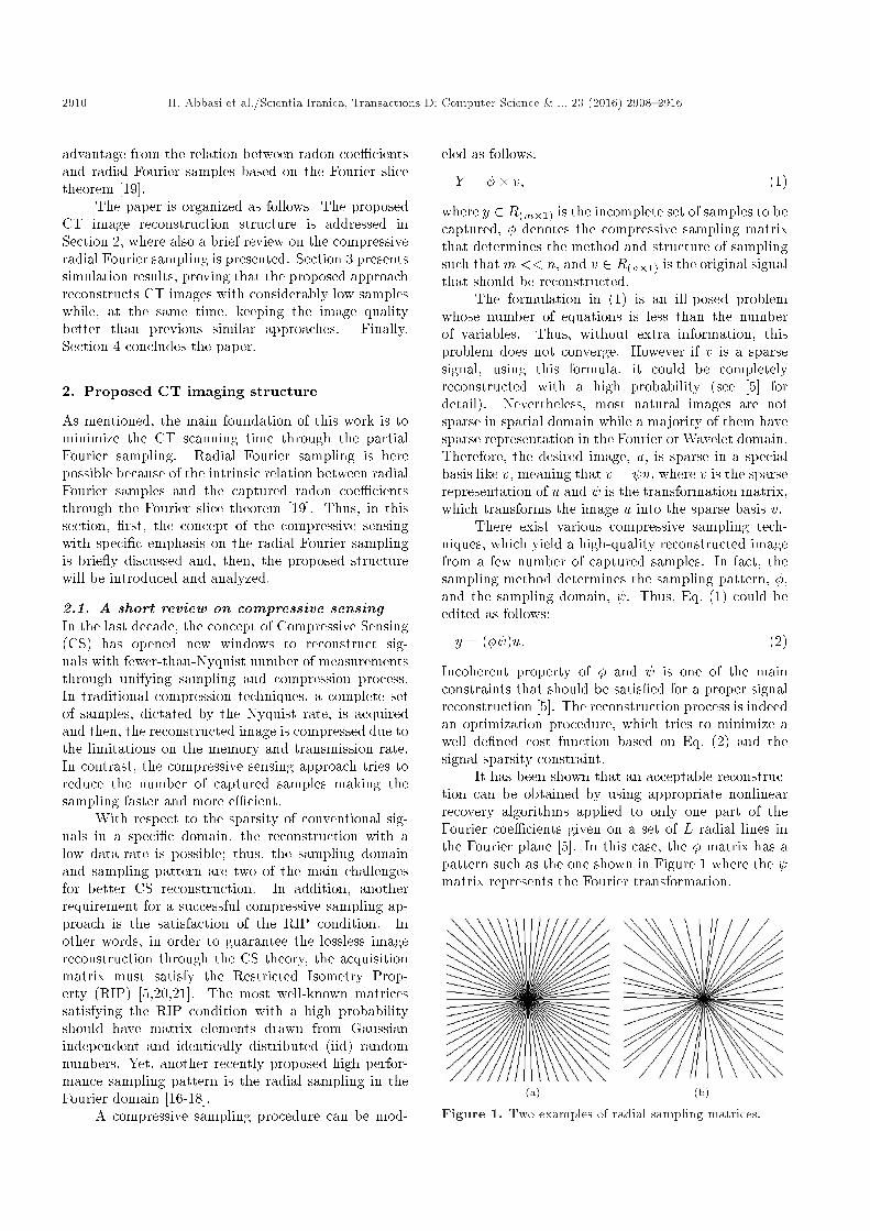
It has been shown that an acceptable reconstruc-tion can be obtained by using appropriate nonlinearrecovery algorithms applied to only one part of theFourier coe�cients given on a set of L radial lines inthe Fourier plane [5]. In this case, the � matrix has apattern such as the one shown in Figure 1 where the matrix represents the Fourier transformation.
Figure 1. Two examples of radial sampling matrices.

H. Abbasi et al./Scientia Iranica, Transactions D: Computer Science & ... 23 (2016) 2908{2916 2911
Apart from the satisfaction of RIP and IP con-straints, in a Radial Fourier sampling scheme, low-frequency components are sampled more densely thanhigh-frequency components. This fact resembles theconcept behind the common digital compression tech-niques where the most useful information from anobject is gathered around the center of the Fourierplane.
Given that in a CT scan structure data aregathered as radon coe�cients, the Radial Fouriersampling could be utilized due to the direct relationsbetween radial Fourier samples and radon coe�cientsbased on FST. This property is here utilized in orderto perform CS-based CT imaging through the radialFourier sampling where the RIP condition could beanalyzed more systematically. The presented approachis described in the sequel.
2.2. Proposed structureIn this paper, the main goal is to implement a fast CS-based CT imaging structure based on radial Fouriersampling in order to satisfy the RIP condition moresystematically. This is done through exploiting therelation between radon coe�cients and radial Fouriersamples. The main framework of the proposed struc-ture for Radial Fourier CT scan is illustrated inFigure 2.
Let I(x; y) represent a compactly supported con-tinuous function on R2 that shows an m�n image. TheCT imaging system works based on capturing the radonsamples. The radon transform, <I(:), is a functionde�ned on the space of straight lines L in R2 by theline integral along each such line:
<I(L) =ZLI(x)jdxj: (3)
Concretely, parametrization of any straight line, L,with respect to arc length, t, can always be writtenas:
(x(t); y(t))=((t sin � + s cos �);(�t cos �+s sin �)); (4)
Figure 2. Proposed structure of the compressivesensing-based CT imaging structure.
where s is the distance of L from the origin and � is theangle between the normal vector to L and the x axis.It follows that the quantities (s; �) can be consideredas coordinates on the space of all lines in R2, and theradon transform can be expressed in these coordinatesby:
<I(s; �)=Z Z
I(x; y)�(x cos � + y sin � � s)dxdy: (5)
Based on this, in order to make the CT scan procedurefaster, we propose to reduce the number of samplesthrough the partial Fourier sampling. To this end, weshould �rst select the required radial Fourier samples ofthe object based on the chosen radial Fourier samplingpattern. This pattern has di�erent models such asuniform or golden-angle as shown in Figure 1 [17]. Thenext step is to relate the selected radii in the Fourierdomain to the corresponding radon coe�cients throughthe Fourier slice theorem. With respect to FST, the 1DFourier transform of line projections in an arbitraryangle, �, (named as FR(!; �)) is equal to a 1D sliceof the 2D Fourier transform of the image in the sameangular direction. This theorem could be modeled asfollows:
F<(!; �) = FI(! cos �; ! sin �); (6)
where FI(:) represents the 2D Fourier transform of theoriginal image. With this relation in hand, the cor-responding required radon coe�cients, <I(s; �), couldbe obtained from the selected radial Fourier samples.Then, the CT structure scans the object to capture theobtained desired radon coe�cients.
The schematic of the recording procedure is shownin Figure 3. These captured radon coe�cients shouldbe converted to the corresponding radial Fourier sam-ples in order to run the CS optimization step andreconstruct the full image. However, before performingthe conversion procedure, the radon coe�cients shouldundergo a zero-padding step as shown in Figure 3,where the schematic of the capturing process forobtaining the required Fourier radii is demonstrated.The 1D Fourier transforms of the zero-padded radoncoe�cients are computed in the next step, which yieldthe required Fourier radii.
With these radial Fourier samples in hand, the op-timization procedure based on the wellknown methodof alternating direction of multiplier [22] is run in orderto reconstruct the whole image in the Cartesian coor-dinate. After choosing the optimization method, the�nal goal is to �nd the image, which has the minimumtotal variation with the maximum accuracy. This goalresults in the following minimization problem:
min �ikDiuk2 + � j uj+ �=2k� u� yk22; (7)

2912 H. Abbasi et al./Scientia Iranica, Transactions D: Computer Science & ... 23 (2016) 2908{2916
Figure 3. The obtained radial Fourier samples from theradon coe�cients.
where the �rst term is the discretization of the TotalVariation (TV) of u over its all pixels across verticaland horizontal directions, the second term is l1-normof the sparse representation of u under , and thelast term denotes the relaxation term. Moreover,� and � are regularization parameters. The mainchallenge to �nd the solution of Relation (7) is thenon-di�erentiability of its �rst and second terms. Byde�ning two auxiliary variables z and w, Relation (7)can be rewritten as:
min �ikwik2 + � j zj+ �=2k� u� yk22;s.t. wi = Diu 8 i; z = Tu: (8)
In order to solve Relation (8) linearly, the augmentedLagrangian term is used as the minimization goal. Bymeans of the augmented Lagrangian of Relation (8)and using an Alternating Direction Method (ADM),
this equation could be simply converted to a leastsquares problem. Therefore, the reconstruction timeis decreased by using ADM algorithm for solving theoptimization problem. The algorithm uses one- andtwo-dimensional shrinkage to compute w and z in eachiteration in parallel. Then, the value of u is updateduntil the relative error becomes small enough (less thana prede�ned threshold value) (see [22] for details).
Therefore, there are three main tasks in eachiteration for the ADMM algorithm: computing thevariables z and w, obtaining a new output, and up-dating the required variables for the next iteration. Tosupport our proposed CT imaging structure, we designa VLSI architecture for the reconstruction algorithm,which provides a real-time prototype. The blockdiagram of its top module is depicted in Figure 4(a).It is composed of three processing units and onememory management unit, working in accordance witha control unit. The control unit sends the necessarycontrol signals to the processing units to initiate theiroperations, and generates the address values to dothe read and write operations on the memory unit.In addition, this design includes the infrastructureoverheads such as memory and Ethernet controller torun on the Xilinx Virtex-6 FPGA ML605 EvaluationKit. The overall data-path of this design is shown inFigure 4(b).
Thus, the proposed structure reduces the sam-pling points signi�cantly compared to common scan-ning structures such as FBP, which in turn reduces thewhole scanning time.
In order to quantitatively demonstrate the im-proved performance of the proposed structure, threemetrics are utilized in this paper. The �rst metric isthe Mean Square Error (MSE) between the original,IO, and reconstructed, IR, images with the followingformula:
MSE = E[(IR � IO)2]: (9)
The second evaluation metric is the Peak Signal-to-
Figure 4. Hardware design: (a) The block diagram of the hardware design; (b) the overall data-path of the hardwaredesign.

H. Abbasi et al./Scientia Iranica, Transactions D: Computer Science & ... 23 (2016) 2908{2916 2913
Noise Ratio (PSNR) of the reconstructed image. ThePSNR metric is de�ned as follows:
PSNR = 20 log
Ip
(MSE)
!; (10)
where I represents the maximum intensity of thereference image. PSNR results are complemented byanother well-known evaluation metric, i.e. StructureSimilarity Index Metric (SSIM), which measures thesimilarity between the original and reconstructed im-ages [23]. The SSIM metric is de�ned as follows:
SSIM =(2mI1mI2 + c1)(2�I1I2 + c2)
(m2I1 +m2
I2 + c1)(�2I1 + �2
I2 + c2); (11)
where mI and �I represent the image mean andvariance, respectively, �I1I2 shows the covariance of theoriginal and reconstructed images, and c1 and c2 arestabilization parameters.
In order to demonstrate superiority of the pro-posed structure in reducing the CT scan time while,at the same time, yielding a high quality image,exhaustive simulations are performed, reported in thenext section. The performed simulations comparethe quality of the proposed method with those ofother interpolation-based and CS-based approaches,intended to reduce the CT scan time.
3. Results and compariosn
In this section, the proposed structure is compared withthree other CT image reconstruction methods, whichtry to lessen the CT scan time. These methods areCSTV [9], interpolation [3], and WL1 [13]. The CTimage of a sample SheppLogan phantom is obtainedthrough MATLAB simulations. The considered sampleis an image of size 256 � 256 pixels while the valueof the regularization parameters in each algorithm isselected empirically. The reconstructed image resultedfrom each algorithm is shown in Figure 5. As it is clearfrom the colormap results of MATLAB and enlargedpart of each image, shown in Figure 6, for CS-basedalgorithms, the proposed structure performs better insaving the image high-frequency contents while, at thesame time, not adding considerable distortion.
In order to evaluate the reconstructed imagesbased on these metrics, two simulation scenarios wereperformed. In the �rst simulation, the reconstructionprocess was done with 5 di�erent algorithms includingCSTV, WL1, FBP, interpolation, and the proposed al-gorithm, where the quality of the reconstructed imagesversus the number of projections was fully compared.The evaluated PSNR for this simulation is shown inFigure 7. This �gure clearly demonstrates the superiorperformance of the proposed method based on PSNRmetric.
Figure 5. Reconstructed images with 24 projections: (a)Proposed structure with uniform angelprojection (PSNR= 42.42); (b) CSTV result (PSNR = 40.47); (c) WL1result (PSNR = 40.83); and (d) interpolation result(PSNR = 19.47).
Figure 6. Reconstructed images with JET colormap: (a)Proposed structure with uniform angel projection; (b)CSTV result with distorted high frequency contents; and(c) WL1 result with added distortions apparent in uppercircle.
Figure 7. PSNR of di�erent algorithms with changingprojection numbers.
In the second simulation scenario, the resultsof the CS-based CT image reconstruction algorithmsincluding CSTV, WL1, and our proposed method arecompared for various numbers of iterations. Themeasured PSNR values for all algorithms are depictedversus the iteration numbers in Figure 8. It is clear

2914 H. Abbasi et al./Scientia Iranica, Transactions D: Computer Science & ... 23 (2016) 2908{2916
Figure 8. PSNR of di�erent algorithms with changingiteration numbers.
that the proposed structure has better quality with thesame number of iterations, i.e. in 400 iterations.
With regard to this �gure, it is obvious that theproposed method performs better based on the PSNRmetric for di�erent iteration numbers. Furthermore,the superior performance of the image reconstructedfrom the proposed structure is also con�rmed throughSSIM and MSE metrics. The numerical results areshown in Table 1. As it is clear in this table, theresult of the proposed structure is better than the otherresult (about 4% in PSNR). Also, this structure has theminimum MSE and better SSIM.
As mentioned in the Introduction, the high sensi-tivity to noise is one of the important shortcomings ofWL1. In contrast, CSTV and the proposed structurehave low sensitivity to noise by using the total variationand minimizing the gradient of image. Figure 9 showsthe results of reconstructed images with noisy data.As can be seen clearly, distortion in the texture ofthe result of WL1 is obvious while CSTV and theproposed structure perform better in the presence ofnoise.
Finally, in order to have a more reliable com-parison between the performances of the proposedstructure and other CS-based methods, real projectiondata are employed and analyzed. Figure 10 showsthe reconstructed images with real dataset [24]. Asis clear from this �gure, the proposed algorithm hasa better reconstruction quality than CSTV and WL1algorithms. CSTV has di�culty in the reconstructionof edges due to the nature of the total variation andWL1 has distortion in the speci�ed parts.
Figure 9. Reconstructed images with noisy data: (a)Proposed structure with uniform angel projection (PSNR= 36.45); (b) CSTV result (PSNR = 38.38); (c) WL1result (PSNR = 31.11); and (d) DFT result (PSNR =18.39).
Figure 10. Reconstruction results with real dataset (250iteration): (a) Proposed structure with uniform angelprojection (PSNR = 32.95); (b) CSTV result (PSNR =29.45); and (c) WL1 result (PSNR= 31.08).
In summary, both the qualitative visual perfor-mance and the quantitative metrics con�rm the supe-riority of the proposed CS-based CT imaging structurein preserving the image quality while, at the same time,lessening the CT scan time.
4. Conclusion
In this paper, an e�cient CT imaging structure basedon compressive sensing is proposed in order to miti-gate the CT scanning time. In order to satisfy therequisite of the compressive sampling algorithms ina systematic manner, partial Fourier sampling wasutilized. Thanks to the intrinsic relation betweencaptured radon samples and radial Fourier samples,
Table 1. Comparison of the results of di�erent CS-based algorithms (24 line projection and 400 iteration).
This Work CSTV [9] WL1 [13] FBP Interpolation
PSNR 42.42 40.47 40.83 13.43 17.81MSE 5:6989e�05 8:9741e�05 7:9831e�05 0.0315 0.0746SSIM 0.9999 0.9998 0.9999 -1 0.9992

H. Abbasi et al./Scientia Iranica, Transactions D: Computer Science & ... 23 (2016) 2908{2916 2915
Partial (radial) Fourier sampling could be implementedmore systematically. This systematic implementationhelped in better satisfaction of CS conditions, suchas Restricted Isometry Property (RIP). Simulationswere performed, in which the performance of theproposed structure was compered with those of 3 otherapproaches that also tried to lessen CT scan timethrough either CS or interpolations. The presentedqualitative and quantitative results showed the superiorperformance of the proposed structure.
Acknowledgment
We would like to thank Amir Ashkan Farsaei for hisvery helpful comments on a draft of hardware designparts of this article.
References
1. Bracewell, R.H. and Riddle, A.C. \Inversion of fanbeam scans in radio astronomy", Astrophysics Journal,150, pp. 427-434 (1967).
2. Ramanchandran, G.N. and Lakshminarayanan, A.V.\Three dimensional reconstructions from radiographsand electron micrographs: Application of convolutioninstead of Fourier transforms", Proceedings of the Na-tional Academy of Sciences, 68, pp. 2236-2240 (1971).
3. Stark, H., Woods, J. and Paul, I., \Direct Fourier re-construction in computer tomography", IEEE Trans.,Speech and Signal Processing, 29, pp. 237-245 (Apr.1981).
4. Donoho, D.L. \Compressed sensing", IEEE Trans.Inform. Theory, 52, pp. 1289-1306 (2006).
5. Candes, E., Romberg, J. and Tao, T. \Robust un-certainty principles: exact signal reconstruction fromhighly incomplete frequency information", Informa-tion Theory, IEEE Transactions on, 52, pp. 489-509(Feb. 2006).
6. Candes, E. and Tao, T. \Near-optimal signal recoveryfrom random projections: Universal encoding strate-gies?", Information Theory, IEEE Transactions on,52, pp. 5406-5425 (Dec. 2006).
7. LaRoque, S.J., Sidky, E.Y. and Pan, X. \Accurateimage reconstruction from few-viewand limited-angledata in di�raction tomography", Journal of the OpticalSociety ofAmerica, 25(7), pp. 1772-1782 (Jun 2008).
8. Sidky, E.Y. and Pan, X.C. \Image reconstructionin circular cone-beam computed tomography by con-strained, total-variation minimization", Physics inMedicine and Biology, 53, pp. 777-807 (2008).
9. Bian, J., Wang, J., Han, X., Sidky, E.Y., Shao, L.and Pan, X. \Optimization-based image reconstruc-tion from sparse-view data in o�set-detector CBCT",Physics in Medicine and Biology, 58, pp. 205-230(2013).
10. Lauzier, P.T. and Chen, G.H. \Characterization of sta-tistical prior image constrained compressed sensing. I.Applications to time-resolved contrast-enhanced CT",Medical Physics, 39, pp. 5930-5948 (2012).
11. Lauzier, PT. and Chen, GH. \Characterization ofstatistical prior image constrained compressed sensing(PICCS): II. application to dose reduction", MedicalPhysics, 40 (2013).
12. Barkan, O., Weill, J., Averbuch, A. and Dekel, S.\Adaptive compressed tomography sensing", IEEEConferance, Computer Vision and Pattern Recognition(CVPR), pp. 2195-2202 (2013).
13. Hou, W. and Zhang, C. \A Compressed sensingapproach to low-radiation CT reconstruction", Com-munication Systems, Networks & Digital Signal Pro-cessing (CSNDSP), pp. 793-797 (2014).
14. Jrgensen, J.H., Sidky, E.Y. and Pan, X. \Quantify-ing admissible undersampling for sparsity-exploitingiterative image reconstruction in X-ray CT", IEEETransactions on Medical Imaging, 32, pp. 460-473(2013).
15. Jrgensen, J.H., Sidky, E.Y., Hansen, P.C. and Pan, X.\Quantitative study of undersampled recoverability forsparse images in computed tomography", Arxiv, pp. 1-20 (2012).
16. Stern, A. \Compressed imaging system with linearsensors", Opt. Lett., 32, pp. 3077-3079 (Nov. 2007).
17. Evladov, S., Levi, O. and Stern, A. \Progressivecompressive imaging from radon projections", Opt.Xpress, 20, pp. 4260-4271 (Feb. 2012).
18. Yang, J., Zhang, Y. and Yin, W. \A fast alternatingdirection method for tvl1-l2 signal reconstruction frompartial fourier data", Selected Topics in Signal Process-ing, IEEE Journal of, 4, pp. 288-297 (April 2010).
19. Hsieh, J., Computed Tomography: Principles, Design,Artifacts, and Recent Advances, Bellingham, WA:SPIE (2009).
20. Duarte, M., Davenport, M., Takhar, D., Laska, J.,Sun, T., Kelly, K. and Baraniuk, R. \Single-pixelimaging via compressive sampling", Signal ProcessngMagazine, IEEE, 25, pp. 83-91 (March 2008).
21. Willett, R.M., Marcia, R.F. and Nichols, J.M. \Com-pressed sensing for practical optical imaging systems:a tutorial", Optical Engineering, 50, no. 7, pp. 072601-072601-13 (2011).
22. Yang, J., Zhang, Y. and Yin, W. \A fast alternatingdirection method for tvl1-l2 signal reconstruction frompartial fourier data", Selected Topics in Signal Process-ing, IEEE Journal of, 4, pp. 288-297 (April 2010).
23. Wang, Z., Bovik, A.C., Sheikh, H.R. and Simoncelli,E.P. \Image quality assessment: From error visibilityto structural similarity", Trans. Img. Proc., 13, pp.600-612 (Apr. 2004).

2916 H. Abbasi et al./Scientia Iranica, Transactions D: Computer Science & ... 23 (2016) 2908{2916
24. DICOM sample image sets (http://www.osirix-viewer.com/datasets/).
Biographies
Hasan Abbasi received his BS degree in ElectricalEngineering in 2013 from Shahid Beheshti University,Tehran, Iran, and MSc degree in 2015 from SharifUniversity of Technology. He is currently PhD studentat Sharif University of Technology. His areas ofinterest and experience are medical imaging and signalprocessing.
Zahra Kavehvash received her BS degree in 2005 andPhD degree in 2013, both in Electrical Engineeringfrom Sharif University of Technology, Iran. She is
interested in 3D imaging, millimeter-wave imaging andholography, and biomedical imaging systems. Dr.Kaveh vash joined the Department of Electrical En-gineering at Sharif University of Technology, Tehran,as an Assistant Professor in 2013.
Mahdi Shabany received his BS degree in ElectricalEngineering from Sharif University of Technology, Iran,in 2002. He �nished his MS and PhD degrees in Elec-trical Engineering at Toronto University in 2009. He isinterested in VLSI implementation of biomedical signalprocessing algorithms, architecture/algorithm designfor 5G communication systems, and ASIC/RTL digitalcircuit design. Dr. Shabany joined the Department ofElectrical Engineering, Sharif University of Technology,Tehran, as an Assistant Professor in 2010.